Note: Descriptions are shown in the official language in which they were submitted.
77761-2 CA 02250603 2000-01-17
Title of the Invention
Treatirlg Urinary and Other Body Strictures
Background of the Invention
1. Field ol= the Invention
This invention relates to techniques for treating
urinary strictures.
2. Related Art
A stricture is an abnormally narrowed segment of an
otherwise patent biological tube or conduit, such as the
gastrointestinal t:ract, genito-urinary tract, pulmonary system,
vascular system, or other systems in the body. Strictures may
occur at various places within these systems in the body, such
as in or near a blood vessel, the bronchial tree, the colon, a
gastrointestinal body structure, a genital body structure, a
kidney, a post-operative stricture, a pulmonary body structure,
the rectum, or the sphirlcter, or a urethral body structure.
The degree of narrowing, the length, and the significance of
the stricture may differ greatly between particular strictures,
and is responsive to the nature of the conduit which is subject
to the stricture. Various etiological factors might be
responsible for th.e development or exacerbation of any
particular stricture; these may include, for example,infection,
inflammation, trauma (whether external, internal, or iatrogenic
or other surgical trauma), or cancer. One or more of these
factors causes the lumeri of the affected conduit to narrow,
that is, to stricture, with consequential obstruction of the
lumen which would compromise the function of the conduit.
Treatment of strictures is aimed at restoration of
intraluminal patency ancl physiological function. Because of
1
CA 02250603 2000-01-17
77761-2
the presence of abnormal or diseased tissue at the stricture,
surgical treatment by endoscopic or by open surgical techniques
often poses extra difficulties and has significant morbidity.
Moreover, because the tissue of the stricture wall is already
diseased, it often generates further scarring and fibrosis when
it heals after surgery, which can lead to recurrence of the
stricture.
Accordirigly, it would be advantageous to provide a
method and system for treatment of strictures, such as for
example urinary st:rictures, which use existing tissue, which
promote healing of: existing tissue, and which help to prevent
recurrence of the stricture. This advantage is achieved in an
embodiment of the invention in which a supporting frame, such
as a cylindrical collagen frame with a diameter comparable to
the normal lumen, is disposed intraluminally in a constricted
region of the stricture, energy is emitted to ablate and harden
the collagen and the tissue, and the supporting frame is used
to maintain patency of t:he lumen and to prevent reformation of
the stricture during a healing period.
Summary of the Invention
The invention provides a method and system for
treatment of body strictures to restore luminal diameter to
within a normal diameter range, in which the stricture is
dilated to stretch its lumen to a desired diameter, collagen is
exuded near to existing tissue of the stricture so as to be
absorbed by that tissue or adhere to that tissue, making a
collagen-enhanced tissue structure, and energy is emitted to
affect the collagen-enhanced tissue, such as by ablation or by
hardening. Ablation and hardening may be repeated so as to
create a set of layers of hardened collagen in the form of a
supporting frame, preferably having a hollow cylindrical shape.
2
CA 02250603 2006-08-03
77458-2
In a preferred embodiment, dilation of the stricture
is achieved by expanding one or more balloons, or by the
pressure of exuded collagen, until the stricture is larger than
a normal diameter range. When energy is emitted into the
collagen, the stricture contracts back to the normal diameter
range, either by ablati_on of excess tissue or by plating of the
stricture wall.
In a preferred embodiment, the stricture's tissue is
also isolated by a set of balloons at either or both ends of
the stricture, so as to isolate the stricture and restrict the
collagen to the stricture's tissue.
In a preferred embodiment, the stricture's tissue is
also supported by a stent, which is preferably tack-welded onto
the stricture's tissue using collagen. Collagen adheres to the
stent, which supports the stricture's tissue until the stent is
absorbed into that tissue.
Another aspect of the invention provides apparatus
for treatment of a stricture within the body, said apparatus
including: a multi-lumen catheter having a proximal end and a
distal end; a catheter tip electrode housing having a proximal
end and a distal end, said electrode housing proximal end being
connected to said catheter distal end; a radiology marker
located in said electrode housing; a first inflatable balloon
located at distal end of said catheter and a second inflatable
balloon located at said electrode housing distal end, both said
first and second balloons being connected to a source of non-
translucent inflation fluid by a lumen running longitudinally
through said catheter, said first and second balloons when
inflated by said inflation fluid being effective to stabilize a
position of said catheter during said treatment and achieve at
least one seal against a surface of said stricture, thereby
confining at least a portion of said stricture between said
3
CA 02250603 2006-08-03
77458-2
first and second balloons; at least one first port located on
surface of said electrode housing between said first and second
balloons, said at least one first port being connected with a
source of non-translucent treatment by a lumen running
longitudinally through said catheter and disposed to exude a
first mass of said treatment fluid into said confined portion
of said stricture under a first pressure and for a first time,
said first pressure and said first time being effective to
dilate said stricture and cause at least a portion of said
first mass of treatment fluid to be suffused and absorbed into
at least a portion of an existing tissue of said confined
portion of said stricture; at least one temperature sensor on
the surface of said electrode housing, said temperature sensor
being connected to a control system by a lumen running
longitudinally through said catheter; a conductor connected
with a source of energy by a lumen running longitudinally
through said catheter; at least one electrode included in said
electrode housing and coupled to said conductor, said electrode
being operative to emit energy and raise a temperature,
substantially proximate to said electrode housing only, to at
least 100 degrees Celsius, for a time effective to couple at
least a portion of said suffused and absorbed treatment fluid
with at least a portion of said mass of existing tissue into a
unified tissue matrix.
Brief Description of the Drawings
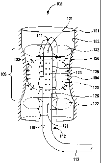
Figure 1 shows a urinary stricture with a catheter
positioned therein.
Figure 2 is a flowchart for a method of operation for
the catheter.
Figure 3 shows a urinary stricture with a stent
positioned and attached therein.
4
CA 02250603 2006-08-03
77458-2
Detailed Description of the Preferred Embodiment
Urinary Stricture
Figure 1 shows a urinary stricture with a catheter
positioned therein.
A stricture 100 comprises a mass of relatively
healthy tissue 101, forming a first portion of a wall 102 for a
lumen 103 or other pathway, and a mass of relatively weakened
tissue 104, forming a second portion of the wall 102 for the
lumen 103 in a constricted region 105. The stricture 100 is
shown with the lumen 103 having at least some flow capability,
but there are structures 100 in which the flow capability has
been reduced to zero, either because the wall 102 in the
constricted region 105 has collapsed completely so as to block
the lumen 103, because the lumen 103 is blocked with a mass of
tissue or other substances (not shown), or some combination of
these two problems.
A catheter 110 comprises a distal end 111 and a
proximal end 112, the latter being coupled to a tube 113 or
other connector for coupling control signals, energy, and
fluids between the catheter and 110 and a control system (not
shown).
In a preferred embodiment, the catheter 110 comprises
a catheter such as shown in United States Patent Application
Serial No. 08/717,612, now United States Patent No. 6,077,257.
In a preferred embodiment, the catheter 110 is
about 5 to about 6 French in width (1 French equals 1/3 of a
millimeter or about 0.18 inch). However, in alternative
embodiments, the catheter 110 may be of lesser or greater width
so as to accommodate strictures of lesser or greater diameter.
5
CA 02250603 2006-08-03
77458-2
The catheter 110 also comprises a first x-ray
marker 121, preferably disposed at or near the distal end 111
of the catheter 110 and a second x-ray marker 121, preferably
disposed at or near the proximal end 1ll of the catheter 110.
With suitable x-ray or fluoroscopy equipment, a radiologist or
surgeon can position the catheter 110 relative to the
constricted region 105 without any requirement for a camera or
other optical equipment disposed in or near the constricted
region 105.
The catheter 110 also comprises a first ring
balloon 122, preferably disposed at or near the distal end 111
of the catheter 110 and a ring balloon 122, preferably disposed
at or near the proximal end 111 of the catheter 110. The ring
balloons 122 are disposed so that, when inflated and in
combination with the body of the catheter 110, they physically
seal off gas or fluids between the constricted region 105 and
other portions of the lumen 103 outside the constricted
region 105.
In alternative embodiments, the ring balloons 122 may
comprise other shapes, and in particular, the ring balloon 122
disposed at the distal end 111 of the catheter 110 may comprise
a spherical or ellipsoidal balloon disposed to seal off the
lumen 103 without need for combination with the body of the
catheter 110. In such alternative embodiments, the spherical
or ellipsoidal balloon is disposed in substantially the same
location as shown for the ring balloon 122, except that the
spherical or ellipsoidal balloon is disposed to substantially
block the lumen 103 and thus seal it off.
In further alternative embodiments, the ring
balloons 122 may be porous, microporous, semiporous, or some
combination thereof, or may be disposed within the lumen 103
slightly imperfectly, so that so that the seal made by the ring
6
CA 02250603 2006-08-03
77458-2
balloons 122 is not necessarily completely gas-tight or even
completely fluid-tight.
The catheter 110 also comprises an expansion
balloon 123, preferably disposed at or near a middle portion of
the catheter 110. The expansion balloon 123 is disposed so
that when inflated it physically forces the constricted
region 105 or the stricture 100 to open to a greater diameter,
such as a diameter within a normal diameter range for the
lumen 103.
In a preferred embodiment, the expansion balloon 123
comprises a porous, microporous, or semiporous membrane through
which a mass of collagen 130, a solution including saline, or
other flowable substances, may flow. The catheter 110
comprises an internal lumen (not shown) which couples flowable
substances from the tube 113, so as to exude those flowable
substances out from a set of holes 124 and to the expansion
balloon 123. When flowable substances are exuded out from the
holes 124 to the expansion balloon 123, pressure from the
flowable substances causes the expansion balloon 123 to expand
and to physically force the constricted region 105 of the
stricture 100 to open to the greater diameter.
In a preferred embodiment, the expansion balloon 123
comprises a spherical or ellipsoidal shape, so as to expand in
a middle region near the stricture 105. In a preferred method
of operation, the expansion balloon 123 is first expanded to
its maximum diameter, then deflated somewhat so as to allow
flowable substances to flow into the region of the
stricture 105.
However, in alternative embodiments, the expansion
balloon 123 may comprise another shape, such as a concave shape
(shaped somewhat like the stricture itself) having a greater
7
CA 02250603 2006-08-03
77458-2
degree of expansion at a distal end of the stricture 105 and at
a proximal end of the stricture 105, and having a lesser degree
of expansion at a middle portion of the stricture 105. The
expansion balloon 123 can take on this concave shape by being
comprised of a relatively thinner (and therefore more
expansible) rubber material at the distal end of the
stricture 105 and at the proximal end of the stricture 105,
while being comprised of a relatively thicker (and therefore
less expansible) rubber material at the middle portion of the
stricture 105.
The catheter 110 also comprises a set of
electrodes 125, preferably disposed at or near a middle portion
of the catheter 110. The electrodes 125 are coupled using the
tube 113 to a power source (not shown). The power source
provides energy to the electrodes 125, which emit that energy
into the constricted region 105 of the stricture 100 so as to
affect the mass of collagen 130, the relatively weakened
tissue 104, and (in some embodiments) the relatively healthy
tissue 101.
The catheter 110 also comprises a set of sensors 126,
preferably disposed at or near a surface of the catheter 110.
The sensors 126 are coupled using the tube 113 to a control
system (not shown) and to an operator display (not shown). The
sensors 126 provide signals to the control system for feedback
controi, and to the operator display for displaying information
to an operator.
In a preferred embodiment, the sensors 126 comprises
a plurality of temperature sensors, such as thermistors or
thermocouples, and the control system provides feedback control
to maintain a temperature of the mass of collagen 130 at a
temperature selected by the operator. In a preferred
embodiment, the operator display comprises a temperature
8
CA 02250603 2006-08-03
77458-2
reporting gauge. However, it would be clear to those skilled
in the art that other and further sensor signals, feedback
control, and display signals, would be useful, and are within
the scope and spirit of the invention.
Method of Operation
Figure 2 is a flowchart for a method of operation for
the catheter.
A method 200 of operation for the catheter 110
comprises a sequence of steps between the flow points 210
and 230. In a preferred embodiment, the method 200 is carried
out using the catheter 110, as well as other and further
equipment which would be clearly deemed necessary or desirable
by those skilled in the art.
At a flow point 210, it is desired to treat the
urinary stricture 100.
At a step 221, the catheter 110 is inserted into the
constricted region 105 of the urinary stricture 100. As noted
herein, the radiologist or surgeon positions the catheter 110
relative to the urinary stricture 100 using the x-ray
markers 121 and an fluoroscope or other x-ray device.
At a step 222, the first ring balloon 122 and the
second ring balloon 122 are expanded to isolate the constricted
region 105 from other portions of the lumen 103 in a gas-tight
and fluid-tight manner.
At a step 223, the mass of collagen 130 and a saline
solution are flowed through the tube 113, through the body of
the catheter 110, through the holes 124, and into the expansion
balloon 123. The flow of the mass of collagen 130 and the
saline solution into the expansion balloon 123 causes the
expansion balloon 123 to expand, physically forcing the
9
CA 02250603 2006-08-03
77458-2
relatively weakened tissue 104 out to a diameter greater than
the normal diameter range for the lumen 103.
At a step 224, the mass of collagen 130 and the
saline solution are flowed through the expansion balloon 123,
into contact with the relatively weakened tissue 104. The mass
of collagen 130 and the saline solution are absorbed into the
relatively weakened tissue 104.
At a step 225, electrical energy is conducted from
the power source through the tube 113, through the body of the
catheter 110, to the electrodes 125. The electrodes 125 emit
RF energy (at a preferred frequency of between about 400
megahertz and about 700 megahertz, but possibly at other
frequencies, such as microwave frequencies), which is received
by the saline solution and thus transmitted to the relatively
weakened tissue 104.
The relatively weakened tissue 104, having been
suffused with the mass of collagen 130, receives the RF energy
emitted by the electrodes 125 and is ablated. As RF energy is
received by the relatively weakened tissue 104, the relatively
weakened tissue 104 is heated to at least about 90 to 120
degrees Celsius, causing oblation to occur by means of cell
death, dehydration, denaturation, or other means.
In a second preferred embodiment, the mass of
collagen 130 forms a surface layer over the wall 102 of the
relatively weakened tissue 104. At the step 224, the mass of
collagen 130 adheres to the surface while the saline solution
is absorbed into the relatively weakened tissue 104. At the
step 225, the mass of collagen 130 is cooked or otherwise
thermoset by the RF energy so as to solidify into a layer of
hardened collagen, preferably about 1 mil (0.001 inch or
about 0.0025 centimeters) in thickness. The step 224 and the
CA 02250603 2006-08-03
77458-2
step 225 are repeated a number of times sufficient to create a
layer of hardened collagen effective to restrain fluid flowing
in the lumen 103 from seeping into the relatively weakened
tissue 104.
The mass of collagen 130, having been heated by
application of RF energy, cooks or otherwise thermosets to a
solidified state.
At a step 226, the relatively weakened tissue 104,
having been ablated, shrinks to a diameter within a normal
diameter range for the lumen 103.
At a step 227, the relatively weakened tissue 104, as
supported by the mass of collagen 130, is allowed to heal by
growth of epithelial cells.
At a flow point 230, the urinary stricture 100 has
been treated and should be in condition for normal operation.
In alternative embodiments, the method 200 may be
applied to other body structures or other places within the
gastrointestinal tract, genito-urinary tract, pulmonary system,
vascular system, or other systems in the body, such as in or
near a blood vessel, the bronchial tree, the colon, a
gastrointestinal body structure, a genital body structure, a
kidney, a postoperative stricture, a pulmonary body structure,
the rectum, the sphincter, or a urethral body structure.
Figure 3 shows a urinary stricture with a stent
positioned and attached therein.
A stent 300 comprises a substantially cylindrical
structure having a distal end 301 and a proximal end 302, and
formed in the shape of a mesh or a woven structure, such as
used in gauze or stretchable fabrics. In a preferred
11
CA 02250603 2006-08-03
77458-2
ernbodiment, the stent 300 comprises a suture material, such as
catgut, polygalactic polymer 910, or PDS.
The stent 300 is disposed in the urinary
stricture 100 by coupling the stent 300 to the catheter 110,
disposing the catheter 110 substantially within the constricted
region 105 of the urinary stricture 100, and coupling the
stent 300 to at least a portion of the urinary stricture 100.
The stent 300 is coupled to the urinary stricture 100
by coupling the distal end 301 of the stent 300 to a coupling
spot 310 on the relatively healthy tissue 101 outside the
constricted region 105 of the urinary stricture 100 by means of
"tack welding". "Tack welding" refers to disposing the distal
end 301 of the stent 300 at the coupling spot 310, exuding
collagen so as to adhere to both the distal end 301 of the
stent 300 and the coupling spot 310, and emitting energy so as
to harden the collagen to permanently or semipermanently couple
the distal end 301 of the stent 300 to the coupling spot 310.
In a preferred embodiment, the coupling spot 310
comprises an 0-shaped ring around the lumen 103.
Alternative Embodiments
Although preferred embodiments are disclosed herein,
many variations are possible which remain within the concept,
scope, and spirit of the invention, and these variations would
become clear to those skilled in the art after perusal of this
application.
12