Note: Descriptions are shown in the official language in which they were submitted.
CA 02250723 1998-09-30
WO 97/44469 PCT/US97/08950 --
-1-
MULTIPLE EPITOPE FUSION PROTEIN
Field of the Invention
This invention relates generally to the fields of protein synthesis and
immunoassays and specifically relates to methods of synthesizing long chains
of amino
acids that contain multiple copies of epitopes for viruses such as HCV, and to
assay
devices that utilize multiple epitopes to detect the presence of antibodies.
Background of the Invention
In general, immunoassays are produced by first determining epitopes that are
specifically associated with a virus and then determining which of the
epitopes is
preferred for the assay being developed. When the immunodominant epitopes are
isolated, their sequences are determined, and genetic material for producing
the
immunodominant epitopes is produced. Methods of producing proteins by either
chemical or biological means are known, as are assays used to detect the
presence of
antibodies to particular epitopes.
In producing immunoassays the overall object is to obtain an immunoassay which
is both highly sensitive and highly selective. More specifically, the
immunoassay must
be designed such that it can detect even very low levels of the material it is
designed to
detect, i.e., it is highly sensitive. An assay having a high degree of
sensitivity ensures
that a sample, which has been tested, is not contaminated with the material
the assay is
designed to detect. For example, a highly sensitive assay that detects even
the slightest
presence of antibodies for a given virus is desirable in that it makes it
possible to detect
and thus discard samples that contain any amount of the antibody indicating
that the
samples contain the virus.
Although a high degree of sensitivity is desirable in an assay, it is not
desirable if
the assay is falsely indicating the presence of the material, i.e. the assay
is providing a
CA 02250723 1998-09-30
WO 97/44469 PCT/US97/08950
-2-
false positive result. Such false positive results can occur when the analyte
has a high
degree of similarity with another material present in the sample. The ability
of an assay
to differentiate between two similar but different materials relates to its
selectivity.
An immunoassay with a high degree of selectivity will detect the presence of a
material being assayed for even when that material is present in the sample in
combination with other materials having a similar structure. Thus, a highly
selective
immunoassay will eliminate most false positive results. In general, as
selectivity
increases, sensitivity decreases. This occurs, in part, due to the high degree
of variability
in viruses. Thus, assays which are designed to be highly sensitive must take
into account
variability between different viruses. As virus variability is accommodated to
improve
sensitivity, the selectivity decreases. Alternatively, as one produces an
immunoassay
that is more and more selective with respect to a particular virus, the
likelihood of the
assay becoming so selective as to have decreased sensitivity, increases.
To a large extent, the problem of providing improved selectivity (less false
positives) is dealt with by searching for and finding the most immunodominant
epitopes.
The problem of sensitivity (low concentration detection) is dealt with by
providing
immunodominant epitopes from a variety of different regions of the virus.
Current assays are designed to utilize relatively few peptides selected as
"major
epitopes" or highly immunodominant epitopes. Assay sensitivity is dependent on
the
number of major epitopes available on the solid support. If the availability
of epitopes is
limited by the number of peptides that can be coated on the solid phase, that
assay will
have reduced sensitivity. These results can be demonstrated as poor assay
dilution
sensitivity, poor seroconversion sensitivities and/or false negative
determinations (Chien,
D.Y. et al. (1993) J. Gastroent. Hepatol. 8:S33-39).
Accordingly, there is currently a need to improve the sensitivity and
selectivity of
assays for antibodies to pathogens in biological fluids and thereby improve
diagnosis of
pathogen infection resulting in improved screening of blood supplies.
Summary of the Invention
Multiple copy fusion antigen (MEFA) immunoassays capable of detecting
antibodies from multiple strains of a pathogen in a single assay are produced
by
CA 02250723 1998-09-30
WO 97/44469 PCT/US97/08950
-3-
(1) identifying nucleotide sequences that encode a plurality of different
epitopes,
including immunodominant components; (2) placing the nucleotide sequences into
an
expression cassette wherein at least two copies of a sequence coding for the
same epitope
region of an organism such as virus or corresponding regions of different
strains of the
virus is placed in a single cassette; (3) transforming a suitable host with
one or more
copies of the cassette in order to express sequences encoding epitopes, which
sequences
will include two or more copies of at least one epitope in a single chain
antigen;
(4) purifying the expressed multiple epitope antigen; and (5) adapting the
purified
multiple epitope antigen for an immunoassay, where adapting may include, but
is not
limited to, the following: coating the multiple epitope antigen on a surface
of a substrate;
covalently attaching a detectable marker to the multiple epitope antigen; and
the like.
The purified epitopes are encompassed by the general structural formula (A),,--
(B)Y-(C), which represents a linear amino acid sequence. B is an amino acid
sequence
of at least five and not more than 1,000 amino acids of an antigenic
determinant or
cluster of antigenic determinants, and y is an integer of 2 or more. Each copy
of B is an
equivalent antigenic determinant (for example, each copy is an epitope from a
different
viral strain). A and C are each independently an amino acid sequence of an
epitope or
cluster of epitopes not immediately adjacent to B in nature; and, x and z are
each
independently an integer of 0 or more, wherein at least one of x and z is 1 or
more.
Preferably the y epitopes of B are equivalent antigenic determinants from
different viral
strains thereby increasing the variety of pathogens detectable by a single
multiple epitope
antigen.
The selectivity is further improved by including immunodominant epitopes from
the same region of two or more different strains of the same virus. More
preferably, the
equivalent antigenic determinants of B have different serotype specificity.
Homology
between the B epitopes is at least 30%, preferably at least 40%. The epitopes
of the
invention are more soluble, and are therefore more easily purified, than
conventional
epitopes. Further, the presence of repeating epitope sequences decreases
masking
problems and improves sensitivity in detecting antibodies by allowing a
greater number
of epitopes on a unit area of substrate. Sensitivity is further improved by
placing the
multiple copy epitopes of the invention on small spherical or irregularly
shaped beads or
CA 02250723 1998-09-30
WO 97/44469 PCTIUS97/08950
-4-
microparticles thereby increasing the exposed surface area per given area of
an assay
device.
An object of the invention is to provide an amino acid sequence comprised of a
plurality of epitopes wherein at least the antigenic determinant portion of at
least one of
the epitopes is repeated two or more times.
Another object of the invention is to provide a method of producing an
immunoassay using multiple epitope fusion antigens.
A feature of the invention is that amino acid sequences that comprise multiple
copies of a given epitope sequence have higher solubility as compared with
amino acid
sequences comprising only a single copy of any given epitope.
Another feature of the invention is that the nucleotide sequences encoding the
epitopes are in a linear order that may be different from their linear order
in the genome
of the pathogen. Thus, the antigenic determinants of A, B, and C may be in a
linear
order different from the naturally occurring antigenic determinants of A, B
and C. The
linear order of the sequences of the invention is preferably arranged for
optimum
antigenicity of the expressed amino acid sequences comprising the multiple
epitope
fusion antigen.
An advantage of the invention is that the multi-epitope antigens of formula
(I) can
be more easily purified as compared with conventional epitopes.
Another advantage of the invention is that masking of an antigenic determinant
can be reduced.
Another advantage of the invention is that the immunoassays utilizing the
multiple epitope fusion antigens have improved sensitivity and selectivity.
Yet another advantage of the invention is that the multiple epitopes,
particularly
the repeated epitopes of B, provide an assay capable of detecting more than
one pathogen
or more than one strain of a single pathogen based on the type specificity of
the epitopes.
Another feature of the invention is that the multiple epitope sequences of
formula
(I) can be designed to include a larger number and/or longer sequences than
are generally
present in epitope sequences containing only a single copy of any given
epitope.
Another advantage of the invention is that the design of the multi-epitope
antigens per formula (I) makes it possible to include a greater number of
antigenic
CA 02250723 1998-09-30
WO 97/44469 PCT/US97/08950
-5-
determinants on a unit area of surface of an immunoassay as compared to
antigens
containing only a single copy of any given epitope.
The invention also provides the advantage of improving the general specificity
and sensitivity of serological tests when multiple epitopes are required and
solid phase
surface area is limiting. Additionally, immunoassay tests based on a single
chimeric
antigen will greatly simplify the manufacturing process, particularly for
tests which
require antigens labelled with detectable markers.
An embodiment of the invention further provides a rapid capture ligand
immunoassay using multiple epitope fusion antigens that is simple and
convenient to
perform because it is a one step simultaneous assay. Detection is by the
attachment of a
detectable marker to a member of the antigen/antibody complex, preferably to
the
antigen. Attachment may be by covalent means or by subsequent binding of
detectably
labeled antibodies, such as a standard sandwich assay, or by enzyme reaction,
the product
of which reaction is detectable. The detectable marker may include, but is not
limited to,
a chromophore, an antibody, an antigen, an enzyme, an enzyme reactive compound
whose cleavage product is detectable, rhodamine or rhodamine derivative,
biotin,
strepavidin, a fluorescent compound, a chemiluminescent compound, such as
dimethyl
acridinium ester (DMAE, Ciba Coming Diagnostics Corp.), derivatives and/or
combinations of these markers.
In another embodiment of the invention, the capture ligand format assay
contains
a MEFA as an antigen, as well as an additional detectable epitope added to the
assay
mixture. The additional detectable epitope may be a single epitope or multiple
epitopes
and may include, but is not limited to, the epitopes included in the MEFA,
preferably
epitopes from regions such as El, E2 and c33c. According to this embodiment of
the
invention, the additional epitope is attached or attachable to a detectable
marker as
described above. Where the additional epitope has preferred characteristics
such as
conformation, glycosylation, and the like, the additional epitope is expressed
as a
recombinant polypeptide from a cell, which expression provides the epitope in
a desired
form. Preferably, the epitope is obtainable from the cell using gentle
isolation conditions
that preserve the desired characteristics of the epitope. The cell may be any
appropriate
CA 02250723 1998-09-30
WO 97/44469 PCT/US97/08950
-6-
cell such as a mammalian cell, preferably a Chinese hamster ovary (CHO), or a
bacterial,
yeast or insect cell from which the additional epitope can be isolated in the
desired form.
These and other objects, advantages and features of the present invention will
become apparent to those persons skilled in the art upon reading the details
of the
multiple copy epitopes, immunoassays, and methods for producing such as more
fully set
forth below, with reference being made to the accompanying general structural
formula
forming a part hereof wherein like symbols refer to like molecular moieties
throughout.
Brief Description of the Figures
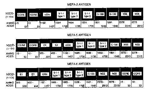
Fig. 1 is a schematic drawing showing the identification, amino acids, and the
arrangements of epitopes, in the MEFA-3, MEFA-5 and MEFA-6 antigens.
Fig. 2 is a schematic drawing showing the MEFA-5 antigen epitopes and their
location within the HCV genome. A diagram of pmefa-5, an expression vector for
MEFA-5, is also provided.
Fig. 3 is a schematic drawing showing the MEFA-6 antigen epitopes and their
location within the HCV genome. A diagram of pmefa-6, an expression vector for
MEFA-6, is also provided.
Fig. 4 is a schematic drawing of an enzyme-linked immunosorption assay
(ELISA) in which a MEFA is adsorbed onto the surface of a solid support.
Fig. 5 is a schematic diagram of an antibody capture format for detection of
anti-
HCV antibodies by chemiluminescence in which a MEFA is attached to a
detectable
marker molecule, DMAE. Also indicated is a format in which a MEFA (MEFA-6) and
an additional epitope (c33c) are the antigens of the assay.
Fig. 6 is a schematic diagram of an antibody capture format for detection by
chemiluminescence of human anti-pathogen antibodies in which an antigen (MEFA)
is
attached to biotin (B) that binds strepavidin labeled with DMAE.
Fig. 7 is a plot comparing the dilution sensitivity of MEFA-6-DMAE and MEFA-
6-DMAE + c33c-DMAE to the dilution sensitivity of a commercial ELISA, HCV 2.OG
(second generation) ELISA.
CA 02250723 1998-09-30
WO 97/44469 PCT/US97/08950
-7-
Fig. 8 is a plot comparing the seroconversion sensitivity of a commercial
ELISA
(Abbott Laboratories), MEFA-6, MEFA-6 + c33c, and RIBA 3Ø Samples were
taken
from a chronically infected patient over time (bleed dates).
Fig. 9 is a diagram correlating HCV antibody detection (positive or negative)
in
samples by HCV Second Generation ELISA to detection by an MEFA
chemiluminescence immunoassay (CLIA).
Fig. 10 is a chart illustrating the accuracy of the MEFA-6-DMAE CLIA of the
invention. All known negative samples exhibited relative light units (RLU)
below the
cutoff value, while known positive samples exhibited RLUs well above the
cutoff value.
Detailed Description of Embodiments
The practice of the present invention will employ, unless otherwise indicated,
conventional methods of virology, immunology, microbiology, molecular biology
and
recombinant DNA techniques within the skill of the art. Such techniques are
explained
fully in the literature. See, e.g., Sambrook, et al., Molecular Cloning: A
Laboratory
Manual (2nd Edition, 1989); DNA Cloning: A Practical Approach, vol. I & II (D.
Glover, ed.); Methods In Enzymology (S. Colowick and N. Kaplan eds., Academic
Press,
Inc.); and Handbook of Experimental Immunology, Vols. I-IV (D.M. Weir and C.C.
Blackwell eds., Blackwell Scientific Publications); Fundamental Virology, 2nd
Edition,
vol. I & II (B.N. Fields and D.M. Knipe, eds.).
Before the present multiple epitope fusion proteins, immunoassays and methods
for producing and using such are described, it is to be understood that this
invention is
not limited to the particular amino acid sequences, immunoassays or methods of
production as such may, of course, vary. It is also to be understood that the
terminology
used herein is for the purpose of describing particular embodiments only, and
is not
intended to be limiting since the scope of the present invention will be
limited only by
the appended claims.
Unless defined otherwise, all technical and scientific terms used herein have
the
same meaning as commonly understood by one of ordinary skill in the art in the
field in
which this invention belongs. Although any methods and materials similar or
equivalent
CA 02250723 1998-09-30
WO 97/44469 PCTIUS97/08950
-8-
to those described herein can be used in the practice or testing of the
present invention,
the preferred methods and materials are now described.
Definitions
As used herein, the term "multiple copy" specifies a sequence of amino acids
which contains at least about five and not more than about 1,000 amino acids
in a linear
fashion, repeated two or more times within a linear molecule. The repeating
sequence
need not be directly connected to itself, is not repeated in nature in the
same manner and,
further, may be present within a larger sequence which includes other amino
acids not
repeated or "copied." The sequence of at least five and not more than 1,000
amino acids
comprises an epitope as defined below. For the purposes of this invention, a
"copy" of
an amino acid sequence may be either an exact sequence copy or a sequence
which
corresponds to the same epitope of a different viral strain, i.e. copies are
either exact
copies or sequences which are "equivalent antigenic determinants" as defined
below.
The term "epitope" as used herein refers to a sequence of at least about five,
and
not more than about 1,000 amino acids connected in a linear fashion, which
amino acids,
by themselves or as part of a larger sequence, bind to an antibody generated
in response
to such sequence. An epitope for use in the subject invention is not limited
to a
polypeptide having the exact sequence of the portion of the parent protein
from which it
is derived. Indeed, viral genomes are in a state of constant flux and contain
several
variable domains which exhibit relatively high degrees of variability between
isolates.
Thus the term "epitope" encompasses sequences identical to the native
sequence, as well
as modifications to the native sequence, such as deletions, additions and
substitutions
(generally conservative in nature).
As used herein, the term "conformational epitope" refers to a recombinant
epitope
having structural features native to the amino acid sequence encoding the
epitope within
the full-length natural protein. Native structural features include, but are
not limited to,
glycosylation and three dimensional structure. Generally, a conformational
epitope is
added to the MEFA-containing immunoassay mixture to enhance assay sensitivity
and
selectivity. Preferably, a recombinant conformational epitope is expressed in
a cell from
which it is extractable under conditions which preserve its desired structural
features, e.g.
CA 02250723 1998-09-30
WO 97/44469 PCTIUS97/08950
-9-
without denaturation of the epitope. Such cells include bacteria, yeast,
insect, and
mammalian cells. Preferably, the cell in which a conformational epitope is
expressed is a
mammalian cell, such as a Chinese hamster ovary cell (CHO). Expression and
isolation
of recombinant conformational epitopes from the E1 and E2 regions of HCV are
described in WO 96/04301, WO 94/01778, WO 95/33053, WO 92/08734.
The term "expression cassette" as used herein refers to a DNA sequence which
contains a coding region operably linked to one or more suitable control
sequences
capable of effecting expression of the coding region in a compatible host.
Expression
systems invariably comprise a promoter, but, depending on the host intended,
may
contain additional critical nucleotide sequences such as a ribosome binding
site or CAP
site, termination sequence, and optional enhancer sequences upstream from the
promoter
or in other operable locations. The recombinant expression cassettes of the
invention
herein comprise a DNA of the invention encoding a MEFA operably linked to
additional
DNA sequences that are capable of effecting its expression. The expression
cassette may
reside on a transfer vector such as a plasmid or other vector that is self-
replicating
independently of the chromosome of the host cell, or may be constructed so
that when
inserted into a host cell it is able to integrate into the chromosome.
By "equivalent antigenic determinant" is meant an antigenic determinant from
different sub-species or strains of a given organism e.g., a different strain
of a virus such
as from strains 1, 2, or 3 of hepatitis C virus. More specifically for a virus
such as
hepatitis C, epitopes are known, such as 5-1-1, and such epitopes vary between
the
known strains 1, 2, and 3. Thus, the epitope 5-1-1 from the three different
strains are
equivalent antigenic determinants and thus are "copies" even though their
sequences are
not identical. In general the amino acid sequences of equivalent antigenic
determinants
will have a high degree of sequence homology, e.g., amino acid sequence
homology of
more than 30%, preferably more than 40%, when the two sequences are aligned.
The term "tracer" shall mean any detectable marker molecule attachable to an
epitope or a MEFA. Attachment is preferably by covalent means. Detectable
marker
molecules useful as tracers in the invention include, but are not limited to,
dimethyl
acridinium ester (DMAE), a chromophore, biotin, strepavidin, an antibody, an
antigen,
CA 02250723 1998-09-30
WO 97/44469 PCT/US97/08950
-10-
enzymes fluorogenic compounds, rhodamine compounds, fluorescein, FITC, and the
like.
Immunoassays-General
Highly sensitive and selective immunoassays can be produced using the multiple
epitope fusion antigens of the present invention. In order to produce such
immunoassays, it is first necessary to identify a target for which a sample is
to be
assayed, e.g., a particular virus in a body fluid sample. After identifying
the virus of
interest, the preferred immunodominant epitopes of the virus are isolated,
sequenced and
nucleotide sequences encoding the amino acid sequences of the epitopes are
determined
and produced. The nucleotide sequences encoding the amino acid sequences can
be
fused together using standard recombinant methodology. The sequences can also
be
fused to additional polypeptides to facilitate expression and purification
thereof.
The fused sequence must include at least two copies of nucleotide sequences
that
encode a given epitope. The nucleotide sequence is then placed within an
expression
cassette and a suitable host is transformed with the cassette. The host is
allowed to
express the sequences to provide the multiple copy epitopes (multiple epitope
fusion
antigen, MEFA). The multiple copy epitopes produced are then purified, for
example, by
affinity chromatography, which process is expedited to a certain degree due to
the
presence of the multiple copies of a given epitope. The purified MEFAs are
then coated
onto the surface of the substrate for ELISA-type assays. Alternatively, the
purified
MEFAs are attached to a detectable marker tracer molecule for detection of
antibody
binding, such as in a chemiluminescence assay (CLIA).
The essence of the invention is the purified multiple copy epitopes, i.e.,
purified
fusion proteins that include multiple copies of a given epitope fused, in a
linear fashion
in nature, to other epitopes that are not normally connected to each other in
this fashion
(MEFAs). The purified epitopes are encompassed by the general structural
formula (I) as
follows: (A),,-(B),,-(Q,
, which represents a linear amino acid sequence. B is an amino
acid sequence of an epitope or cluster of epitopes and each B contains at
least five and
not more than 1,000 amino acids, y is an integer of 2 or more, A and C are
each
independently an amino acid sequence of an epitope or cluster of epitopes not
CA 02250723 1998-09-30
WO 97/44469 PCTIUS97/08950
-11-
immediately adjacent to B in nature, and x and z are each independently an
integer of 0
or more wherein at least one of x and z is 1 or more. When each of x, y, or z
is greater
than 1, or when each of x, y, and z are greater than 1, the multiple copies of
A, B and C
may be identical, i.e., each copy of A (different from B and C) is the exact
same amino
acid sequence, each copy of B (different from A and C) is the exact same amino
acid
sequence, and each copy of C (different from A and B) is the exact same amino
acid
sequence. Alternatively, each A, B or C copy may be an equivalent antigenic
determinant from different strains of the same virus. Thus, for example, if y
is 3, each B
may be an identical amino acid sequence or three different sequences from
equivalent
antigenic determinants from HCV strain 1, 2, and 3. The invention may utilize
genetic
material encoding known epitopes or groups of epitopes by connecting the
material in a
nucleic acid construct that produces a multiple copy epitope of the formula
(I).
HCV antibody capture assays in which the individual single epitopes are coated
on a solid support are less sensitive than capture assays in which a chimeric
multiple
epitope polyprotein, such as (C25) containing epitopes from the immunodominant
core,
c33c (NS3), and c100 (NS4) region sequences (Chien, D.Y., et al (1992) Proc.
Natl.
Acad. Sci. USA 89:10011-10015), is coated on a solid support. In turn, a
capture assay
using the C25 chimeric polyprotein is less sensitive than an HCV antibody
capture assay
using a MEFA of the invention, which MEFA contains multiple copies of at least
one
epitope and at least one copy is from a different HCV strain. Thus, a
preferred MEFA of
the invention having the general formula Ax-By-Cz, contains more than one copy
of an
epitope (i.e., y is an integer of 2 or more), and at least one of the epitopes
of B is a
different equivalent antigenic determinant (e.g. an epitope from a different
pathogen
strain).
The invention disclosed herein utilizes recombinant DNA technology and protein
engineering to design a recombinant polyprotein which fuses a variety of
different
immunodominant epitopes from a variety of pathogens or pathogen strains as the
chimeric antigen for immunoassay development. Further, the invention utilizes
multiple
copies of selected epitopes from structural as well as non-structural coding
regions of a
gene combined and expressed as a recombinant polyprotein to significantly
improve the
sensitivity and selectivity of an immunoassay.
CA 02250723 1998-09-30
WO 97/44469 PCT/US97/08950
-12-
Epitopes used in making a multiple copy epitope of the invention can be from a
variety of different organisms. For example, the epitope may be an amino acid
sequence
from a bacteria, protozoa, virus, rickettsiae, parasite or fungus. A preferred
embodiment
of the invention uses epitopes that are derived from a bacteria or virus, with
particularly
preferred epitopes being those derived from a virus, such as from human
immunodeficiency virus (HIV) and, most preferably, from hepatitis C virus
(HCV). For
example, HIV epitopes may be derived from any of the various viral regions
which
display immunoreactivity such as, but not limited to, any of the various
envelope
proteins such as gp120, gp160 and gp4l, gag antigens such as p24gag and
p55gag, as
well as proteins derived from the pol region. Similarly, HCV epitopes can be
derived
from any of the various viral regions, such as, but not limited to, the C, E
1, E2/NS 1,
NS2, NS3, NS4, and NS5 regions.
Figure 1 shows representative MEFA antigens for use in the present invention
which are derived from HCV. However, it is to be understood that other
epitopes
derived from the HCV genome will also find use with the present assays. For
example,
additional epitopes, derived from, e.g., the hypervariable region of E2, such
as a region
spanning amino acids 384-410 or 390-410, can be included in the MEFA antigen.
A
particularly effective E2 epitope is one which includes a consensus sequence
derived
from this region, such as the consensus sequence Gly-Ser-Ala-Ala-Arg-Thr-Thr-
Ser-Gly-
Phe-Val-Ser-Leu-Phe-Ala-Pro-Gly-Ala-Lys-Gln-Asn, which represents a consensus
sequence for amino acids 390-410 of the HCV type 1 genome. A representative E2
epitope present in a MEFA antigen of the invention can comprise a hybrid
epitope
spanning amino acids 390-444. Such a hybrid E2 epitope can include a consensus
sequence representing amino acids 390-410 fused to the native amino acid
sequence for
amino acids 411-444 of HCV E2.
It is well known that any given organism varies from one individual organism
to
another and further that a given organism such as a virus can have a number of
different
strains. For example, numerous HIV isolates exist and hepatitis C virus
includes at least
strains 1, 2, and 3. Each of these strains will include equivalent antigenic
determinants.
More specifically, each strain will include a number of antigenic determinants
that will
be present on all strains of the virus but will be slightly different from one
viral strain to
CA 02250723 1998-09-30
WO 97/44469 PCTIUS97/08950
-13-
another. For example, hepatitis C includes the antigenic determinant known as
5-1-1 (in
the NS3 region of the viral genome). This particular antigenic determinant
appears in
three different forms on the three different viral strains of hepatitis C.
Accordingly, in a
preferred embodiment of the invention all three forms of 5-1-1 appear on the
multiple
epitope fusion antigen of the invention. A MEFA of the invention has the above
structural formula I, wherein y is 3 and thus each of the three "Bs" are
equivalent
antigenic determinants of 5-1-1 taken from the three different viral strains
of hepatitis C.
The multiple copy epitope of the present invention can also include multiple
copies which are exact copies of the same epitope. For example, it is
desirable to include
two copies of an epitope from the core region of hepatitis C. A particularly
preferred
embodiment of the present invention is the multiple copy epitope as shown
within Fig. 3.
This multiple copy epitope includes two exact copies of an epitope from the
core region
and three copies of an epitope from the 5-1-1 region, which copies are
equivalent
antigenic determinants meaning that they are antigenic determinants taken from
the three
different viral strains of hepatitis C. In general, equivalent antigenic
determinants have a
high degree of homology in terms of amino acid sequence which degree of
homology is
generally 30% or more or more preferably 40% or more, when aligned.
CA 02250723 1998-09-30
WO 97/44469 PCT/US97/08950
-14-
HCV Immunoassays
Highly selective and sensitive immunoassays generally contain major
immunodominant epitopes of the pathogen suspected of infecting a patient.
Previously,
immunoassays made use of individual epitopes to bind anti-HCV antibodies in
biological
samples.
For the virus HCV, major immunodominant linear epitopes were identified from
the core, NS3 (nonstructural), NS4, and NS5 regions of the virus polyprotein.
Sallberg et
al. assayed HCV core protein and putative matrix proteins against human serum
samples
containing antibodies to HCV and defined several immunodominant regions within
the
HCV proteins (Sallberg, M. et al. (1992) J. Clin. Microbiol. 30:1989-1994).
Protein
domains of HCV-1 polyproteins including domains C, E1, E2/NS1, NS2, NS3, NS4,
and
NS5 were identified and their approximate boundaries provided by Chien and
Rutter
(Chien, D.Y. and Rutter, W., WO 93/00365, international publication date
January 7,
1993). Kotwal et al. designed individual polypeptides having sequences derived
from
the structural region of HCV in order to obtain an immunodominant epitope
useful in
testing sera of HCV patients (Kotwal, G.J., et al. (1992) Proc. Natl. Acad.
Sci. 89:4486-
4489).
Serologically definable subtypes of HCV were identified by Chien et al. as
viral
subtypes exhibiting varied antigenicity (presented at the Third International
Hepatitis
Meeting, Tokyo, May, 1993 and in Chien, D.Y. et al. (1994) Viral Hepatitis and
Liver
Disease, pp. 320-324). HCV-1 core, NS4, and NS5 regions were found to contain
serotype-specific epitopes. Individual putative core proteins from HCV-1 and
HCV-2
were used as individual antigens to produce antibodies for enzyme-linked
immunosorbent assays to detect HCV infection using serologically
distinguishable core
antigen subtypes (Machida, A. et al. (1992) Hepatology 16:886-891). Simmonds
et al.
investigated the effect of sequence variability between different types of HCV
upon the
antigenicity of the NS4 protein by epitope mapping and by enzyme-linked
immunosorbent assay (ELISA). These authors mapped two major antigenic regions
in
the HCV NS4 polyprotein that were recognized by antibody elicited upon natural
infection by HCV. Type-specific antibody to particular HCV types was also
detected
CA 02250723 1998-09-30
WO 97/44469 PCT/US97/08950 -
-15-
(Simmonds, P. et al. (1993) J. Clin. Microbiol. 31:1493-1503). Ching et al.
prepared a
series of synthetic peptides based on the sequence of a highly conserved
region of the
HCV putative nucleocapsid (core) protein and found an immunodominant region
that
was recognized by human and chimpanzee sera (Ching, W.-M. et al. (1992) Proc.
Natl.
Acad. Sci. 89:3190-3194).
Assays involving single epitopes as test antigens have the disadvantage that
it is
difficult to control solid phase coating of the support surface by large
numbers of
individual epitopes containing short peptides. In such cases, where the assay
involves
deposition of an immunogenic antigen on a solid support, the sensitivity of
the assay is
limited by the amount of antigen that can be coated on the surface of the
solid support.
An example of an immunoassay that includes immunodominant epitopes from
different regions of a single virus subtype is disclosed within Chien et al.
(Prot. Natl.
Acad. Sci. USA 89:10011-10015 (1992)). The assay described by Chien utilizes
recombinant HCV polypeptides derived from many different regions of the HCV
type 1
polyprotein, including that of chimeric recombinant polyprotein, C25,
comprises
immunodominant components evident in both the structural and non-structural
regions.
The polyproteins produced are recombinantly derived viral polypeptides and are
included
on the surface of an immunoassay in order to capture antibodies, i.e., detect
the presence
of antibodies generated in response to infection with HCV. However, these
polyproteins
contain epitopes from a single viral strain thereby limiting the ability to
detect anti-HCV
antibodies from different strains of the virus.
EXAMPLES
The following examples are put forth so as to provide those of ordinary skill
in
the art with a complete disclosure and description of how to make the multiple
copy
epitopes and reagents for use in immunoassays of the invention, as well as use
of such,
and are not intended to limit the scope of what the inventors regard as their
invention.
Efforts have been made to ensure accuracy with respect to numbers used (e.g.
amounts,
temperature, etc.) but some experimental error and deviation may be inherent
in the
description. Unless indicated otherwise, parts are parts by weight, molecular
weight is
CA 02250723 1998-09-30
WO 97/44469 PCTIUS97/08950
-16-
weight average molecular weight, temperature is in degrees Centigrade and
pressure is at
or near atmospheric.
Example 1: Construction and Expression of an HCV epitope Polyprotein
Expression
Cassette
The following example illustrates the concept of preparing a polyprotein
cassette
of major epitopes, particularly a cassette of multiple epitopes. The example
further
illustrates the success of using epitopes from different strains of a
pathogen. It is also
shown that a hydrophilic multiple epitope antigen increases the solubility of
the
polyprotein. The epitopes are shown to maintain their native local
conformation for
binding to antibodies as evidenced by the antigenicity of the polyprotein.
The polyprotein expressed from the multiple epitope cassette is referred to
herein
as a Multiple Epitope Fusion Antigen (MEFA).
Preferably, where an epitope is repeated, the extra copy or copies are
tandemly
arrayed in the same orientation. It is understood that the region of a viral
coding
sequence used as an epitope may be varied slightly and still retain antigenic
activity, and
that the amino acid numbering designation may vary from strain to strain.
Thus, the
repeated epitopes may vary one from another in amino acid sequence due to
strain
sequence variations and/or numbering designation. Preferably, the amino acid
sequences
of repeated epitopes within a MEFA are at least 30% homologous at the amino
acid
level, more preferably at least 40% homologous at the amino acid level.
Unique restriction enzyme sites were introduced in order to connect the
epitopes
in the prescribed order and enhance the usefulness of the invention by
facilitating
modifications in design of a chimeric antigen. The choice of restriction
enzyme sites and
cloning procedures are readily determined by one of ordinary skill in the art
of
recombinant DNA technology. Preferably, the epitope junctions (amino acid
sequences
created between epitopes due to cloning) do not generate non-specific
epitopes. Non-
specific epitopes are, for example, non-HCV sequences which do not exist
adjacent to
the HCV epitopes in nature. Non-specific epitopes may bind antibodies in a
test sample
causing false positive assay results. Preferably, the multiple epitope fusion
protein is
tested for false positive results due to such sequences generated at the
epitope junctions.
CA 02250723 1998-09-30
WO 97/44469 PCTIUS97/08950 -
-17-
To avoid non-specific interactions with the MEFA due to junction sequences,
the DNA
sequence encoding the junction may, for example, be mutated such that non-
specific
interactions with the mutant amino acid sequence are reduced, and cloning of
the epitope
fragments is possible.
Construction of a MEFA expression cassette of HCV epitopes
The HCV MEFA-3 expression cassette was constructed by cloning the coding
nucleotide sequences containing major epitopes in a tandem array as shown in
Fig. 1. A
major epitope was chosen based on antibody reaction frequency and reaction
intensity
(titer) to the epitope (Chein, D.Y. et at. (1994) Viral Hepatitis and Liver
Disease, pp.
320-324). The various DNA segments coding for the HCV epitopes were
constructed by
PCR amplification or by synthetic oligonucleotides. The amino acid codons
encoded in
each segment are shown below each segment. The complete HCV-1 amino acid
sequence (3011 amino acids) was determined by Choo, et al. (1991) Proc. Natl.
Acad.
Sci. USA 88:2451-2455. Oligonucleotides capable of binding to HCV are
described in
U.S. Patent No. 5,350,671. The numbering of the amino acids in epitopes of the
invention follows the numbering designation provided in Choo, et al., supra,
in which
amino acid #1 is the first methionine encoded by the coding sequence of the
core region.
For example, an epitope segment from the core region is encoded by amino acid
codons
10 to 53 of the HCV core protein. An epitope from the c33c region is encoded
by amino
acid codons 1192 to 1457.
The MEFA-3 construct contains in the expression cassette two copies of the
core
segment epitope amino acids 10-35; one copy of the c33c epitope segment from
amino
acids 1192-1457; three copies of equivalent antigenic determinants from the
HCV NS4
region, specifically the 5-1-1 region, where two of the epitopes are a segment
(amino
acids 1694 to 1735) from the NS4 5-1-1 region of HCV type 1, while one copy is
a
segment from the NS4 5-1-1 region of HCV type-2 (Nomoto) from amino acids 1694
to
1735; two copies of the NS4 (C100) C-terminal region major epitopes from amino
acids
1901-1940; and two copies of major epitopes from amino acids 2278-23 10 of the
NS5
region. The MEFA-3 expression cassette has the general structural formula 2-1-
2-1-2-2
CA 02250723 1998-09-30
WO 97/44469 PCTIUS97/08950
-18-
for Ax-By-Cz, where A = core-core-c33c, x = 1; B = (5-1-1), y = 3; and C =
(cl00)-
(c100)-(NS5)-(NS5), z = 1.
Other HCV MEFAs include MEFA-5 and MEFA-6 for which expression
cassettes were constructed by cloning the coding nucleotide sequences
containing major
epitopes in a tandem array as shown in Fig. 2 and Table 1, and Fig. 3 and
Table 2,
respectively. It is noted that all of the epitopes in MEFA-5 and MEFA-6 are
from HCV
type 1 except for two of the three equivalent antigenic determinants of the 5-
1 -1 epitope.
Epitopes from the 5-1-1 region have been found to vary between serotypes of
HCV. A
copy of each of the HCV type-specific 5-1-1 epitopes present in the MEFAs
described
herein allows binding of any of the HCV types that may be present in the test
biological
sample. It is a feature of the invention that an epitope useful in
distinguishing serotypes
of a virus such as HCV is provided as repeated equivalent antigenic
determinants in order
to detect multiple types of a virus or pathogen in a single assay. Methods of
determining
HCV serotype are found in WO 96/27153.
CA 02250723 1998-09-30
WO 97/44469 PCT/US97/08950
-19-
Table 1
MEFA-5 Antigen Epitopes and Their Location
Within the HCV Genome
mefa aa# 5' End Site Epitope HCV aa# Strain
1-154 Ncol hSOD
159-202 EcoRI core 10-53 1
205-246 Sacl core 10-53 1
251-268 Pstl El 303-320 1
271-309 Hindlll E2 405-444 1
310-576 Dralll c33c 1192-1457 1
579-625 Sphl 5-1-1 1689-1735 1
628-674 Nrul 5-1-1 1689-1735 3
677-723 C/al 5-1-1 1689-1735 2
726-765 Aval c100 1901-1940 1
768-803 Xbal NS5 2278-2313 1
806-841 Bg/II NS5 2278-2313 1
CA 02250723 1998-09-30
WO 97/44469 PCTIUS97/08950
-20-
Table 2
MEFA-6 Antigen Eyitopes and Their Location
Within the HCV Genome
mefa aa# 5' End Site Epitope HCV aa# Strain
1-154 Ncol hSOD
159-176 EcoRl El 303-320 1
179-217 Hindlll E2 405-444 1
218-484 Dralll c33c 1192-1457 1
487-533 Sphl 5-1-1 1689-1735 1
536-582 Nrul 5-1-1 1689-1735 3
585-631 Clal 5-1-1 1689-1735 2
634-673 Aval c loo 1901-1940 1
676-711 Xbal NS5 2278-2313 1
714-749 B /II NS5 2278-2313 1
750-793 Ncol core 10-53 1
796-839 Sacl core 10-53 1
CA 02250723 1998-09-30
WO 97/44469 PCT/US97/08950 -
-21 -
The cloning procedures for preparation of MEFA-5 and MEFA-6 were similar to
those used for preparing MEFA-3, above. MEFA-5 and MEFA-6 contained epitopes
from the core, envelope, NS3, NS4 and NS5 regions of the hepatitis C
polyprotein,
including equivalent antigenic determinants from HCV strains 1, 2, and 3. The
various
DNA segments coding for the HCV epitopes were constructed by PCR amplification
or
by synthetic oligonucleotides. Figs. 2 and 3 are diagrammatic representations
showing
the location of epitopes within the HCV genome used in MEFA-5 and -6, as well
as the
MEFA vector construction. Tables 1 and 2 describe the amino acid segments of
each
epitope, the linear arrangement of the various epitopes and the number of
copies in the
MEFA-5 and MEFA-6 cassettes, respectively. The amino acids between each
epitope
(junction amino acids) are derived from the restriction sites used for
cloning. Preferably,
a MEFA is tested in an immunoassay for a false positive result due to the non-
pathogen
(HCV, for example) junction amino acids. MEFA-5 differs from MEFA-6 in linear
arrangement in that the core segments are near the N-terminus in MEFA-5, but
are at the
C-terminus in MEFA-6. As the amino acid 10-53 core epitope is highly
antigenic, its
placement at the C-terminus improves the antigenicity of the MEFA, possibly by
improved interaction of epitopes from core proteins and antigens from El and
E2 regions
with the anti-HCV antibodies of the sample.
The epitopes were subeloned into a yeast expression vector and the sequences
verified before assembling the entire fusion antigen as an EcoRI-Sall fragment
of 2060
bp (for MEFA-5 and MEFA-6) and as an EcoRI-Sall fragment of approximately 1927
bp
(for MEFA-3). MEFA-5 and MEFA-6 were cloned as human superoxide dismutase
(hSOD) fusion proteins (157 amino acids of hSOD) under the regulation of the
hybrid
ADH2-GAPDH promoter. The MEFA-5 and MEFA-6 expression cassettes were ligated
to the yeast shuttle vector pAB24 (Chiron Corporation) to produce pMEFA-5 and
pMEFA-6, as shown in Fig. 2 and Fig. 3, respectively.
As shown in Fig. 3, the MEFA-6 antigen includes multiple copies of HCV
epitopes from the core and NS5 region; different serotype epitopes from the
NS4 5-1-1
region; a single copy of major linear epitopes from the c100 C-terminal
regions, El, and
E2 regions, as well as the HCV NS3 (c33c) region. The general structural
formula for
MEFA-6 is hSOD--E1-E2-c33c-5-1-1(type 1)-5-1-I(type 3)-5-1-1(type 2)-cl00-
NS5(2
CA 02250723 1998-09-30
WO 97/44469 PCTIUS97/08950
-22-
copies)-core(2 copies). This antigen has a very high expression level in
yeast, purifies to
a high degree of homogeneity, and exhibits high sensitivity and high
selectivity in the
immunoassays described below.
Expression of a MEFA in Yeast
The following example of the expression of a MEFA in yeast may be applied to
the expression of any MEFA of the invention. It is within the scope of the
instant
invention to vary the conditions, as necessary, to express a particular MEFA
construct.
Cells preferred for expression of a MEFA of the invention include bacteria,
yeast, and
insect cells. The expression of MEFA-6 in yeast is provided as a non-limiting
example
of the instant invention.
Yeast strains AB 122, JSC3 10, and AD2 (Chiron Corporation) were transformed
with the appropriate MEFA expression plasmid (such as pMEFA-6) using a lithium
acetate protocol. Ura transformants were streaked for single colonies and
patched onto
Leu /8% glucose plates to increase plasmid copy number. Leu starter cultures
were
grown for 24 hours at 30 C and then diluted 1:20 in YEPD (yeast extract
bactopeptone
glucose) media. The cells were grown for 48 hours at 30 C and harvested. To
test for
expression of the MEFA-6 recombinant antigen, an aliquot of the cells (0.5 OD
unit
equivalent) was boiled in SDS (sodium dodecylsulfate) gel electrophoresis
sample buffer
(e.g. Lammli buffer) containing 50 mM DTT and the protein components of the
cell
mixture were separated by gel electrophoresis on an Tris-glycine
polyacrylamide gel.
MEFA-6 was highly enriched in the insoluble pellet fraction.
Purification of a MEFA Protein in Yeast
The following procedure describes the purification of a specific MEFA, MEFA-6.
The techniques and conditions are not intended to limit the invention, as one
of ordinary
skill in the art may find it necessary to adjust conditions for the
purification of another
MEFA of the invention. Unless otherwise indicated, purification of a MEFA is
conducted at approximately 0 C.
MEFA-6 was expressed in S. cerevisiae and cells were harvested as described
above. The cells were suspended in lysis buffer (50 mM Tris, 0.15 M NaCl, 1 mM
CA 02250723 2005-01-27
-23-
EDTA, 1 mM PMSF, pH 8.0) and lysed in a Dyno-Mill (Wab Willy A. Bachofon,
Basel,
Switzerland) or equivalent apparatus using glass beads. The lysate was
centrifuged at
low speed conditions (3,000 to 5,000 rpm, 15 min) and the pellet containing
the insoluble
protein fraction was washed with increasing concentrations of urea (1 M, 2 M,
3 M) in
lysis buffer. Protein was solubilized from the centrifugation pellet with 0.1
N NaOH, 4
M urea in lysis buffer. Cell debris was removed by low speed centrifugation at
3,000 to
5,000 rpm, 15 min. The supernatant was adjusted to pH 8.0 with 6 N HCl to
precipitate
proteins insoluble under these conditions.
The precipitate was removed by centrifugation and the supernatant was adjusted
to 2.3% SDS, 50 mM DTT, pH 8.0 and boiled' for 3 min. Proteins in the mixture
were
TM
fractionated by gel filtration on a Pharmacia Sephacryl S-400 in phosphate
buffered
saline containing 0.1% SDS, 1 mM EDTA and adjusted to pH 7.4. Column eluate
fractions containing MEFA-6 were collected, pooled, and concentrated on an
Amicon
YM-30 membrane. Gel filtration was repeated on the pooled fractions using the
same
column and conditions.
Evaluation of the Antigenicity of a MEFA
In order to evaluate the antigenicity of a chimeric antigen of the invention,
the
epitopes of MEFA-3 were exposed to polyclonal or monoclonal antibodies raised
to
specific individual epitopes. Purified recombinant multiple epitope fusion
antigens
(MEFA) were diluted to optimal coating concentration in phosphate-buffered
saline (pH
7.4) and coated on Immulon I plates (Dynatech). Monoclonal antibodies to core,
NS3
(c33c), NS4 (c100 and 5-1-1), NS5 and polyclonal antisera anti-El and E2 from
rabbits
were prepared by standard techniques (BIOS-Chile, Maraton 1943, Santiago,
Chile) and
were diluted 200-fold in sample diluent on the plate and incubated for 1 hr at
37 C, and
TM
washed with plate wash buffer (PBS, 0.075% Tween-20, pH 7.2). Either goat anti-
mouse F(ab')2 or affinity purified goat anti-rabbit IgG heavy and light chain
specific
antibody conjugated to horseradish peroxidase (diluted 1:5000 for anti-mouse
conjugate;
diluted 1:10,000 for anti-rabbit conjugate) were added to each assay well. The
plates
were incubated for 1 hr at 37 C and washed. o-Phenylenediamine dihydrochloride
(OPD) and hydrogen peroxide were added for horse radish peroxidase (HRPO)
reaction
CA 02250723 1998-09-30
WO 97/44469 PCT/US97/08950
-24-
color development. The optical density readings were determined using a plate
reader at
492/620 nm.
The results indicated that all of the antigen epitopes within the designed
MEFA
were easily detected by the specific HCV antibodies for all of the MEFAs of
the
invention. For example, Table 3 provides data on the immunoreactivity of the
individual
epitopes, as well as the chimeric antigen, MEFA-3, to monoclonal antibodies of
HCV-
specific epitopes. As shown in Table 3, core, c33c, c100, 5-1-1, and NS5
epitopes of
MEFA-3 were immunoreactive with HCV-specific antibodies. Table 4 shows that
the
epitopes c33c, c22, 5-1-1, clOO, NS5, El, and E2 of MEFA-5 and MEFA-6 were
immunoreactive with HCV-specific antibodies.
CA 02250723 1998-09-30
WO 97/44469 PCT/US97/08950
-25-
Table 3
HCV Specific Epitopes of MEFA-3 Antigen:
Evaluation by Anti-HCV Monoclonal Antibodies
HCV Mab ID# 3G1-1 4D1-1 22AFG3 20AGF3 5A1/F5 Comment
Results
Mab Specificity anti-core anti-c33c anti-5-1-1 anti-c100 anti-ns-5
Recombinant OD OD OD OD OD
Test antigens
SOD (non- 0.001 0.001 0.002 0.002 0.003 No reaction with
recombinant) SOD
C25 2.755(+) 2.813(+) 2.726(+) 0.028(-) 0.023(-) React with
epitopes of core,
c33c & 5-1-1
c22 (core) 2.700(+) 0.043(-) 0.035(-) 0.036(-) 0.038(-) React with
e ito a of core
c33c (NS3) 0.029(-) 2.646(+) 0.018(-) 0.020)-) 0.014(-) React with
epitope of c33c
c100 (NS4) 0.020(-) 0.022(-) 2.907(+) 3.021(+) 0.016(-) React with
epitopes of 5-1-1
and C-terminal
epitope of 000
NS5 0.012(-) 0.029(-) 0.009(-) 0.009(-) 2.513(+) React with
epitope of NS5
Test Antigen 3.2361 +) 3.236(+) 3.467(+) 0.713(+) 0.024(-) React with
MEFA-3 epitopes of core
c33c, 5-1-1 and
c100
CA 02250723 1998-09-30
WO 97/44469 PCT/US97/08950
-26-
Table 4
HCV Epitope Exposure Within MEFA-5 and MEFA-6
Antibody ID Antibody Antigenic to HCV MEFA-6 MEFA-5
Specifity sequence region epitope exposure epitope exposure
OD OD
Mab 3G1-1 anti-core (c22c) (aa# 10-50) 3.018 (R) 2.702 (R)
Mab 4D1-1 anti-NS3 (c33c) linear epitope of c33c 3.119 (R) 2.952 (R)
Mab anti-NS4 (c100) (aa# 1901-1940) 3.853 (R) 2.998 (R)
6C1 0/D 1
Mab anti-NS4 (5-1-1) (aa# 1689-1735) 3.006 (R) 3.192 (R)
22A5/C 12
Mab anti-NS5 (aa# 2297-2313) 2.808 (R) 2.863 (R)
3E1/F1
Mab anti-NS5 (aa# 2297-2313) 2.892 (R) 2.784 (R)
1E5/F10
polyclonal anti-El (aa# 192-380) 4.375 (R) 1.908 (R)
R667
polyclonal anti-E2 (aa# 404-662) 1.76 (R) 0.963 (R)
R669
Cutoff 0.45 OD 0.45 OD
value
R = Reaction
NR = No Reaction
CA 02250723 1998-09-30
WO 97/44469 PCT/US97/08950 -
-27-
Inhibition Assays: Peptide inhibition assays were performed to test whether
serotype specific epitopes on a MEFA antigen detect HCV type-specific
antibodies in
serum. The assay evaluated the degree to which a MEFA in solution would bind
to
serum HCV type-specific antibodies, thereby inhibiting the subsequent ELISA
reaction
in which the serotype-specific peptides are the antigenic species on a solid
support. Fig.
4 is a schematic drawing of a standard ELISA procedure in which binding to the
solid
support-bound antigen is detected by enzyme catalyzed hydrolysis.
Inhibition assays were performed by multi-antigen ELISA. Recombinant HCV
antigens were prepared as described in Chien et al. (1992) Proc. Natl. Acad.
Sci.
89:10011-10015. The c22 (119 amino acids), El (130 aa), NS5 (942 aa), and
chimeric
C25 (858 aa) antigens were expressed as internal antigens within the yeast S.
cerevisiae
as C-terminal fusions with human superoxide dismutase (SOD) using methods
described
previously for the generation of the c100-3 (363 aa) antigen (Kuo, G. et al.
(1989)
Science 244:362-364; and Cousens, L.S. et al. (1987) Gene 61:265-275). The
c33c
antigen (363 amino acids) was expressed as an internal SOD fusion polypeptide
in E. coli
by methods described for the synthesis of the 5-1-1 antigen (Choo, O.-L. et
al. (1989)
Science 244:359-362). The recombinant HCV antigens were purified as described
in
Chien, D.Y. et al. ((1989) Proc. Natl. Acad. Sci. 89:10011-10015, supra))
Prior to performing the inhibition assays, the patient sample dilution
breaking
points were determined (Table 5). Patient samples were serially diluted and
tested for
reaction to recombinant c22, c33c, 000 and NS-5 antigens immobilized
separately onto
a solid support (see, for example, Van der Poel, C.L. et al. (1991) Lancet
337:317-319).
The dilution breaking point was the greatest dilution at which binding was
still
detectable. For optimal detection in subsequent inhibition assays, the patient
samples
were less dilute than the dilution breaking point dilution, as indicated in
Table 6.
CA 02250723 1998-09-30
WO 97/44469 PCT/US97/08950 -
-28-
Table 5
Detection Limit Determination for Patient Samples
MEFA-3 Antigen Epitopes
HCV Patient Sample ID Sample Dilution Breaking Points
Recombinant Antigens
c22 c33c c100 NS5
PAA LL57366
1:8 1:128 neat neat
PAA LL57454 1:32 1:128 1:8 neat
PAA FF25946 1:32 1:256 1:32 NR
PAA FF25912 ND ND ND neat
NR = no reaction
ND = not done
In general, the inhibition assays were performed by the following procedure.
Recombinant HCV antigens and denatured SOD (control) were diluted to optimal
concentration in phosphate-buffered saline (pH 7.4) and coated on Immulon I
plates
(Dynatech). A 200 41 aliquot of either 30% fetal calf serum (FCS) or MEFA-3
peptide
(5 or 10 g per assay as indicated) dissolved in 30% FCS was mixed on the plate
with 5 l
of diluted serum or plasma specimen. The samples were incubated for 1 hr at 37
C and
washed with plate wash buffer. Polyclonal goat anti-human IgG (heavy- and
light-chain-
specific) antibody conjugated to either 1211 or horseradish peroxidase (HRP)
was added to
each well. The plates were incubated for 1 hr at 37 C and then washed. o-
Phenylenediamine dihydrochloride and hydrogen peroxide were added for HRP
color
development. The results were read using a plate reader at 492nm/620 nm
(ELISA).
The ELISA cutoff OD values for antigens from regions SOD, C25, c22, El, E2,
c33c,
and NS-5 were 0.40 plus the mean OD of three negative control sera included in
each
assay. If the control SOD antigen was reactive, then that sample was
considered to be
CA 02250723 1998-09-30
WO 97/44469 PCT/US97/08950 -
-29-
nonreactive or indeterminate. The percentage of binding inhibition was
calculated by the
following formula: 100 x (A492nm for patient sample without added MEFA
antigen) -
(A492nm for patient sample with added MEFA antigen)/(A492nm for patient sample
without added MEFA antigen). The % inhibition of binding to type specific
peptides
caused by added MEFA-3 indicates that the ability of the epitopes within MEFA-
3 to
bind the anti-HCV antibodies of the patient samples (See Table 6).
Table 6
Binding Inhibition by Specific Epitopes of MEFA-3
Patient Sample Control MEFA-3 % Inhibition
Added
ID Dilution OD OD
c22 Antigen
LL57366 1:4 1.614 0.163 90%
LL57454 1:16 1.370 0.212 84.5%
FF25946 1:16 2.013 0.205 90%
c33c Antigen
LL57366 1:64 2.525 0.07 99%
LL57454 1:64 1.839 0.075 96%
FF25946 1:128 0.842 0.061 93%
C100
Antigen
LL57454 1:4 1.666 0.484 71%
FF25946 1:16 2.364 0.092 96%
NS-5 Antigen
LL57454 Neat 2.319 1.820 20%
FF25912 Neat 1.490 0.873 41%
CA 02250723 1998-09-30
WO 97/44469 PCT/US97/08950 -
-30-
The ability of MEFA-3 to interact with anti-HCV type I and anti-HCV type 2
antibodies was demonstrated by inhibition studies using a MEFA ELISA protocol.
Individual synthetic peptides from HCV type 1 a, I b, 2a, and 2b 5-1-1 regions
were
immobilized on separate solid supports. The ability of the synthetic peptides
from the 5-
1-1 region to bind the type specific patient antibodies was determined by
competition
with added MEFA-3. The results in Table 7 show that MEFA-3 inhibits binding of
HCV
1 a, 1 b, 2a, and 2b to the individual type specific epitopes (amino acids
1689-1718 from
the 5-1-1 region). The ability of a MEFA to bind antibodies to two different
strains of
HCV was the same for MEFA-3, MEFA-5, and MEFA-6.
CA 02250723 1998-09-30
WO 97/44469 PCT/US97/08950 -
-31 -
Table 7
HCV Type Specificity: MEFA-3 5-1-1 Epitopes Interact with
Antibodies to HCV Types 1 and 2
Control Inhibition, %
HCV Type Specific Peptides HCV Type Specific Pe tide + MEFA-3
HCV1a HCV1b HCV2a HCV2b HCV1a HCV1b HCV2a HCV2b
(1689- (1689- (1689- (1689-
1718) 1718) 1718) 1718) ELISA ELISA ELISA ELISA
epitope epitope epitope epitope OD OD OD OD
Sample specific specifi specific specific
ELISA c ELISA ELISA
OD ELISA OD OD
OD
(A) HCV-Type 1
Sample
#4(1:10 1.093 0.073 0.002 0.004 0.165 0.136 0.044 0.014
d) 85% 0% 0% 0%
Inhibitio
n
(B) HCV-type 1 b sample
#358 0.964 1.543 0.424 0.235 0.438 0.261 0.284 0.234
% 55% 83% 33% 0%
Inhibitio
n
(C) HCV-type 2 sample
#32(1:1 0.001 0.001 0.839 0.460 0.007 0.018 0.034 0.055
Od) 0% 0% 96% 88%
Inhibitio
n
CA 02250723 1998-09-30
WO 97/44469 PCTIUS97/08950
-32-
Example 2: Sensitivity of ELISA Using a MEFA as the Antigen
A comparison of dilution sensitivity was made between MEFA ELISA (MEFA-
3) and C25 ELISA. HCV polyprotein C-25 (c33c-clOO-3-c22) and assay procedures
were as described by Chien, D.Y. et al. (1992) Proc. Natl. Acad. Sci. USA
89:10011-
10015, supra) using a coating buffer of 1 x phosphate buffered saline (PBS),
pH 7.0-7.2.
Antigens were coated onto the surface of Immulon I plate microliter wells at
100 ng
antigen per well plus 5 g/ml BSA. Sample size was 5 l per assay. The goat
anti-
human IgG (heavy- and light-chain-specific) antibody conjugated to horse
radish
peroxidase was diluted 1:60,000 for the MEFA-3 assay, and 1:40,000 for the C25
assay.
The results in Table 8 show that serum antibodies are detectable using MEFA-3
ELISA
at dilutions at which the C25 ELISA showed no reaction. The sensitivity of
MEFA-3, -5,
and -6 CLIA were compared to each other and to C25 ELISA. The results in Table
9
show that MEFA-5 and MEFA-6 CLIA provided superior sensitivity to MEFA-3 CLIA,
while MEFA-3 CLIA was more sensitive than C25 ELISA.
CA 02250723 1998-09-30
WO 97/44469 PCTIUS97/08950
-33-
Table 8
Dilution Sensitivity:
Comparison Study between MEFA ELISA and C-25 ELISA
MEFA-3 ELISA C-25 ELISA
Immulon I plate Immulon I plate
100 ng/well + 5ug/ml 100 ng/well + 5ug/ml
BSA BSA
Conjugate: 1:60000 Conjugate: 1:40000
Sample Panel ID Sample size: 5 ul/assay Sample size: 5 ul/assay
OD OD
Sample Dilution
LL57454
1:512 0.983 0.734
1:1024 0.652 NR
1:2048 0.463 NR
LL57366
1:512 0.609 0.425(+/-)
1:1024 0.522(+/-) NR
1:2048 0.203 NR
FF25946
1:100 1.818 1.736
1:1000 0.763 0.525
1:2000 0.718 NR
1:4000 0.455 NR
Seroconversion Panel C Bleed Date
C7 (8/29/88)day 1 0.562 NR
C8 (9/01/88)day 4 1.035 0.667
C9 (9/28/88)day 32 2.762 2.145
Men of negative
sample OD 0.124 0.086
Cutoff OD 0.55 0.45
(+/-) = OD near cutoff value
NR = Non-reactive
C-25 ELISA is equivalent to 2G (Second Generation) HCV ELISA
CA 02250723 1998-09-30
WO 97/44469 PCTIUS97/08950
-34-
Table 9
Dilution Sensitivity of MEFA-3 vs. -5 vs. -6 vs. c25
SENSITIVITY PANEL
Patient MEFA-3 CLIA MEFA-5 CLIA MEFA-6 CLIA c25 ELISA
Sample
S/C.O. S/C.O. S/C.O. S/C.O.
FF25946 1:16 1.71 2.72 2.67 1.32
1:32 1.64 2.59 2.48 1.35
1:64 1.50 1.89 2.11 1.20
1:128 1.34 1.92 1.68 0.92
1:256 1.11 1.48 1.68 0.91
1:512 0.84 1.14 1.28 0.69
1:1024 0.58 0.82 1.11 0.63
LL57385 1:16 1.73 2.74 2.68 1.49
1:32 1.56 2.41 2.18 1.04
1:64 1.20 1.76 1.79 1.00
1:128 0.87 1.10 1.03 0.61
1:256 0.76 0.93 0.90 0.57
1:512 0.51 0.68 0.64 0.48
1:1024 0.38 0.47 0.45 0.39
1:2048 0.23 0.33 0.29 0.20
FF25879 1:16 1.70 2.79 2.54 1.46
1:32 1.66 2.73 2.38 1.03
1:64 1.30 1.82 1.88 0.86
1:128 1.21 1.35 1.17 0.73
1:256 0.96 1.20 1.14 0.66
1:512 0.60 0.88 0.73 0.52
1:1024 0.48 0.76 0.36 0.50
1:2048 0.42 0.65 0.44 0.40
LL57366 1:16 1.67 2.71 2.59 1.59
1:32 1:32 2.30 1.92 1.15
1:64 1.11 1.65 1.57 0.96
1:128 1.19 1.35 1.09 0.77
1:256 0.84 1.02 1.11 0.63
1:512 0.55 0.83 0.88 0.50
1:1024 0.55 0.60 0.54 0.47
1:2048 0.38 0.49 0.58 0.37
LL57454 1:16 1.87 3.10 2.59 1.80
1:32 1.57 2.82 2.16 1.33
1:64 1.30 2.17 1.38 1.14
1:128 1.11 1.66 1.38 0.79
1:256 0.63 1.07 1.04 0.60
1:512 0.51 0.76. 0.74 0.43
1:1024 0.41 0.52 0.54 0.34
1:2048 0.22 0.45 0.56 0.30
S/CO = sensitivity (0D)/cutoff (OD)
CA 02250723 1998-09-30
WO 97/44469 PCTIUS97/08950
-35-
A seroconversion sensitivity assay measures the sensitivity of the method to
detecting pathogen-specific antibodies as the titers increase in response to
infection. The
sensitivity of MEFA-3 ELISA compared to C25 ELISA for blood samples from a
single
HCV-infected patient over time is provided in Table 8. MEFA-3 detected
antibodies
with greater sensitivity at an earlier time post-infection that the C25 ELISA.
Sensitivity and Convenience of a Chemiluminescence Immunoassay Using
MEFA Relative to an Existing Commercial Assay
MEFA as tracer.
MEFA-6 recombinant antigen was used to design a manual chemiluminescence
immunoassay (CLIA) as well as an automated CLIA on the Ciba Corning ACS-NG
system (F-model).
A CLIA, designated the HCV r-Ag-DMAE CLIA (HCV recombinant antigen-
dimethyl acridinium ester chemiluminescence immunoassay) was developed (Fig.
5). A
polypeptide or synthetic peptide antigen was labeled with DMAE by reaction of
amino
acid side chains (e.g. lysine side chain or cysteine thiol) with a reactive
moiety
covalently linked to DMAE (see WO 95/27702, published October 19, 1995, Ciba
Corning Diagnostics Corp.). The HCV MEFAs described herein were labeled by
reaction with the amino groups of lysine side chains with NSP-DMAE-NHS (2',6'-
Dimethyl-4'-(N-succinimidyloxycarbonyl)phenyl 10-(3'-Sulfopropyl)-acridinium-9-
carboxylate) obtained from Ciba Corning. Thiols of amino acid side chains can
be
labeled using DMAE-ED-MCC or NSP-DMAE-PEG-BrAc (Ciba Corning). Labeling
procedures were generally as described in WO 95/27702 (supra) with variations
in
conditions as necessary for each antigen to provide optimal detection and
antigenicity. It
is understood that other detectable markers are useful in the invention, such
as
fluorescent compounds, rhodamine compounds, antibodies, antigens, enzymes, and
the
like. Labeling with any marker is carried out under conditions for obtaining
optimal
detection and antigenicity of the of MEFA or other epitope.
Where DMAE is the detectable marker in an assay, the resultant HCV r-Ag-
DMAE conjugate is the tracer, with DMAE detectable by light emission when
reacted
CA 02250723 1998-09-30
WO 97/44469 PCT/US97/08950
-36-
with NaOH/H202. When a particular MEFA, such as MEFA-6, was used in the assay,
it
was designated the MEFA-6-DMAE CLIA.
Manual assay. A manual HCV r-Ag-DMAE CLIA protocol used for the studies
disclosed herein is first described. A Magic Lite Analyzer System II (MLA II)
was used
for the manual assay. Parameters such as volume, concentration, time, and
temperature
are provided for guidance. Variation of these parameters to obtain antibody
detection is
within the scope of the invention. A 2 - 10 l aliquot of test sample was added
to
corresponding tubes. The test sample was preferably a biological fluid (plasma
or serum,
for example) containing anti-HCV antibodies. To each tube was added 50 l of
water
followed by 100 l biotinylated recombinant antigens, synthetic peptides, or
directly
conjugate DMAE to the polypeptides (MEFA-6-DMAE, c33c-DMAE, c200-DMAE, and
c22-DMAE, for example). The antigens were diluted in ligand reagent (LR)
diluent to
concentrations from approximately 0.1 g/assay to 1 pg/assay. Preferably, an
amount of
ligand reagent was added to each sample such that approximately 25 x 106 light
unit
equivalents (relative light units, RLU) were present per assay. This
approximate amount
of light unit equivalents was preferred for the addition of a single ligand,
or for multiple
ligands. LR diluent contained Tris buffer, pH 8.0, 150 mM NaCl, 1.0% BSA, 0.1%
Tween-20, 0.09% NaN31 1 mM EDTA. A 100-150 pl aliquot of PMP (paramagnetic
particles) attached to anti-human IgG Fc was added to each tube for a final
concentration
of approximately 60 .Lg/assay. Preferably, the paramagnetic particles were
less than
approximately 10 m in diameter. The anti-IgGFc-PMP particles were diluted in
a
diluent containing Tris buffer, pH 8.0, 150 mM NaCl, 2.75% BSA, 0.1 % casein,
0.1 %
Tween-20, 0.1% yeast extract, 0.25% E. coli extract, 0.005% SOD, 0.09% NaN3, 1
mM
EDTA. To ensure complete mixing, the tubes were shaken on a Vortex mixer 6
times at
5-10 seconds each time. The sample tubes were incubated at 37 C for 18
minutes. The
sample tubes were placed on a magnet for 3 minutes, for sufficient time to
sediment the
PMP particles. The samples were decanted using a magnet to retain the PMP
particles.
The PMP particles were washed twice with vortexing in 1 ml of PBS. The wash
solution
was PBS, 0.1 % Tween-20, 0.09% NaN31 1 mM EDTA. The steps of mixing,
incubating,
sedimenting and decanting may be repeated at least one time. To each tube 100
l of
CA 02250723 1998-09-30
WO 97/44469 PCT/US97/08950
-37-
water was added to resuspend the PMP particles. The tubes were then placed in
an
MLA-II instrument and light emission was measured for 2 seconds.
The manual MEFA-6-DMAE CLIA method provided enhanced detection
sensitivity relative to the MEFA-6 ELISA. Following the study of eight
dilution
sensitivity panels, it was found that the MEFA-6-DMAE CLIA demonstrated a
better
dilution sensitivity than ELISA in six out of eight panels.
Importantly, the MEFA-6-DMAE CLIA method detected the presence of HCV
antibodies in all samples from chronically infected HCV patients tested. For
example, of
29 chronic hepatitis C infected individuals, 26 tested positive using a C25
ELISA, while
all 29 tested positive using the MEFA-6-DMAE CLIA of the invention. In
addition, no
false positive results were found during the testing of 200 random samples by
MEFA-6-
DMAE CLIA. Other advantages of the CLIA method are inter-assay and intra-assay
precision with covariences of less than 10%. In addition, the CLIA had a wider
response
range and improved linearity relative to ELISA.
Automated Assay. An automated MEFA-DMAE assay having the following
protocol was also used. An F model automated analyzer was used for the assay.
A 10 l
sample (such as a biological fluid containing human anti-HCV antibodies) was
added to
each sample tube. The automated sampler then simultaneously dispensed into
each
sample tube the following: 100 l of HCV r-Ag-DMAE conjugate (having a total
of
approximately 25 x 106 light unit equivalents per test) plus 150 1 anti-human
IgGFc
attached to paramagnetic particles (60 g IgGFc per assay) plus a 40 l water
backing.
The ligand diluent and the IgG-PMP diluent were as described above for the
manual
assay. No mixing by vortex was required. The samples were heated to 37 C for
18 min
on a heating block. The anti-human IgG FC PMP particles which bound to the HCV
antibodies present in the serum sample were washed three times with
resuspension in a
wash buffer of PBS containing 0.1 % Tween 20, 0.09% NaN31 1 mM EDTA. A magnet
was used to retain the PMP particles while the sample supernatants were
aspirated. The
particles were resuspended in 500 l wash buffer. Using the automated method,
it was
not necessary to repeat the mixing, incubating, sedimenting, and decanting
steps thereby
CA 02250723 1998-09-30
WO 97/44469 PCTIUS97/08950
-38-
making the HCV r-Ag-DMAE CLIA assay both efficient (20 minutes versus 40
minutes), sensitive, and accurate relative to existing commercial assays.
The MEFA-6-DMAE CLIA and the MEFA-6-DMAE + c33c-DMAE CLIA had
better or equivalent sensitivities and specificities when compared to the
multiantigen
HCV 2.OG ELISA tests (Chiron Corp., Emeryville, CA), which contain the
separate
recombinant peptides c100-3, c22-3, and c200 (c33c linked to c100-3) (see Fig.
7).
Further, the assay method of the invention is easy to perform because it is a
one-step
simultaneous assay on a single instrument using one convenient, recombinant
capture
antigen. According to further embodiments of the invention the additional
epitope may a
different epitope of the MEFA, such as conformational epitopes CHO E1 or CHO
E2
(HCV epitopes E l or E2 expressed from Chinese hamster ovary cells) and
labeled with a
detectable marker as described for additional epitope c33c in the above
example. Such
conformational epitopes from HCV and immunoassays involving them are described
in
WO 96/04301, WO 94/01778, WO 95/33053, WO 92/08734, supra.
Seroconversion Sensitivity
The seroconversion sensitivity of the MEFA-6 chimeric antigen was also
determined by CLIA (DMAE as detectable marker) and compared to commercial
ELISA
methods. In addition to using the MEFA-6-DMAE alone as an antigen, a mixture
of
.20 MEFA-6-DMAE + c33c-DMAE was tested for seroconversion sensitivity as
another
embodiment of the invention. Blood samples were obtained from a chronically
infected
HCV patient over time, tested by CLIA using the procedure described above, and
compared with the performance of Ortho 3.0 EIA (ELISA) (Table 10, only) and
Abbott
2.0 ELISA (see Fig. 8 and Table 10). Sensitivity was reported as the optical
density of
the assay sample divided by the assay detection cut off in optical density
units (S/CO).
The detection of HCV antibody in these samples was also performed by a
commercial strip immunoblot assay (RIBA 3.0 Chiron Corporation), which assay
is
used clinically as a confirmatory test for HCV antibody detection. According
to the
RIBA method, recombinant HCV antigens are separated by gel electrophoresis
and
contacted with patient serum. Reactivity with the separated antigens is
performed by
CA 02250723 1998-09-30
WO 97/44469 PCT/US97/08950 --
-39-
immunoblot assay using secondary labeled antibodies (Eheling, F. et al. (1991)
Lancet
337:912-913).
The results of the comparison in Fig. 8 and Table 10 indicate that the MEFA-6-
DMAE + c33c-DMAE assay was able to detect HCV antibodies with greater
sensitivity
at an earlier bleed date. The MEFA-6-DMAE and MEFA-6-DMAE + c33c-DMAE
assays were more sensitive at earlier bleed times than either the commercial
assays or the
confirmatory RIBA test.
The MEFA CLIA method of the invention was compared to ELISAs from
commercial sources to confirm that the MEFA CLIA reliably detects true
positive and
true negative samples. The results in Fig. 9 show that the HCV antibody
detection using
MEFA CLIAs of the invention is consistently correlated with the antibody
detection of
the HCV Second Generation ELISA used commercially (Abbott Laboratories). In
the
cases where a sample was assayed as positive for HCV antibodies by the
commercial
assay and negative by the MEFA CLIA, the sample was found to be negative (non-
reactive) by the confirmatory RIBA' test, further supporting the accuracy of
the MEFA
CLIA of the invention.
CA 02250723 1998-09-30
WO 97/44469 PCT/US97/08950
-40-
Table 10
Seroconversion Sensitivity
Patient MEFA-6 MEFA-6 + Ortho 3.0 Abbott RIBA 3.0
Bleed CLIA c33c ELISA 2.0
Day CLIA ELISA
1 0.63 0.93 0.02 0.2 0 (Nonreactive)
2 0.63 0.94 0.02 0.2 0 (Nonreactive)
7 0.63 1.17 1.45 0.4 I (Intermediate)
9 0.74 1.27 2.74 0.8 I (Intermediate)
14 1.99 3.54 4.11 3.9 I (Intermediate)
16 3.64 6.38 4.11 5 I (Intermediate)
20 6.84 10.9 4.11 5.3 4 (Reactive)
Seroconversion panel ID: Boston Biomedical, Inc. anti-HCV Serconversion panel
(PHV902)
CA 02250723 1998-09-30
WO 97/44469 PCT/US97/08950
-41-
The accuracy of detection of HCV antibodies was further demonstrated using
MEFA-6-DMAE CLIA (see Fig. 10). Two hundred random negative samples from
blood donation centers and 42 known HCV positive samples were tested using the
MEFA-DMAE CLIA protocol described above. As Fig. 10 indicates, no false
positives
were found when testing the negative samples, and no negative results were
obtained
when testing the known positive samples.
Biotinylated MEFA.
A chemiluminescence immunoassay (CLIA) was developed in which a MEFA
was attached to biotin as a detectable marker and indirectly attached to DMAE
via a
biotin-strepavidin-DMAE link. According to this method, anti-human IgGFc-PMP
particles as described above were contacted with a biological fluid containing
human
anti-HCV antibodies. The human antibodies were bound to the anti-human IgGFc-
PMP
particles and the MEFA-biotin was bound to the human anti-HCV antibodies.
Strepavidin-DMAE conjugate was then bound to the MEFA-biotin. Approximately 25
x
106 light unit equivalents of the strepavidin-DMAE were added to each test
sample.
Unbound material was washed from the sample and the light emitted by the
reaction of
the PMP particle bound DMAE with NaOH/HZO2 was measured for 2 seconds.
This MEFA CLIA method differs from the MEFA-DMAE CLIA also described
herein in that the latter has the DMAE tracer molecule attached directly to
the MEFA,
whereas the biotinylated MEFA CLIA involves an additional biotin/strepavidin
link to
bind the DMAE tracer molecule to the anti-HCV/MEFA complex. A diagrammatic
representation of the assay procedure is provided in Fig. 6.
The CLIA in which a MEFA is attached to biotin can be automated as described
for the MEFA-DMAE CLIA described above. Under these circumstances, strepavidin-
DMAE would be added to the sample for binding and detection. Approximately 25
x 106
light unit equivalents of the strepavidin-DMAE conjugate are preferably added
to the test
mixture.
The instant invention has been shown and described herein and was considered
to
be the most practical, and preferred embodiments. It is recognized, however,
that
CA 02250723 1998-09-30
WO 97/44469 PCTIUS97/08950
-42-
departures may be made therefrom which are within the scope of the invention,
and that
obvious modifications will occur to one skilled in the art upon reading this
disclosure.