Note: Descriptions are shown in the official language in which they were submitted.
CA 02254866 1998-11-12
WO 98/41615 PCTIUS98/05376
METHODS FOR CREATING TRANSGENIC ANIMALS
This invention was made with United States government support awarded by the
U.S.
Department of Agriculture Hatch Project #3669. The Government of the United
States of
America has certain rights in the invention.
FIELD OF THE INVENTION
The present invention relates to improved methods for the generation of
transgenic
non-human animals. In particular, the present invention relates to the
introduction of
retroviral particles into the perivitelline space of gametes, zygotes and
early stage embryos to
allow the insertion of genetic material into the genome of the recipient
gamete or embryo.
BACKGROUND
The ability to alter the genetic make-up of animals, such as domesticated
mammals
such as cows, pigs and sheep, allows a number of commercial applications.
These
applications include the production of animals which express large quantities
of exogenous
proteins in an easily harvested form (e.g., expression into the milk), the
production of animals
which are resistant to infection by specific microorganisms and the production
of animals
having enhanced growth rates or reproductive performance. Animals which
contain
exogenous DNA sequences in their genome are referred to as transgenic animals.
The most widely used method for the production of transgenic animals is the
microinjection of DNA into the pronuclei of fertilized embryos. This method is
efficient for
the production of transgenic mice but is much less efficient for the
production of transgenic
animals using large mammals such as cows and sheep. For example, it has been
reported that
1,000 to 2,000 bovine embryos at the pronuclear stage must be microinjected to
produce a
single transgenic cow at an estimate cost of more than $500,000 [Wall et al.
(1992) J. Cell.
Biochem. 49:113]. Furthermore, microinjection of pronuclei is more difficult
when embryos
from domestic livestock (e.g., cattle, sheep, pigs) is employed as the
pronuclei are often
obscured by yolk material. While techniques for the visualization of the
pronuclei are known
(i.e., centrifugation of the embryo to sediment the yolk), the injection of
pronuclei is an
invasive technique which requires a high degree of operator skill.
Alternative methods for the production include the infection of embryos with
retroviruses or with retroviral vectors. Infection of both pre- and post-
implantation mouse
-1-
CA 02254866 1998-11-12
WO 98/41615 PCT/US98/05376
embryos with either wild-type or recombinant retroviruses has been reported
[Janenich
(1976) Proc. Natl. Acad. Sci. USA 73:1260-1264; Janenich et al. (1981) Cell
24:519;
Stuhlmann et al. (1984) Proc. Natl. Acad. Sci. USA 81:7151; Jahner et al.
(1985) Proc. Natl.
Acad Sci. USA 82:6927-693 1; Van der Putten, et al. (1985) Proc. Natl. Acad
Sci. USA
82:6148-6152; Stewart, et al. (1987) EMBO J. 6:383-388]. The resulting
transgenic animals
are typically mosaic for the transgene since incorporation occurs only in a
subset of cells
which form the transgenic animal. The consequences of mosaic incorporation of
retroviral
sequences (i.e., the transgene) include lack of transmission of the transgene
to progeny due to
failure of the retrovirus to integrate into the germ line, difficulty in
detecting the presence of
viral sequences in the founder mice in those cases where the infected cell
contributes to only
a small part of the fetus and difficulty in assessing the effect of the genes
carried on the
retrovirus.
In addition to the production of mosaic founder animals, infection of embryos
with
retrovirus (which is typically performed using embryos at the 8 cell stage or
later) often
results in the production of founder animals containing multiple copies of the
retroviral
provirus at different positions in the genome which generally will segregate
in the offspring.
Infection of early mouse embryos by co-culturing early embryos with cells
producing
retroviruses requires enzymatic treatment to remove the zona pellucida [Hogan
et al. (1994)
in Manipulating the Mouse Embryo: A Laboratory Manual, 2nd Ed., Cold Spring
Harbor
Laboratory Press, Cold Spring Harbor, NY, pp. 251-252]. In contrast to mouse
embryos,
bovine embryos dissociate when removed from the zona pellucida. Therefore,
infection
protocols which remove the zona pellucida cannot be employed for the
production of
transgenic cattle or other animals whose embryos dissociate or suffer a
significant decrease in
viability upon removal of the zona pellucida (e.g., ovine embryos).
An alternative means for infecting embryos with retroviruses is the injection
of virus
or virus-producing cells into the blastocoele of mouse embryos [Jahner, D. et
al. (1982)
Nature 298:623-628]. As is the case for infection of eight cell stage embryos,
most of the
founders produced by injection into the blastocoele will be mosaic. The
introduction of
transgenes into the germline of mice has been reported using intrauterine
retroviral infection
of the midgestation mouse embryo [Jahner, D. et al. (1982) supra]. This
technique suffers
from a low efficiency of generation of transgenic animals and in addition
produces animals
which are mosaic for the transgene.
-2-
CA 02254866 2005-11-14
73534-1
Infection of bovine and ovine embryos with
retroviruses or retroviral vectors to create transgenic
animals has been reported. These protocols involve the
micro-injection of retroviral particles or growth arrested
(i.e., mitomycin C-treated) cells which shed retroviral
particles into the perivitelline space of fertilized eggs or
early embryos [PCT International Application WO 90/08832
(1990) and Haskell and Bowen (1995) Mol. Reprod. Dev.
40:386]. PCT International Application WO 90/08832
describes the injection of wild-type feline leukemia virus B
into the perivitelline space of sheep embryos at the 2 to 8
cell stage. Fetuses derived from injected embryos were
shown to contain multiple sites of integration. The
efficiency of producing transgenic sheep was low (efficiency
is defined as the number of transgenics produced compared to
the number of embryos manipulated); only 4.2% of the
injected embryos were found to be transgenic.
Haskell and Bowen (supra) describe the micro-
injection of mitomycin C-treated cells producing retrovirus
into the perivitelline space of 1 to 4 cell bovine embryos.
The use of virus-producing cells precludes the delivery of a
controlled amount of viral particles per embryo. The
resulting fetuses contained between 2 and 12 proviruses and
were shown to be mosaic for proviral integration sites, the
presence of provirus, or both. The efficiency of producing
transgenic bovine embryos was low; only 7% of the injected
embryos were found to be transgenic.
The art needs improved methods for the production
of transgenic animals, particularly for the production of
transgenics using large domestic livestock animals. The
ideal method would be simple to perform and less invasive
than pronuclear injection, efficient, would produce mosaic
transgenic founder animals at a low frequency and would
- 3 -
CA 02254866 2006-11-03
73534-1
result in the integration of a defined number of copies of
the introduced sequences into the genome of the transgenic
animal.
SiJNIlKARY OF THE INVENTION
In one aspect, the present invention provides a
non-human unfertilized oocyte comprising a recombinant
retrovirus integrated into the genome of said oocyte.
In another aspect of the present invention, there
is provided a non-human unfertilized oocyte having a zona
pellucida and a plasma membrane which together define a
perivitelline space, said oocyte comprising a heterologous
oligonucleotide contained within the genome of a recombinant
replication incompetent retrovirus integrated into the
genome of said oocyte in vitro by microinjection into the
perivitelline space.
In another aspect, the present invention provides
a method (which may be carried out ex vivo) for introducing
a heterologous polynucleotide into the genome of a non-human
unfertilized oocyte, comprising: a) providing: i) a non-
human unfertilized egg comprising an oocyte having a plasma
membrane and a zona pellucida, said plasma membrane and said
zona pellucida defining a perivitelline space; ii) an
aqueous solution comprising a recombinant retrovirus, which
retrovirus comprises a heterologous polynucleotide; and b)
introducing said solution comprising said recombinant
retrovirus into said perivitelline space under conditions
which permit the introduction of said heterologous
polynucleotide into the genome of said oocyte.
In a further aspect, the present invention
provides a method (which may be carried out ex vivo) for the
- 3a -
CA 02254866 2006-11-03
73534-1
production of a transgenic non-human embryo comprising: a)
providing: i) an unfertilized egg comprising an oocyte
having a plasma membrane and a zona pellucida, said plasma
membrane and said zona pellucida defining a perivitelline
space; ii) an aqueous solution containing infectious
retrovirus; b) introducing said solution containing
infectious retrovirus into said perivitelline space under
conditions which permit the infection of said oocyte; and c)
contacting said infected oocyte with sperm under conditions
which permit the fertilization of said infected oocyte to
produce an embryo.
According to a further aspect of the present
invention, there is provided the ex vivo method as described
above further comprising following the introduction of said
solution containing infectious retrovirus into a pre-
maturation oocyte, the further step of culturing said
infected pre-maturation oocyte under conditions which permit
the maturation of said pre-maturation oocyte.
In yet another aspect, the present invention
provides a method for the production of a transgenic non-
human animal comprising: a) providing: i) an unfertilized
egg comprising an oocyte having a plasma membrane and a zona
pellucida, said plasma membrane and said zona pellucida
defining a perivitelline space; ii) an aqueous solution
containing infectious retrovirus; b) introducing said
solution containing infectious retrovirus into said
perivitelline space under conditions which permit the
infection of said oocyte; c) contacting said infected oocyte
with sperm under conditions which permit the fertilization
of said infected oocyte to produce an embryo; d)
transferring said embryo into a hormonally synchronized non-.
- 3b -
CA 02254866 2009-07-22
73534-1
human recipient animal; and e) allowing said embryo to
develop to term.
The present invention provides improved methods
and compositions for the production of transgenic non-human
animals. In one embodiment, the present invention provides
a composition comprising a non-human unfertilized oocyte
comprising a heterologous oligonucleotide (i.e., a
heterologous polynucleotide) integrated into the genome of
the oocyte. In a preferred embodiment the unfertilized
oocyte is a pre-maturation oocyte. In another preferred
embodiment the unfertilized oocyte is a pre-fertilization
oocyte. The present invention is not limited by the nature
of the heterologous oligonucleotide contained within the
genome of the oocyte. In a preferred embodiment, the
heterologous oligonucleotide is the proviral form of a
retroviral vector.
In yet another aspect, the present invention
relates to an in vitro composition comprising (1) a non-
human unfertilized oocyte having a zona pellucida and a
plasma membrane which together define a perivitelline space,
said oocyte comprising a heterologous oligonucleotide
contained within the genome of a recombinant replication
incompetent retrovirus introduced in vitro into the
perivitelline space of said oocyte, in (2) aqueous solution.
In yet another aspect, the present invention
relates to an ex vivo method for introducing a heterologous
polynucleotide into the genome of a non-human unfertilized
oocyte, comprising: a) providing i) a non-human unfertilized
egg comprising an oocyte having a plasma membrane and a zona
pellucida, said plasma membrane and said zona pellucida
defining a perivitelline space; ii) an aqueous solution
- 3c -
CA 02254866 2009-07-22
73534-1
comprising a recombinant replication incompetent retrovirus,
which retrovirus comprises a heterologous polynucleotide;
and b) introducing said solution comprising said recombinant
replication incompetent retrovirus into said perivitelline
space under conditions which permit the introduction of said
heterologous polynucleotide into the genome of said oocyte.
In yet another aspect, the present invention
relates to an ex vivo method for the production of a
transgenic non-human embryo comprising: a) providing i) an
unfertilized egg comprising an oocyte having a plasma
membrane and a zona pellucida, said plasma membrane and said
zona pellucida defining a perivitelline space; ii) an
aqueous solution containing infectious replication
incompetent retrovirus; b) introducing said solution
containing infectious replication incompetent retrovirus
into said perivitelline space under conditions which permit
the infection of said oocyte; and c) contacting said
infected oocyte with sperm under conditions which permit the
fertilization of said infected oocyte to produce an embryo.
- 3d -
CA 02254866 2008-01-18
73534-1
The invention is not limited by tne nature of the retroviral vector employed.
Retroviral vectors containing a variety of genes may be employed. For example,
the
retroviral vector may contain sequences encoding proteins which modify growth
rate, size
and/or carcass composition (e. g., bovine growth hormone or other growth
hormones) or
foreign proteins of commercial value that are expressed in, and harvested
from, a particular
tissue component (e.g., blood or milk). The retroviral vector may contain
genes that confer
disease resistance to viruses or other microorganisms, including DNA sequences
that are
transcribed into RNA sequences that catalytically cleave specific RNAs (i.e.,
ribozymes) such
as viral RNAs and DNA sequences that are transcribed into anti-sense RNA of an
essential
i 0 gene of a pathogenic microorganism. The above protein-encoding genes and
DNA sequences
are examples of "genes of interest."
The compositions of the present invention are not limited by the nature of the
non-
human animal emploved to provide oocytes. In a preferred embodiment, the non-
human
animal is a mammal (e.g., cows, pigs, sheep, goats, rabbits, rats, mice,
etc.). In a particularly
preferred embodiment, the non-human animal is a cow.
The present invention further provides a method for introducing a heterologous
polynucleotide into the genome of a non-human unfertilized oocyte, comprising:
a)
providing: i) a non-human unfertilized egg comprising an oocyte having a
plasma membrane
and a zona pellucida, the plasma membrane and the zona pellucida defining a
perivitelline
space; ii) a_n aqueous solution comprising a heterologous polynucleotide; and
b) introducing
the solution comprising the heterologous polynucleotide into the perivitelline
space under
conditions which permit the introduction of the heterologous polynucleotide
into the genome
of the oocyte. The method of the present invention is not limited by the
nature of the
heterologous polvnucleotide employed. In a preferred embodiment, the
heterologous
polynucleotide encodes a protein of interest. In a particularly preferred
embodiment, the
heterologous polynucleotide is contained within genome of a recombinant
retrovirus.
The method of the present invention may be practiced using unfertilized eggs
comprising a pre-maturation oocyte. Alternatively, the method of the present
invention may
employ pre-fertilization oocytes as the unfertilized egg.
When a recombinant retrovirus is employed infectious retroviral particles
comprising
the heterologous polymacleotide are preferentially employed. The method of the
present
-4-
CA 02254866 1998-11-12
WO 98/41615 PCTIUS98/05376
invention is not limited by the nature of the infectious retrovirus employed
to deliver nucleic
acid sequences to an oocyte. Any retrovirus which is capable of infecting the
species of
oocyte to be injected may be employed. In a preferred embodiment, the
infectious retrovirus
comprises a heterologous membrane-associated protein. In a preferred
embodiment, the
heterologous membrane-associated protein is a G glycoprotein selected from a
virus within
the family Rhabdoviridae. In another preferred embodiment, the heterologous
membrane-
associated protein is selected from the group consisting of the G glycoprotein
of vesicular
stomatitis virus, Piry virus, Chandipura virus, Spring viremia of carp virus
and Mokola virus.
In a particularly preferred embodiment, the heterologous membrane-associated
protein is the
G glycoprotein of vesicular stomatitis virus.
The method of the present invention is not limited by the nature of the non-
human
animal employed to provide oocytes. In a preferred embodiment, the non-human
animal is a
mammal (e.g., cows, pigs, sheep, goats, rabbits, rats, mice, etc.). In a
particularly preferred
embodiment, the non-human animal is a cow.
The present invention further provides a method for the production of a
transgenic
non-human animal comprising: a) providing: i) an unfertilized egg comprising
an oocyte
having a plasma membrane and a zona pellucida, the plasma membrane and the
zona
pellucida defining a perivitelline space; ii) an aqueous solution containing
infectious
retrovirus; b) introducing the solution containing infectious retrovirus into
the perivitelline
space under conditions which permit the infection of the oocyte; and c)
contacting the
infected oocyte with sperm under conditions which permit the fertilization of
the infected
oocyte to produce an embryo. In a preferred embodiment, the method of the
present
invention further comprises, following the fertilization of the infected
oocyte, the step of
transferring the embryo into a hormonally sychronized non-human recipient
animal (i.e., a
female animal hormonally sychronized to stimulate early pregnancy). In another
preferred
embodiment, the method comprises the step of allowing the transferred embryo
to develop to
term. In still another referred embodiment, at least one transgenic offspring
is identified from
the offspring allowed to develop to term.
The method of the present invention may be practiced using unfertilized eggs
comprising a pre-maturation oocyte. Alteratively, the method of the present
invention may
employ pre-fertilization oocytes as the unfertilized egg.
When pre-maturation oocytes are employed in the method of the present
invention, the
method may further comprise, following the introduction of the solution
containing infectious
-5-
CA 02254866 2005-11-14
73534-1
retrovirus into the pre-maturation oocyte, the further step
of culturing the infected pre-maturation oocyte under
conditions which permit the maturation of the pre-maturation
oocyte. The art is well aware of culture conditions which
permit the in vitro maturation of pre-maturation oocytes
from a variety of mammalian species.
The method of the present invention is not limited
by the nature of the infectious retrovirus employed to
deliver nucleic acid sequences to an oocyte. Any retrovirus
which is capable of infecting the species of oocyte to be
injected may be employed. In a preferred embodiment, the
infectious retrovirus comprises a heterologous membrane-
associated protein. In a preferred embodiment, the
heterologous membrane-associated protein is a G glycoprotein
selected from a virus within the family Rhabdoviridae. In
another preferred embodiment, the heterologous membrane-
associated protein is selected from the group consisting of
the G glycoprotein of vesicular stomatitis virus, Piry
virus, Chandipura virus, Spring viremia of carp virus and
Mokola virus. In a particularly preferred embodiment, the
heterologous membrane-associated protein is the G
glycoprotein of vesicular stomatitis virus.
The method of the present invention is not limited
by the nature of the non-human animal employed to provide
oocytes. In a preferred embodiment, the non-human animal is
a mammal (e.g., cows, pigs, sheep, goats, rabbits, rats,
mice, etc.). In a particularly preferred embodiment, the
non-human animal is a cow.
6
CA 02254866 2005-11-14
73534-1
DESCRIPTION OF THE DRAWINGS
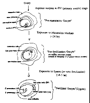
Figure 1 provides a schematic showing the
production of pre-maturation oocytes, pre-fertilization
oocytes and fertilized oocytes (zygotes).
Figure 2 shows an autoradiogram of a Southern blot
of genomic DNA isolated from the skin (A) and blood (B) of
calves derived from pre-fertilization oocytes and zygotes
which were injected with pseudotyped LSRNL retrovirus. The
calf DNA was obtained as shown in (C), by digesting it with
HindIII which cut the pLSRNL vector twice to generate the
1.6 kb fragment of calf DNA used in the Southern blots of 2A
and 2B.
Figure 3 shows an ethidium bromide stained agarose
gel containing electrophoresed PCR products which were
amplified using neo gene primers (A) or HBsAg primers (B)
from the blood and skin of calves derived from pre-
fertilization oocytes and zygotes injected with pseudotyped
LSRNL retrovirus.
6a
CA 02254866 1998-11-12
WO 98/41615 PCT/US98/05376
DEFINITIONS
To facilitate understanding of the invention, a number of terms are defined
below.
As used herein, the term "egg" when used in reference to a mammalian egg,
means an
oocyte surrounded by a zona pellucida and a mass of cumulus cells (follicle
cells) with their
associated proteoglycan. The term "egg" is used in reference to eggs recovered
from antral
follicles in an ovary (these eggs comprise pre-maturation oocytes) as well as
to eggs which
have been released from an antral follicle (a ruptured follicle).
As used herein, the term "oocyte" refers to a female gamete cell and includes
primary
oocytes, secondary oocytes and mature, unfertilized ovum. An oocyte is a large
cell having a
large nucleus (i.e., the germinal vesicle) surrounded by ooplasm. The ooplasm
contains non-
nuclear cytoplasmic contents including mRNA, ribosomes, mitochondria, yolk
proteins, etc.
The membrane of the oocyte is referred to herein as the "plasma membrane."
The term "pre-maturation oocyte" as used herein refers to a female gamete cell
following the oogonia stage (i.e., mitotic proliferation has occurred) that is
isolated from an
ovary (e.g., by aspiration) but which has not been exposed,. to maturation
medium in vitro.
Those of skill in the art know that the process of aspiration causes oocytes
to begin the
maturation process but that completion of the maturation process (i.e.,
formation of a
secondary oocyte which has extruded the first polar body) in vitro requires
the exposure of
the aspirated oocytes to maturation medium. Pre-maturation oocytes will
generally be
arrested at the first anaphase of meiosis.
The term "pre-fertilization oocyte" as used herein refers to a female gamete
cell such
as a pre-maturation oocyte following exposure to maturation medium in vitro
but prior to
exposure to sperm (i.e., matured but not fertilized). The pre-fertilization
oocyte has
completed the first meiotic division, has released the first polar body and
lacks a nuclear
membrane (the nuclear membrane will not reform until fertilization occurs;
after fertilization,
the second meiotic division occurs along with the extrusion of the second
polar body and the
formation of the male and female pronuclei). Pre-fertilization oocytes may
also be referred to
as matured oocytes at metaphase II of the second meiosis.
The terms "unfertilized egg" or "unfertilized oocyte" as used herein refers to
any
female gamete cell which has not been fertilized and these terms encompass
both pre-
maturation and pre-fertilization oocytes.
The term "perivitelline space" refers to the space located between the zona
pellucida
and the plasma membrane of a mammalian egg or oocyte.
_7 -
CA 02254866 1998-11-12
WO 98/41615 PCT/US98/05376
The term "infectious retrovirus" refers to a retroviral particle which is
capable of
entering a cell (i.e., the particle contains a membrane-associated protein
such as an envelope
protein or a viral G glycoprotein which can bind to the host cell surface and
facilitate entry of
the viral particle into the cytoplasm of the host cell) and integrating the
retroviral genome (as
a double-stranded provirus) into the genome of the host cell.
Retroviral vectors can be used to transfer genes efficiently into host cells
by exploiting
the viral infectious process. Foreign or heterologous genes cloned (i.e.,
inserted using
molecular biological techniques) into the retroviral genome can be delivered
efficiently to
host cells which are susceptible to infection by the retrovirus. Through well
known genetic
manipulations, the replicative capacity of the retroviral genome can be
destroyed. The
resulting replication-defective vectors can be used to introduce new genetic
material to a cell
but they are unable to replicate. A helper virus or packaging cell line can be
used to permit
vector particle assembly and egress from the cell.
The terms "vector particle" or "retroviral particle" refer to viral-like
particles that are
capable of introducing nucleic acid into a cell through a viral-like entry
mechanism.
The host range of a retroviral vector (i.e., the range of cells that these
vectors can
infect) can be altered by including an envelope protein from another closely
related virus.
The term "membrane-associated protein" refers to a protein (e.g., a viral
envelope
glycoprotein or the G proteins of viruses in the Rhabdoviridae family such as
VSV, Piry,
Chandipura and Mokola) which are associated with the membrane surrounding a
viral
particle; these membrane-associated proteins mediate the entry of the viral
particle into the
host cell. The membrane associated protein may bind to specific cell surface
protein
receptors, as is the case for retroviral envelope proteins or the membrane-
associated protein
may interact with a phospholipid component of the plasma membrane of the host
cell, as is
the case for the G proteins derived from members of the Rhabdoviridae family.
The term "heterologous membrane-associated protein" refers to a membrane-
associated
protein which is derived from a virus which is not a member of the same viral
class or family
as that from which the nucleocapsid protein of the vector particle is derived.
"Viral class or
family" refers to the taxonomic rank of class or family, as assigned by the
International
Committee on Taxonomy of Viruses.
The term "Rhabdoviridae" refers to a family of enveloped RNA viruses that
infect
animals, including humans, and plants. The Rhabdoviridae family encompasses
the genus
Vesiculovirus which includes vesicular stomatitis virus (VSV), Cocal virus,
Piry virus,
-8-
CA 02254866 1998-11-12
WO 98/41615 PCTIUS98/05376
Chandipura virus, and Spring viremia of carp virus (seqeunces encoding the
Spring viremia of
carp virus are available under GenBank accession number U18101). The G
proteins of
viruses in the Vesiculovirus genera are virally-encoded integral membrane
proteins that form
externally projecting homotrimeric spike glycoproteins complexes that are
required for
receptor binding and membrane fusion. The G proteins of viruses in the
Vesiculovirus genera
have a covalently bound palmititic acid (C16) moiety. The amino acid sequences
of the G
proteins from the Vesiculoviruses are fairly well conserved. For example, the
Piry virus G
protein share about 38% identity and about 55% similarity with the VSV G
proteins (several
strains of VSV are known, e.g., Indiana, New Jersey, Orsay, San Juan, etc.,
and their G
proteins are highly homologous). The Chandipura virus G protein and the VSV G
proteins
share about 37% identity and 52% similarity. Given the high degree of
conservation (amino
acid sequence) and the related functional characteristics (e.g., binding of
the virus to the host
cell and fusion of membranes, including syncytia formation) of the G proteins
of the
Vesiculoviruses, the G proteins from non-VSV Vesiculoviruses may be used in
place of the
VSV G protein for the pseudotyping of viral particles. The G proteins of the
Lyssa viruses
(another genera within the Rhabdoviridae family) also share a fair degree of
conservation
with the VSV G proteins and function in a similar manner (e.g., mediate fusion
of
membranes) and therefore may be used in place of the VSV G protein for the
pseudotyping
of viral particles. The Lyssa viruses include the Mokola virus and the Rabies
viruses (several
strains of Rabies virus are known and their G proteins have been cloned and
sequenced). The
Mokola virus G protein shares stretches of homology (particulary over the
extracellular and
transmembrane domains) with the VSV G proteins which show about 31% identity
and 48%
similarity with the VSV G proteins. Preferred G proteins share at least 25%
identity,
preferably at least 30% identity and most preferably at least 35% identity
with the VSV G
proteins. The VSV G protein from which New Jesery strain (the sequence of this
G protein
is provided in GenBank accession numbers M27165 and M21557) is employed as the
reference VSV G protein.
The term "conditions which permit the maturation of a pre-maturation oocyte"
refers
to conditions of in vitro cell culture which permit the maturation of a pre-
maturation oocyte
to a mature ovum (e.g., a pre-fertilization oocyte). These culture conditions
permit and
induce the events which are associated with maturation of the pre-maturation
oocyte including
stimulation of the first and second meiotic divisions. In vitro culture
conditions which permit
the maturation of pre-maturation oocytes from a variety of mammalian species
(e.g., cattle,
-9-
CA 02254866 1998-11-12
WO 98/41615 PCT/US98/05376
hamster, pigs and goats) are well know to the art [see e.g., Parrish et al.
(1985)
Theriogenology 24:537; Rosenkrans and First (1994) J. Ani. Sci. 72:434;
Bavister and
Yanagimachi (1977) Biol. Reprod. 16:228; Bavister et al. (1983) Biol. Reprod.
28:235;
Leibfried and Bavister (1982) J. Reprod. Fert. 66:87; Keskintepe et al. (1994)
Zygote 2:97
Funahashi et al. (1994) J. Reprod. Fert. 101:159 and Funahashi et al. (1994)
Biol. Reprod
50:10721.
DESCRIPTION OF THE INVENTION
The present invention provides improved methods for the production of
transgenic
animals. The methods of the present invention provide, for the first time, the
production of
transgenic animals by the introduction of exogenous DNA into pre-maturation
oocytes and
mature, unfertilized oocytes (i.e., pre-fertilization oocytes) using
retroviral vectors which
transduce dividing cells [e.g., vectors derived from murine leukemia virus
(MLV)].
The Description of the Invention is divided into the following sections: I.
Retroviruses
and Retroviral Vectors; II. Integration of Retroviral DNA; III. Introduction
of Retroviral
Vectors Into Gametes Before the Last Meiotic Division; and IV. Detection of
the Retrovirus
Following Injection Into Oocytes or Embryos.
I. Retroviruses and Retroviral Vectors
Retroviruses (family Retroviridae) are divided into three groups: the
spumaviruses
(e.g., human foamy virus); the lentiviruses (e.g., human immunodeficiency
virus and sheep
visna virus) and the oncoviruses (e.g., MLV, Rous sarcoma virus).
Retroviruses are enveloped (i.e., surrounded by a host cell-derived lipid
bilayer
membrane) single-stranded RNA viruses which infect animal cells. When a
retrovirus infects
a cell, its RNA genome is converted into a double-stranded linear DNA form
(i.e., it is
reverse transcribed). The DNA form of the virus is then integrated into the
host cell genome
as a provirus. The provirus serves as a template for the production of
additional viral
genomes and viral mRNAs. Mature viral particles containing two copies of
genomic RNA
bud from the surface of the infected cell. The viral particle comprises the
genomic RNA,
reverse transcriptase and other pol gene products inside the viral capsid
(which contains the
viral gag gene products) which is surrounded by a lipid bilayer membrane
derived from the
host cell containing the viral envelope glycoproteins (also referred to as
membrane-associated
proteins).
-10-
CA 02254866 1998-11-12
WO 98/41615 PCT/US98/05376
The organization of the genomes of numerous retroviruses is well known to the
art
and this has allowed the adaptation of the retroviral genome to produce
retroviral vectors.
The production of a recombinant retroviral vector carrying a gene of interest
is typically
achieved in two stages. First, the gene of interest is inserted into a
retroviral vector which
contains the sequences necessary for the efficient expression of the gene of
interest [including
promoter and/or enhancer elements which may be provided by the viral long
terminal repeats
(LTRs) or by an internal promoter/enhancer and relevant splicing signals],
sequences required
for the efficient packaging of the viral RNA into infectious virions [e.g.,
the packaging signal
(Psi), the tRNA primer binding site (-PBS), the 3' regulatory sequences
required for reverse
transcription (+PBS)] and the viral LTRs. The LTRs contain sequences required
for the
association of viral genomic RNA, reverse transcriptase and integrase
functions, and
sequences involved in directing the expression of the genomic RNA to be
packaged in viral
particles. For safety reasons, many recombinant retroviral vectors lack
functional copies of
the genes which are essential for viral replication (these essential genes are
either deleted or
disabled); the resulting virus is said to be replication defective.
Second, following the construction of the recombinant vector, the vector DNA
is
introduced into a packaging cell line. Packaging cell lines provide viral
proteins required in
trans for the packaging of the viral genomic RNA into viral particles having
the desired host
range (i.e., the viral-encoded gag, pol and env proteins). The host range is
controlled, in part,
by the type of envelope gene product expressed on the surface of the viral
particle.
Packaging cell lines may express ecotrophic, amphotropic or xenotropic
envelope gene
products. Alternatively, the packaging cell line may lack sequences encoding a
viral envelope
(env) protein. In this case the packaging cell line will package the viral
genome into particles
which lack a membrane-associated protein (e.g., an env protein). In order to
produce viral
particles containing a membrane associated protein which will permit entry of
the virus into a
cell, the packaging cell line containing the retroviral sequences is
transfected with sequences
encoding a membrane-associated protein [e.g., the G protein of vesicular
stomatitis virus
(VSV)]. The transfected packaging cell will then produce viral particles which
contain the
membrane-associated protein expressed by the transfected packaging cell line;
these viral
particles which contain viral genomic RNA derived from one virus encapsidated
by the
envelope proteins of another virus are said to be pseudotyped virus particles.
Viral vectors, including recombinant retroviral vectors, provide a more
efficient
means of transferring genes into cells as compared to other techniques such as
calcium
-11-
CA 02254866 1998-11-12
WO 98/41615 PCT/US98/05376
phosphate-DNA co-precipitation or DEAE-dextran-mediated transfection,
electroporation or
microinjection of nucleic acids. It is believed that the efficiency of viral
transfer is due in
part to the fact that the transfer of nucleic acid is a receptor-mediated
process (f. e., the virus
binds to a specific receptor protein on the surface of the cell to be
infected). In addition, the
virally transferred nucleic acid once inside a cell integrates in controlled
manner in contrast to
the integration of nucleic acids which are not virally transferred; nucleic
acids transferred by
other means such as calcium phosphate-DNA co-precipitation are subject to
rearrangement
and degradation.
The most commonly used recombinant retroviral vectors are derived from the
amphotropic Moloney murine leukemia virus (MoMLV) [Miller and Baltimore (1986)
Mol.
Cell. Biol. 6:2895]. The MoMLV system has several advantages: 1) this specific
retrovirus
can infect many different cell types, 2) established packaging cell lines are
available for the
production of recombinant MoMLV viral particles and 3) the transferred genes
are
permanently integrated into the target cell chromosome. The established MoMLV
vector
systems comprise a DNA vector containing a small portion of the retroviral
sequence (the
viral long terminal repeat or "LTR" and the packaging or "psi" signal) and a
packaging cell
line. The gene to be transferred is inserted into the DNA vector. The viral
sequences present
on the DNA vector provide the signals necessary for the insertion or packaging
of the vector
RNA into the viral particle and for the expression of the inserted gene. The
packaging cell
line provides the viral proteins required for particle assembly [Markowitz et
al. (1988) J.
Virol. 62:1120].
Despite these advantages, existing retroviral vectors based upon MoMLV are
limited
by several intrinsic problems: 1) they do not infect non-dividing cells
[Miller et al., (1990)
Mol. Cell. Biol. 10:4239], 2) they produce low titers of the recombinant virus
[Miller and
Rosman (1989) BioTechniques 7: 980 and Miller (1992) Nature 357: 455] and 3)
they infect
certain cell types (e.g., human lymphocytes) with low efficiency [Adams et al.
(1992) Proc.
Natl. Acad. Sci. USA 89:8981 ]. The low titers associated with MoMLV-based
vectors has
been attributed, at least in part, to the instability of the virus-encoded
envelope protein.
Concentration of retrovirus stocks by physical means (e.g.,
ultracentrifugation and
ultrafiltration) leads to a severe loss of infectious virus.
The low titer and inefficient infection of certain cell types by MoMLV-based
vectors
has been overcome by the use of pseudotyped retroviral vectors which contain
the G protein
of VSV as the membrane associated protein. Unlike retroviral envelope proteins
which bind
-12-
CA 02254866 1998-11-12
WO 98/41615 PCT/US98/05376
to a specific cell surface protein receptor to gain entry into a cell, the VSV
G protein interacts
with a phospholipid component of the plasma membrane [Mastromarino et al.
(1977) J. Gen.
Virol. 68:2359]. Because entry of VSV into a cell is not dependent upon the
presence of
specific protein receptors, VSV has an extremely broad host range. Pseudotyped
retroviral
vectors bearing the VSV G protein have an altered host range characteristic of
VSV (i.e., they
can infect almost all species of vertebrate, invertebrate and insect cells).
Importantly, VSV
G-pseudotyped retroviral vectors can be concentrated 2000-fold or more by
ultracentrifugation
without significant loss of infectivity [Bums et al. (1993) Proc. Natl. Acad.
Sci. USA
90:8033].
The VSV G protein has also been used to pseudotype retroviral vectors based
upon the
human immunodeficiency virus (HIV) [Naldini et al. (1996) Science 272:263].
Thus, the
VSV G protein may be used to generate a variety of pseudotyped retroviral
vectors and is not
limited to vectors based on MoMLV.
The present invention is not limited to the use of the VSV G protein when a
viral G
protein is employed as the heterologous membrane-associated protein within a
viral particle.
The G proteins of viruses in the Vesiculovirus genera other than VSV, such as
the Piry and
Chandipura viruses, that are highly homologous to the VSV G protein and, like
the VSV G
protein, contain covalently linked palmitic acid [Brun et al. (1995)
Intervirol. 38:274 and
Masters et al. (1990) Virol. 171:285]; thus, the G protein of the Piry and
Chandipura viruses
can be used in place of the VSV G protein for the pseudotyping of viral
particles. In
addition, the VSV G proteins of viruses within the Lyssa virus genera such as
Rabies and
Mokola viruses show a high degree of conservation (amino acid sequence as well
as
functional conservation) with the VSV G proteins. For example, the Mokola
virus G protein
has beeft shown to function in a manner similar to the VSV G protein (i.e., to
mediate
membrane fusion) and therefore may be used in place of the VSV G protein for
the
pseudotyping of viral particles (Mebatsion et al. (1995) J. Virol. 69:1444].
The nucleotide
sequence encoding the Piry G protein is provided in SEQ ID NO:5 and the amino
acid
sequence of the Piry G protein is provided in SEQ ID NO:6. The nucleotide
sequence
encoding the Chandipura G protein is provided in SEQ ID NO:7 and the amino
acid sequence
of the Chandipura G protein is provided in SEQ ID NO:8. The nucleotide
sequence encoding
the Mokola G protein is provided in SEQ ID NO:9 and the amino acid sequence of
the
Mokola G protein is provided in SEQ ID NO: 10. Viral particles may be
pseudotyped using
either the Piry, Chandipura or Mokola G protein as described in Example 2 with
the
-13-
CA 02254866 1998-11-12
WO 98/41615 PCT/US98/05376
exception that a plasmid containing sequences encoding either the Piry,
Chandipura or
Mokola G protein under the transcriptional control of a suitable promoter
element [e.g., the
CMV intermediate-early promoter; numerous expression vectors containing the
CMV IE
promoter are available, such as the pcDNA3.1 vectors (Invitrogen)] is used in
place of
pHCMV-G. Sequences encoding other G proteins derived from other members of the
Rhabdoviridae family may be used; sequences encoding numerous rhabdoviral G
proteins are
available from the GenBank database.
II. Integration of Retroviral DNA
The majority of retroviruses can transfer or integrate a double-stranded
linear form of
the virus (the provirus) into the genome of the recipient cell only if the
recipient cell is
cycling (i.e., dividing) at the time of infection. Retroviruses which have
been shown to infect
dividing cells exclusively, or more efficiently, include MLV, spleen necrosis
virus, Rous
sarcoma virus and human immunodeficiency virus (HIV; while HIV infects
dividing cells
more efficiently, HIV can infect non-dividing cells).
It has been shown that the integration of MLV virus DNA depends upon the host
cell's progression through mitosis and it has been postulated that the
dependence upon mitosis
reflects a requirement for the breakdown of the nuclear envelope in order for
the viral
integration complex to gain entry into the nucleus [Roe et al. (1993) EMBO J.
12:2099].
However, as integration does not occur in cells arrested in metaphase, the
breakdown of the
nuclear envelope alone may not be sufficient to permit viral integration;
there may be
additional requirements such as the state of condensation of the genomic DNA
(Roe et al.,
supra).
III. Introduction of Retroviral Vectors Into Gametes Before the Last Meiotic
Division
The nuclear envelope of a cell breaks down during meiosis as well as during
mitosis.
Meiosis occurs only during the final stages of gametogenesis. The methods of
the present
invention exploit the breakdown of the nuclear envelope during meiosis to
permit the
integration of recombinant retroviral DNA and permit for the first time the
use of unfertilized
oocytes (i.e., pre-fertilization and pre-maturation oocytes) as the recipient
cell for retroviral
gene transfer for the production of transgenic animals. Because infection of
unfertilized
-14-
CA 02254866 1998-11-12
WO 98/41615 PCTIUS98/05376
oocytes permits the integration of the recombinant provirus prior to the
division of the one
cell embryo, all cells in the embryo will contain the proviral sequences.
Oocytes which have not undergone the final stages of gainetogenesis are
infected with
the retroviral vector. The injected oocytes are then permitted to complete
maturation with the
accompanying meiotic divisions. The breakdown of the nuclear envelope during
meiosis
permits the integration of the proviral form of the retrovirus vector into the
genome of the
oocyte. When pre-maturation oocytes are used, the injected oocytes are then
cultured in vitro
under conditions which permit maturation of the oocyte prior to fertilization
in vitro.
Conditions for the maturation of oocytes from a number of mammalian species
(e.g., bovine,
ovine, porcine, murine, caprine) are well known to the art. In general, the
base medium used
herein for the in vitro maturation of bovine oocytes, TC-M199 medium, may be
used for the
in vitro maturation of other mammalian oocytes. TC-M199 medium is supplemented
with
hormones (e.g., luteinizing hormone and estradiol) from the appropriate
mammalian species.
The amount of time a pre-maturation oocyte must be exposed to maturation
medium to permit
maturation varies between mammalian species as is known to the art. For
example, an
exposure of about 24 hours is sufficient to permit maturation of bovine
oocytes while porcine
oocytes require about 44-48 hours.
Occytes may be matured in vivo and employed in place of oocytes matured in
vitro in
the practice of the present invention. For example, when porcine oocytes are
to be employed
in the methods of the present invention, matured pre-fertilization oocytes may
be harvested
directly from pigs that are induced to superovulate as is known to the art.
Briefly, on day 15
or 16 of estrus the female pig(s) is injected with about 1000 units of
pregnant mare's serum
(PMS; available from Sigma and Calbiochem). Approximately 48 hours later, the
pig(s) is
injected with about 1000 units of human chorionic gonadotropin) (hCG; Sigma)
and 24-48
hours later matured oocytes are collected from oviduct. These in vivo matured
pre-fertlization
oocytes are then injected with the desired retroviral preparation as described
herein. Methods
for the superovulation and collection of in vivo matured (i.e., oocytes at the
metaphase 2
stage) oocytes are known for a variety of mammals [e.g., for superovulation of
mice, see
Hogan et al. (1994), supra at pp. 130-133; for superovulation of pigs and in
vitro fertilzation
of pig oocytes see Cheng, W. (1995) Doctoral Dissertation, Cambridge
University,
Cambridge, United Kingdom].
Retroviral vectors capable of infecting the desired species of non-human
animal which
can be grown and concentrated to very high titers (e.g., _ 1 x 10g cfu/ml) are
preferentially
-15-
CA 02254866 1998-11-12
WO 98/41615 PCT/US98/05376
employed. The use of high titer virus stocks allows the introduction of a
defined number of
viral particles into the perivitelline space of each injected oocyte. The
perivitelline space of
most mammalian oocytes can accommodate about 10 picoliters of injected fluid
(those in the
art know that the volume that can be injected into the perivitelline space of
a mammalian
oocyte or zygote varies somewhat between species as the volume of an oocyte is
smaller than
that of a zygote and thus, oocytes can accommodate somewhat less than can
zygotes).
The vector used may contain one or more genes encoding a protein of interest;
alternatively, the vector may contain sequences which produce anti-sense RNA
sequences or
ribozymes. The infectious virus is microinjected into the perivitelline space
of oocytes
(including pre-maturation oocytes) or one cell stage zygotes. Microinjection
into the
perivitelline space is much less invasive than the microinjection of nucleic
acid into the
pronucleus of an embryo. Pronuclear injection requires the mechanical puncture
of the
plasma membrane of the embryo and results in lower embryo viability. In
addition, a higher
level of operator skill is required to perform pronuclear injection as
compared to perivitelline
injection. Visualization of the pronucleus is not required when the virus is
injected into the
perivitelline space (in contrast to injection into the pronucleus); therefore
injection into the
perivitelline space obviates the difficulties associated with visualization of
pronuclei in species
such as cattle, sheep and pigs.
The virus stock may be titered and diluted prior to microinjection into the
perivitelline
space so that the number of proviruses integrated in the resulting transgenic
animal is
controlled. The use of a viral stock (or dilution thereof) having a titer of 1
x 108 cfu/ml
allows the delivery of a single viral particle per oocyte. The use of pre-
maturation oocytes or
mature fertilized oocytes as the recipient of the virus minimizes the
production of animals
which are mosaic for the provirus as the virus integrates into the genome of
the oocyte prior
to the occurrence of cell cleavage.
In order to deliver, on average, a single infectious particle per oocyte, the
micropipets
used for the injection are calibrated as follows. Small volumes (e.g., about 5-
10 pl) of the
undiluted high titer viral stock (e.g., a titer of about 1 x 10⁸ cfu/ml) are
delivered to the wells
of a microtiter plate by pulsing the micromanipulator. The titer of virus
delivered per a given
number of pulses is determined by diluting the viral stock in each well and
determining the
titer using a suitable cell line (e.g., the 208F cell line) as described in
Ex. 2. The number of
pulses which deliver, on average, a volume of virus stock containing one
infectious viral
-16-
CA 02254866 2004-07-02
73534-1
particle (i.e., gives a MOI of 1 when titered on 208F cells) are used for
injection of the viral
stock into the oocytes.
Prior to microinjection of the titered and diluted (if required) virus stock,
the cumulus
cell layer is opened to provide access to the perivitelline space. The cumulus
cell layer need
not be completely removed from the oocyte and indeed for certain species of
animals (e.g.,
cows, sheep, pigs, mice) a portion of the cumulus cell layer must remain in
contact with the
oocyte to permit proper development and fertilization post-injection.
Injection of viral
particles into the perivitelline space allows the vector RNA (f. e., the viral
genome) to enter
the cell through the plasma membrane thereby allowing proper reverse
transcription of the
viral RNA.
IV. Detection of the Retrovirus Following Injection Into Oocytes or Embryos
The presence of the retroviral genome in cells (e.g., oocytes or embryos)
infected with
pseudotyped retrovirus may be detected using a variety of means. The
expression of the gene
product(s) encoded by the retrovirus may be detected by detection of mRNA
corresponding to
the vector-encoded gene products using techniques well known to the art (e.g.,
Northern blot,
dot blot, in situ hybridization and RT-PCR analysis). Direct detection of the
vector-encoded
gene product(s) is employed when the gene product is a protein which either
has an
enzymatic activity (e.g., (3-galactosidase) or when an antibody capable of
reacting with the
vector-encoded protein is available.
Alternatively, the presence of the integrated viral genome may be detected
using
Southern blot or PCR analysis. For example, the presence of the LZRNL or LSRNL
genomes may be detected following infection of oocytes or embryos using PCR as
follows.
Genomic DNA is extracted from the infected oocytes or embryos (the DNA may be
extracted
from the whole embryo or alternatively various tissues of the embryo may be
examined)
using techniques well known to the art. The LZRNL and LSRNL viruses contain
the neo
gene and the following primer pair can be used to amplify a 349-bp segment of
the neo gene:
upstream primer: 5'-GCATTGCATCAGCCATGATG-3' (SEQ ID NO:1) and downstream
primer: 5'-GATGGATTGCACGCAGGTTC-3' (SEQ ID NO:2). The PCR is carried out
using well known techniques [e.g., using a GeneAmp kit according to the
manufacturer's
instructions (Perkin-Elmer)]. The DNA present in the reaction is denatured by
incubation at
94 C for 3 min followed by 40 cycles of 94 C for 1 min, 60 C for 40 sec and 72
C for 40
sec followed by a final extension at 72 C for 5 min. The PCR products may be
analyzed by
*Trade-mark
-17-
CA 02254866 1998-11-12
WO 98/41615 PCT/US98/05376
electrophoresis of 10 to 20% of the total reaction on a 2% agarose gel; the
349-bp product
may be visualized by staining of the gel with ethidium bromide and exposure of
the stained
gel to UV light. If the expected PCR product cannot be detected visually, the
DNA can be
transferred to a solid support (e.g., a nylon membrane) and hybridized with a
32P-labeled neo
probe.
Southem blot analysis of genomic DNA extracted from infected oocytes and/or
the
resulting embryos, offspring and tissues derived therefrom is employed when
information
concerning the integration of the viral DNA into the host genome is desired.
To examine the
number of integration sites present in the host genome, the extracted genomic
DNA is
typically digested with a restriction enzyme which cuts at least once within
the vector
sequences. If the enzyme chosen cuts twice within the vector sequences, a band
of known
(i.e., predictable) size is generated in addition to two fragments of novel
length which can be
detected using appropriate probes.
EXPERIMENTAL
The following examples serve to illustrate certain preferred embodiments and
aspects
of the present invention and are not to be construed as limiting the scope
thereof.
In the experimental disclosure which follows, the following abbreviations
apply: M
(molar); mM (millimolar); M (micromolar); nM (nanomolar); mol (moles); mmol
(millimoles); mol (micromoles); nmol (nanomoles); gm (grams); mg
(milligrams); g
(micrograms);pg (picograms); L (liters); ml (milliliters); l (microliters);
cm (centimeters);
mm (millimeters); m (micrometers); nm (nanometers); C (degrees Centigrade);
AMP
(adenosine 5'-monophosphate); BSA (bovine serum albumin); cDNA (copy or
complimentary
DNA); CS (calf serum); DNA (deoxyribonucleic acid); ssDNA (single stranded
DNA);
dsDNA (double stranded DNA); dNTP (deoxyribonucleotide triphosphate); LH
(luteinizing
hormone); NIH (Natioal Institues of Health, Besthesda, MD); RNA (ribonucleic
acid); PBS
(phosphate buffered saline); g (gravity); OD (optical density); HEPES
(N-[2-Hydroxyethyl]piperazine-N-[2-ethanesulfonic acid]); HBS (HEPES buffered
saline);
PBS (phosphate buffered saline); SDS (sodium dodecylsulfate); Tris-HCl
(tris[Hydroxymethyl]aminomethane-hydrochloride); Klenow (DNA polymerase I
large
(Klenow) fragment); rpm (revolutions per minute); EGTA (ethylene glycol-bis(13-
aminoethyl
ether) N, N, N', N'-tetraacetic acid); EDTA (ethylenediaminetetracetic acid);
bla (13-lactamase
or ampicillin-resistance gene); ORI (plasmid origin of replication); lacI (lac
repressor); X-gal
-18-
CA 02254866 2004-07-02
73534-1
(5-bromo-4-chloro-3-indolyl-[i-D-galactoside); ATCC (American Type Culture
Collection,
Rockville, MD); GIBCOBRL (GIBCOBRL, Grand Island, NY); Perkin-Elmer (Perkin-
Elmer, Norwalk, CT); and Sigma (Sigma Chemical Company, St. Louis, MO).
EXAMPLE 1
Generation of Cell Lines Stably Expressing the MoMLV gag and pol Proteins
The expression of the fusogenic VSV G protein on the surface of cells results
in
syncytium formation and cell death. Therefore, in order to produce retroviral
particles
containing the VSV G protein as the membrane-associated protein a three step
approach was
taken. First, stable cell lines expressing the gag and pol proteins from MoMLV
at high levels
were generated (e.g., 293GP cells; Example 1). These stable cell lines were
then infected
using the desired retroviral vector which is derived from an amphotrophic
packaging cell
(e.g., PA317 cells transfected with the desired retroviral vector; Example
2a). The infected
stable cell line which expresses the gag and pol proteins produces
noninfectious viral particles
lacking a membrane-associated protein (e.g., a envelope protein). Third, these
infected, cell
lines are then transiently transfected with a plasmid capable of directing the
high level
expression of the VSV G protein (Example 2b). The transiently transfected
cells produce
VSV G-pseudotyped retroviral vectors which can be collected from the cells
over a period of
3 to 4 days before the producing cells die as a result of syncytium formation.
The first step in the production of VSV G-pseudotyped retroviral vectors, the
generation of stable cell lines expressing the MoMLV gag and pol proteins is
described
below.
The human adenovirus 5-transfonmed embryonal kidney cell line 293 (ATCC CRL
1573) was cotransfected with the pCMVgag-pol and pFR400 plasmids using a ratio
of 10:1
(pCMVgag-pol and pFR400). pCMV gag-pol contains the MoMLV gag and pol genes
under
the control of the CMV promoter (pCMV gag-pol is available from the ATCC):
pFR400
encodes a mutant dihydrofolate reductase which has a reduced affinity for
methotrexate
[Simonsen et al., Proc. Natl. Acad. Sci. 80:2495 (1983)].
The plasmid DNA was introduced into the 293 cells using calcium phosphate co-
precipitation [Graham and Van der Eb, Virol. 52:456 (1973)]. Approximately 5 x
105 293
cells were plated into a 100 mm tissue culture plate the day before the DNA co-
precipitate
-19-
CA 02254866 1998-11-12
WO 98/41615 PCT/US98/05376
was added. A total of 20 g of plasmid DNA (18 g pCMV gag-pol and 2 g
pFR400) was
added as a calcium-DNA co-precipitate to each 100 mm plate. Stable
transformants were
selected by growth in DMEM-high glucose medium containing 10% FCS, 0.5 M
methotrexate and 5 M dipyridimole (selective medium). Colonies which grew in
the
selective medium were screened for extracellular reverse transcriptase
activity [Goff et al., J.
Virol. 38:239 (1981)] and intracellular p309n expression. p30P9 expression was
determined
by Western blotting using a goat-anti p30 antibody (NCI antiserum 77S000087).
A clone
which exhibited stable expression of the retroviral genes in the absence of
continued
methotrexate selection was selected. This clone was named 293GP (293 gag-pol).
The
293GP cell line, a derivative of the human Ad-5-transformed embryonal kidney
cell line 293,
was grown in DMEM-high glucose medium containing 10% FCS. The 293GP cell line
is
commercially available from Viagen, Inc., San Diego, CA.
EXAMPLE 2
Preparation of Pseudotyped Retroviral Vectors Bearing the G Glycoprotein of
VSV
In order to produce VSV G protein pseudotyped retrovirus the following steps
were
taken. First, the 293GP cell line was infected with virus derived from the
amphotrophic
packaging cell line PA317. The infected cells packaged the retroviral RNA into
viral
particles which lack a membrane-associated protein (because the 293GP cell
line lacks an env
gene or other gene encoding a membrane-associated protein). The infected 293GP
cells were
then transiently transfected with a plasmid encoding the VSV G protein to
produce
pseudotyped viral particles bearing the VSV G protein.
a) Cell Lines and Plasmids
The amphotropic packaging cell line, PA317 (ATCC CRL 9078) was grown in
DMEM-high glucose medium containing 10% FCS. The 293GP cell line was grown in
DMEM-high glucose medium containing 10% FCS. The titer of the pseudo-typed
virus may
be determined using either 208F cells [Quade (1979) Virol. 98:461] or NIH/3T3
cells (ATCC
CRL 1658); 208F and NIH/3T3 cells are grown in DMEM-high glucose medium
containing
10% CS.
-20-
CA 02254866 2004-07-02
73534-1
The plasmid pLZRNL [Xu et al. (1989) Virol. 171:331] contains the gene
encoding E.
colf P-galactosidase (LacZ) under the transcriptional control of the LTR of
the Moloney
murine sarcoma virus (MSV) followed by the gene encoding neomycin
phosphotransferase
(Neo) under the transcriptional control of the Rous sarcoma virus (RSV)
promoter. The
plasmid pLSRNL contains the gene encoding the hepatitis B surface antigen gene
(HBsAg)
under the transcriptional control of the MSV LTR followed by the Neo gene
under the control
of the RSV promoter (U.S. Patent No. 5,512,421).
The plasmid pHCMV-G contains the VSV G gene under the
transcriptional control of the human cytomegalovirus intermediate-early
promoter [Yee et al.
(1994) Meth. Cell Biol. 43:99].
b) Production and Titering of Pseudotyped LZRNL Virus
pLZRNL DNA was transfected into the amphotropic packaging line PA317 to
produced LZRNL virus. The resulting LZRNL virus was then used to infect 293GP
cells to
produce pseudotyped LZRNL virus bearing the VSV G.protein (following transient
transfection of the infected 293GP cells with a plasmid encoding the VSV G
protein). The
procedure for producing pseudotyped LZRNL virus was carried out as described
[Yee gt al.
(1994) Meth. Cell Biol. 43:99].
Briefly, on day 1, approximately 5 x 10⁵ PA317 cells were placed in a 100 mm
tissue
culture plate. On the following day (day 2), the PA317 cells were transfected
with 20 g of
pLZRNL plasmid DNA (plasmid DNA was purified using CsCI gradients) using the
standard
calcium phosphate co-precipitation procedure [Graham and Van der Eb (1973)
Virol. 52:456].
A range of 10 to 40 g of plasmid DNA may be used. Because 293GP cells may
take
more than 24 hours to attach firmly to tissue culture plates, the 293GP cells
may be placed in
100 mm plates 48 hours prior to transfection. The transfected PA317 cells
provide
amphotropic LZRNL virus.
On day 3, approximately I x 10S 293GP cells were placed. in a 100 mm tissue
culture
plate 24 hours prior to the harvest of the amphotropic virus from the
transfected PA317 cells.
On day 4, culture medium was harvested from the transfected PA317 cells 48
hours after the
application of the pLZRNL DNA. The culture medium was filtered through a 0.45
m filter
and polybrene was added to a final concentration of 8 g/ml. A stock solution
of polybrene
was prepared by dissolving 0.4 gm hexadimethrine bromide (polybrene; Sigma) in
100 ml
sterile water; the stock solution was stored at 4 C. The culture medium
containing LZRNL
-21-
CA 02254866 1998-11-12
WO 98/41615 PCTIUS98/05376
virus (containing polybrene) was used to infect the 293GP cells as follows.
The culture
medium was removed from the 293GP cells and was replaced with the LZNRL virus
containing culture medium. The virus containing medium was allowed to remain
on the
293GP cells for 16 hours. Following the 16 hour infection period (on day 5),
the medium
was removed from the 293GP cells and was replaced with fresh medium containing
400
g/ml G418 (GIBCOBRL). The medium was changed every 3 days until G418-resistant
colonies appeared two weeks later. Care was taken not to disturb the G418-
resistant colonies
when the medium was changed as 293GP cells attach rather loosely to tissue
culture plates.
The G418-resistant 293 colonies were picked using an automatic pipettor and
transferred directly into 24-well plates (i.e., the colonies were not removed
from the plates
using trypsin). The G418-resistant 293 colonies (as termed "293GP/LZRNL"
cells) were
screened for the expression of the LacZ gene in order to identify clones which
produce high
titers of pseudotyped LZRNL virus. Clones in 24-well plates were transferred
to 100 mm
tissue culture plates and allowed to grow to confluency. Protein extracts are
prepared from
the confluent plates by washing the cells once with 10 ml PBS (137 mM NaCl,
2.6 mM KCI,
8.1 mM Na2HPO411.5 mM KH2PO4). Two ml of 250 mM Tris-HCI, pH 7.8 was added and
the cells were scrapped off the plate using a rubber policeman. The cells were
then collected
by centrifugation at room temperature and resuspended in 100 l 250 mM Tris-
HCI, pH 7.8.
The cells were subjected to four rapid freeze/thaw cycles followed by
centrifugation at room
temperature to remove cell debris. The P-galactosidase activity present in the
resulting
protein extracts was determined as follows. Five microliters of protein
extract was mixed
with 500 l P-gal buffer (50 mM Tris-HCI, pH 7.5, 100 mM NaCI, 10 mM MgCIZ)
containing 0.75 ONPG (Sigma). The mixtures were incubated at 37 C until a
yellow color
appeared. The reactions were stopped by the addition of 500 l 10 mM EDTA and
the
optical density of the reactions was determined at 420 nm.
The 293GP/LZRNL clone which generated the highest amount of (3-galactosidase
activity was then expanded and used subsequently for the production of
pseudotyped LZNRL
virus as follows. Approximately I x 106 293GP/LZRNL cells were placed into a
100 mm
tissue culture plate. Twenty-four hours later, the cells were transfected with
20 g of
pHCMV-G plasmid DNA using calcium phosphate co-precipitation. Six to eight
hours after
the calcium-DNA precipitate was applied to the cells, the DNA solution was
replaced with
fresh culture medium (lacking G418). Longer transfection times (overnight)
have been found
-22-
CA 02254866 1998-11-12
WO 98/41615 PCTIUS98/05376
to result in the detachment of the majority of the 293GP/LZRNL cells from the
plate and are
therefore avoided. The transfected 293GP/LZRNL cells produce pseudotyped LZRNL
virus.
The pseudotyped LZRNL virus generated from the transfected 293GP/LZRNL cells
can be collected at least once a day between 24 and 96 hr after transfection.
The highest
virus titer was generated approximately 48 to 72 hr after initial pHCMV-G
transfection.
While syncytium formation became visible about 48 hr after transfection in the
majority of
the transfected cells, the cells continued to generate pseudotyped virus for
at least an
additional 48 hr as long as the cells remained attached to the tissue culture
plate. The
collected culture medium containing the VSV G-pseudotyped LZRNL virus was
pooled,
filtered through a 0.45 m filter and stored at -70 C.
The titer of the VSV G-pseudotyped LZRNL virus was then determined as follows.
5
x 10S rat 208F fibroblasts or NIH 3T3 cells were plated in a 100 mm culture
plate. Twenty-
fours hours after plating, the cells were infected with serial dilutions of
the LZRNL virus-
containing culture medium in the presence of 8 g/ml polybrene. Sixteen hours
after
infection with virus, the medium was replaced with fresh medium containing 400
jig/ml G418
and selection was continued for 14 days until G418-resistant colonies became
visible. Viral
titers were typically about 0.5 to 5.0 x 10⁶ colony forming units (cfu)/ml.
The titer of the
virus stock could be concentrated to a titer of greater than 109 cfu/ml as
described below.
EXAMPLE 3
Concentration of Pseudotyped Retroviral Vectors
The VSV G-pseudotyped LZRNL virus was concentrated to a high titer by two
cycles
of ultracentrifugation. The frozen culture medium collected as described in
Example 2 which
contained pseudotyped LZRNL virus was thawed in a 37 C water bath and was then
transferred to ultraclear centrifuge tubes (14 x 89 mm; Beckman, Palo Alto,
CA) which had
been previously sterilized by exposing the tubes to UV light in a laminar flow
hood
overnight. The virus was sedimented in a SW41 rotor (Beckman) at 50,000 x g
(25,000 rpm)
at 4 C for 90 min. The culture medium was then removed from the tubes in a
laminar flow
hood and the tubes were well drained. The virus pellet was resuspended to 0.5
to 1% of the
original volume of culture medium in either TNE (50 mM Tris-HCI, pH 7.8; 130
mM NaC1;
1 mM EDTA) or 0.1X Hank's balanced salt solution [1X Hank's balanced salt
solution
contains 1.3 mM CaC12, 5 mM KCI, 0.3 mM KHZPO4, 0.5 mM MgC12=6H2), 0.4 mM
-23-
CA 02254866 1998-11-12
WO 98/41615 PCT/US98/05376
MgSO4=7H20, 138 mM NaCl, 4 mM NaHCO310.3 mM NaH2P04=H20; 0.1X Hank's is made
by mixing 1 parts 1X Hank's with 9 parts PBS]. The resuspended virus pellet
was incubated
overnight at 4 C without swirling. The virus pellet could be dispersed with
gentle pipetting
after the overnight incubation without significant loss of infectious virus.
The titer of the
virus stock was routinely increase 100- to 300-fold after one round of
ultracentrifugation. The
efficiency of recovery of infectious virus varied between 30 and 100%.
The virus stock was then subjected to low speed centrifugation in a microfuge
for 5
min at 4 C to remove any visible cell debris or aggregated virions that were
not resuspended
under the above conditions (if the virus stock is not to be used for injection
into oocytes or
embryos, this centrifugation step may be omitted).
The virus stock was then subjected to another round of ultracentrifugation to
concentrate the virus stock further. The resuspended virus from the first
round of
centrifugation was pooled and pelleted by a second round of
ultracentrifugation which was
performed as described above. Viral titers were increased approximately 2000-
fold after the
second round of ultracentrifugation (titers of the pseudotyped LZRNL virus
were typically
greater than or equal to I x l09 cfu/ml after the second round of
ultracentrifugation).
The titers of the pre- and post-centrifugation fluids were determined by
infection of
208F (NIH 3T3 or Mac-T cells can also be employed) followed by selection of
G418-
resistant colonies as described above in Example 2. The concentrated viral
stock was stable
(i.e., did not lose infectivity) when stored at 4 C for several weeks.
EXAMPLE 4
Preparation of Pseudotyped Retrovirus For Infection of Oocytes and Embryos
The concentrated pseudotyped retrovirus were resuspended in 0.1X HBS (2.5 mM
HEPES, pH 7.12, 14 mM NaCI, 75 M Na2HPO4=H20) and 18 l aliquots were placed
in 0.5
ml vials (Eppendorf) and stored at -80 C until used. The titer of the
concentrated vector was
determined by diluting l l of the concentrated virus 10-'- or 10'8-fold with
0.1X HBS. The
diluted virus solution was then used to infect 208F and Mac-T cells and viral
titers were
determined as described in Example 2.
Prior to infection of oocytes or embryos (by microinjection), 1 41 of
polybrene [25
ng/ l; the working solution of polybrene was generated by diluting a stock
solution having a
concentration of 1 mg/ml (in sterile H20) in 0.1 HBS, pH 7.12] was mixed with
4 l of
-24-
CA 02254866 1998-11-12
WO 98/41615 PCT/US98/05376
concentrated virus to yield a solution containing 103-104 cfu/ l and 8 g/ml
polybrene. This
solution was loaded into the injection needle (tip having an internal diameter
of
approximately 2-4 m) for injection into the perivitelline space of gametes
(pre-maturation
oocytes, matured oocytes) or one cell stage zygotes (early stage embryo). An
Eppendorf
Transjector 5246 was used for all microinjections.
EXAMPLE 5
Preparation and Microinjection of Gametes and Zygotes
Gametes (pre-maturation and pre-fertilization oocytes) and zygotes (fertilized
oocytes)
were prepared and microinjected with retroviral stocks as described below.
a) Solutions
Tyrodes-Lactate with HEPES (TL-HEPES): 114 mM NaC1, 3.2 mM KCI, 2.0 mM
NaHCO31 0.4 mM Na2H2PO4=H2O, 10 mM Na-lactate, 2 mM CaC12=2H20, 0.5 mM
MgC12=6H20, 10 mM HEPES, 100 IU/ml penicillin, 50 g/ml phenol red, I mg/ml
BSA
fraction V, 0.2 mM pyruvate and 25 g/ml gentamycin.
Maturation Medium: TC-199 medium (GIBCO) containing 10% FCS, 0.2 mM
pyruvate, 5 g/ml NIH o-LH (NIH), 25 g/ml gentamycin and l g/ml estradiol-
17P.
Sperm-Tyrodes-Lactate (Sperm-TL): 100 mM NaCI, 3.2 mM KCI, 25 mM NaHCO31
0.29 mM NaZH2PO4=H20, 21.6 mM Na-lactate, 2.1 mM CaC12=2H20, 0.4 mM
MgCI2=6H20,
10 mM HEPES, 50 g/ml phenol red, 6 mg/ml BSA fraction V, 1.0 mM pyruvate and
25
g/ml gentamycin.
Fertilization Medium: 114 mM NaCI, 3.2 mM KCI, 25 mM NaHCO31 0.4 mM
Na2H2PO4=H20, 10 mM Na-lactate, 2 mM CaCi2=2H20, 0.5 mM MgC12=6H20, 100 IU/ml
penicillin, 50 g/ml phenol red, 6 mg/ml BSA fatty acid free, 0.2 mM pyruvate
and 25 g/ml
gentamycin.
PHE: 1 mM hypotaurine, 2 mM penicillamine and 250 M epinephrine.
Embryo Incubation + Amino Acids (EIAA): 114 M NaCl, 3.2 M KC1, 25 M
NaHCO31 1.6 g/ml L(+)-lactate, 10.7 g/ml L-glutamine, 300 g/ml BSA fatty
acid free,
0.275 g/ ml pyruvate, 25 g/ml gentamycin, 10 l of 100X MEM amino acids
stock
(M7145, Sigma) per ml and 20 1 of 50X BME amino acids stock (B6766, Sigma)
per ml.
0.1X HBS: 2.5 mM HEPES (pH 7.12), 14 mM NaCI and 75 M Na2HPO4=H20.
-25-
CA 02254866 2004-07-02
73534-1
b) Preparation, Injection, Maturation and Fertilization of Pre-
Maturation Oocytes
Oocytes were aspirated from small antral follicles on ovaries from dairy
cattle
obtained from a slaughterhouse. Freshly aspirated oocytes at the germinal
vesicle (GV) stage,
meiosis arrested, with the cumulus mass attached were selected (f. e., pre-
maturation oocytes).
The oocytes were then washed twice in freshly prepared TL-HEPES and
transferred into a
100 1 drop of TL-HEPES for microinjection.
Concentrated retroviral particles (prepared as described in Example 3) were
resuspended in 0.1X HBS, mixed with polybrene and loaded into the
injection'needle as
described in Example 4. Approximately. 10 pl of the virus solution was then
injected into the
perivitelline space of pre-maturation oocytes.
Following injection, the pre-maturation oocytes were washed twice in fresh TL-
HEPES and transferred into maturation medium (10 oocytes in 50 l). The pre-
maturation
oocytes were then incubated in- Maturation Medium for 24 hours at 37 C which
permits the
oocytes to mature to the metaphase II stage. The matured oocytes were then
washed twice in
Sperm-TL and 10 oocytes were then transferred into 44 l of Fertilization
Medium. The
mature oocytes (10 oocytes/44 l Fertilization Medium) were then fertilized by
the addition
of 2 l of sperm at a concentration of 2.5 x 10'/ml, 2 l of PHE and 2 l of
heparin
(fertilization mixture). Sperm was prepared by discontinuous percoll gradient
separation of
frozen-thawed semen as described [Kim et al. (1993) Mol. Reprod. Develop.
35:105].
Briefly, percoll*gradients were formed by placing 2 ml of each of 90% and 45%
percoll in a
15 ml conical tube. Frozen-thawed semen was layered on top of the gradient and
the tubes
were centrifuged for 10 minutes at 700xg. Motile sperm were collected from the
bottom of
the tube.
The oocytes were incubated for 16 to 24 hours at 37 C in the fertilization
mixture.
Following fertilization, the cumulus cells were removed by vortexing the cells
(one cell stage
zygotes, Pronucleus Stage) for 3 minutes to produce "nude" oocytes. The nude
oocytes were
then washed twice in embryo culture medium (EIAA) and 20 to 25 zygotes were
then
cultured in 50 1 drop of EIAA (without serum until Day 4 at which time the
zygotes were
placed in EIAA containing 10% serum) until the desired developmental stage was
reached:
approximately 48 hours or Day 2 (Day 0 is the day when the matured oocytes are
co-cultured
with sperm) for morula stage (8 cell stage) or Day 6-7 for blastocyst stage.
Embryos at the
morula stage were analyzed for expression of p-gala.ctosidase as described in
Example 6.
*Trade-mark
-26-
CA 02254866 1998-11-12
WO 98/41615 PCTIUS98/05376
Embryos derived from injected pre-maturation oocytes were also analyzed for (3-
galactosidase
expression at the 2 cell, 4 cell, and blastocyst stage and all developmental
stages examined
were positive.
c) Preparation, Injection and Fertilization of Pre-Fertilization Oocytes
Pre-maturation oocytes were harvested, washed twice with TL-HEPES as described
above. The oocytes were then cultured in Maturation Medium (10 oocytes per 50
l
medium) for 16 to 20 hours to produce pre-fertilization oocytes (Metaphase II
Stage). The
pre-fertilization or matured oocytes were then vortexed for 3 minutes to
remove the cumulus
cells to produce nude oocytes. The nude oocytes were washed twice in TL-HEPES
and then
transferred into a 100 l drop of TL-HEPES for microinjection. Microinjection
was
conducted as described above.
Following microinjection, the pre-fertilization oocytes were washed twice with
TL-
HEPES and then placed in Maturation Medium until fertilization. Fertilization
was conducted
as described above. Following fertilization, the zygotes were then washed
twice in EIAA and
to 25 zygotes were then cultured per 50 l drop of EIAA until the desired
developmental
stage was reached. The embryos were then examined for 0-galactosidase
expression (Ex. 6)
or transferred to recipient cows (Ex. 7).
20 d) Preparation and Injection of One-Cell Stage Zygotes
Matured oocytes (Metaphase II stage) were generated as described above. The
matured oocytes were then co-cultured in the presence of sperm for 16 to 20
hours as
described above to generate zygotes at the pronucieus stage. Zygotes at the
pronucleus stage
were vortexed for 3 minutes to remove the cumulus cell layer prior to
microinjection.
Microinjection of retrovirus was conducted as described above. Following
microinjection, the
zygotes were washed four times in EIAA and then placed in an EIAA culture drop
(25
zygotes per 50 l drop of EIAA). The zygotes were cultured in EIAA (20 to 25
zygote per
50 l drop of EIAA) until the desired developmental stage was reached. The
embryos were
then examined for (3-galactosidase expression (Ex. 6) or transferred to
recipient cows (Ex. 7).
-27-
CA 02254866 1998-11-12
WO 98/41615 PCT/US98/05376
EXAMPLE 6
Injection of Pseudotyped Retrovirus Into the Perivitelline Space of Maturing
Bovine Oocytes
Results in the Efficient Transfer of Vector Sequences
Oocytes and one-cell zygotes which had been microinjected with pseudotyped
LZRNL
virus and cultured in vitro were examined for expression of vector sequences
by staining for
P-galactosidase activity when the embryos had reached the morula stage. P-
galactosidase
activity was examined as follows. Embryos were washed twice in PBS then fixed
in 0.5%
glutaraldehyde in PBS containing 2mM MgC12 for 40 min. at 4 C . The fixed
embryos were
then washed three times with PBS containing 2mM MgCIZ and then incubated at 37
C
overnight in X-gal solution (20mM K3Fe(CN)6, 20mM K4Fe(CN)6-HZO, 2 mM MgCIZ
and I
mg/mi X-gal). The presence of a blue precipitate indicates expression of P-
galactosidase
activity. The results are shown in Table I below.
TABLE 1
Stage at Injection Stage at Analysis % Positive For
P-galactosidase
Expression
Pre-Fertilization Oocyte Morula 47 (80/172)a
(injected 20-24 hrs after
exposure to Maturation
Medium)
Pronuclei Stage (injected Morula 25 (20/80)
18-20 hrs after exposure
to sperm)
One-Cell Zygote Morula 25 (20/80)
8 Number positive/number injected.
From the results shown in Table 1, it is clear that infection of pre-
fertilization oocytes
and zygotes using the methods of the present invention results in the transfer
and expression
of retrovirally encoded nucleic acid. While not limiting the present invention
to any
particular theory, it is currently believed that only half of the daughter
cells from an initial
founder cell infected with a retrovirus will contain the provirus because the
retroviral provirus
integrates into post-replication host DNA [Hajihosseini et al. (1993) EMBO J.
12:4969].
Therefore, the finding that 47% of the injected pre-fertilization oocytes are
positive for (3-
-28-
CA 02254866 1998-11-12
WO 98/41615 PCT/US98/05376
galactosidase expression suggests that 100% of these injected oocytes were
infected with the
recombinant retrovirus. Therefore, the methods of the present invention
provide an efficiency
of generating transgenic embryos which is superior to existing methods.
EXAMPLE 7
Generation of Transgenic Cows Containing Integrated
Retroviral Nucleic Acid Sequences
Embryos derived from infected pre-fertilization oocytes and early zygotes were
transferred into recipient cows which were allowed to progress to term as
described below.
a) Treatment of Embryos Derived From Infected Oocytes and Zygotes
Pre-fertilization oocytes (infected about 17 hours after exposure to
Maturation
Medium) and early stage zygotes (<_ 8 cell stage) were prepared and infected
as described in
Example 5 with the exceptions that 1) the VSV-G-pseudotyped virus used was the
LSRNL
virus which was prepared as described for the LZRNL virus in Ex. 2 and 2) at
day 4 post-
fertilization, embryos derived from injected pre-fertilization oocytes and
zygotes were placed
in freshly prepared EIAA medium containing 10% FCS and allowed to develop in
vitro until
transfer into recipient cows. Embryos at Day 7 were transferred into recipient
females which
were prepared as described below.
b) Preparation of Recipient Cows and Embryo Transfer
Recipients cows were synchronized by injecting 100 g of gonadotropin-
releasing
hormone (GnRH; Sanofi Winthrop Pharmaceutical Inc., New York, NY) (Day 0).
Seven
days later, the recipients were injected with 25 mg of PGF2a (Upjohn Co.,
Kalamazoo, MI).
Thirty to 48 hours after injection of PGF2a, a second injection of 100 g of
GnRH was
given. Ovulation occurs about 24-32 hours post injection. Seven days after
ovulation
occurred, embryos derived from infected oocytes and zygotes (Day 7 embryos)
were then
transferred nonsurgically to the uteri the recipient cows. Two embryos were
transferred into
each recipient (it is expected that only one calf will be born from the
transfer of two embryos
into a single recipient).
-29-
CA 02254866 1998-11-12
WO 98/41615 PCT/US98/05376
A total of 20 embryos were transferred into recipients on three separate days.
In the
first transfer 8 embryos derived from infected pre-fertilization oocytes were
transferred into 4
recipients; four calves were born to these recipients and all four were found
to be positive for
the presence of vector proviral DNA (i.e., 100% were transgenic). In the
second transfer 8
embryos derived from pre-fertilization oocytes were transferred into 4
recipients; 2 calves
were born to these recipients and one of these animals was found to be
transgenic (in the
second transfer, one pregnancy was lost in the first month and another
pregnancy comprising
twins was lost in the eighth month; neither embryo from the 8 month pregnancy
was
transgenic). In the third transfer 4 embryos derived from infected zygotes
(infected at the 4-8
cell stage) were transferred into 2 recipients; 3 calves were born to these
recipients and none
were transgenic.
The nine calves appeared healthy at birth and continue to appear healthy at
the age of
6 months. Following the birth of offspring derived from the injected oocytes
and zygotes, the
offspring were examined by Southern blot and PCR analyses to determine whether
they
contained the retroviral transgenes and whether they exhibited somatic cell
mosaicism. Skin
tissue and white blood cells (buffy coat) was collected from the calves.
Genomic DNA was
extracted using standard techniques. Briefly, the tissue samples were digested
with 50 g/ml
proteinase K (GIBCO) at 55 C. The samples were then extracted sequentially
twice with an
equal volume of phenol, once with phenol:chloroform (1:1) and once with
chloroform. The
DNA present in the aqueous layer was then precipitated by the addition of 2
volumes of
isopropanol. The DNA was collected by centrifugation and the DNA pellet was
resuspended
in TE buffer (10mM Tris-Cl, 1 mM EDTA, pH 8.0) and the concentration was
determined
spectrophotometrically. The DNA was then analyzed by Southern blotting and PCR
analysis.
The results are shown in Figures 2 and 3.
Figure 2 shows an autoradiography of a Southern blot of genomic DNA isolated
from
the skin (Fig. 2A) and blood (Fig. 2B) of the six calves derived from either
pre-fertilization
oocytes infected with VSV G-pseudotyped LSRNL virus at about 17 hours after
exposure to
Maturation Medium (calves numbered 17, 18, 20 and 21) or one cell zygotes
infected at about
12 hrs post-fertilization (calves numbered 15 and 16). The calf DNA was
digested with
HindIII which cuts the pLSRNL vector twice to generate a 1.6 kb fragment (Fig.
2C).
HindIII-digested DNA from the blood (lane labelled * 12 derived from a randon,
nontransgenic calf), ovary and semen of nontransgenic cows (derived random
adult females
and males) were also included. Lanes labeled "3989 M and F" represent DNA
derived from
-30-
- - --------- ---
CA 02254866 1998-11-12
WO 98/41615 PCT/US98/05376
two late term embryos that were born one month prematurely (these calves were
generated
from injected fertilized eggs and both are nontransgenic). Lanes labelled
"LSRNL pDNA"
contain HindI1I-digested pLSRNL plasmid DNA and provide controls for the
quantitiation of
the copy number of the integrated proviruses in the offspring (DNA equivalent
to 5, 10 or 25
copies of LSRNL were applied in these lanes).
Approximately 10 g of the HindIII-digested DNAs were electrophoresed on 0.8%
agarose gels, and blotted onto a nylon membrane. The membrane was hybridized
with a 32P-
labelled probe which hybridizes to the HBsAg gene present in the pLZRNL vector
(Fig. 2C).
The HBsAG probe was generated by PCR amplification of pLSRNL plasmid DNA using
the
upstream primer S-1 [5'-GGCTATCGCTGGATGTGTCT-3' (SEQ ID NO:3)] and the
downstream primer S-3 [5'-ACTGAACAAATGGCACTAGT-3' (SEQ ID NO:4)]. The PCR-
generated probe (334 bp) was labeled using a Rediprime kit (Amersham,
Arlington Heights,
IL) according to the manufactuer's instructions. The autoradiographs shown in
Fig. 2 were
generated by exposure of the blots to X-ray film for 3 weeks at -80 C.
The results shown in Figure 2 demonstrates that calves 16, 17, 18, 20 and 21
contained retroviral vector DNA in both the skin (Fig. 2A) and blood (Fig.
2B). As blood
cells (buffy coat) are derived from the mesoderm and skin cells are derived
from the
ectoderm, these results show that the transgenic animals do not display
somatic cell
mosaicism. Southern blotting analysis has shown that the majority (i.e., 7/9)
of the
transgenic calves contain a single copy of the proviral sequence; a few (i.e.,
2/9) animals
appear to contain two copies of the integrated proviral sequence. These
results further
demonstrate that retroviral infection of both pre-fertilization oocytes and
early stage zygotes
was successful in integrating the viral sequences into the genome of the
resulting transgenic
animals.
In order to confirm the presence of integrated retroviral sequences in the
genome of
the transgenic animals' somatic cells, PCR analysis (Fig. 3) was performed
using genomic
DNA isolated from the five transgenic calves which were determined by Southern
blot
analysis to be transgenic for the retroviral sequences. Figure 3 shows the
results of the PCR
analysis following amplification of two different regions (i.e., the neo gene
and the HBsAg
gene) of the LZRNL retroviral genome which was injected into the oocytes.
Genomic DNA
from the skin and blood of each of the five transgenic calves was amplified
using the
upstream and downstream primers (SEQ ID NOS: 1 and 2 and NOS:3 and 4;
described
supra) for the neo (Fig. 3A) and HBsAg (Fig. 3B) genes, respectively. The PCRs
were
-31-
CA 02254866 2004-07-02
73534-1
conducted using the following thermocycling conditions: 94 C ( 4 min); [94 C
(2 min); 50 C
(2 min); 72 C (2 min)] 30,yd.; 72 C (10 min). Amplification yielded the
expected size of
amplified sequence with the neo (349 bp) and HBsAg (334 bp) primers in both
the blood and
skin of each of the five transgenic calves. Genomic DNA isolated from the
blood of non-
transgenic calves as well as from semen and ovary of non-transgenic cattle
were used as
negative controls in the PCRs. pLSRNL DNA was used as the positive control.
These data demonstrate that the infection of pre-fertilization oocytes results
in the
efficient transfer of retroviral vector DNA (100% or 4 transgenic calves/4
calves born from
embryos derived from infected pre-fertilization oocytes). In addition to
providing a means for
efficiently generating transgenic animals. The methods of the present
invention provide a
means for generating transgenic animals which do not display somatic cell
mosaicism.
Further, these methods permit the production of transgenic animals which
contain a single
copy of the transgene.
In order to confirm germ line transmission of the integrated viral sequences,
the
transgenic offspring are bred with non-transgenic cattle and the presence of
the viral
sequences (i.e., the transgene) is deterinined using Southern blot analysis or
PCR
amplification as described above. Animals which are heterozygous or homozygous
for the
transgene are produced using methods well known to the art (e.g.,
interbreeding of animals
heterozygous for the transgene).
From the above it is clear that the invention provides improved methods and
compositions for the production of transgenic non-human animals. The methods
of the
present invention provide for the production of transgenic non-human animals
with improved
efficiency and a reduced incidence of generating animals which are mosaic for
the presence
of the transgene.
Various modifications and variations of the described method and
system of the invention will be apparent to those skilled in the, art without
departing from the
scope and spirit of the invention. Although the invention has been described
in connection
with specific preferred embodiments, it should be understood that the
invention as claimed
should not be unduly limited to such specific embodiments. Indeed, various
modifications of
the described modes for carrying out the invention which are obvious to those
skilled in
molecular biology or related fields are intended to be within the scope of the
following
claims.
-32-
CA 02254866 1999-05-19
SEQUENCE LISTING
(1) GENERAL INFORMATION:
(i) APPLICANT: WISCONSIN ALUMNI RESEARCH FOUNDATION
(ii) TITLE OF INVENTION: METHODS FOR CREATING TRANSGENIC ANIMALS
(iii) NUMBER OF SEQIJENCES: 10
(iv) CORRESPONDENCE ADDRESS:
(A) ADDRESSEE: SMART & BIGGAR
(B) STREET: P.O. BOX 2999, STATION D
(C) CITY: OTTAWA
(D) STATE: ONT
(E) COUNTRY: CANADA
(F) ZIP: K1P 5Y6
(v) COMPUTER READABLE FO]RM:
(A) MEDIUM TYPE: Floppy disk
(B) COMPUTER: IBM PC compatible
(C) OPERATING SYSTEM: PC-DOS/MS-DOS
(D) SOFTWARE: ASCII (text)
(vi) CURRENT APPLICATION DATA:
(A) APPLICAT:CON NUM]3ER: CA 2,254,866
(B) FILING DATE: 18-MAR-1998
(C) CLASSIFICATION:
(vii) PRIOR APPLICATION DATA:
(A) APPLICAT:CON NUM]3ER: US 08/821,984
(B) FILING DATE: 20-MAR-1997
(viii) ATTORNEY/AGENT INFORMATION:
(A) NAME: SMART & BIGGAR
(B) REGISTRATION NUMBER:
(C) REFERENCE/DOCKET NUMBER: 73534-1
(ix) TELECOMMUNICATION INFORMATION:
(A) TELEPHONE: (613)-232-2486
(B) TELEFAX: (613)-232-8440
(2) INFORMATION FOR SEQ ID NO:1:
(i) SEQUENCE CHARACTERISTICS:
- 33 -
73534-1
CA 02254866 1999-05-19
(A) LENGTH: :20 base pairs
(B) TYPE: nucleic acid
(C) STRANDEDNESS: single
(D) TOPOLOGY: linear
(ii) MOLECULE TYPE: other nucleic acid
(A) DESCRIPTION: /desc = "DNA
(xi) SEQUENCE DESC:E2IPTION: SEQ ID NO:l:
GCATTGCATC AGCCATGATG 20
(2) INFORMATION FOR SEQ ID NO:2:
( i ) SEQUENCE CHARkCTERIS'TICS :
(A) LENGTH: :20 base pairs
(B) TYPE: nucleic acid
(C) STRANDEDNESS: single
(D) TOPOLOGY: linear
(ii) MOLECULE TYPE: other nucleic acid
(A) DESCRIPTION: /desc = "DNA"
(xi) SEQUENCE DESCRIPTION: SEQ ID NO:2:
GATGGATTGC ACGCAGGTTC 20
- 33a -
73534-1
CA 02254866 1998-11-12
WO 98/41615 PCTIUS98/05376
(2) INFORMATION FOR SEQ ID NO:3:
(i) SEQUENCE CHARACTERISTICS:
(A) LENGTH: 20 base pairs
(B) TYPE: nucleic acid
(C) STRANDEDNESS: single
(D) TOPOLOGY: linear
(ii) MOLECULE TYPE: other nucleic acid
(A) DESCRIPTION: /desc = "DNA"
(xi) SEQUENCE DESCRIPTION: SEQ ID NO:3:
GGCTATCGCT GGATGTGTCT 20
(2) INFORMATION FOR SEQ ID NO:4:
(i) SEQUENCE CHARACTERISTICS:
(A) LENGTH: 20 base pairs
(B) TYPE: nucleic acid
(C) STRANDEDNESS: single
(D) TOPOLOGY: linear
(ii) MOLECULE TYPE: other nucleic acid
(A) DESCRIPTION: /desc = "DNA"
(xi) SEQUENCE DESCRIPTION: SEQ ID NO:4:
ACTGAACAAA TGGCACTAGT 20
(2) INFORMATION FOR SEQ ID NO:5:
(i) SEQUENCE CHARACTERISTICS:
(A) LENGTH: 1590 base pairs
(B) TYPE: nucleic acid
(C) STRANDEDNESS: double
(D) TOPOLOGY: linear
(ii) MOLECULE TYPE: other nucleic acid
(A) DESCRIPTION: /desc = "DNA"
(ix) FEATURE:
(A) NAME/KEY: CDS
(B) LOCATION: 1..1587
(xi) SEQUENCE DESCRIPTION: SEQ ID NO:5:
ATG GAT CTC TTT CCC ATT TTG GTC GTG GTG CTC ATG ACA GAT ACT GTC 48
Met Asp Leu Phe Pro Ile Leu Val Val Val Leu Met Thr Asp Thr Val
1 5 10 15
TTA GGG AAG TTT CAA ATT GTC TTC CCG GAT CAG AAT GAA CTG GAG TGG 96
Leu Gly Lys Phe Gln Ile Val Phe Pro Asp Gln Asn Glu Leu Glu Trp
20 25 30
AGA CCA GTT GTG GGT GAC TCT CGG CAT TGC CCA CAG TCA TCA GAA ATG 144
Arg Pro Val Val Gly Asp Ser Arg His Cys Pro Gln Ser Ser Glu Met
35 40 45
CAA TTC GAT GGA AGC AGA TCC CAG ACC ATA CTG ACT GGG AAA GCT CCC 192
Gln Phe Asp Gly Ser Arg Ser Gln Thr Ile Leu Thr Gly Lys Ala Pro
50 55 60
GTG GGG ATC ACG CCC TCT AAA TCA GAT GGA TTT ATC TGC CAT GCC GCA 240
Val Gly Ile Thr Pro Ser Lys Ser Asp Gly Phe Ile Cys His Ala Ala
65 70 75 80
AAA TGG GTG ACA ACA TGT GAT TTC AGG TGG TAT GGG CCG AAA TAC ATC 288
Lys Trp Val Thr Thr Cys Asp Phe Arg Trp Tyr Gly Pro Lys Tyr Ile
85 90 95
-34-
CA 02254866 1998-11-12
WO 98/41615 PCT/US98/05376
ACT CAT TCA ATA CAT CAT CTG AGA CCG ACA ACA TCA GAC TGT GAG ACA 336
Thr His Ser Ile His His Leu Arg Pro Thr Thr Ser Asp Cys Glu Thr
100 105 110
GCT CTC CAA AGG TAT AAA GAT GGG AGC TTA ATC AAT CTT GGA TTC CCC 384
Ala Leu Gln Arg Tyr Lys Asp Gly Ser Leu Ile Asn Leu Gly Phe Pro
115 120 125
CCA GAA TCC TGC GGT TAT GCA ACA GTC ACA GAT TCT GAG GCA ATG TTG 432
Pro Glu Ser Cys Gly Tyr Ala Thr Val Thr Asp Ser Glu Ala Met Leu
130 135 140
GTC CAA GTG ACT CCC CAC CAC GTT GGG GTG GAT GAT TAT AGA GGT CAC 480
Val Gln Val Thr Pro His His Val Gly Val Asp Asp Tyr Arg Gly His
145 150 155 160
TGG ATC GAC CCA CTA TTT CCA GGA GGA GAA TGC TCC ACC AAT TTT TGT 528
Trp Ile Asp Pro Leu Phe Pro Gly Gly Glu Cys Ser Thr Asn Phe Cys
165 170 175
GAT ACA GTC CAC AAT TCA TCG GTG TGG ATC CCC AAG AGT CAA AAG ACT 576
Asp Thr Val His Asn Ser Ser Val Trp Ile Pro Lys Ser Gln Lys Thr
180 185 190
GAC ATC TGT GCC CAG TCT TTC AAA AAT ATC AAG ATG ACC GCA TCT TAC 624
Asp Ile Cys Ala Gln Ser Phe Lys Asn Ile Lys Met Thr Ala Ser Tyr
195 200 205
CCC TCA GAA GGA GCA TTG GTG AGT GAC AGA TTT GCC TTC CAC AGT GCA 672
Pro Ser Glu Gly Ala Leu Val Ser Asp Arg Phe Ala Phe His Ser Ala
210 215 220
TAT CAT CCA AAT ATG CCG GGG TCA ACT GTT TGC ATA ATG GAC TTT TGC 720
Tyr His Pro Asn Met Pro Gly Ser Thr Val Cys Ile Met Asp Phe Cys
225 230 235 240
GAA CAA AAG GGG TTG AGA TTC ACA AAT GGA GAG TGG ATG GGT CTC AAT 768
Glu Gln Lys Gly Leu Arg Phe Thr Asn Gly Glu Trp Met Gly Leu Asn
245 250 255
GTG GAG CAA TCC ATC CGA GAG AAG AAG ATA AGT GCC ATC TTC CCA AAT 816
Val Glu Gln Ser Ile Arg Glu Lys Lys Ile Ser Ala Ile Phe Pro Asn
260 265 270
TGT GTT GCA GGG ACT GAA ATC CGA GCC ACA CTA GAA TCA GAA GGG GCA 864
Cys Val Ala Gly Thr Glu Ile Arg Ala Thr Leu Glu Ser Glu Gly Ala
275 280 285
AGA ACT TTG ACG TGG GAG ACT CAA AGA ATG CTA GAT TAC TCT TTG TGT 912
Arg Thr Leu Thr Trp Glu Thr Gln Arg Met Leu Asp Tyr Ser Leu Cys
290 295 300
CAG AAC ACC TGG GAC AAA GTT TCC AGG AAA GAA CCT CTC AGT CCG CTT 960
Gln Asn Thr Trp Asp Lys Val Ser Arg Lys Glu Pro Leu Ser Pro Leu
305 310 315 320
GAC TTG AGC TAT CTG TCA CCA AGG GCT CCA GGG AAA GGC ATG GCC TAT 1008
Asp Leu Ser Tyr Leu Ser Pro Arg Ala Pro Gly Lys Gly Met Ala Tyr
325 330 335
ACC GTC ATA AAC GGA ACC CTG CAT TCG GCT CAT GCT AAA TAC ATT AGA 1056
Thr Val Ile Asn Gly Thr Leu His Ser Ala His Ala Lys Tyr Ile Arg
340 345 350
ACC TGG ATT GAT TAT GGA GAA ATG AAG GAA ATT AAA GGT GGA CGT GGA 1104
Thr Trp Ile Asp Tyr Gly Glu Met Lys Glu Ile Lys Gly Gly Arg Gly
355 360 365
-35-
CA 02254866 1998-11-12
WO 98/41615 PCT/US98/05376
GAA TAT TCC AAG GCT CCT GAG CTC CTC TGG TCC CAG TGG TTC GAT TTT 1152
Glu Tyr Ser Lys Ala Pro Glu Leu Leu Trp Ser Gln Trp Phe Asp Phe
370 375 380
GGA CCG TTC AAA ATT GGA CCG AAT GGA CTC CTG CAC ACA GGG AAA ACC 1200
Gly Pro Phe Lys Ile Gly Pro Asn Gly Leu Leu His Thr Gly Lys Thr
385 390 395 400
TTT AAA TTC CCT CTT TAT TTG ATC GGA GCA GGC ATA ATT GAC GAA GAT 1248
Phe Lys Phe Pro Leu Tyr Leu Ile Gly Ala Gly Ile Ile Asp Glu Asp
405 410 415
CTG CAT GAA CTA GAT GAG GCT GCT CCC ATT GAT CAC CCA CAA ATG CCT 1296
Leu His Glu Leu Asp Glu Ala Ala Pro Ile Asp His Pro Gln Met Pro
420 425 430
GAC GCG AAA AGC GTT CTT CCA GAA GAT GAA GAG ATA TTC TTC GGA GAC 1344
Asp Ala Lys Ser Val Leu Pro Giu Asp Glu Glu Ile Phe Phe Gly Asp
435 440 445
ACA GGT GTA TCC AAA AAC CCT ATC GAG TTG ATT CAA GGA TGG TTC TCA 1392
Thr Gly Val Ser Lys Asn Pro Ile Glu Leu Ile Gln Gly Trp Phe Ser
450 455 460
AAT TGG AGA GAG AGT GTA ATG GCA ATA GTC GGA ATT GTT CTA CTC ATC 1440
Asn Trp Arg Glu Ser Val Met Ala Ile Val Gly Ile Val Leu Leu Ile
465 470 475 480
GTT GTG ACA TTT CTG GCG ATC AAG ACG GTC CGG GTG CTT AAT TGT CTC 1488
Val Val Thr Phe Leu Ala Ile Lys Thr Val Arg Val Leu Asn Cys Leu
485 490 495
TGG AGA CCC AGA AAG AAA AGA ATC GTC AGA CAA GAA GTA GAT GTT GAA 1536
Trp Arg Pro Arg Lys Lys Arg Ile Val Arg Gln Glu Val Asp Val Glu
500 505 510
TCC CGA CTA AAC CAT TTT GAG ATG AGA GGC TTT CCT GAA TAT GTT AAG 1584
Ser Arg Leu Asn His Phe Glu Met Arg Gly Phe Pro Glu Tyr Val Lys
515 520 525
AGA TAA 1590
Arg
(2) INFORMATION FOR SEQ ID NO:6:
(i) SEQUENCE CHARACTERISTICS:
(A) LENGTH: 529 amino acids
(B) TYPE: amino acid
(D) TOPOLOGY: linear
(ii) MOLECULE TYPE: protein
(xi) SEQUENCE DESCRIPTION: SEQ ID NO:6:
Met Asp Leu Phe Pro Ile Leu Val Val Val Leu Met Thr Asp Thr Val
1 5 10 15
Leu Gly Lys Phe Gln Ile Val Phe Pro Asp Gln Asn Glu Leu Glu Trp
20 25 30
Arg Pro Val Val Gly Asp Ser Arg His Cys Pro Gln Ser Ser Glu Met
35 40 45
Gln Phe Asp Gly Ser Arg Ser Gln Thr Ile Leu Thr Gly Lys Ala Pro
50 55 60
Val Gly Ile Thr Pro Ser Lys Ser Asp Gly Phe Ile Cys His Ala Ala
65 70 75 80
-36-
CA 02254866 1998-11-12
WO 98/41615 PCT/US98/05376
Lys Trp Val Thr Thr Cys Asp Phe Arg Trp Tyr Gly Pro Lys Tyr Ile
85 90 95
Thr His Ser Ile His His Leu Arg Pro Thr Thr Ser Asp Cys Glu Thr
100 105 110
Ala Leu Gln Arg Tyr Lys Asp Gly Ser Leu Ile Asn Leu Gly Phe Pro
115 120 125
Pro Glu Ser Cys Gly Tyr Ala Thr Val Thr Asp Ser Glu Ala Met Leu
130 135 140
Val Gln Val Thr Pro His His Val Gly Val Asp Asp Tyr Arg Gly His
145 150 155 160
Trp Ile Asp Pro Leu Phe Pro Gly Gly Glu Cys Ser Thr Asn Phe Cys
165 170 175
Asp Thr Val His Asn Ser Ser Val Trp Ile Pro Lys Ser Gln Lys Thr
is0 185 190
Asp Ile Cys Ala Gln Ser Phe Lys Asn Ile Lys Met Thr Ala Ser Tyr
195 200 205
Pro Ser Glu Gly Ala Leu Val Ser Asp Arg Phe Ala Phe His Ser Ala
210 215 220
Tyr His Pro Asn Met Pro Gly Ser Thr Val Cys Ile Met Asp Phe Cys
225 230 235 240
Glu Gln Lys Gly Leu Arg Phe Thr Asn Gly Glu Trp Met Gly Leu Asn
245 250 255
Val Glu Gln Ser Ile Arg Glu Lys Lys Ile Ser Ala Ile Phe Pro Asn
260 265 270
Cys Val Ala Gly Thr Glu Ile Arg Ala Thr Leu Glu Ser Glu Gly Ala
275 280 285
Arg Thr Leu Thr Trp Glu Thr Gln Arg Met Leu Asp Tyr Ser Leu Cys
290 295 300
Gln Asn Thr Trp Asp Lys Val Ser Arg Lys Glu Pro Leu Ser Pro Leu
305 310 315 320
Asp Leu Ser Tyr Leu Ser Pro Arg Ala Pro Gly Lys Gly Met Ala Tyr
325 330 335
Thr Val Ile Asn Gly Thr Leu His Ser Ala His Ala Lys Tyr Ile Arg
340 345 350
Thr Trp Ile Asp Tyr Gly Glu Met Lys Glu Ile Lys Gly Gly Arg Gly
355 360 365
Glu Tyr Ser Lys Ala Pro Glu Leu Leu Trp Ser Gln Trp Phe Asp Phe
370 375 380
Gly Pro Phe Lys Ile Gly Pro Asn Gly Leu Leu His Thr Gly Lys Thr
385 390 395 400
Phe Lys Phe Pro Leu Tyr Leu Ile Gly Ala Gly Ile Ile Asp Glu Asp
405 410 415
Leu His Glu Leu Asp Glu Ala Ala Pro Ile Asp His Pro Gln Met Pro
420 425 430
Asp Ala Lys Ser Val Leu Pro Glu Asp Glu Glu Ile Phe Phe Gly Asp
435 440 445
-37-
CA 02254866 1998-11-12
WO 98/41615 PCT/US98/05376
Thr Gly Val Ser Lys Asn Pro Ile Glu Leu Ile Gln Gly Trp Phe Ser
450 455 460
Asn Trp Arg Glu Ser Val Met Ala Ile Val Gly Ile Val Leu Leu Ile
465 470 475 480
Val Val Thr Phe Leu Ala Ile Lys Thr Val Arg Val Leu Asn Cys Leu
485 490 495
Trp Arg Pro Arg Lys Lys Arg Ile Val Arg Gln Glu Val Asp Val Glu
500 505 510
Ser Arg Leu Asn His Phe Glu Met Arg Gly Phe Pro Glu Tyr Val Lys
515 520 525
Arg
(2) INFORMATION FOR SEQ ID NO:7:
(i) SEQUENCE CHARACTERISTICS:
(A) LENGTH: 1590 base pairs
(B) TYPE: nucleic acid
(C) STRANDEDNESS: double
(D) TOPOLOGY: linear
(ii) MOLECULE TYPE: other nucleic acid
(A) DESCRIPTION: /desc = "DNA"
(ix) FEATURE:
(A) NAME/KEY: CDS
(B) LOCATION: 1..1587
(xi) SEQUENCE DESCRIPTION: SEQ ID NO:7:
ATG GAT CTC TTT CCC ATT TTG GTC GTG GTG CTC ATG ACA GAT ACT GTC 48
Met Asp Leu Phe Pro Ile Leu Val Val Val Leu Met Thr Asp Thr Val
1 5 10 15
TTA GGG AAG TTT CAA ATT GTC TTC CCG GAT CAG AAT GAA CTG GAG TGG 96
Leu Gly Lys Phe Gln Ile Val Phe Pro Asp Gln Asn Glu Leu Glu Trp
20 25 30
AGA CCA GTT GTG GGT GAC TCT CGG CAT TGC CCA CAG TCA TCA GAA ATG 144
Arg Pro Val Val Gly Asp Ser Arg His Cys Pro Gln Ser Ser Glu Met
35 40 45
CAA TTC GAT GGA AGC AGA TCC CAG ACC ATA CTG ACT GGG AAA GCT CCC 192
Gln Phe Asp Gly Ser Arg Ser Gln Thr Ile Leu Thr Gly Lys Ala Pro
50 55 60
GTG GGG ATC ACG CCC TCT AAA TCA GAT GGA TTT ATC TGC CAT GCC GCA 240
Val Gly Ile Thr Pro Ser Lys Ser Asp Gly Phe Ile Cys His Ala Ala
65 70 75 80
AAA TGG GTG ACA ACA TGT GAT TTC AGG TGG TAT GGG CCG AAA TAC ATC 288
Lys Trp Val Thr Thr Cys Asp Phe Arg Trp Tyr Gly Pro Lys Tyr Ile
85 90 95
ACT CAT TCA ATA CAT CAT CTG AGA CCG ACA ACA TCA GAC TGT GAG ACA 336
Thr His Ser Ile His His Leu Arg Pro Thr Thr Ser Asp Cys Glu Thr
100 105 110
GCT CTC CAA AGG TAT AAA GAT GGG AGC TTA ATC AAT CTT GGA TTC CCC 384
Ala Leu Gln Arg Tyr Lys Asp Gly Ser Leu Ile Asn Leu Gly Phe Pro
115 120 125
CCA GAA TCC TGC GGT TAT GCA ACA GTC ACA GAT TCT GAG GCA ATG TTG 432
Pro Glu Ser Cys Gly Tyr Ala Thr Val Thr Asp Ser Glu Ala Met Leu
130 135 140
-38-
CA 02254866 1998-11-12
WO 98/41615 PCTIUS98/05376
GTC CAA GTG ACT CCC CAC CAC GTT GGG GTG GAT GAT TAT AGA GGT CAC 480
Val Gln Val Thr Pro His His Val Gly Val Asp Asp Tyr Arg'Gly His
145 150 155 160
TGG ATC GAC CCA CTA TTT CCA GGA GGA GAA TGC TCC ACC AAT TTT TGT 528
Trp Ile Asp Pro Leu Phe Pro Gly Gly Glu Cys Ser Thr Asn Phe Cys
165 170 175
GAT ACA GTC CAC AAT TCA TCG GTG TGG ATC CCC AAG AGT CAA AAG ACT 576
Asp Thr Val His Asn Ser Ser Val Trp Ile Pro Lys Ser Gln Lys Thr
180 185 190
GAC ATC TGT GCC CAG TCT TTC AAA AAT ATC AAG ATG ACC GCA TCT TAC 624
Asp Ile Cys Ala Gln Ser Phe Lys Asn Ile Lys Met Thr Ala Ser Tyr
195 200 205
CCC TCA GAA GGA GCA TTG GTG AGT GAC AGA TTT GCC TTC CAC AGT GCA 672
Pro Ser Glu Gly Ala Leu Val Ser Asp Arg Phe Ala Phe His Ser Ala
210 215 220
TAT CAT CCA AAT ATG CCG GGG TCA ACT GTT TGC ATA ATG GAC TTT TGC 720
Tyr His Pro Asn Met Pro Gly Ser Thr Val Cys Ile Met Asp Phe Cys
225 230 235 240
GAA CAA AAG GGG TTG AGA TTC ACA AAT GGA GAG TGG ATG GGT CTC AAT 768
Glu Gln Lys Gly Leu Arg Phe Thr Asn Gly Glu Trp Met Gly Leu Asn
245 250 255
GTG GAG CAA TCC ATC CGA GAG AAG AAG ATA AGT GCC ATC TTC CCA AAT 816
Val Glu Gln Ser Ile Arg Glu Lys Lys Ile Ser Ala Ile Phe Pro Asn
260 265 270
TGT GTT GCA GGG ACT GAA ATC CGA GCC ACA CTA GAA TCA GAA GGG GCA 864
Cys Val Ala Gly Thr Glu Ile Arg Ala Thr Leu Glu Ser Glu Gly Ala
275 280 285
AGA ACT TTG ACG TGG GAG ACT CAA AGA ATG CTA GAT TAC TCT TTG TGT 912
Arg Thr Leu Thr Trp Glu Thr Gln Arg Met Leu Asp Tyr Ser Leu Cys
290 295 300
CAG AAC ACC TGG GAC AAA GTT TCC AGG AAA GAA CCT CTC AGT CCG CTT 960
Gin Asn Thr Trp Asp Lys Val Ser Arg Lys Glu Pro Leu Ser Pro Leu
305 310 315 320
GAC TTG AGC TAT CTG TCA CCA AGG GCT CCA GGG AAA GGC ATG GCC TAT 1008
Asp Leu Ser Tyr Leu Ser Pro Arg Ala Pro Gly Lys Gly Met Ala Tyr
325 330 335
ACC GTC ATA AAC GGA ACC CTG CAT TCG GCT CAT GCT AAA TAC ATT AGA 1056
Thr Val Ile Asn Gly Thr Leu His Ser Ala His Ala Lys Tyr Ile Arg
340 345 350
ACC TGG ATT GAT TAT GGA GAA ATG AAG GAA ATT AAA GGT GGA CGT GGA 1104
Thr Trp Ile Asp Tyr Gly Glu Met Lys Glu Ile Lys Gly Gly Arg Gly
355 360 365
GAA TAT TCC AAG GCT CCT GAG CTC CTC TGG TCC CAG TGG TTC GAT TTT 1152
Glu Tyr Ser Lys Ala Pro Glu Leu Leu Trp Ser Gln Trp Phe Asp Phe
370 375 380
GGA CCG TTC AAA ATT GGA CCG AAT GGA CTC CTG CAC ACA GGG AAA ACC 1200
Gly Pro Phe Lys Ile Gly Pro Asn Gly Leu Leu His Thr Gly Lys Thr
385 390 395 400
TTT AAA TTC CCT CTT TAT TTG ATC GGA GCA GGC ATA ATT GAC GAA GAT 1248
Phe Lys Phe Pro Leu Tyr Leu Ile Gly Ala Gly Ile Ile Asp Glu Asp
405 410 415
-39-
CA 02254866 1998-11-12
WO 98/41615 PCT/US98/05376
CTG CAT GAA CTA GAT GAG GCT GCT CCC ATT GAT CAC CCA CAA ATG CCT 1296
Leu His Glu Leu Asp Glu Ala Ala Pro Ile Asp His Pro Gln Met Pro
420 425 430
GAC GCG AAA AGC GTT CTT CCA GAA GAT GAA GAG ATA TTC TTC GGA GAC 1344
Asp Ala Lys Ser Val Leu Pro Glu Asp Glu Glu Ile Phe Phe Gly Asp
435 440 445
ACA GGT GTA TCC AAA AAC CCT ATC GAG TTG ATT CAA GGA TGG TTC TCA 1392
Thr Gly Val Ser Lys Asn Pro Ile Glu Leu Ile Gln Gly Trp Phe Ser
450 455 460
AAT TGG AGA GAG AGT GTA ATG GCA ATA GTC GGA ATT GTT CTA CTC ATC 1440
Asn Trp Arg Glu Ser Val Met Ala Ile Val Gly Ile Val Leu Leu Ile
465 470 475 480
GTT GTG ACA TTT CTG GCG ATC AAG ACG GTC CGG GTG CTT AAT TGT CTC 1488
Val Val Thr Phe Leu Ala Ile Lys Thr Val Arg Val Leu Asn Cys Leu
485 490 495
TGG AGA CCC AGA AAG AAA AGA ATC GTC AGA CAA GAA GTA GAT GTT GAA 1536
Trp Arg Pro Arg Lys Lys Arg Ile Val Arg Gln Glu Val Asp Val Glu
500 505 510
TCC CGA CTA AAC CAT TTT GAG ATG AGA GGC TTT CCT GAA TAT GTT AAG 1584
Ser Arg Leu Asn His Phe Glu Met Arg Gly Phe Pro Glu Tyr Val Lys
515 520 525
AGA TAA 1590
Arg
(2) INFORMATION FOR SEQ ID NO:8:
(i) SEQUENCE CHARACTERISTICS:
(A) LENGTH: 529 amino acids
(B) TYPE: amino acid
(D) TOPOLOGY: linear
(ii) MOLECULE TYPE: protein
(xi) SEQUENCE DESCRIPTION: SEQ ID NO:B:
Met Asp Leu Phe Pro Ile Leu Val Val Val Leu Met Thr Asp Thr Val
1 5 10 15
Leu Gly Lys Phe Gln Ile Val Phe Pro Asp Gln Asn Glu Leu Glu Trp
20 25 30
Arg Pro Val Val Gly Asp Ser Arg His Cys Pro Gln Ser Ser Glu Met
35 40 45
Gln Phe Asp Gly Ser Arg Ser Gln Thr Ile Leu Thr Gly Lys Ala Pro
50 55 60
Val Gly Ile Thr Pro Ser Lys Ser Asp Gly Phe Ile Cys His Ala Ala
65 70 75 80
Lys Trp Val Thr Thr Cys Asp Phe Arg Trp Tyr Gly Pro Lys Tyr Ile
85 90 95
Thr His Ser Ile His His Leu Arg Pro Thr Thr Ser Asp Cys Glu Thr
100 105 110
Ala Leu Gln Arg Tyr Lys Asp Gly Ser Leu Ile Asn Leu Gly Phe Pro
115 120 125
Pro Glu Ser Cys Gly Tyr Ala Thr Val Thr Asp Ser Glu Ala Met Leu
130 135 140
-40-
CA 02254866 1998-11-12
WO 98/41615 PCT/US98/05376
Val Gln Val Thr Pro His His Val Gly Val Asp Asp Tyr Arg Gly His
145 150 155 160
Trp Ile Asp Pro Leu Phe Pro Gly Gly Glu Cys Ser Thr Asn Phe Cys
165 170 175
Asp Thr Val His Asn Ser Ser Val Trp Ile Pro Lys Ser Gln Lys Thr
180 185 190
Asp Ile Cys Ala Gln Ser Phe Lys Asn Ile Lys Met Thr Ala Ser Tyr
195 200 205
Pro Ser Glu Gly Ala Leu Val Ser Asp Arg Phe Ala Phe His Ser Ala
210 215 220
Tyr His Pro Asn Met Pro Gly Ser Thr Val Cys Ile Met Asp Phe Cys
225 230 235 240
Glu Gln Lys Gly Leu Arg Phe Thr Asn Gly Glu Trp Met Gly Leu Asn
245 250 255
Val Glu Gln Ser Ile Arg Glu Lys Lys Ile Ser Ala Ile Phe Pro Asn
260 265 270
Cys Val Ala Gly Thr Glu Ile Arg Ala Thr Leu Glu Ser Glu Gly Ala
275 280 285
Arg Thr Leu Thr Trp Glu Thr Gln Arg Met Leu Asp Tyr Ser Leu Cys
290 295 300
Gln Asn Thr Trp Asp Lys Val Ser Arg Lys Glu Pro Leu Ser Pro Leu
305 310 315 320
Asp Leu Ser Tyr Leu Ser Pro Arg Ala Pro Gly Lys Gly Met Ala Tyr
325 330 335
Thr Val Ile Asn Gly Thr Leu His Ser Ala His Ala Lys Tyr Ile Arg
340 345 350
Thr Trp Ile Asp Tyr Gly Glu Met Lys Glu Ile Lys Gly Gly Arg Gly
355 360 365
Glu Tyr Ser Lys Ala Pro Glu Leu Leu Trp Ser Gln Trp Phe Asp Phe
370 375 380
Gly Pro Phe Lys Ile Gly Pro Asn Gly Leu Leu His Thr Gly Lys Thr
385 390 395 400
Phe Lys Phe Pro Leu Tyr Leu Ile Gly Ala Gly Ile Ile Asp Glu Asp
405 410 415
Leu His Glu Leu Asp Glu Ala Ala Pro Ile Asp His Pro Gln Met Pro
420 425 430
Asp Ala Lys Ser Val Leu Pro Glu Asp Glu Glu Ile Phe Phe Gly Asp
435 440 445
Thr Gly Val Ser Lys Asn Pro Ile Glu Leu Ile Gln Gly Trp Phe Ser
450 455 460
Asn Trp Arg Glu Ser Val Met Ala Ile Val Gly Ile Val Leu Leu Ile
465 470 475 480
Val Val Thr Phe Leu Ala Ile Lys Thr Val Arg Val Leu Asn Cys Leu
485 490 495
Trp Arg Pro Arg Lys Lys Arg Ile Val Arg Gln Glu Val Asp Val Glu
500 505 510
-41 -
CA 02254866 1998-11-12
WO 98/41615 PCTIUS98/05376
Ser Arg Leu Asn His Phe Glu Met Arg Gly Phe Pro Glu Tyr Val Lys
515 520 525
Arg
(2) INFORMATION FOR SEQ ID NO:9:
(i) SEQUENCE CHARACTERISTICS:
(A) LENGTH: 1569 base pairs
(B) TYPE: nucleic acid
(C) STRANDEDNESS: double
(D) TOPOLOGY: linear
(ii) MOLECULE TYPE: other nucleic acid
(A) DESCRIPTION: /desc = "DNA"
(ix) FEATURE:
(A) NAME/KEY: CDS
(B) LOCATION: 1..1566
(xi) SEQUENCE DESCRIPTION: SEQ ID NO:9:
ATG AAT ATA CCT TGC TTT GCT GTG ATC CTC AGC TTA GCT ACT ACA CAT 48
Met Asn Ile Pro Cys Phe Ala Val Ile Leu Ser Leu Ala Thr Thr His
1 5 10 15
TCT CTG GGA GAA TTC CCC TTG TAT ACG ATT CCC GAG AAA ATA GAG AAA 96
Ser Leu Gly Glu Phe Pro Leu Tyr Thr Ile Pro Glu Lys Ile Glu Lys
20 25 30
TGG ACC CCC ATA GAC ATG ATC CAT CTT AGT TGC CCT AAT AAC ATG CTG 144
Trp Thr Pro Ile Asp Met Ile His Leu Ser Cys Pro Asn Asn Met Leu
35 40 45
TCT GAG GAA GAA GGT TGC AAT ACA GAG TCT CCT TTC ACC TAC TTC GAG 192
Ser Glu Glu Glu Gly Cys Asn Thr Glu Ser Pro Phe Thr Tyr Phe Glu
50 55 60
CTC AAG AGT GGT TAC CTA GCC CAT CAG AAG GTC CCA GGA TTT ACA TGC 240
Leu Lys Ser Gly Tyr Leu Ala His Gln Lys Val Pro Gly Phe Thr Cys
65 70 75 80
ACT GGG GTT GTG AAT GAG GCA GAG ACA TAC ACA AAC TTT GTC GGA TAT 288
Thr Gly Val Val Asn Glu Ala Glu Thr Tyr Thr Asn Phe Val Gly Tyr
85 90 95
GTC ACC ACC ACC TTC AAA AGG AAG CAC TTT AAA CCT ACA GTG GCT GCT 336
Val Thr Thr Thr Phe Lys Arg Lys His Phe Lys Pro Thr Val Ala Ala
100 105 110
TGT CGT GAT GCC TAC AAC TGG AAA GTA TCA GGG GAC CCC CGA TAT GAA 384
Cys Arg Asp Ala Tyr Asn Trp Lys Val Ser Gly Asp Pro Arg Tyr Glu
115 120 125
GAA TCT CTA CAC ACC CCG TAT CCC GAC AGC AGC TGG TTA AGG ACT GTG 432
Glu Ser Leu His Thr Pro Tyr Pro Asp Ser Ser Trp Leu Arg Thr Val
130 135 140
ACC ACA ACC AAA GAA GCC CTT CTT ATA ATA TCG CCA AGC ATT GTA GAG 480
Thr Thr Thr Lys Glu Ala Leu Leu Ile Ile Ser Pro Ser Ile Val Glu
145 150 155 160
ATG GAC ATA TAT GGC AGG ACC CTT CAC TCT CCC ATG TTC CCT TCG GGG 528
Met Asp Ile Tyr Gly Arg Thr Leu His Ser Pro Met Phe Pro Ser Gly
165 170 175
AAA TGT TCC AAG CTC TAT CCT TCT GTC CCC TCT TGT ACA ACC AAC CAT 576
Lys Cys Ser Lys Leu Tyr Pro Ser Val Pro Ser Cys Thr Thr Asn His
180 185 190
-42-
CA 02254866 1998-11-12
WO 98/41615 PCT/US98/05376
GAT TAC ACA TTG TGG TTG CCA GAA GAT TCT AGT CTG AGT TTG ATT TGC 624
Asp Tyr Thr Leu Trp Leu Pro Glu Asp Ser Ser Leu Ser Leu Ile Cys
195 200 205
GAC ATC TTC ACT TCC AGC AGT GGA CAG AAG GCC ATG AAT GGG TCT CGC 672
Asp Ile Phe Thr Ser Ser Ser Gly Gln Lys Ala Met Asn Gly Ser Arg
210 215 220
ATC TGC GGA TTC AAG GAT GAA AGG GGA TTT TAC AGA TCC TTG AAG GGA 720
Ile Cys Gly Phe Lys Asp Glu Arg Gly Phe Tyr Arg Ser Leu Lys Gly
225 230 235 240
TCC TGT AAG CTG ACA TTG TGC GGG AAA CCT GGA ATT AGG CTG TTC GAC 768
Ser Cys Lys Leu Thr Leu Cys Gly Lys Pro Gly Ile Arg Leu Phe Asp
245 250 255
GGA ACT TGG GTC TCT TTT ACA AAG CCG GAC GTT CAT GTG TGG TGC ACT 816
Gly Thr Trp Val Ser Phe Thr Lys Pro Asp Val His Val Trp Cys Thr
260 265 270
CCC AAC CAG TTA GTC AAT ATA CAT AAC GAC AGA CTA GAT GAG GTT GAA 864
Pro Asn Gln Leu Val Asn Ile His Asn Asp Arg Leu Asp Glu Val Glu
275 280 285
CAT CTG ATC GTG GAC GAT ATC ATC AAG AAG AGA GAG GAG TGT TTA GAC 912
His Leu Ile Val Asp Asp Ile Ile Lys Lys Arg Glu Glu Cys Leu Asp
290 295 300
ACG CTG GAA ACT ATA CTT ATG TCT CAA TCA GTT AGT TTT AGA CGG TTG 960
Thr Leu Glu Thr Ile Leu Met Ser Gln Ser Val Ser Phe Arg Arg Leu
305 310 315 320
AGC CAT TTC AGA AAG TTA GTT CCA GGA TAT GGA AAA GCT TAC ACT ATT 1008
Ser His Phe Arg Lys Leu Val Pro Gly Tyr Gly Lys Ala Tyr Thr Ile
325 330 335
TTG AAC GGC AGC TTA ATG GAA ACA AAT GTC TAC TAC AAA AGA GTT GAC 1056
Leu Asn Gly Ser Leu Met Glu Thr Asn Val Tyr Tyr Lys Arg Val Asp
340 345 350
AGG TGG GCG GAC ATT TTG CCT TCT AGG GGA TGT CTG AAA GTC GGA CAA 1104
Arg Trp Ala Asp Ile Leu Pro Ser Arg Gly Cys Leu Lys Val Gly Gln
355 360 365
CAG TGC ATG GAC CCT GTC AAP. GGG GTC CTC TTC AAC GGA ATT ATC AAG 1152
Gln Cys Met Asp Pro Val Lys Gly Val Leu Phe Asn Gly Ile Ile Lys
370 375 380
GGT CCG GAT GGA CAA ATA TTG ATT CCA GAG ATG CAG TCA GAG CAG CTC 1200
Gly Pro Asp Gly Gln Ile Leu Ile Pro Glu Met Gln Ser Glu Gln Leu
385 390 395 400
AAA CAG CAT ATG GAT CTG TTG AAA GCA GCT ATG TTT CCT CTC CGT CAT 1248
Lys Gln His Met Asp Leu Leu Lys Ala Ala Met Phe Pro Leu Arg His
405 410 415
CCT TTA ATC AAC AGA GAG GCA GTC TTC AAG AAG GAT GGA AAT GCC GAT 1296
Pro Leu Ile Asn Arg Glu Ala Val Phe Lys Lys Asp Gly Asn Ala Asp
420 425 430
GAT TTT GTT GAT CTC CAT ATG CCT GAT GTT CAA AAA TCT GTG TCG GAT 1344
Asp Phe Val Asp Leu His Met Pro Asp Val Gln Lys Ser Val Ser Asp
435 440 445
GTC GAC CTG GGC CTG CCT CAT TGG GGG TTC TGG TTG TTA GTC GGG GCA 1392
Val Asp Leu Gly Leu Pro His Trp Gly Phe Trp Leu Leu Val Gly Ala
450 455 460
-43-
-- - ---------
CA 02254866 1998-11-12
WO 98/41615 PCTIUS98/05376
ACA GTA GTA GCC TTT GTG GTC TTG GCG TGC TTG CTC CGT GTA TGT TGT 1440
Thr Val Val Ala Phe Val Val Leu Ala Cys Leu Leu Arg Val Cys Cys
465 470 475 480
AGG AGA ATG AGA AGG AGA AGG TCA CTG CGT GCC ACT CAG GAT ATC CCC 1488
Arg Arg Met Arg Arg Arg Arg Ser Leu Arg Ala Thr Gln Asp Ile Pro
485 490 495
CTC AGC GTT GCC CCT GCC CCT GTC CCT CGT GCC AAA GTG GTG TCA TCA 1536
Leu Ser Val Ala Pro Ala Pro Val Pro Arg Ala Lys Val Val Ser Ser
500 505 510
TGG GAG TCT TCT AAA GGG CTC CCA GGT ACT TGA 1569
Trp Glu Ser Ser Lys Gly Leu Pro Gly Thr
515 520
(2) INFORMATION FOR SEQ ID NO:10:
(i) SEQUENCE CHARACTERISTICS:
(A) LENGTH: 522 amino acids
(B) TYPE: amino acid
(D) TOPOLOGY: linear
(ii) MOLECULE TYPE: protein
(xi) SEQUENCE DESCRIPTION: SEQ ID NO:10:
Met Asn Ile Pro Cys Phe Ala Val Ile Leu Ser Leu Ala Thr Thr His
1 5 10 15
Ser Leu Gly Glu Phe Pro Leu Tyr Thr Ile Pro Glu Lys Ile Glu Lys
20 25 30
Trp Thr Pro Ile Asp Met Ile His Leu Ser Cys Pro Asn Asn Met Leu
35 40 45
Ser Glu Glu Glu Gly Cys Asn Thr Glu Ser Pro Phe Thr Tyr Phe Glu
50 55 60
Leu Lys Ser Gly Tyr Leu Ala His Gln Lys Val Pro Gly Phe Thr Cys
65 70 75 80
Thr Gly Val Val Asn Glu Ala Glu Thr Tyr Thr Asn Phe Val Gly Tyr
85 90 95
Val Thr Thr Thr Phe Lys Arg Lys His Phe Lys Pro Thr Val Ala Ala
100 105 110
Cys Arg Asp Ala Tyr Asn Trp Lys Val Ser Gly Asp Pro Arg Tyr Glu
115 120 125
Glu Ser Leu His Thr Pro Tyr Pro Asp Ser Ser Trp Leu Arg Thr Val
130 135 140
Thr Thr Thr Lys Glu Ala Leu Leu Ile Ile Ser Pro Ser Ile Val Glu
145 150 155 160
Met Asp Ile Tyr Gly Arg Thr Leu His Ser Pro Met Phe Pro Ser Gly
165 170 175
Lys Cys Ser Lys Leu Tyr Pro Ser Val Pro Ser Cys Thr Thr Asn His
180 185 190
Asp Tyr Thr Leu Trp Leu Pro Glu Asp Ser Ser Leu Ser Leu Ile Cys
195 200 205
Asp Ile Phe Thr Ser Ser Ser Gly Gln Lys Ala Met Asn Gly Ser Arg
210 215 220
-44-
CA 02254866 1998-11-12
WO 98/41615 PCT/US98/05376
Ile Cys Gly Phe Lys Asp Glu Arg Gly Phe Tyr Arg Ser Leu Lys Gly
225 230 235 240
Ser Cys Lys Leu Thr Leu Cys Gly Lys Pro Gly Ile Arg Leu Phe Asp
245 250 255
Gly Thr Trp Val Ser Phe Thr Lys Pro Asp Val His Val Trp Cys Thr
260 265 270
Pro Asn Gln Leu Val Asn Ile His Asn Asp Arg Leu Asp Glu Val Glu
275 280 285
His Leu Ile Val Asp Asp Ile Ile Lys Lys Arg Glu Glu Cys Leu Asp
290 295 300
Thr Leu Glu Thr Ile Leu Met Ser Gln Ser Val Ser Phe Arg Arg Leu
305 310 315 320
Ser His Phe Arg Lys Leu Val Pro Gly Tyr Gly Lys Ala Tyr Thr Ile
325 330 335
Leu Asn Gly Ser Leu Met Glu Thr Asn Val Tyr Tyr Lys Arg Val Asp
340 345 350
Arg Trp Ala Asp Ile Leu Pro Ser Arg Gly Cys Leu Lys Val Gly Gin
355 360 365
Gln Cys Met Asp Pro Val Lys Gly Val Leu Phe Asn Gly Ile Ile Lys
370 375 380
Gly Pro Asp Gly Gin Ile Leu Ile Pro Glu Met Gln Ser Glu Gln Leu
385 390 395 400
Lys Gln His Met Asp Leu Leu Lys Ala Ala Met Phe Pro Leu Arg His
405 410 415
Pro Leu Ile Asn Arg Glu Ala Val Phe Lys Lys Asp Gly Asn Ala Asp
420 425 430
Asp Phe Val Asp Leu His Met Pro Asp Val Gin Lys Ser Val Ser Asp
435 440 445
Val Asp Leu Gly Leu Pro His Trp Gly Phe Trp Leu Leu Val Gly Ala
450 455 460
Thr Vai Val Ala Phe Val Val Leu Ala Cys Leu Leu Arg Val Cys Cys
465 470 475 480
Arg Arg Met Arg Arg Arg Arg Ser Leu Arg Ala Thr Gln Asp Ile Pro
485 490 495
Leu Ser Val Ala Pro Ala Pro Val Pro Arg Ala Lys Val Val Ser Ser
500 505 510
Trp Glu Ser Ser Lys Gly Leu Pro Gly Thr
515 520
-45-