Note: Descriptions are shown in the official language in which they were submitted.
CA 02256526 2004-03-02
MISMATCH ENDONUCLEASEB=AND-USES THEREOF IN IDENTIFYING
MUTATIONS IN TARGETED POLYNUCLEOTIDE STRANDS
It is hereby acknowledged that the
U.S. Government has certain rights in the invention
described herein, which was made in part with funds
from the National Institutes of Health, National Cancer
Institute.
FIELD OF T8E INVENTION
This invention relates to materials and
methods for the detection of mutations in targeted
nucleic acids. More specifically, the invention
provides novel mismatch specific nucleases and methods
of use of the enzyme that facilitate the genetic
screening of hereditary diseases and cancer. The
method is also useful for the detection of genetic
polymorphisms.
~ACRGROUND OF THE INVENTION
Several publications are referenced in this
application by numerals in parenthesis in order to
more fully describe the state of the art to which this
invention pertains. Full citations for these
references are found at the end of the specification.
_
The sequence of nucleotides within a gene
can be mutationally altered or "mismatched" in any of
several ways, the most frequent of which being base-
pair substitutions, frame-shift mutations and
deletions or insertions. These mutations can be
induced by environmental factors, such as radiation
CA 02256526 1998-11-23
WO 97/46701 PCT/US97/08705
- 2 -
and mutagenic chemicals; errors are also occasionally
committed by DNA polymerases during replication. Many
human disease states arise because fidelity of DNA
replication is not maintained. Cystic fibrosis,
sickle cell anemia and some cancers are caused by
single base changes in the DNA resulting in the
synthesis of aberrant or non-functional proteins.
The high growth rate of plants and the
abundance of DNA intercalators in plants suggests an
enhanced propensity for mismatch and frameshift
lesions. Plants and fungi are known to possess an
abundance of single-stranded specific nucleases that
attack both DNA and RNA (9-14). Some of these, like
the Nuclease a of Ustilago maydis, are suggested to
take part in gene conversion during DNA recombination
(15,16). Of these nucleases, S1 nuclease from
Aspergillus oryzue (17), and P1 nuclease from
Penicillium citr.inum (18), and Mung Bean Nuclease from
the sprouts of Vigna radiata (19-22) are the best
characterized. S1, P1 and the Mung Bean Nuclease are
Zn proteins active mainly near pH 5.0 while Nuclease a
is active at pH 8Ø The single strandedness property
of DNA lesions appears to have been used by a plant
enzyme, SP nuclease, for bulky adduct repair. The
nuclease SP, purified from spinach, is a single-
stranded DNase, an RNase, and able to incise DNA at
TC6_q dimers and cisplatin lesions, all at neutral pH
(23,24). It is not yet known whether SP can incise
DNA at mismatches.
In Escherichia coli, lesions of
base-substitution and unpaired DNA loops are repaired
by a methylation-directed long patch repair system.
The proteins in this multienzyme system include Mutes,
Mutt and MutS (1, 2). This system is efficient, but
the C/C lesion and DNA loops larger than 4 nucleotides
are not repaired. The MutS and Mutt proteins are
CA 02256526 1998-11-23
WO 97/46701 PCT/US97/08705
- 3 -
conserved from bacteria to humans, and appear to be
able to perform similar repair roles in higher
organisms. For some of the lesions not well repaired
by the MutS/MutL system, and for gene conversion where
short-patch repair systems may be more desirable,
other mismatch repair systems with novel capabilities
are needed.
Currently, the most direct method for
mutational analysis is DNA sequencing, however it is
also the most labor intensive and expensive. It is
usually not practical to sequence all potentially
relevant regions of every experimental sample.
Instead some type of preliminary screening method is
commonly used to identify and target for sequencing
only those samples that contain mutations. Single
stranded conformational polymorphism (SSCP) is a
widely used screening method based on mobility
differences between single-stranded wild type and
mutant sequences on native polyacrylamide gels. Other
methods are based on mobility differences in wild
type/mutant heteroduplexes {compared to control
homoduplexes) on native gels (heteroduplex analysis)
or denaturing gels (denaturing gradient gel
electrophoresis). While sample preparation is
relatively easy in these assays, very exacting
conditions for electrophoresis are required to
generate the often subtle mobility differences that
form the basis for identifying the targets that
contain mutations. Another critical parameter is the
size of the target region being screened. In general,
SSCP is used to screen target regions no longer than
about 200-300 bases. The reliability of SSCP for
detecting single-base mutations is somewhat uncertain
but is probably in the 70-90% range for targets less
than 200 bases. As the size of the target region
increases, the detection rate declines, for example in
CA 02256526 1998-11-23
WO 97/46701 PCT/CTS97/08705
- 4 -
one study from 87% for 183 by targets to 57% for
targets 307 by in length (35). The ability to screen
longer regions in a single step would enhance the
utility of any mutation screening method.
Another type of screening technique
currently in use is based on cleavage of unpaired
bases in heteroduplexes formed between wild type
probes hybridized to experimental targets containing
point mutations. The cleavage products are also
analyzed by gel electrophoresis, as subfragments
generated by cleavage of the probe at a mismatch
generally differ significantly in size from full
length, uncleaved probe and are easily detected with a
standard gel system. Mismatch cleavage has been
effected either chemically (osmium tetroxide,
hydroxylamine) or with a less toxic, enzymatic
alternative, using RNase A. The RNase A cleavage
assay has also been used, although much less
frequently, to screen for mutations in endogenous mRNA
targets for detecting mutations in DNA targets
amplified by PCR. A mutation detection rate of over
50% was reported for the original RNase screening
method (36).
A newer method to detect mutations in DNA
relies on DNA ligase which covalently joins two
adjacent oligonucleotides which are hybridized on a
complementary target nucleic acid. The mismatch must
occur at the site of ligation. As with other methods
that rely on oligonucleotides, salt concentration and
temperature at hybridization are crucial. Another
consideration is the amount of enzyme added relative
to the DNA concentration.
The methods mentioned above cannot reliably
detect a base change in a nucleic acid which is
contaminated with more than 80% of a background
nucleic acid, such as normal or wild type sequences.
CA 02256526 1998-11-23
WO 97/46701 PCT/fJS97/08705
- 5 -
Contamination problems are significant in cancer
detection wherein a malignant cell, in circulation for
example, is present in extremely low amounts. The
methods now in use lack adequate sensitivity to be
practically applied in the clinical setting.
A method for the detection of gene mutations
with mismatch repair enzymes has been described by Lu-
Chang and Hsu. See WO 93/20233. The product of the
Mutt gene which recognizes mispaired A/G residues is
employed in conjunction with another enzyme described
in the reference as an "all type enzyme" which can
nick at all base pair mismatches. The enzyme does not
detect insertions and deletions. Also, the all type
enzyme recognizes different mismatches with differing
efficiencies and its activity can be adversely
affected by flanking DNA sequences. This method
therefore relies on a cocktail of mismatch repair
enzymes and DNA glycosylases to detect the variety of
mutations that can occur in a given DNA molecule.
Often, in the clinical setting, the nature
of the mutation or mismatch is unknown so that the use
of specific DNA glycosylases is precluded. Thus,
there is a need for a single enzyme system that is
capable of recognizing all mismatches with equal
efficiency and also detecting insertions and
deletions, regardless of the flanking DNA sequences.
It would be beneficial to have a sensitive and
accurate assay for detecting single base pair
mismatches which does not require a large amount of
sample, does not require the use of toxic chemicals,
is neither labor intensive nor expensive and is
capable of detecting not only mismatches but deletions
and insertions of DNA as well.
Such a system, coupled with a method that
would facilitate the identification of the location of
the mutation in a given DNA molecule would be clearly
CA 02256526 1998-11-23
WO 97/46701 PCT/US97/08705
- 6 -
advantageous for genetic screening applications. It
is the purpose of the present invention to provide
this novel mutation detection system.
SUMMARY OF THE INVENTION
The present invention provides materials and
methods for the detection of mutations or mismatches
in a targeted polynucleotide strand. Detection is
achieved using novel endonucleases in combination with
a gel assay system that facilitates the screening and
identification of altered base pairing in targeted
nucleic acid strands.
According to one aspect of the invention,
there is provided a novel plant-based nuclease which
is useful in the detection of mutations or mismatches
in target DNA or RNA. Celery, for example, (Apium
graveolens var. dulce) contains abundant amounts of
the nuclease of the invention which is highly specific
for insertional/deletional DNA loop lesions and
mismatches. This enzyme, designated herein as CEL I,
incises at the phosphodiester bond at the 3' side of
the mismatched nucleotide. CEL I has been purified
about 10,000 fold, so as to be substantially
homogeneous.
In a preferred embodiment of the invention,
a method is provided for determining a mutation in a
target sequence of single stranded mammalian
polynucleotide with reference to a non-mutated
sequence of a polynucleotide that is hybridizable with
the polynucleotide including the target sequence. The
sequences are amplified by polymerase chain reaction
(PCR), labeled with a detectable marker, hybridized to
one another, exposed to CEL I of the present
invention, and analyzed on gels for the presence of
the mutation.
The plant-based endonuclease of the
CA 02256526 1998-11-23
WO 97/46701 PCT/US97/08705
invention has a unique combination of properties.
These include the ability to detect all possible
mismatches between the hybridized sequences formed in
performing the method of the invention; recognize
polynucleotide loops and insertions between such
hybridized sequences; detect polymorphisms between
such hybridized strands; recognize sequence
differences in polynucleotide strands between about
100 by and 3 kb in length and recognize such mutations
in a target polynucleotide sequence without
substantial adverse effects of flanking DNA sequences.
The plant-based endonuclease, CEL I of the
invention is not unique to celery. Functionally
similar enzymatic activities have been demonstrated in
fourteen different plant species. Therefore, the
enzyme is likely to be conserved in the plant ,kingdom
and may be purified from plants other than celery.
The procedure to purify this endonuclease activity
from a plant other than celery is well known to those
skilled in the art and is contemplated to be within
the scope of the present invention. Such enzymes have
been purified to substantial homogeneity from the
plant species Arabidopsis thaliana, for example, in
accordance with the present invention. This novel
enzyme, designated AR.A I, is like CEL I in its
enzymatic activities and thus may be used to advantage
in the genetic mutation screening assays of the
invention.
The plant-based endonuclease may not be limited
to the plant kingdom but may be found in other Life
forms as well. Such enzymes may serve functions
similar to that of CEL I in celery or be adapted for
other special steps of DNA metabolism. Such enzymes
or the genes encoding them may be used or modified to
produce enzymatic activities that can function the
same or similar to CEL I. The isolation of such genes
CA 02256526 1998-11-23
WO 97146701 PCT/US97/08705
_ g _
and their modification is also within the scope of the
present invention.
In another embodiment of the invention, the
above-described method is employed in conjunction with
S the addition of DNA ligase, DNA polymerase or a
combination thereof thereby reducing non-specific DNA
cleavage.
In yet another embodiment of the invention,
the simultaneous analysis of multiple samples is
performed using the above-described enzyme and method
of the invention by a technique referred to herein as
multiplex analysis.
In order to more clearly set forth the
parameters of the present invention, the following
definitions are provided:
The term "endonuclease" refers to an enzyme
that can cleave DNA internally.
The term "isolated nucleic acid" refers to a
DNA or RNA molecule that is separated from sequences
with which it is normally immediately contiguous (in
the 5' and 3' directions) in the naturally occurring
genome of the organism in which it originates.
The term "base pair mismatch" indicates a
base pair combination that generally does not form in
nucleic acids according to Watson and Crick base
pairing rules. For example, when dealing with the
bases commonly found in DNA, namely adenine, guanine,
cytosine and thymidine, base pair mismatches are those
base combinations other than the A-T and G-C pairs
normally found in DNA. As described herein, a
mismatch may be indicated, for example as C/C meaning
that a cytosine residue is found opposite another
cytosine, as opposed to the proper pairing partner,
guanine.
The phrase "DNA insertion or deletion"
refers to the presence or absence of "matched" bases
CA 02256526 1998-11-23
WO 97/46701 PCT/US97/08705
_ g _
between two strands of DNA such that complementarily
is not maintained over the region of inserted or
deleted bases.
The term "complementary" refers to two DNA
strands that exhibit substantial normal base pairing
characteristics. Complementary DNA may contain one or
more mismatches, however.
The term "hybridization" refers to the
hydrogen bonding that occurs between two complementary
DNA strands.
The phrase "flanking nucleic acid sequences"
refers to those contiguous nucleic acid sequences that
are 5' and 3' to the endonuclease cleavage site.
The term "multiplex analysis" refers to the
simultaneous assay of pooled DNA samples according to
the above described methods.
The term "substantially pure" refers to a
preparation comprising at least 50-60% by weight of
the material of interest. More preferably, the
preparation comprises at least 75% by weight, and most
preferably 90-99% by weight of the material of
interest. Purity is measured by methods appropriate
for the material being purified, which in the case of
protein includes chromatographic methods, agarose or
polyacrylamide gel electrophoresis, HPLC analysis and
the like.
C>T indicates the substitution of a cytosine
residue for a thymidine residue giving rise to a
mismatch. Inappropriate substitution of any base for
another giving rise to a mismatch or a polymorphism
may be indicated this way.
N, N, N', N'-tetramethyl-6-carboxyrhodamine
(TAMR.A? is a fluorescent dye used to label DNA
molecular weight standards which are in turn utilized
as an internal standard for DNA analyzed by automated
DNA sequencing.
CA 02256526 1998-11-23
WO 97!46701 PCT/US97/08705
- 10 -
Primers may be labeled fluorescently with 6-
carboxyfluorescein (6-FAM). Alternatively primers may
be labeled with 4, 7, 2', 7'-Tetrachloro-6-
carboxyfluorescein (TET). Other alternative DNA
labeling methods are known in the art and are
contemplated to be within the scope of the invention.
CEL I has been purified so as to be
substantially homogeneous, thus, peptide sequencing of
the amino terminus is envisioned to provide the
corresponding specific oligonucleotide probes to
facilitate cloning of the enzyme from celery.
Following cloning and sequencing of the gene, it may
be expressed in any number of recombinant DNA systems.
This procedure is well known to those skilled in the
art and is contemplated to be within the scope of the
present invention.
BRIEF DESCRIPTION OF THE DRAWINGS
Figure 1 shows the results of sodium dodecyl
sulfate (SDS) polyacrylamide gel analysis of the
purified enzyme, CEL I. The positions of molecular
weight markers are shown on the side. T indicates the
top of the resolving gel.
Figure 2 depicts certain heteroduplex DNA
substrates used in performing nucleic acid analyses in
accordance with the present invention. Figure 2A
depicts a 64-mer which can be terminally labeled at
either the 5'-P or the 3'-OH. The nucleotide
positions used as a reference in this analysis are
indicated irrespective of the number of nucleotide
insertions at X in the top strand. The inserted
sequences and substrate numbers are indicated in the
table. Figure 2B illustrates mismatched basepair
substrates used in this analysis, with the identities
of nucleotides Y and Z varied as in the accompanying
CA 02256526 1998-11-23
WO 97/46701 PCT/US97108705
- 11 -
table to produce various mispaired substrates.
Figure 3 is an autoradiogram demonstrating
the effect of temperature on CEL I incisions in
different substrates.
Figure 4 is an autoradiogram illustrating
the relative incision preferences of CEL I at DNA
loops of one nucleotide. Figure 4A shows that in
addition to the X=G, the X=C also allows two alternate
basepairing conformations. Figure 4B demonstrates
that the bottom strand of the substrate is competent
for CEL I incision as in the C/C mismatch, #10, in
lane 16.
Figure 5 is an autoradiogram of denaturing
15% polyacrylamide gels showing the AmpliTaq DNA
polymerase mediated stimulation of purified CEL I
incision at DNA mismatches of a single extrahelical
nucleotide. F indicates the full length substrate, 64
nucleotides long, labeled at the 5' terminus (*) of
the top strand. In panels 5A, 5B and 5C, substrates
were treated with varying quantities of CEL I in the
presence or absence of DNA polymerase.
Figure 6 is an autoradiogram showing the pH
optimum of CEL I incision at the extrahelical G
residue in the presence or absence of AmpliTaq DNA
polymerase. The top panel shows the CEL I activity in
the absence of AmpliTaq DNA polymerase. The bottom
panel shows CEL I activity in the presence of
polymerase.
Figure 7 is an autoradiogram demonstrating
the recognition of base substitution mismatches by
purified CEL I in the presence of AmpliTaq DNA
CA 02256526 1998-11-23
WO 97/46701 PCT/US97/08705
- 12 -
polymerase. (I) indicates the primary incision site
at the phosphodiester bond 3' of a mismatched
nucleotide. Panel 7A illustrates cleavage of the
substrate in the presence of both CEL I and DNA
polymerase. In panel 7B, CEL I was omitted.
Figure 8 is an autoradiogram illustrating
the ability of CEL I to recognize mutations in pooled
DNA samples in the presence of excess wild-type DNA.
Lanes 3, 5, 6, 10, 11, 12, and 13 contain single
samples containing wild type heteroduplexes. Lanes 4
and 6 contain an AG deletion. Lanes 8 and 9 contain a
substrate with an 11 base-pair loop. The samples
described above were pooled and treated with CEL I.
The results of this "multiplex analysis" are shown in
Lane 14.
Figure 9 is an autoradiogram further
illustrating the ability of CEL I to recognize
mutations in the presence of excess wild-type DNA. 1,
2, 3, 4, 10 or 30 heteroduplexed, radiolabeled PCR
products (amplified from exon 2 of the BRCAI gene)
were exposed to CEL I in a single reaction tube and
the products run on a 6% polyacrylamide gel. Lanes 1
and 2 are negative controls run in the absence of CEL
I. Lane 3 to 11 contain 1 sample with the AG deletion
in the presence of increasing amounts of wild-type
non-mutated heteroduplexes.
Figure 10 shows a schematic representative
diagram of the BRCA1 gene and the exon boundaries in
the gene.
Figure 11 is a histogram of a sample showing
the localization of a 5 base deletion in the 11D exon
of BRCA1 following PCR amplification and treatment
CA 02256526 1998-11-23
WO 97/46701 PCT/US97/08705
- 13 -
with CEL I. A spike indicates a DNA fragment of a
specific size generated by cleavage by CEL I at the
site of a mismatch. Panel A shows the results
obtained with a 6-FAM labeled primer annealed at
nucleotide 3177 of BRCA1. Panel B shows the results
obtained with a TET labeled primer annealed 73 bases
into the intron between exon 11 and exon 12. Panel C
represents the TAMRA internal lane size standard.
Note that the position of the mutation can be assessed
on both strands of DNA.
Figure 12 is a histogram of a sample showing
the localization of nonsense mutation, A>T, at
position 2154 and a polymorphism C>T at nucleotide
2201 in the 11C exon of BRCAI following PCR
amplification and treatment with CEL I. Panel A shows
a spike at base #700 and Panel B shows a spike at #305
corresponding to the site of the nonsense mutation.
Panel C is the TAMRA internal lane standard.
Figure 13 shows the results obtained from
four different samples analyzed for the presence of
mutations in exon 11A using the methods of the instant
invention. Results from the 6-FAM samples are shown.
Panel A shows a polymorphism T>C at nucleotide 2430
and a second spike at position #483 corresponding to
the site of another polymorphism C>T at nucleotide
2731. Panel B shows only the second polymorphism
described in panel A. Panel C shows no polymorphism
or mutation. Panel D shows the two polymorphisms seen
in panel A.
Figure 14 depicts a gel showing the
purification scheme for ARA I mismatch endonuclease of
Ara~bidopsis thaliana. Lane 1: Crude extract of cells
broken by French Press; Lane 2: 25% - 85°s saturated
CA 02256526 1998-11-23
WO 97!46701 PCT/US97J08705
- 14 -
ammonium sulfate fractionation; Lane 3: Con A-
Sepharose affinity column, ARA I was eluted by a-
methyl mannoside; Lane 4: Phosphocellulose P-11
column AR.A I peak; Lane 5: DEAF Sephacel anion
exchange column ARA I peak. The molecular weight
standards are shown in lanes indicated with "S".
Figure 15 shows an autoradiogram of a denaturing
DNA sequencing gel analysis demonstrating that ARA I
cuts mismatched substrates throughout the purification
scheme. Lane numbers correspond to those of the
purification steps in Figure 14. Panels A, B, C
illustrate the ARA I cutting of substrate #2,
substrate #4 and substrate #18 (no-mismatch control
substrate), respectively. F = full length, I = ARA I
cut.
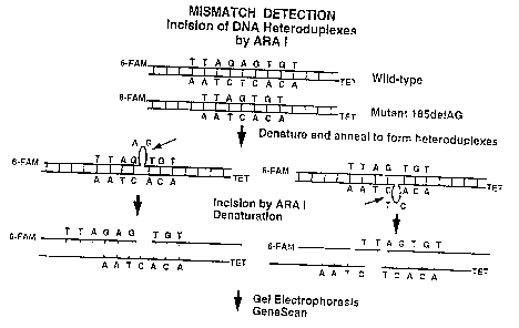
Figure 16 is a schematic diagram of the ARA
I based mismatch detection assay.
Figure 17 is an illustration of data
obtained from GeneScan analysis of endonucleolytic
activity of ARA I on a heteroduplex containing a
mismatch.
Figure 18 shows a comparison of the GeneScan
mutation detection of ARA I versus CEL I, involving a
series of control reactions using the wild type allele
of exon 19 of the BRCAI gene. This fragment of DNA
does not contain any mutations and accordingly no
mismatch nicking was observed. Panels A and B show
the two strands treated with 7 ng of CEL I and
stimulated in mismatch cutting by Amplitaq DNA
polymerase. B = (6-FAM); G = (TET). Panels C and D
are the two strands treated with 20 ng of purified ARA
I without stimulation by Amplitaq DNA polymerase.
CA 02256526 1998-11-23
WO 97/46701 PCT/LTS97/08705
- 15 -
Panels E and F show the two strands treated with 20 ng
of ARA I stimulated for mismatch cutting by the
presence of Amplitaq DNA polymerase.
Figure 19 depicts a side-by-side GeneScan
analysis of CEL I and ARA I mismatch detection
activity in exon 19 of the BRCA1 gene. Panels A and B
show mismatch cutting using 7 ng of CEL I in the
presence of 0.5 units of Amplitaq DNA polymerase.
Panels C and D show the cutting of an A nucleotide
deletion mismatch by 20 ng of ARA I without Amplitaq
DNA polymerase. Panels E and F show the cutting of
the same substrate by 2 ng of ARA I stimulated in
mismatch cutting by the presence of 0.5 ng units of
Amplitaq DNA polymerase. All mutations and
polymorphisms detected were confirmed by automated
sequencing. These results suggest that AR.A I, like
the CEL I mutation detection method, can identify
mutations that are difficult to detect with SSCP or
DNA sequencing.
Figure 20 shows a comparison of the GeneScan
mutation detection of ARA I versus CEL I, involving a
series of control reactions employing the wild type
allele of exon 2 of the BRCA1 gene. As in figure 18,
this gene segment does not contain any mutations, thus
no mismatch nicking is observed. Panel A and B show
the two strands treated with 7 ng of CEL I stimulated
in mismatch cutting by 0.5 units of Amplitaq DNA
polymerase. Panels C and D show the two strands
treated with 20 ng of purified ARA I without
stimulation by Amplitaq DNA polymerase. Panels E and
F show the two strands treated with 20 ng of ARA I
stimulated for mismatch cutting by the presence of
Amplitaq DNA polymerase. Panels G and H show the two
strands treated with 2 ng of AR.A I stimulated for
CA 02256526 1998-11-23
WO 97/46701 PCT/US97/08705
- 16 -
mismatch cutting by the presence of 0.5 units of
Amplitaq DNA polymerase.
Figure 21 depicts GeneScan analysis of CEL I and
ARA I mismatch detection in Exon 2 of the BRCA1 gene.
Panels A and B show an AG-deletion mismatch cutting by
7 ng of CEL I in the presence of 0.5 units of Amplitaq
DNA polymerase. Panels C and D show the cutting of
the AG nucleotide deletion mismatch by 20 ng of ARA I
without Amplitaq DNA polymerase. Panels E and F show
ZO the cutting of the same substrate by 20 ng of ARA I
stimulated in mismatch cutting by the presence of 0.5
ng units of Amplitaq DNA polymerase. Panels G and H
show the cutting of the same substrate by 2 ng of ARA
I stimulated in mismatch cutting by the presence of
0.5 units of Amplitaq DNA polymerase.
Figure 22 is an autoradiogram demonstrating
that mismatch endonuclease activity similar to that of
ARA I and CEL I is present in the extracts of 10 other
plants.
Figure 23 is an autoradiogram showing that
mismatch endonuclease activity similar to ARA I and
CEL I is present in the extracts of 11 other plants.
DETAILED DESCRIPTION OF THE INVENTION
The enzymatic basis for the maintenance of
correct base sequences during DNA replication has been
extensively studied in E. coli. This organism has
evolved a mismatch repair pathway that corrects a
variety of DNA basepair mismatches in hemimethylated
DNA as well as insertions/deletions up to four
nucleotides long. Cells deficient in this pathway
mutate more frequently, hence the genes are called
MutS, Mutt and Mutes etc. MutS protein binds to the
CA 02256526 1998-11-23
WO 97!46701 PCT/US97/08705
- 17 -
mismatch and Mutes is the endonuclease that incises the
DNA at a GATC site on the strand in which the A
residue is not methylated. Mutt forms a complex with
Mutes and MutS during repair. Homologs of MutS and
Mutt, but not Mutes exist in many systems. In yeast
MSH2 (MutS homology can bind to a mismatch by itself,
but a complex of two Mutt homologs (MLH and PMS1) plus
a MSH2 has been observed. The human homolog hMSH2 has
evolved to bind to larger DNA insertions up to 14
nucleotides in length, which frequently arise by
mechanisms such as misalignment at the microsatelite
repeats in humans. A role for hMLHl in loop repair is
unclear. Mutations in any one of these human homologs
were shown to be responsible for the hereditary form
of non-polyposis colon cancer (27, 28).
Celery contains over 40 ~g of psoralen, a
photoreactive intercalator, per gram of tissue (3).
As a necessity, celery may possess a high capability
for the repair of lesions of insertion, deletion, and
other psoralen photoadducts. Single-strandedness at
the site of the lesion is common to base substitution
and DNA loop lesions. The data in the following
examples demonstrate that celery, Arabidopsis thaliana
and other plant species possess ample mismatch-
specific endonucleases to deal with these potentially
mutagenic events.
It has been found that the incision at a mismatch
site by CEL I is greatly stimulated by the presence of
a DNA polymerase. For a DNA loop containing a single
nucleotide insertion, CEL I substrate preference is
A > G > T > C. For base-substitution mismatched
basepairs, CEL I preference is C/C > C/A ~ C/T > G/G >
A/C ~ A/A ~ T/C > T/G -. G/T ~ G/A ~ A/G > T/T. CEL I
shows a broad pH optimum from pH 6 to pH 9. To a
lesser extent compared with loop incisions, CEL I is
also a single-stranded DNase, and a weak exonuclease.
CA 02256526 1998-11-23
WO 97!46701 PCT/US97/08705
- 18 -
CEL I possesses novel biochemical activities when
compared to other nucleases. Mung Bean Nuclease is a
39 kd nuclease that is a single-stranded DNase and
RNase, and has the ability to nick DNA at destabilized
regions and DNA loops (19-22). However, it has a pH
optimum at 5Ø It is not known whether Mung Bean
Nuclease activity can be stimulated by a DNA
polymerase as in the case of CEL I. Thus CEL I and
Mung Bean Nuclease appear to be different enzymes;
however this has not yet been conclusively confirmed.
The mechanism responsible for the AmpliTaq
DNA polymerase stimulation of the CEL I activity is
presently unknown. One possibility is that the DNA
polymerase has a high affinity for the 3'-OH group
produced by the CEL I incision at the mismatch and
displaces CEL I simply by competition for the site.
CEL I may have different affinities for the 3'-OH
termini generated by incisions at different
mismatches, thereby attenuating the extent that
AmpliTaq DNA polymerase can stimulate its activity.
The use of a DNA polymerase to displace a repair
endonuclease in DNA repair was also observed for the
UvrABC endonuclease mechanism (25). It was shown that
the UvrABC endonuclease does not turnover unless it is
in the presence of DNA polymerase I. The protein
factors in vivo that can stimulate the CEL I activity
may not be limited to DNA polymerases. It is possible
that DNA helicases, DNA ligases, 3'-5' exonucleases or
proteins that bind to DNA termini may perform that
function.
It is important to note that a 5'-labeled
substrate can be used to show a CEL I incision band in
a denaturing polyacrylamide gel. Recently, a putative
human all-type mismatch incision activity (24) was
shown to be related to the human topoisomerase I.
This enzyme is unable to release itself from a 5'-
CA 02256526 1998-11-23
WO 97/46701 PCT/US97/08705
- 19 -
labeled substrate after mismatch nicking due to the
formation of a covalent enzyme-DNA intermediate with
the 3' terminus of the DNA nick (26). This covalent
protein-DNA complex cannot migrate into the denaturing
polyacrylamide gel to form a band. CEL I mismatch
nicking has been demonstrated with 5' labeled
substrates. Therefore, CEL I is not a plant
equivalent of the topoisomerase I-like human all-type
mismatch repair activity.
CEL I appears to be a mannopyranosyl
glycoprotein as judged by its tight binding to
Concanavalin A-Sepharose resin and by the staining of
CEL I with the Periodic acid-Schiff glycoprotein
stain. Insofar as is known, no repair enzyme has been
demonstrated to be a glycoprotein. Glycoproteins are
often found to be excreted from the cell, on cellular
membranes or secreted into organelles. However,
glycoproteins have also been shown to exist in the
nucleus for important functions. The level of a 100
kDa stress glycoprotein was found to increase in the
nucleus when Gerbil fibroma cells are subjected to
heat shock treatment {27). Transcription factors for
RNA polymerase II in human cells are known to be
modified with N-acetylglucosamine residues (28, 29).
Recently, lactoferrin, an iron-binding glycoprotein,
was found to bind to DNA in the nucleus of human cells
and it activated transcription in a sequence-specific
manner {30). The nuclei of cells infected with some
viruses are known to contain viral glycoproteins {31-
33). These examples where glycoproteins are known to
exist inside the nucleus, not merely on the nuclear
membrane or at the nuclear pores, tend to show that
glycosylated proteins may be important in the nucleus.
CEL I appears to be an example of a glycoprotein that
can participate in DNA repair.
The properties of the celery mismatch
CA 02256526 2002-05-31
- 20 -
endonuclease CEL I resemble those of single-stranded
nucleases. The best-suited substrates for CEL I are
DNA loops and base-substitution mismatches such as the
C/C mismatch. In contrast, loops greater than 4
nucleotides and the C/C mismatch are the substrates
worst-suited for the E. coli mutHLS mismatch repair
system (1,2). Thus CEL I is an enzyme that possesses
novel mismatch endonuclease activity.
The following examples are provided to
describe the invention in further detail. These
examples, which set forth the best mode presently
contemplated for carrying out the invention, are
intended to illustrate and not to limit the invention.
Exaanple I
Purification of CEL I
Two different CEL I preparations were made
up as described below. Their properties are similar
except that the less pure preparation (Mono Q"
fraction) may contain protein factors that can
stimulate the CEL I activity.
(i) Preparation of CEL I Mono 0 fraction
100 gm of celery stalk was homogenized in a
blaring*blender with 100 ml of a buffer of 0.1 M
Tris-HC1 pH 7.0 with 10 ~M phenylmethanesulfonyl
fluoride (PMSF) (Buffer A) at 4 °C for 2 minutes. The
mixture was cleared by centrifugation, and the
supernatant was stored at -70°C. The extract was
fractionated by anion exchange chromatography on a
FPLC Mono Q HR5/10 column. The bound CEL I nuclease
activity was eluted with a linear gradient of salt at
about 0.15 M KC1.
*Trade-mark
CA 02256526 2002-05-31
- 21 -
(ii) Preparation of highly purified CEL I
7 Kg of celery at 4 °C was extracted with a
juicer and adjusted with lOX Buffer A to give a final
concentration of 1X Buffer A. The extract was
concentrated with a 25% to 85o saturation ammonium
sulfate precipitation step. The final pellet was
dissolved in 250 ml of Huffer A and dialyzed against
0.5 M KC1 in Buffer A. The solution was incubated
with 10 ml of Concanavalin A-Sepharose*resin (Sigma)
overnight at 4 °C. The slurry was packed into a 2.5
cm diameter column and washed with 0.5 M KC1 in Buffer
A. The bound CEL I was eluted with 60 ml of 0.3 M a-D
mannose, 0.5 M KC1 in Buffer A at 65 °C. The CEL I
was dialyzed against a solution of 25 mM KP04, 10 ACM
PMSF, pH 7.4 (Buffer B), and applied to a
phosphocellulose column that had been equilibrated in
the Buffer B. The bound enzyme was eluted with a
linear gradient of KC1 in Buffer B. The peak of CEL I
activity from this column was further fractionated by
size on a Superose 12*FPLC column in 0.2 M KC1, 1 mM
ZnClz, 10 ~,M PMSF, 50 mM Tris-HC1 pH 7.8. The center
of the CEL I peak from this gel filtration step was
used as the purified CEL I in this study. A protein
band of about 34,000 daltons is visible when 5
micrograms of CEL I of the Superose 12 fraction was
visualized with Coomassie Blue staining or
carbohydrate staining (Periodic acid-Schiff base
mediated staining kit, SIGMA Chemicals (5)) on a 15%
polyacrylamide SDS PAGE gel as shown in Figure 1. A
second band of approximately 36,000 daltons was also
visible in the gel. Both bands were stained with the
glycoprotein specific stain. The subtle mobility
differences observed in the two bands may be due to
differential glycosylation. Alternatively, there may
be a contaminant in the preparation which co-purifies
with CEL I.
*Trade-mark
CA 02256526 1998-11-23
WO 97/46701 PCT/US97/08705
- 22 -
Protein determination
Protein concentrations of the samples were
determined by the Bicinchoninic acid protein assay (4,
Pierce ) .
Following purification of CEL I enzyme,
mutational analysis on experimental and clinical DNA
substrates were performed in a suitable gel system.
CEL I recognized and cleaved DNA at a variety of
mismatches, deletions and insertions. The following
examples describe in greater detail the manner in
which mutational analysis is practiced according to
this invention.
EXAMPLE II
Preparation of heteroduplexes
containina various mismatches
DNA heteroduplex substrates of 64 basepairs
long were constructed containing mismatched basepairs
or DNA loops which were. prepared using similar methods
reported in Jones and Yeung (34). The DNA loops are
composed of different nucleotides and various loop
sizes as illustrated in Fig. 2. The DNA duplexes were
labeled at one of the four termini so that DNA
endonuclease incisions at the mispaired nucleotides
could be identified as a truncated DNA band on a
denaturing DNA sequencing gel. The oligonucleotides
were synthesized on an Applied Biosystems DNA
synthesizer and purified by using a denaturing PAGE
gel in the presence of 7M urea at 50 °C. The purified
single-stranded oligonucleotides were hybridized with
appropriate opposite strands. The DNA duplex,
containing mismatches or not, was purified by using a
nondenaturing PAGE gel. DNA was eluted from the gel
slice by using electro-elution in a Centricon unit in
an AMICON model 57005 electroeluter. The upper
reservoir of this unit has been redesigned to include
CA 02256526 1998-11-23
WO 97/46701 PCT/US97/08705
- 23 -
water-tight partitions that prevent cross-
contamination.
EXAMPLE III
Mismatch endonuclease assay
Fifty to 100 fmol of 5' [32P] -labeled
substrate described in Example II were incubated with
the Mono Q CEL I preparation in 20 mM Tris-HC1 pH 7.4,
25 mM KC1, 10 mM MgCl2 for 30 minutes at temperatures
of 0 °C to 80 °C. From one half to 2.5 units of
AmpliTaq DNA polymerase was added to the nuclease
assay reaction. Ten ~,M dNTP was included in the
reaction mixture where indicated (Figures 2 & 5). The
JCL reaction was terminated by adding 10 ~,L of 1.5 %
SDS, 47 mM EDTA, and 75% formamide plus tracking dyes
15 and analyzed on a denaturing 15% PAGE gel in 7M urea
at 50°C. An autoradiogram was used to visualize the
radioactive bands. Chemical DNA sequencing ladders
were included as size markers. Incision sites were
accurately determined by co-electrophoresis of the
20 incision band and the DNA sequencing ladder in the
same lane.
Example IV
The Effect of Temperature on CEL I Incision
Activity at single-nucleotide DNA
loop and nucleotide substitutions
The CEL I fraction eluted from the Mono Q
chromatography of the celery extract was found to
specifically nick DNA heteroduplexes containing DNA
loops with a single extrahelical guanine (substrate
#2) or thymine residue (#3), but not the perfectly
basepaired DNA duplex #1 as shown in Fig. 3. In these
experiments fifty fmol of heteroduplex #2 (lanes 3-9),
#3 (lanes 10-16), perfectly basepaired duplex #1
(lanes 17-23) and single-stranded DNA substrate (lanes
CA 02256526 1998-11-23
WO 97/46701 PCT/LTS97/08705
- 24 -
24-30) , each labeled at the 5' -terminus with 'y- [3zp]
ATP and T4 polynucleotide kinase at about 6000
Ci/mmol, were incubated with 0.5 ~L (10 ~.g) of the
Mono Q fraction of the CEL I preparation in 20 mM
Tris-HCl pH 7.4, 25 mM KC1, 10 mM MgCl2 for 30 minutes
at various temperatures. Each 20 ~,L reaction was
terminated by adding 10 ~,L of 1.5% SDS, 47 mM EDTA,
and 75% formamide containing xylene cyanol and
bromophenol blue. Ten ~.L of the sample was loaded
onto a 15% polyacrylamide, 7 M urea denaturing DNA
sequencing gel at about 50 °C, and subjected to
electrophoretic separation and autoradiography as
previously reported (7). The G+A and the T chemical
sequencing reactions were performed as described (7)
and used as size markers. CEL I incision produced
bands at about 35 nucleotides long. Lines are drawn
from the positions of the incision bands to the
phosphodiester bonds (I and II) nicked by the
endonuclease in the reference sequencing ladder. For
a 5'-labeled substrate, when a nuclease nicks 5' of a
nucleotide and produces a 3'-OH terminus, the
truncated band runs half a nucleotide spacing slower
than the band for that nucleotide in the chemical DNA
sequencing reaction product lane (34).
Substrate #2 can basepair in two
conformations because the inserted G is within a CGCG
sequence. Therefore either the G residue in the
second or the third nucleotide position can become
unpaired, possibly extrahelical in conformation, when
this duplex is hybridized:
5'-CGGCG-3' or 5'-CGGCG-3'
3'-G-CGC-5' S'-GC-GC-5'
Accordingly, two mismatch incision bands were
observed, each correlating to the phosphodiester bond
CA 02256526 1998-11-23
WO 97/46701 PCT/LTS97/08705
- 25 -
immediately 3' of the unpaired nucleotide. See Fig.
3, lanes 3-9. This slippage can occur in the target
sequence only when G or C is in the mismatched top
strand. Therefore, the non-paired T residue in
substrate #3 gave one incision band at the same
relative position as the upper band derived from the
substrate #2. See Fig. 3, lanes 10-16. These gel
mobilities are consistent with the production of a 3'-
OH group on the deoxyribose moiety (6). CEL I
increases in activity with temperature up to 45°C as
illustrated by the increase in band intensity, see
Fig. 3. However, from 65°C to 80°C, specificity is
diminished due to DNA duplex denaturation.
EXAMPLE V
Relative Incision Preferences of CEL I
To ascertain whether there is a single
endonuclease incision at each DNA duplex, the
experiment described in Fig. 3 was repeated with DNA
labeled on the 3' terminus of the top strand. If
there were only one incision site, initial incision
positions revealed by substrates labeled at the 5' or
the 3' termini should be at the same phosphodiester
bond. In these experiments, substrates were labeled
at the 3' termini with [32P] a-dCTP, cold dGTP and the
Klenow fragment of DNA polymerase I to about 6000
Ci/mmol. The sample preparation, denaturing gel
resolution and autoradiogram analysis are the same as
described in Fig. 3 except incubation of 50 fmole of
substrate with 10 ug of the CEL I Mono Q fraction was
for 30 minutes at a single temperature, 37°C. The DNA
sequencing ladders for substrates #4 and #5 are shown
in lanes 1-4 to illustrate the DNA sequences used.
Lanes 5-8 had no enzyme during the incubation. Lanes
9-12 are mismatch endonuclease incisions of the
substrates #2, #4, #5, #3, respectively. A line is
CA 02256526 1998-11-23
WO 97/46701 PCT/ITS97/08705
- 26 -
drawn from the position of the incision band to the
phosphodiester bond (I) nicked by the endonuclease in
the reference sequencing ladder. Lanes 13 and 14
demonstrate the coelectrophoresis of the CEL I
incision band with a chemical DNA sequencing ladder to
accurately determine the incision position.
Relative incision preferences for substrates #2, #3,
#4, and #5 are shown in Fig. 4 for the 3' labeled
substrates. The mobilities of the incision bands in
lanes 9-12 of Fig. 4 indicate that the incision
reactions had occurred at the phosphodiester bond
immediately 3' of the unpaired nucleotide. Therefore,
the incision site is the same for substrates labeled
either at the 5' or the 3' terminus. The fact that
the DNA incision was found to occur at the same bond
position, whether the substrate DNA was labeled at the
5' termini or the 3' termini shows that CEL I is not a
DNA glycosylase. A DNA glycosylase mechanism would
cause the DNA incision position in the two DNA
substrates to be one base apart because a base is
excised by the DNA glycosylase.
Precise determination of the incision site was
performed as in the example in lane 14 in which the T
residue chemical sequencing reaction of the labeled
top strand of substrate #2 (lane 13) was mixed with
the CEL I incision product of lane 9 and analyzed in
the same lane. For a 3'-labeled substrate, when a
nuclease nicks 3' of a nucleotide and produces a 5' P04
terminus, the truncated band runs with the band for
that nucleotide in the chemical DNA sequencing
reaction product lane (7). Moreover, the gel
mobility, relative to the size standards of chemical
DNA sequencing, illustrated that the DNA nick produced
a 5'-phosphorylated terminus (6). For a DNA loop with
a single nucleotide insertion, the nuclease
specificity is A > G> T > C. It can be seen in Fig.
CA 02256526 1998-11-23
WO 97!45701 PCT/US97/08705
- 27 -
4A that a small amount of 5' to 3' exonuclease
activity is present in this CEL I preparation.
To test whether CEL I can cut in the bottom
strand across from a DNA loop of one nucleotide in the
top strand, or whether nicking of the loop-containing
strand may lead to secondary CEL I incision across
from the nick, the bottom strand that contains no
unpaired nucleotides in substrate #2 was labeled at
the 3' end and incubated in the presence of CEL I.
The extrahelical nucleotide in the top strand, or the
DNA nick made by CEL I in the top strand of substrate
#2, seen in lane 9 of Fig. 4, did not lead to
significant nicking of the bottom strand (lane 18).
As a control against the possibility that DNA sequence
effect may favor CEL I incision in the top strand and
not the bottom strand, CEL I was tested for incision
of the bottom strand in the C/C mismatch substrate in
lanes 15 and 16. Mismatch incision was made when CEL
I was present in lane 16.
In the characterization of the incision site
of a repair endonuclease, it is important to determine
whether one or two incisions have been made for each
lesion. This is normally accomplished by using
lesion-containing substrates that have been labeled,
in turn, at the four termini of a DNA duplex. This
test has been satisfied in the analysis of substrate
#2 by using three labeled substrates because of the
near absence of incision in the bottom strand. In
Fig. 3, lane 4-7 and Fig. 4, lane 9, respectively, the
incision of this substrate in both the 5' labeled and
the 3' labeled substrates have been compared. The
incision site was found to be at the 3' side of the
mismatched nucleotide in both cases. The lack of
incision on the bottom strand for substrate #2 was
demonstrated in lane 18 of Fig. 4. Only the 5'
labeled substrate was needed in this case since no
CA 02256526 1998-11-23
WO 97/46701 PCT/LTS97/08705
- 28 -
significant bottom strand incision had occurred.
Example yI
Effect of AmpliTaq DNA polymerase on the
incisions at DNA loop mismatches
CEL I activity is stimulated by the presence
of a DNA polymerase. In Fig. 5, the CEL I incisions
at single-nucleotide loop substrates were stimulated
by AmpliTaq DNA polymerase to different extents
depending on which nucleotides are present in the
loop. It was necessary to use different amounts of
CEL I to illustrate the AmpliTaq DNA polymerase
stimulation. The stimulation of the incision at
extrahelical C and extrahelical T substrates are best
illustrated in Figs. 5 A & B (compare lanes 4 with
lanes 9, and lanes 5 with lanes 10, in the respective
panels) where higher CEL I levels are required to show
good incision at these mismatches. For extrahelical G
and extrahelical A substrates that are among the best
substrates for CEL I, AmpliTaq DNA polymerase
stimulation can best be illustrated by using a much
lower level of CEL I as in Fig. 5. The amounts of
AmpliTaq stimulation of CEL I in Fig. 5 were
quantified and presented in Table I.
CA 02256526 1998-11-23
WO 97/46701 PCT/US97/08705
- 29 -
Table I
Quantification of the CEL I incision bands
shown in the autoradiogram in Fig. 5.
'~pliTaq
Substrate Panel Counts Panel Counts
lane# lane#
Extrahelical G,bandA,2 20894 A,7 22101 1.1
I
Extrahelical A,bandA,3 19451 A,8 26357 1.4
I
Extrahelical C,bandA,4 4867 A,9 12009 2.5
I
Extrahelical T,bandA,5 2297 A,10 25230 11.0
I
Extrahelical G,bandB,2 12270 B,7 19510 1.6
I
Extrahelical A,bandB,3 10936 B,8 24960 2.3
I
Extrahelical C,bandB,4 1180 B,9 2597 2.2
I
Extrahelical T,bandB,5 700 B,10 21086 30.1
I
Extrahelical G,bandC,11 10409 C,13 18649 1.8
I
Extrahelical G,bandIIC,11 9020 C,13 19912 2.2
Extrahelical A,bandC,12 7165 C,14 14983 2.1
I
The Autoradiograms were quantified in two dimensions with an
AMBIS densitometer and the amount of signal in each band is given
as counts.
Example VII
Optimum pIi of CEL I Activity
The pH optimum of CEL I for the extrahelical
G substrate was investigated in the absence or
presence of the AmpliTaq DNA polymerase. CEL I
(9.5 ng) was incubated with 100 fmol of the substrate
in a 20 ~,L reaction in buffers of pH 5-6.5 (imidazole)
and pH 7-9.5 (Tris-HCl) for 30 minutes at 37 °C. When
used, one half unit of AmpliTaq DNA polymerase was
present in the incubation in the top (- polymerase) or
bottom panels (+ polymerase), respectively. As shown
in Fig. 6, CEL I was found to be active from pH 5.0 to
pH 9.5, and showed a broad pH optimum centered about
CA 02256526 1998-11-23
WO 97/46701 PCT1US97/08705
- 30 -
pH 7.5 (top panel). When AmpliTaq DNA polymerase was
present, the incision was stimulated across the whole
pH range (bottom panel). The assay method did not use
initial kinetics and thus precluded quantitative
conclusions on this pH profile of CEL I. However, it
is clear that the enzyme works very well in the
neutral pH ranges.
Example VIII
Incisions by CEL I at basepair substitutions
Other combinations of mismatched substrates
are also recognized by CEL I and incised on one of the
two DNA strands of each DNA duplex. Some of these
substrates are less efficiently incised compared with
those containing DNA loops; therefore 45°C was used
for incubation instead of 37 °C. Substrates with the
5' termini of the top strands labeled were used in
this study. The autoradiogram of Fig. 7 shows that
mismatches containing a C residue are the preferred
mismatch substrates with C/C often better than C/A and
C/T. The incisions at these mismatches tend to
produce two alternate incision positions, one at the
phosphodiester bond 3' of the mismatched C residue,
one at the phosphodiester bond one nucleotide further
removed in the 3' direction. Whether alternate
incision sites will be observed for these mismatches
within another DNA sequence context has not been
investigated. One possible explanation for this
phenomenon may be greater basepair destabilization
next to a mismatch that contains a C residue than for
other base-substitutions. Alternatively, the specific
mismatched nucleotide may shift one position to the 3'
side because the next nucleotide is also a C residue
and the two residues can exchange their roles in the
pairing with the G residue in the opposite DNA strand.
For base substitution mismatched basepairs, CEL I
CA 02256526 1998-11-23
WO 97/46701 PCT/IJS97/08705
- 31 -
specificity in the presence of AmpliTaq DNA
polymerase, with respect to the top strand, is C/C >
C/A -. C/T > G/G > A/C ~ A/A ~ T/C > T/G ~ G/T ~ G/A
A/G > T/T (Fig. 7A). Because eubacterial DNA
polymerases are known to incise at unusual DNA
structures (8), a test was conducted to determine
whether AmpliTaq DNA polymerase by itself will incise
at the 13 substrates used in Fig. 7. Under extended
exposure of the autoradiogram, no mismatch incision by
the AmpliTaq DNA polymerase was observed (Fig. 7B).
Example IX
Detection of DNA mutations
Usina CEL-I and Multiplex Analysis
The sensitivity of CEL I for mismatch
detection is illustrated by its ability to detect
mutations in pooled DNA samples. DNA was obtained
from peripheral blood lymphocytes from individuals
undergoing genetic screening at the Fox Chase Cancer
Center. Samples were obtained from breast cancer-
only, ovarian cancer-only, breast/ovarian cancer
syndrome families or from non-breast/ovarian cancer
control samples. Unlabeled primers specific for exon
2 of BRCA1 were utilized to PCR amplify this region of
the gene. The wild-type PCR products of exon 2 were
labeled with gamma 32P-ATP. Briefly, 10 picomoles of
PCR product were purified by the Wizard procedure
(Promega). Exon 2 wild-type products were then
phosphorylated using T4 kinase and 15 picomoles of
gamma 32P-ATP at 6,000 Ci/mmol in 30 ~1 1X kinase
buffer (70 mM Tris-HCl (pH 7.6), 10 mM MgCl2, 5 mM
dithiothreitol) at 37°C for 1 hour. The reactions
were stopped with 1 ~,1 0.5 M EDTA. The reaction
volume was brought up to 50 ~1 with 1X STE buffer (100
mM NaCl, 20 mM Tris-HC1, pH 7.5, 10 mM EDTA) and
processed through a Pharmacia Probe Quant column.
Labeled DNA (1 pmol/~,1 in 100 ~1) was then used for
CA 02256526 1998-11-23
WO 97/46701 PCT/US97/08705
- 32 -
hybridization with individual unlabeled PCR amplified
experimental samples. For each individual sample, 100
fmol of the unlabeled PCR amplified product was
incubated with 200 fmol of the 3zP-labeled wild-type
PCR product in CEL I reaction buffer (25 mM KCl, 10 mM
MgCl2, 20 mM Tris-HC1, pH 7.5). Following denaturation
and renaturation, heteroduplexed, radiolabeled PCR
products were exposed to CEL I for 30 minutes at 37°C
in 1X CEL reaction buffer and stopped via the addition
of 10 ~.1 stop mix (75o formamide, 47 mM EDTA, 1.50
SDS, xylene cyanol and bromophenol blue). The
heteroduplexes were treated with the enzyme
individually (lanes 4-13) or pooled in one sample tube
(lane 14) and treated. The products of the reaction
were loaded onto a 15o polyacrylamide gel containing 7
M urea and the results are shown in Fig. 8. Out of
the 10 samples analyzed, 2 contained an AG deletion
(lanes 4 and 7), 2 contained an 11 base-pair loop
(lanes 8 and 9), and the other 6 were wild type (lanes
5, 6, 10, 11, 12, and 13). Cleavage by CEL I at the
AG deletion resulted in the formation of two bands,
one of approximately 151 nucleotides from the top
strand, the other at 112 nucleotides from the bottom
strand (lanes 4 and 7). Cleavage by CEL I at 11 base-
pair loops resulted in the formation of one band at
147 nucleotides from the top strand, and a group of
bands at 109 nucleotides in the bottom strand (lanes 8
and 9). Lanes 1, 2 and 3 contain DNA that was not
exposed to CEL I as negative controls, lane 15
contains 64 and 34 by nucleotide markers. As can be
seen in lane 14 of the gel, when the samples were
pooled and exposed simultaneously to CEL I, the enzyme
cleaved at all of the above listed mutations with no
loss of specificity. Also, the PCR products of the
wild-type samples showed no non-specific DNA nicking.
To further illustrate the ability of CEL-I
CA 02256526 1998-11-23
WO 97/46701 PCT/US97/08705
- 33 -
to detect mutations in pooled DNA samples, 1, 2, 3, 5,
or 30 heteroduplexed, radiolabelled PCR products,
(again amplified from exon 2 of the BRCAI gene), were
exposed to CEL-I in a single reaction tube and the
5 products run on a 6% polyacrylamide gel containing 7M
urea. Samples were amplified and radiolabeled as
described above. Each pool contained only one sample
which had a mutation (AG deletion). The other samples
in each pool were wild-type. Lanes 1 and 2 contain
10 control samples which were not exposed to CEL I. In
the pooled samples where a mutation was present, CEL-I
consistently cleaved the PCR products illustrating the
sensitivity of the enzyme in the presence of excess
wild-type, non-mutated DNA {Lanes 4, 5, 6, 7, 8, 9,
and 11). As a control, heteroduplexed PCR products
containing no mutations were analyzed and no cut band
corresponding to a mutation appeared (Fig. 9, lanes 3
and 10 ) .
EXAMPLE X
Detection of Mutations and Polymorphisms by
CEL-I in Samples Obtained from Hiah Risk Families
PCR primer sets specific for the exons in
the BRCA1 gene have been synthesized at Fox Chase
Cancer Center. The gene sequence of BRCA1 is known.
The exon boundaries and corresponding base numbers are
shown in table II. Primers to amplify desired
sequences can be readily designed by those skilled in
the art following the methodology set forth in Current
Protocols in Molecular Bioloav, Ausubel et al., eds,
John Wiley and Sons, Inc. (1995). These primers were
planned such than in each PCR reaction, one primer is
labeled at the 5' termini with a fluorescent-label, 6-
FAM, while the other primer is similarly labeled with
a label of another color, TET. A PCR product will
thus be labeled with two colors such that DNA nicking
events in either strand can be observed independently
CA 02256526 1998-11-23
WO 97/46701 PCT/US97/08705
- 34 -
and the measurements corroborated. A summary of the
results is presented in Table III.
TABLE II
EXON BOUNDARIES AND CORRESPONDING
BASED NUMBERS IN BRCA1
EXON BASE #'s
1 1 -100
2 101 - 199
3 200 - 253
5 254 - 331
6 332 - 420
7 421 - 560
8 561 - 665
9 666 - 712
10 713 - 788
11 789 - 4215
11B 789 - 1591
11C 1454 - 2459
11A 2248 - 3290
11D 3177 - 4215
12 4216 - 4302
13 4303 - 4476
14 4477 - 4603
15 4604 - 4794
16 4795 - 5105
17 5106 - 5193
lg 5194 - 5273
lg 5274 - 5310
20 5311 - 5396
21 5397 - 5451
22 5452 - 5526
23 5527 - 5586
24 5587 - 5711
CA 02256526 1998-11-23
WO 97/46701 PCT/LTS97/08705
- 35 -
Fig. 10 depicts a schematic of the exons present
in the BRCAI gene. Peripheral blood samples from
individuals in high risk families were collected and
the DNA isolated. The PCR products were amplified
using Elongase (BRL) and purified using Wizard PCR
Preps (Promega). The DNA was heated to 94°C and
slowly cooled in 1X CEL I buffer (20 mM Tris-HC1 pH
7.4, 25 mM KC1, 10 mM MgCl2) to form heteroduplexes.
The heteroduplexes were incubated in 20 ~1 1X CEL I
buffer with 0.2 ~1 of CEL I and 0.5 units of AmpliTaq
at 45°C for 30 minutes. The reactions were stopped
with 1 mM phenanthroline and incubated for an
additional 10 minutes at 45°C. The sample was
processed through a Centricep column (Princeton
Separations) and dried down. One microliter of ABI
loading buffer (25 mM EDTA, pH 8.0, 50 mg/ml Blue
dextran), 4 ~,l deionized formamide and 0.5 ~,1 TAMRA
internal lane standard were added to the dried DNA
pellet. The sample was heated at 90°C for 2 minutes
and then quenched on ice prior to loading. The sample
was then loaded onto a 4.25% denaturing 34 cm well-to-
read acrylamide gel and analyzed on an ABI 373
Sequencer using GENESCAN 672 software. The 6-FAM
labelled primer in this experimental sample was at
nucleotide 3177 of the BRCA1 cDNA (region 11D), the
TET labelled primer was 73 nucleotides into the intron
between exon 11 and exon 12. Each spike represents
the presence of a DNA band produced by the cleavage of
the heteroduplex by CEL-I where a mutation or a
polymorphism is present. One spike represents the
size of the CEL I produced fragment from the 3' side
of the mismatch site to the 5' 6-FAM label of the top
strand. The other spike represents the corresponding
fragment in the bottom strand from the 3' side of the
mismatch to the 5' TET label. The sum of the two
CA 02256526 1998-11-23
WO 97/46701 PCT/US97/08705
- 36 -
fragments equals one base longer than the length of
the PCR product. The 6-FAM panel shows a spike at
base #645 from the 6-FAM label and the TET panel shows
a spike at base #483 from the TET label, both
corresponding to the site of the 5 base deletion at
nucleotide 3819 of the BRCA1 cDNA (Fig. 11).
Analysis of exon 11 in another individual
was performed using a 6-FAM-labelled primer at
nucleotide 1454 of the BRCA1 cDNA (Fig. 12). The TET-
l0 labelled primer was at nucleotide 2459 (region 11C).
The PCR amplified products were amplified and prepared
as described above. In this individual, the 6-FAM
panel shows a spike at base #700 and the TET panel
shows a spike at #305, each spike corresponding to the
site of CEL I incision in the respective DNA strand at
a nonsense mutation of A>T at nucleotide 2154 of the
BRCA7. cDNA. The 6-FAM panel also shows a spike at
base #747 and the TET panel shows a spike at #258
corresponding to the site of a polymorphism C>T at
nucleotide 2201 of the BRCA1 cDNA. The nonsense
mutation and polymorphism have been confirmed by
sequencing of this particular sample (KO-11) using the
ABI 377 Sequencer. Spikes that are marked with an
asterisk are alsc present in the no enzyme control
lane and represent PCR product background.
Certain individuals have mutations in
another region of exon 11, region 11A, on the
schematic in Fig. 10. A 6-FAM-labelled primer at
nucleotide 2248 of the BRCA1 cDNA and a TET labeled
primer at nucleotide 3290 were used to amplify this
region of exon 11. Following amplification, the
samples were processed as described above. The four
6-FAM panels represent CEL-I reactions with 4
different individual samples. The first panel in Fig.
13A, sample #KO-2, shows one spike at #182
corresponding to the site of a polymorphism T>C at
CA 02256526 1998-11-23
WO 97/46701 PCTlUS97/08705
- 37 -
nucleotide 2430 and a second spike at nucleotide #483
corresponding to the site of another polymorphism C>T
at nucleotide 2731. The second panel, Fig. 13B,
sample #KO-3, shows only the second polymorphism. The
third panel, Fig. 13C, sample #KO-7 shows no
polymorphism. The fourth panel, Fig. 13D, sample #KO-
11, shows two spikes corresponding to the two
polymorphisms. It is interesting to note that this
sample, KO-11, shows up positive for a nonsense
mutation and a polymorphism in the region of exon 11C
corresponding to nucleotides 1454-2459 as described
above.
TABLE III
SUMMARY OF BRCA1 MUTATIONS
AND POLYMORPHISMS DETECTED BY CEL I
EXON NUCLEOTIDE TYPE OF
POSITION # MUTATION
2 185 AG deletion
2 188 11 base
deletion
11 C 2154 A > T
11 D 3819 5 base deletion
11 D 4168 A > G
11 D 4153 A deletion
11 D 4184 4 base deletion
20 5382 C insertion
CA 02256526 1998-11-23
WO 97/46701 PCT/US97/08705
- 38 -
EXON NUCLEOTIDE TYPE OF
POSITION # POLYMORPHISM
11 B 1186 A > G
11 C 2201 T > C
11 A 2430 T > C
11 A 2731 C > T
11 D 3667 A > G
Table IV sets forth the 5' and 3' flanking
sequences surrounding the mutations detected by CEL I
in the present invention. While not exhaustive, it
can be seen from the variability of the flanking
sequences surrounding these mutations and
polymorphisms that CEL I sensitivity and recognition
of mismatched DNA heteroduplexes does not appear to be
adversely affected by flanking sequences.
CA 02256526 1998-11-23
WO 97/46701 PCT/US97/08705
- 39 -
TABLE Iv
EFFECT OF FLANRINC3 SEQBENCES ON ENDONUCLEASE ACTIVITY OF CEL I
nucleotide EXON type of 5' flanking 3' flanking
position change sequence se quence
i
185 2 AG deletion5' ATCTT 5' AGTGT3'
3'
TAGGA TCACA
188 2 llbp 5' TTAGA3' S' G
deletion AATCT th e 4 by
next
ar e
in
intron
1186 11 B A--> G 5' TAAGC3' S' GAAAC3'
ATTCG CTTG
2154 11 C A--> T 5' GAGCC3' 5' AGAAG3'
CTCGG TCTTC
2201 11 C T--> C 5' GACAG3' 5' GATAC3'
CTGTC CTATG
1 0 2430 11 A T--> C 5' AGTAG3' 5' AGTAT3'
TCATC TCATA
2731 11 A C--> T 5' TGCTC3' S' GTTTT3'
ACGAG CAAAA
3667 11 D A--> G 5' CAGAA3' 5' GGAGA3'
CTCTT CCTCT
3819 11 D 5 by 5' GTAAA3' S' CAATA3'
deletion CATTT GTTAT
4153 11 D A deletion5' TGATG3' S' AGAAA3'
ACTAC TCTTT
I 5 4184 11 D 4 by 5' AATAA3' S' GAAGA3'
deletion TTATT CTTCT
4168 11 D A--> G 5' AACGG3' S' CTTGA3'
TTGCC GAACT
5382 20 C insertion5' ATCCC3' 5' AGGAC3'
TAGGG TCCTG
As can be seen from the above described
20 examples, utilization of CEL I has distinct advantages
over methods employing other mismatch repair systems
during analysis of mutations in the clinical setting.
These advantages are summarized in Table V.
CA 02256526 1998-11-23
WO 97/46701 PCT/US97/08705
- 40 -
11 H N TI1N H N N N N N N
N W dl 11 CI W C1 W CI W pl W
W 7 ~ > >. >. >. >. !.
II r . . .
H ~
_
> a a g " ~ a g c c
a
~
IIl . . . = i
,~
~I r~.. w
J ~ a
g
d O p O O O
~ 'lW.~ C C . ~ C C C
<
N
W ~z~ s
v ~ c y c ~ c j c c c
... ~ c ,
z "
E
a
H O 3
L
V ~ W N H H H H = N H L O
~ U r,
7
~' ,~ W ~. ~ ~. )' ~ !. ~' s
~ ~
w
N
U_ _
E
~
J
L
_
~~
U N H ~ W p O N N p N
~
V ~ W ~' i a C 'Y ~ ~ C 7.
r ~ W
.. _..
V. a N
~C
6
1
N ~ y N N H
7 7
W n H p p
v W ~ >. ~. C C .
~ ~ 4-
W Z Z - Z
N . . ?
w v-
w w w w
'O ' 9 ~
L O
L
~
a s
~ W C C C ~ T C C
, lv-
~~
E ~J 7
L
c c
I1 W N O N p p H H p O
ii
H ~ C ~ y T C c >' ~' c c -~c
~ ~
7 7
V
'~ ' ~ a
o
~
N ~ d
~
V a ~~
"
~ i t z < o i
' ~i ~ 1.M
< ~ a. ac .~. z i. ..
.. 6 a ,
- i
'Y!~~ ~ ~~ _
N ~ ~
~U
~ 1
0 '
O O O N .'' ~ .
~ v- = N ~
W
'
w O O W r- r.
W ~ O
w
. dl W V ~ N H Ci dl
1ra r a r V .- ~ W O 0
a V~ OI 01~.~ Of 01
7 ~
W W W W W W W >. !. W W
N C .. .~ .. W N N
.. ... i, ..~ _~ ..
. a ..- c ii
~ _ ...U.r C~ CW ~1,1,~ C'r .
~ rIJ C ' . .
QIU
W01
~r ~r .r W W W ..r ~ iL ~
Y ~ ~ .r N W _ i
~ ~ ~~ ~ W
C
N ~ ~ ~ .~ <~~ <~~ N 4
t .N
CA 02256526 1998-11-23
WO 97/46701 PCT/US97/08705
- 41 -
EXAMPLE XI
As noted above, many plant species
synthesize efficient endonuclease enzymes. In
accordance with the present invention, a novel
endonuclease, AR.A I has been isolated from Arabidopsis
thaliana. This endonuclease is quite similar to CEL I
in many respects. Arabidopsis tha3iana is considered
to provide a model system for studies in plant
molecular biology and biochemistry. Advantages of
the Arabidopsis system include a short life cycle of
about 26 days, the small size of the plant, the
diploid nature of the genome, and especially, the
small size of the genome compared with most higher
plants and animals. The Arabidopsis genome, at 7 x 10~
basepairs, is only about 10 times larger than that of
E. coli (4 x lOsbasepairs), making genetic cloning of
the mismatch endonuclease and genetic manipulation
substantially easier than in higher plants and humans,
both containing about 2 x 10⁹basepairs. Thus the
finding of the mismatch endonuclease ARA I in
Arabidopsis, and the ability to use AR.A I to perform
mutation detection, are important steps leading to the
the application of these mismatch endonuclease in
mutation detection.
Preparation of highly purified ARA I
The purification procedure of AR.A I is very
similar to that disclosed for CEL I. This is not
unexpected as the data indicate that the two enzymes
are substantially similar.
Two hundred and fifty grams of callus of
Arabidopsis thaliana ecotype Columbia were grown on
minimal salt agar and stored frozen. The callus was
resuspended in a blaring blender in Buffer A (0.1M
Tris-HC1, pH 8.0 with 10 micromolar
phenylmethanesulfonyl fluoride (PMSF)). The suspended
CA 02256526 1998-11-23
WO 97/46701 PCT/US97/08705
- 42 -
cells were broken by two passages through a French
Pressure cell at 20,000 PSIG to produce the crude
lysate. The crude lysate was cleared by
centrifugation, and the supernatant was adjusted to
25% saturation in ammonium sulfate with solid ammonium
sulfate at 4°C. After two hours, the solution was
centrifuged, and the supernatant was adjusted to 850
saturation in ammonium sulfate. The solution was again
centrifuged and the pellet was dissolved in 160 ml of
Buffer A containing 0.5M NaCl. Five ml of Con A resin
was added to this solution and the mixture was tumbled
overnight at 4° for 3 hours. The slurry was packed
into a 2.5 cm diameter column and washed with 0.5M KC1
in buffer A. The bound ARA I was eluted with about 60
ml of 0.5 M alpha-methylmannoside, 0.5M NaCl, in
Buffer A at 4°C. The eluted ARA I was dialyzed
against a solution of Buffer B (25mM KPO4, 10
micromolar PMSF, pH 7.4) and applied to a
phosphocellulose column equilibrated in Buffer B. The
bound enzyme was eluted with a gradient of KCl in
Buffer B. The eluted peak from P-11 was dialyzed
against buffer A and concentrated by passage through a
column of Mono Q anion exchanger. The ARA I eluted
from the Mono Q step was purified several
thousandfold, however, the preparation was not yet
homogeneous.
The protein composition of the various
purification steps was analyzed by a 4% to 20%
polyacrylamide gradient SDS gel electrophoresis. In
the gel, which is shown in Figure 14, the lanes are as
follows: 1. extract preparation, 2. ammonium sulfate
precipitation, 3. Con-A Sepharose affinity column
chromatography, 4. phosphocellulose P-11
chromatography; and 5. final purification over a DEAF
Sephacel anion exchange column give rise to a over a
10,000 fold purification of ARA I. Protein in the gel
CA 02256526 2002-05-31
- 43 -
was visualized with staining with Coomassie Blue 8-
250.
PCR products were amplified using Amplitaq*
(Perkin-Elmer) and purified using Wizard PCR Preps*
(Promega). The DNA was heated to 94 °C and slowly
cooled in 1X ARA I buffer (20 mM Tris-HC1, pH 7.4, 25
mM KC1, 10 mM MgCl2) to form heteroduplexes. The
heteroduplexes were incubated in 20 ~.1 1X ARA I buffer
with 0.2 ~C1 ARA I (0.01 fig) and 0.5 units of Amplitaq
(Perkin-Elmer) at 45°C for 30 minutes. The reactions
were stopped with 1 mM phenanthroline and incubated
for an additional 10 minutes at 45 °C. The samples
were processed through a Centricep column (Princeton
Separations) and dried down. One microliter of ABI
loading buffer (25 mM EDTA, pH 8.0, 50 mg/ml Blue
dextran) 4 ~.1 deionized formamide and o.5 ~.1 TAMRA
internal lane standard were added to the dried DNA
pellet. The sample was heated at 90°C for 2 minutes
and then quenched on ice prior to loading. The sample
was then loaded onto a 4.25 % denaturing 34 cm well-
to-read acrylamide gel and analyzed on an ABI 373
Sequences using GeneScan 672 Software. The vertical
axis of the electropherogram is relative fluorescence
units. The horizontal axis of the electropherogram is
DNA length in nucleotides.
Throughout purification, the presence of
mismatch endonuclease activity catalyzed by ARA I was
readily observable. Figure 15 shows an autoradiogram
of denaturing DNA sequencing gel analysis of ARA I-cut
mismatched substrates. The lanes in the gel
correspond to the lanes containing the various stages
of purified ARA I shown in Figure 14. In Figure 15,
Panels A, B, C illustrate the ARA I cutting of
substrate #2 with the extrahelical G nucleotide,
substrate #4 with the extrahelical A nucleotide, and
substrate #18, a no-mismatch control substrate,
*Trade-mark
CA 02256526 1998-11-23
WO 97/46701 PCT/US97108705
- 44 -
respectively. F refers to full length substrates
whereas I refers to ARA I cut substrates which
produced a fragment 35 nucleotides long.
Preparation of GeneScan targets
Genescan targets were prepared as described
for the CEL I studies.
Peripheral blood samples from individuals at
high risk for breast/ovarian cancer were collected and
the DNA isolated used as PCR templates. PCR primers
specific for the exons in the BRCA1 gene were
synthesized with a 6-FAM dye at the 5' end of the
forward primer, and a TET dye at the 5' end of the
reverse primer. PCR products for an exon were
hybridized to form heteroduplexes and reacted with ARA
I. The products were resolved by the ABI 373
Automated DNA Sequencer and analyzed by GeneScan 672
software.
A schematic diagram of the ARA I mismatch
detection assay is shown in Figure 16. The PCR
products of a wild type BRCA1 allele and a mutant
BRCA1 allele (with an AG deletion in this example)
were mixed. After denaturation by heat, and
reannealing, heteroduplexes were formed such that in
some of them, the extra AG bases formed a loop in the
top strand. In others, the extra CT bases formed a
loop in the bottom strand. The looped strands were
color labeled by having a color dye marker at the 5'
termini of each PCR primer used to create the DNA
fragment. ARA I cuts the loops at the 3' side of the
mismatch, similar to the mismatch cutting by CEL I. As
a result, a truncated blue (6-FAM) band and a
truncated green (TET) band are produced. The lengths
of these two bands independently pin point the
location of the mutation in the fragment.
An illustration of the data obtained from
CA 02256526 1998-11-23
WO 97/46701 PCT/US97/08705
- 45 -
GeneScan analysis based ARA I cutting of a
heteroduplex containing a mismatch is shown in Figure
17. The resolution of the ARA I treated DNA fragments
is done in a denaturing polyacrylamide gel in an
automated DNA sequencer Model 373 of Perkin Elmer. In
the denaturing polyacrylamide gel, the fastest running
fluorescent signals are the residual PCR primers. The
two ARA I fragments, one blue (6-FAM) and one green
(TET) follow at their respective size positions. The
slowest migrating band is the uncut full length PCR
product. These bands are displayed by the GeneScan
software as peaks in the fluorogram at the bottom of
Figure 17. The GeneScan software, using molecular
weight standards of red color (TAMRA) run within the
same lane, identifies the sizes of the blue band and
the green band. The length of the ARA I generated
fragments from the two colored ends of the PCR product
independently pinpoint the location of the mutation.
Figures 18-21, provide a simultaneous
comparison of Genescan mutation detection of ARA I
versus CEL I, using the same BRCAl gene PCR products
described in the previous examples. As can be seen
from the data, CEL I and ARA I appear to have
identical enzymatic activity on the substrates tested.
Thus ARA I, like CEL I is an important new
endonuclease that can be used to facilitate the
identification of mutations in DNA. As such, the
enzyme provides a valuable addition to the arsenal of
reagents utilized in genetic screening assays.
EXAMPLE XII
In support of the claim that the mismatch
endonucleases in Arabidopsis, and indeed in most
plants, are substantially similar to that claimed for
CEL I of celery, the data for the mismatch detection
by the extracts of 10 plants, besides Arabidopsis, is
CA 02256526 1998-11-23
WO 97!46701 PCT/US97/08705
- 46 -
presented. The indicated plant extracts, namely,
alfalfa, mung bean, cabbage, cauliflower, Cha ha,
lettuce, parsley, celery-cabbage, tomato and broccoli
were made by homogenizing one part plant with one part
buffer A in a blaring blender. As shown in Figure 22,
one microliter of the crude homogenate was assayed
with top strand 5' 32P labeled substrate #4
(extrahelical A substrate). Lanes G+A and T are Maxam
and Gilbert DNA sequencing ladders used to determine
l0 the exact cut site in the top strand. The mismatch
endonuclease cut produced a 35 nucleotide long
fragment visible in lanes 1 to 11. The activity was
seen in root, shoot, stalk, leaves, flower, and fruit
of these plants, illustrating its ubiquitous nature.
Danes 12-22, which correspond to lanes 1-11, serve as
negative controls using no-mismatch substrate #1. Cha
ha is a Vietnamese vegetable that resembles celery
stalk and is rich in nucleases.
To further illustrate the ubiquitous nature
of mismatch endonuclease activity similar to ARA I and
CEL I, extracts of a group of 11 plants (including all
of the plants from Figure 22, plus asparagus) were
analyzed for enzymatic activity. See Figure 23. The
plant extracts identified in Figure 22 were used to
cut a mismatch substrate #2, (extrahelical G
substrate). One microliter of the crude homogenate was
assayed with top strand 5' 32P labeled substrate #2.
Lanes G, G+A, C, and T are Maxam and Gilbert DNA
sequencing ladders used to determine the exact cut
site in the top strand. The mismatch endonuclease cut
produced 34 and 35 nucleotide long fragments visible
in lanes 1 to 17. Two bands were seen for the
mismatch cut because of mismatch slippage at the two
consecutive G residues in the mismatched substrate.
The activity was seen in root, shoot, stalk, leaves,
flower, and fruit of these plants. Lanes 12-22
CA 02256526 1998-11-23
WO 97/46701 PCT/US97/08705
- 47 -
correspond to lanes 2-11, but with the no-mismatch
substrate #1. Lanes 14-17 illustrate that the mismatch
endonuclease activities from four of the plants are
shown to be mannosyl proteins similar to CEL I and ARA
S I. These activities were bound to a ConA-Sepharose
resin and then eluted with mannose buffer. Lanes 18-21
are controls of lanes 14-17 except substrate #1, with
no mismatch, was used. It is clear from Figures 22 and
23 that the CEL I and ARA I mismatch endonucleases are
substantially similar and conserved among plants both
in terms of mismatch cutting ability, abundance of
activity, and the mannosyl nature of the
glycoproteins.
The description and examples set forth above
relate to preferred embodiments of the invention.
Other embodiments may be apparent to those skilled in
the art. Therefore, the invention is not limited to
the particular embodiments described and exemplified,
but may be capable of modification or variation
without departing from the spirit of the invention,
the full scope of which is delineated by the appended
claims.
CA 02256526 1998-11-23
WO 97/46701 PCT/US97/08705
- 48 -
REFERENCES
1. Modrich, P. (1994) Science 266, 1959- 1960.
2. Su, S.-S., Lahue, R.S., Au, K.G., and Moldrich, P.
(1988) J. Biol. Chem. 263, 5057-5061.
3. Finkelstein, E., Afek U., Gross, E., Aharoni, N.,
Rosenberg, L., and Halevy, S. (1994) International
Journal of Dermatology 33, 116-118.
4. Smith, P. K., Krohn, R. I., Hermanson, G. T.,
Mallia, A. K., Gartner, F. H., Provenzano, M. D.,
Fujimoto, E. K., Goeke, N. M., Olson, B. J., and
Klenk, D. C. (1985) Analytical Chemistry 150, 76-85.
5. Gregory D. J., Culp, D. J., and Jahnke, M. R.
(1990) Analytical Biochem. 185, 324-330.
6. Yeung, A.T., Mattes, W.B., Oh, E.Y., and Grossman,
L. (1983) Proc. Nat. Acad Sci. USA, 80, 6157-6161.
7. Yeung, A. T., Dinehart, W. J. and Jones, B. K.
(1988) Nucleic Acids Res, 16, 4539-4554.
8. Lyamichev, V., Brow, M. A. D., and Dahlberg, J. E.
(1993) Science 260, 778-783.
9. Ramotar, D., Auchincloss, A. H., and Fraser, M. J.
(1987) J. Biol. Chem. 262, 425-31.
10. Chow, T. Y.-K., and Resnick, M. A. (1987) J.
Biol. Chem. 262, 17659-17667.
11. Wyen, N.V., Erdei, S., and Farkas, G.L. (1971)
Biochem Biophys. Acta. 232, 472-83.
12. Brown, P. H., and Ho, D. T. (1987) Eur. J.
Biochem. 168, 357-364.
13. Hanson, D.M. and Fairley, J. L. (1969) J. Biol.
Chern. 244, 24~'~-2449.
14. Nucleases, ads. Linn, S. M., Lloyd, R. S., and
Roberts, R. J. Cold Spring Harbor Laboratory Press,
1993 .
15. Holloman, W. K., Rowe, T. C., and Rusche, J. R
(1981) J. Biol. Chem. 256, 5087-5094.
16. Badman, R. (1972) Genetic Res., Camb. 20,
213-229.
CA 02256526 1998-11-23
WO 97/46701 PCT/US97/08705
- 49 -
17. Shank T. E., Rhodes, C. Rigby, P. W. J., and
Berg, P. (1975) Proc. Nat. Acad. Sci. USA, 72,
989-993.
18. Maekawa, K., Tsunasawa, S., Dibo, G., and
Saklyama, F. (1991) Eur. J. Biochem. 200, 651-661.
19. Kowalski, D., Kroeker, W. D., and Laskowski, M.
Sr. (1976) Biochemistry 15, 4457-4462.
20. Kroeker, W. D., Kowalski, D., and Laskowski, M.
Sr. (1976) Biochemistry 15, 4463-4467.
21. Ardelt, W., and Laskowski, M., Sr. (1971)
Biochem. Biophys. Res., Commun. 44, 1205- 1211.
22. Kowalski, D. (1984) Nucleic Acids. Res. 12,
7071-7086.
23. Strickland, J. A., Marzilli L. G., Puckett, Jr.,
J. M., and Doetsch, P. W. (1991) Biochemistry 30,
9749-9756.
24. Doetsch, P. W., McCray, W. H., Lee, K., Bettler,
D. R., and Valenzuela, M. R. L. (1988) Nucleic Acids
Res. 16, 6935-6952.
25. Caren, P. R., Kushner, S. R., and Grossman, L,
(1985) Proc. Nat. Acad. Sci. USA 82, 4925-4929.
26. Yeh, Y.-C., Liu, H.-F., Ellis, C.A., and Lu,
A.-L. (1994) J. Biol. Chem. 269, 15498-15504.
27. Welch, W. J., Gerrels, J. I., Thomas, G. P., Lin,
J. J.-L., and Feramisco, J. R (1983) J. Biol. Chem.
258, 7102-7111.
28. Jackson, S. P., and Tjian, R (1989) Proc. Nat.
Acad. Sci. USA 86, 1781-1785.
29. Jackson, S. P., and Tjian, R (1988) Cell 55,
125-133.
30. He, J., and Furmanski, P. (1995) Nature 373,
721-724.
31. Peepies, M. E. (1988) Yirology 162, 255-259.
32. Buckley, A., and Gould, E. A. (1988) J. Gen.
Virology, 69, 1913-1920.
33. Gauffre, A., Viron, A., Barel, M., Hermann, J.,
Puvion, E., and Frade, R. (1992) Molecular Immunology
29, 1113-1120.
CA 02256526 1998-11-23
WO 97146701 PCT/US97/08705
- 50 -
34. Jones, B. K. and Yeung, A. T. (1988) Proc. Natl.
Acad. Sci. USA 85, 8410-8414.
35. Sarker, et al., (1992) Nucleic Acids Research
20:871-878.
36. Meyers, R.M. et al., (1986) CSHSQB 52:275.