Note: Descriptions are shown in the official language in which they were submitted.
CA 02259465 1998-12-30
WO 98/00732 PCT/US97/11272
- 1 -
° ULTRASOUND-HALL EFFECT
IMAGING SYST~l AND METHOD
TECHNICAL FIELD
The present invention relates generally to
ultrasound imaging, and more particularly to an imaging
method and system based on the interaction of ultrasound
pulses with a static magnetic field, preferably used to
image the human body.
BACKGROUND OF THE INVENTION
Conventional ultrasound imaging techniques rely
essentially only on the acoustic properties of the object
or subject being imaged as the basic contrast mechanism
for producing an image. Specifically, in such
conventional ultrasonic imaging the progression of the
pulse is monitored by detecting the echoes of the pulse
IS reflected back at the tissue-tissue interfaces, and is
therefore entirely a characterization of the acoustic
impedances of the tissues. The acoustic path involved
starts from the transducer that generates the ultrasonic
pulse, reaches the tissue-tissue interfaces, and back to
the transducer if it is also used to receive the echoes,
or to another receiving transducer. The overall efficacy
of such conventional ultrasound techniques is often
hindered by the limited sizes of the acoustic windows in
the body. Moreover, there is an inherent problem of beam
expansion and low angular resolution away from the origin
of the beam in these conventional ultrasound imaging
methods.
Thus, although conventional ultrasound
techniques provide a very useful imaging modality, further
advancements in ultrasound techniques would be
advantageous, particularly to provide an improved
ultrasound-based imaging method which is not limited to
contrast based solely on acoustic impedance, is not
limited by beam expansion, and is not limited by the sizes
CA 02259465 1998-12-30
WO 98/00732 PCT/US97/11272
- 2 -
° of acoustic windows.
SZJi~iARY OF THE INVENTION
It is, therefore, an object of the present
invention to provide a new ultrasound-based imaging
modality.
A related object of the present invention is to
provide a new ultrasound-based imaging modality that is
based on the interaction among a static magnetic field and
conductive moieties or media having a motion or
displacement that is associated with acoustic energy.
Another object of the present invention is to
provide a new ultrasound-based imaging modality that
provides a contrast mechanism which includes the
conductivity of the medium being imaged.
The present invention achieves these and other
objects, and overcomes the above mentioned and other
limitations of the prior art, by providing a method and
system for imaging a subject or object having conductive
properties, such that a static magnetic field is applied
to the object or subject, and an ultrasound pulse is
propagated into the object, and an electrical signal is
detected which is related to the interaction of the
ultrasound pulse local displacement of the conductive
object and the magnetic field. Alternatively, and
equivalently, a static magnetic field is applied to the
object or subject, an electrical pulse is propagated into
the object, and an ultrasound signal is detected which is
related to the interaction of the electrical pulse
generated in the conductive object and the magnetic field.
The acquired acoustic signals or the acquired electrical
signals are processed to provide an image of the object.
The acquired signals are dependent on local conductivity
as well as local acoustic properties. Imaging in
accordance with the present invention is hereinafter also
referred to as ultrasound-Hall effect imaging or Hall
effect imaging (HEI).
CA 02259465 2005-O1-19
66597-194
- 2a -
One broad aspect of the invention provides a
method for acquiring information on the conductivity
distribution in an object, said method comprising the steps
of: applying a magnetic field to said object; applying an
excitation signal along a direction non-parallel to said
magnetic field and which induces a local charge displacement
in said object as said excitation signal propagates in said
object, said local charge displacement capable of being
induced in the bulk of said object; and acquiring a signal
encoded with information indicative of local conductivity of
said object for regions along path traversed by said
excitation signal, said signal related to an interaction
between said magnetic field and said local charge
displacement as said excitation signal propagates in said
object, said regions capable of being located in the bulk of
said object.
Another broad aspect of the invention provides a
system for acquiring information on the conductivity
distribution in an object, said system comprising: a magnet
which applies a magnetic field to said object; means for
generating an excitation signal applied along a direction
non-parallel to said magnetic field and which induces a
local charge displacement in said object as said excitation
signal propagates in said object, said local charge
displacement capable of being induced in the bulk of said
object; and means for acquiring a signal encoded with
information indicative of local conductivity of said object
for regions along path traversed by said excitation signal,
said signal related to a Lorentz interaction between said
magnetic field and said local charge displacement as said
excitation signal propagates in said object, said regions
capable of being located in the bulk of said object.
CA 02259465 2005-O1-19
66597-194
- 2b -
Another broad aspect of the invention provides an
apparatus for acquiring information on the conductivity
distribution in an object, comprising: an acoustic
transducer coupled to said object and which generates an
incident acoustic signal that propagates into the bulk of
said object; a magnetic element that generates a magnetic
field in said object; an electrical signal receiver that
receives an electrical signal related to an interaction
between said acoustic signal and said magnetic field as said
acoustic signal propagates through the bulk of said object.
Another broad aspect of the invention provides an
apparatus for acquiring information on the conductivity
distribution in an object, comprising: a magnetic element
that generates a magnetic field in said object; an
electrical signal source that generates a time-varying
electrical signal which is applied along a direction non-
parallel to said magnetic field and which propagates through
the bulk of said object; and an acoustic transducer coupled
to said object and which detects an acoustic signal that
propagates through the bulk of said object and relates to an
interaction between said time-varying electrical signal and
said magnetic field.
Another broad aspect of the invention provides a
method for imaging the conductivity distribution in an
object, said method comprising the steps of: applying an
electrical excitation signal and a magnetic field to said
object, tr:e electrical excitation signal being along a
direction non-parallel to said magnetic field, to induce a
local charge displacement in said object as said excitation
signal propagates in said object; acquiring an acoustic
signal using a transducer that includes an array of elements
that concurrently detect said acoustic signal, said acoustic
signal encoded with information indicative of local
CA 02259465 2005-O1-19
66597-194
- 2c -
conductivity of said object for regions along path traversed
by said excitation signal, said acoustic signal
corresponding to a Lorentz interaction between said magnetic
field and said local charge displacement as said excitation
signal propagates in said object; and generating an image
from said acoustic signal, said image weighted by a function
of conductivity of said object, said image thereby
representing the conductivity distribution of said object.
Another broad aspect of the invention provides a
method for imaging an object, comprising the steps of:
applying an electrical excitation to said object in the
presence of a magnetic field to cause Hall effect induction
of an ultrasonic signal distributed in the bulk of said
object; acquiring the induced distributed ultrasonic signal
using an array of ultrasound transducer elements that
concurrently detect the induced distributed ultrasonic
signal; and processing the acquired induced distributed
ultrasound signal concurrently detected by the array of
ultrasound transducer elements to reconstruct an image.
Another broad aspect of the invention provides a
method for acquiring information on the conductivity
distribution in an object, said method comprising the steps
of: applying a magnetic field to said object; applying an
excitation signal along a direction non-parallel to said
magnetic field and which induces a local charge displacement
in said object as said excitation signal propagates in said
object; acquiring a signal encoded with information
indicative of local conductivity of said object for regions
along path traversed by said excitation signal, said signal
related to an interaction between said magnetic field and
said local charge displacement as said excitation signal
propagates in said object; and generating an image from said
signal, said image weighted by a function of conductivity of
CA 02259465 2005-O1-19
66597-194
- 2d -
said object, said image thereby representing the
conductivity distribution of said object.
Another broad aspect of the invention provides a
method for acquiring information on the conductivity
distribution in an object, said method comprising the steps
of: applying a magnetic field to said object; applying an
excitation signal along a direction non-parallel to said
magnetic field and which induces a local charge displacement
in said object as said excitation signal propagates in said
object; acquiring a signal encoded with information
indicative of local conductivity of said object for regions
along path traversed by said excitation signal, said signal
related to an interaction between said magnetic field and
said local charge displacement as said excitation signal
propagates in said object; and wherein said excitation
signal is an acoustic signal localized along a direction of
propagation into said object, and said signal is an
electrical signal corresponding to a Lorentz interaction
between said magnetic field and local displacement of said
object as said acoustic signal propagates through said
object; and wherein multiple acoustic signals are directed
along respective propagation directions into said object,
thereby spatially scanning said object with multiple
acoustic signals.
Another broad aspect of the invention provides a
method for acquiring information on the conductivity
distribution in an object, said method comprising the steps
of: applying a magnetic field to said object; applying an
excitation signal along a direction non-parallel to said
magnetic field and which induces a local charge displacement
in said object as said excitation signal propagates in said
object; acquiring a signal encoded with information
indicative of local conductivity of said object for regions
CA 02259465 2005-O1-19
66597-194
- 2e -
along path traversed by said excitation signal, said signal
related to an interaction between said magnetic field and
said local charge displacement as said excitation signal
propagates in said object; wherein said excitation signal is
a time-varying electrical signal, and said signal is an
acoustic signal corresponding to a Lorentz interaction
between said magnetic field and local electrical current in
said object as said time-varying electrical signal
propagates through said object; and wherein said acoustic
signal is detected along a given direction, and wherein said
given direction is scanned.
Another broad aspect of the invention provides a
method for acquiring information on the conductivity
distribution in an object, said method comprising the steps
of: applying a magnetic field to said object; applying an
excitation signal along a direction non-parallel to said
magnetic field and which induces a local charge displacement
in said object as said excitation signal propagates in said
object; acquiring a signal encoded with information
indicative of local conductivity of said object for regions
along path traversed by said excitation signal, said signal
related to an interaction between said magnetic field and
said local charge displacement as said excitation signal
propagates in said object; wherein said excitation signal is
a time-varying electrical signal, and said signal is an
acoustic signal corresponding to a Lorentz interaction
between said magnetic field and local electrical current in
said object as said time-varying electrical signal
propagates through said object; and wherein said acoustic
signal is detected with a transducer which includes an array
of elements that concurrently detect acoustic energy
respectively localized along respective directions.
CA 02259465 2005-O1-19
66597-194
- 2f -
Another broad aspect of the invention provides a
system for acquiring information on the conductivity
distribution in an object, said system comprising: a magnet
which applies a magnetic field to said object; means for
generating an excitation signal applied along a direction
non-parallel to said magnetic field and which induces a
local charge displacement in said object as said excitation
signal propagates in said object; and means for acquiring a
signal encoded with information indicative of local
conductivity of said object for regions along path traversed
by said excitation signal, said signal related to a Lorentz
interaction between said magnetic field and said local
charge displacement as said excitation signal propagates in
said object; and means for generating an image from said
signal, said image weighted by a function of conductivity of
said object, said image thereby representing the
conductivity distribution of said object.
Another broad aspect of the invention provides an
apparatus for acquiring information on the conductivity
distribution in an object, comprising: an acoustic
transducer coupled to said object and which generates an
incident acoustic signal that propagates into said object; a
magnetic element that generates a magnetic field in said
object; an electrical signal receiver that receives an
electrical signal related to an interaction between said
acoustic signal and said magnetic field as said acoustic
signal propagates through said object; and a processor that
generates an image of said object based on said electrical
signal.
Another broad aspect of the invention provides an
apparatus for acquiring information on the conductivity
distribution in an object, comprising: an acoustic
transducer coupled to said object and which generates an
CA 02259465 2005-O1-19
66597-194
- 2g -
incident acoustic signal that propagates into said object; a
magnetic element that generates a magnetic field in said
object; an electrical signal receiver that receives an
electrical signal related to an interaction between said
acoustic signal and said magnetic field as said acoustic
signal propagates through said object; and wherein said
acoustic transducer generates said acoustic signal as a beam
localized along a direction of propagation into said object,
and wherein said acoustic transducer generates a series of
acoustic signals each directed along a respective
propagation direction into said object, thereby spatially
scanning said object with the series of acoustic signals.
Another broad aspect of the invention provides an
apparatus for acquiring information on the conductivity
distribution in an object, comprising: a magnetic element
that generates a magnetic field in said object; an
electrical signal source that generates a time-varying
electrical signal which is applied along a direction non-
parallel to said magnetic field and which propagates through
said object; an acoustic transducer coupled to said object
and which detects an acoustic signal that propagates through
said object and relates to an interaction between said time-
varying electrical signal and said magnetic field; and
wherein said acoustic transducer detects acoustic energy
localized along a given direction, and wherein said given
direction is scanned.
Another broad aspect of the invention provides an
apparatus for acquiring information on the conductivity
distribution in an object, comprising: a magnetic element
that generates a magnetic field in said object; an
electrical signal source that generates a time-varying
electrical signal which is applied along a direction non-
parallel to said magnetic field and which propagates through
CA 02259465 2005-O1-19
66597-194
- 2h -
said object; an acoustic transducer coupled to said object
and which detects an acoustic signal that propagates through
said object and relates to an interaction between said time-
varying electrical signal and said magnetic field; and
wherein said acoustic transducer includes an array of
elements that concurrently detect acoustic energy from the
object.
Another broad aspect of the invention provides a
method for acquiring information applicable for imaging an
object based on a spatial function of the conductivity
constant and the dielectric constant in the object, said
method comprising the steps of: applying a magnetic field
to said object, said object including dielectric properties;
applying an excitation signal along a direction non-parallel
to said magnetic field and which induces a local charge
displacement in said object as said excitation signal
propagates in said object; and acquiring a signal encoded
with information that is a spatial function of the local
conductivity constant and the local dielectric constant of
said object for a plurality of regions along a path
traversed by said excitation signal, said signal related to
an interaction between said magnetic field and said local
charge displacement along the path as said excitation signal
propagates in said object.
CA 02259465 1998-12-30
WO 98100732 PCT/US97/11272
- 3 -
° BRIEF DESCRIPTION OF THE DRAWINGS
Additional aspects, features, and advantages of
the invention will be understood and will become more
readily apparent when the invention is considered in the
light of the following description made in conjunction
with the accompanying drawings, wherein:
FIG. lA depicts an ultrasound wave packet
propagating along the Z axis, carrying the momentum M(z,
t~ through a sample, in accordance with an illustration of
principles of the present invention;
FIG. 1H shows the ratio of conductivity Q to
mass density p along the Z axis in the sample through
which the ultrasound wave packet of FIG. lA propagates, in
accordance with an illustration of principles of the
present invention;
FIG. 1C shows the Q/p gradient along the Z axis
corresponding to FIG. 1B, in accordance with an
illustration of principles of the present invention;
FIG. 1D shows the Hall voltage acquired over
time as the ultrasound wave packet of FIG. lA propagates
through the sample having the conductivity Q to mass
density p spatial distribution of FIG. 1B, in accordance
with an illustration of principles of the present
invention;
FIG. 2 is a block diagram of an ultrasound-Hall
imaging system in accordance with practicing the present
invention;
CA 02259465 1998-12-30
WO 98/00732 PCT/iJS97111272
- 4 -
FIG. 3A depicts estimated maximum Hall voltage
signal vs. spatial resolution from a fat-muscle interface
in the voltage detection mode of HEI, for different
magnetic field strengths, in accordance with an embodiment
of the present invention;
FIG. 38 depicts estimated maximum ultrasound
pressure signal vs. spatial resolution from a fat-muscle
interface in the ultrasound detection mode, for different
magnetic field strengths, in accordance with an embodiment
of the present invention;
FIG. 4A schematically illustrates an
experimental setup for a simple one-dimensional imaging
experiment demonstrating a principle for an embodiment of
the present invention;
FIG. 4B shows the experimentally measured
results of the signal acquired by the ultrasound probe for
the setup of FIG. 4A, in accordance with the present
invention;
FIG. 4C shows the experimentally measured
results of an ultrasound signal acquired for the setup of
FIG. 4A using conventional ultrasound techniques;
FIG. 4D shows the experimentally measured
results of the signal acquired by the ultrasound probe for
the setup of FIG. 4A, with the beaker placed at the center
of the magnet, in accordance with the present invention;
and
FIG. 4E shows the experimentally measured
CA 02259465 1998-12-30
WO 98/00732 PCT/US97/1I272
- 5 -
° results of the signal acquired by the ultrasound probe for
the setup of FIG. 4A, with the beaker placed near the edge
of the magnet, in accordance with the present invention.
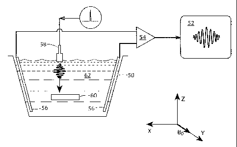
FIG. 5A is a diagram of an experimental setup
for HEI of a sample, in accordance with an embodiment of
the present invention;
FIG. 5H shows an Hall voltage time trace
collected fox a rectangular polystyrene block immersed in
saline using the experimental setup of FIG. SA, and also
includes an inset showing the acquired Hall voltage
magnitude dependence on the applied magnetic field
strength, in accordance with an embodiment of the present
invention;
FIG. 5C is an HE image generated from magnitude
reconstruction of signals acquired using the experimental
setup of FIG. 5A for a polystyrene block immersed in
saline, in accordance with the present invention;
FIG. 6A is a photograph of the cross section of
a block of bacon used with the experimental setup of
FIG. 5A, in accordance with the present invention;
FIG. 6B is an image of the bacon of FIG. 6A
generated by HEI using the experimental setup of FIG. 5A,
in accordance with the present invention; and
FIG. 6C is a conventional echo ultrasound image
of the block of bacon of FIG. 6A.
DETAILED DESCRIPTION OF THE PREFERRED EMBODIMENT
Before further describing embodiments for, and
CA 02259465 1998-12-30
WO 98/00732 PCT/I1S97/11272
- 6 -
° examples of, practicing the present invention, principles
applicable to the present invention are described. The
present invention is directed to a system and method to
form an image of a conductive subject, such as the human
body. The method/system has two basic implementations or
embodiments.
The first implementation is based on the fact
that when a conductive subject moves in a direction
perpendicular to an external magnetic field, the positive
and negative charge carriers in the subject experience the
Lorentz force in opposite directions and therefore tend to
separate, the separation of charge giving rise to an
electric field that emanate from the region of positive
charge concentration and terminates at the region of
negative charge concentration. This electric field can be
detected in the form of a voltage difference between the
positive region and the negative region, the Hall voltage.
The separation of the positive and negative charge is
equivalent to an electric current, the Hall current, which
Can also be detected via wire loops that are inductively
coupled to the subject. The magnitude and phase of the
Hall voltage and Hall current are dependent on the
velocity of the movement of the subject and its
conductivity and dielectric constant.
More particularly, if an ultrasound pulse is
generated at the surface of the subject and propagates
into the subject, wherever the pulse is currently located,
CA 02259465 1998-12-30
WO 98/00732 PCT/ITS97/11272
- 7 -
° the conductive medium at that location vibrates with the
pulse. If the motion of the vibration is perpendicular to
an external magnetic field, a Hall voltage and Hall
current can be detected as described above. The magnitude
and phase of the Hall signal is dependent on the
vibrational velocity and therefore the acoustic impedance
of the medium at that location, as well as the charge
carrier density and mobility and therefore the
conductivity and dielectric constant at that location.
Therefore by continuously monitoring the Hall
signal while the ultrasound pulse travels through the
subject, an image of the portion of the subject along the
path of the ultrasound pulse can be formed based on the
electrical constants and the acoustic impedance. If the
ultrasound path is swept through a series of directions, a
two-dimensional or three-dimensional image can be formed.
A second, and essentially physically equivalent,
basic implementation is based on the fact that in the
presence of a magnetic field, if a subject carries a
current that is perpendicular to the direction of the
magnetic field, the subject experiences a Lorentz force in
the direction perpendicular to the plane formed by the
vector of the magnetic field and the vector of the
current. Therefore, in the presence of a static magnetic
field, if a current distribution is induced in the bulk of
a subject either through direct contact with electrodes or
inductive coupling, and in the form of a short pulse in
CA 02259465 1998-12-30
WO 98/00732 PCT/US97/11272
_ g _
° time, the bulk of the subject will experience a Lorentz
force in the form of a short pulse, and will vibrate under
this force. The vibration from a region in the subject
reaches the surface of the subject at a time proportional
to the acoustic path length between the region and the
surface, and the amplitude of the vibration is dependent
on the electric constants and acoustic impedance of that
region. By sensing these vibrations with an acoustic
transducer at the surface of the subject, which arrive in
sequence according to the depths of the regions from which
they originate, a one-dimensional image can be formed
along the sensitive beam of the transducer. This image
carries information on the electric constants as well as
the acoustic impedances along that beam. Two-dimensional
and three-dimensional images can be formed by using an
array of sensing transducers simultaneously, or by
sweeping the sensitive beam of a transducer over a range
of directions.
Based on applying the electro-mechanical
reciprocity relation, the above two implementations
produce identical images of the subject. Each
implementation can be applied to imaging the human body.
The tissues of the body have different conductivity
constants and dielectric constants. The contrast of the
images acquired with the above methods are based on the
electric constants of tissues, therefore they display the
anatomy of the body. They also provide medical diagnostic
CA 02259465 1998-12-30
WO 98/00732 PCT/US97/11272
_ g _
° information that are related to the electrical properties
of tissues, such as ionic concentration.
To further illustrate the dependence of the Hall
voltage on the electrical and acoustic properties of the
sample, consider a one-dimensional example illustrated in
FIGS. lA-D. An ultrasound transducer generates a
longitudinal wave packet along the "Z" axis (FIG. lA)
perpendicular to a magnetic field Bo (not shown). A step
change in conductivity cr and mass density p occurs between
positions z, and zz (FIG. 1B). If the velocity of the
ultrasound vibration at position z and time t is v(z, t),
IS a charge q at that position experiences a Lorentz force
qv(z, t)Bo. This force is equivalent to that of an
electric field v(z, t)Bo, which in turn establishes a
current density Q(z)v(z, t)Bo in the sample. The net
current derives from integrating this over the ultrasound
beam width W and the ultrasound path:
I ( t) - WBo f Q (z) v(z, t) dz. (1)
soUndpath
If a portion a of the current is collected by electrodes
into a detection circuit of impedance Rd, the detected
Hall voltage V,, (t) is then
vh( t) - aRdWBo f Q (2) v(z, t) dz. (2)
soundpath
Using the equation of wave propagation, the Hall voltage
can be expressed in terms of the ultrasound momentum
CA 02259465 1998-12-30
WO 98/00732 PCT/ITS97/11272
- 10 -
° M(z,t) and the spatial gradient of Q/p. More
particularly, denoting the ultrasound pressure wave as
p(z, t), by using the equation of sound propagation:
P( Z) a~a~ t) + a~aZ t) = 0 (3)
the Hall voltage in equation (2) can be expressed as:
_ Q (z) t 7p(z, z)
a ~~O~soundpath P ( Z) [~ aZ dTJdz
Integration by parts yields:
( ~ oWndpathend
a ~ Q z) ~(z~t) ~ _ a (z) M(z~t)
Vh(t) = a WRdBo f so~d~r~ a f _z
P (z) p (x) oundpatlitxginni~g
IS
where
r
M(z,t) = f ~~z~)dz
_m
is the ultrasound momentum transmitted across position z
at time t. Practical ultrasound transducers emit little
energy in the audio and DC frequency range. The lack of a
DC component means that the net momentum of the wave
packet is zero. Under this condition, it can be shown
that the surface term in equation (6) is zero during the
time the wave packet is somewhere within the ultrasound
path. Hence the Hall voltage can be expressed as:
_c7 a(z)
Yh(t) = a WRdBo f hM(z,t) az ~ P (z) adz. ('1 )
CA 02259465 1998-12-30
WO 98/00732 PCT/US97/11272
- 11 -
This expression shows that a non-zero Hall voltage only
comes from positions where a gradient of Q/p exists. This
point can be visualized by observing the total Hall effect
(HE) current in equation (1), while following the
progression of the ultrasound wave packet. When the wave
packet is in a homogeneous region, the total current is
proportional to the average vibration velocity in the
packet (equation (1)), which is zero due to the absence of
a DC component. When the wave packet passes an interface
of different conductivities, the portion inside the high a
region contributes more current with the same velocity:
thus the integral in equation (1) is no longer zero. When
the wave packet passes an interface of different mass
densities but no change in conductivity, the portion in
the low density region have higher vibration velocities,
therefore the integral in equation (1) is also non-zero.
In both cases the total Hall effect current becomes
non-zero, and the resulting Hall voltage marks the
presence of the interface.
By way of illustration, in applying equation (7)
to the example shown in FIGS. lA-B, the gradient of Q/p is
non-zero only at the interfaces zl and z2 (FIG. 1C). The
ultrasound momentum M(z, t) is carried by the wave packet
as it travels along the Z axis. The Hall voltage V,,, is a
convolution of the ultrasound momentum M(z, t) with the
d/p gradient (equation (7)). When the packet passes the
two interfaces successively, the integrand in equation (7)
CA 02259465 1998-12-30
WO 98/00732 PCT/US97/11272
- 12 -
° two interfaces successively, the integrand in equation (7)
becomes non-zero, giving rise to a Hall voltage. Thus the
time course of the Hall voltage contains two peaks
representing the two interfaces (FIG. 1D). The time of
each peak marks the position of its corresponding
interface. The polarity of the two peaks are opposite,
because the Q/p gradient at z, and z, are in opposite
directions. In this fashion HEI converts spatial
information into the time domain much like conventional
echo ultrasound. Many methods used in echo ultrasound to
collect 2 or 3-dimensional images, such as line scan and
phased array detection, also apply to HEI. Similarly,
motion measurements based on Doppler effect in echo
ultrasound can be readily implemented in HEI.
Referring now to FIG. 2, there is shown a
functional block diagram of a system for practicing the
various embodiments of the present invention. The system
includes a magnet 18 for generating a large static
magnetic field, an ultrasound transducer 16, and
electrical signal transducer 14, an electrical signal
generator/receiver 12, and a controller/processor 10. A
subject (e.g. human body) or object (not shown) is
positioned in the system such that the static magnetic
field traverses the subject/object.
Based on the Lorentz forces upon which the Hall
effect (HE) is based, the electrical signal transducer 14,
ultrasound transducer 16, and magnetic field generated by
CA 02259465 1998-12-30
WO 98/00732 PCT/US97/11272
- 13 -
° magnet 18, are oriented such that they have mutually
orthogonal components for detecting/generating the signals
of interest, and preferably, they are established in an
orthogonal relationship.
Electrical signal transducer 14 may include one
or more (e.g. an array) coils for inductive coupling of
15
electrical signals with the object or body, or
alternatively may include one or more electrodes for
direct contact to the object or subject for direct
conduction of electrical signals with the object or
subject.
In an embodiment of the invention wherein
ultrasound pulses are coupled into object or body and
electrical signals are detected, electrical signal
transducer 14 receives these electrical signals (either
inductively or conductively) and electrical signal
generator/receiver 14 acts as a receiver (e. g.,
radiofrequency detector). Alternatively, in an embodiment
of the invention wherein ultrasound pulses are detected
from the object or body and electrical signals are coupled
into the object or body, then electrical signal transducer
14 transmits (inductively or conductively) the electrical
signal (e. g., pulse) generated by electrical signal
generator 12 (e. g., RF generator) to the object or body.
Similarly, ultrasound transducer 16 is
appropriately employed to either generate or detect
ultrasound signals (e.g., pulses), depending on the
CA 02259465 1998-12-30
WO 98/00732 PCT/US97/II272
- 14 -
° implementation. Ultrasound transducer 16 may be, for
example, a conventional linear array probe which scans a
sector in a plane by steering the direction of the
ultrasonic beam transmitted by ultrasonic transducer 16
according to phase array principles. Alternatively, for
the second implementation for example, ultrasound
transducer 16 may be either a one-dimensional or a
two-dimensional array in which each element of the array
concurrently detects ultrasound energy from the object or
body, and through data processing techniques (e. g.,
Fourier transform) operating on the signals from all the
elements, either a two-dimensional image or three-
dimensional image, respectively, is reconstructed.
Moreover, with regard to transducers, fiber
optic ultrasonic sensors and photoacoustic transducers may
be the basis for high sensitivity sensors and efficient
transmitters which are not affected by the magnetic field,
and are immune to any electromagnetic interference. J. A.
Bucaro, J. H. Cole, A. D. Dandridge, T. G. Giallorenzi and
N, Lagakos. "Fiber optic acoustic sensors," in Optical
testing and metrology. Bellingham, WA. Society of Photo-
Optical Instrumentation Engineers, 1986. pp. 182; Q. X.
Chen, R. J. Dewhurst, P. A. Payne and B. Wood, "A new
laser-ultrasound transducer for medical applications."
Ultrasonics vol. 32, pp. 309-313, 1994; S. Knudsen, A. M.
Yurek, A. B. Tveten and A. D. Dandridge, "High-sensitivity
fiber optic planar ultrasonic microphone," in Proceedings
CA 02259465 1998-12-30
WO 98/00732 PCT/US97/11272
- 15 -
° of the International Society for Optical Engineering. vol.
2360. pp. 396. 1994; J. F. Dorighi. S. Krishnaswamy and J.
D. Achenbach, "Embedded fiber optic ultrasonic sensors and
generators." in Proceedings of the International Society
for Optical Engineering, vol. 2574. pp. 46, 1995.
The controller/processor 10, which may be a
conventional computer, workstation, or adapted ultrasound
generator/receiver 12, ultrasound transducer 16, and
processing system, is coupled to electrical signal
magnet 18, for controlling the overall signal acquisition
sequences and preferably, also for processing and
displaying images according to these acquisitions.
In a first implementation, an ultrasound pulse
is propagated into the body by ultrasound transducer 16.
In the presence of a static magnetic field provided by
magnet 18, the Lorentz force associated with the local
displacement of conductive moieties in the body as the
ultrasonic pulse propagates through the body results in
the generation of an electrical signal. The electrical
signal is detected with electrical signal transducer 14
and electrical signal receiver 12 as the ultrasonic pulse
propagates through the body. Controller/processor 10
acquires the electrical signal and processes this
information to roduce an ima a (e.
p g g., two-dimensional or
three-dimensional) which is weighted by conductivity
(which includes any function of the conductivity). For
instance, the image contrast (e. g., intensity, gray-scale,
CA 02259465 1998-12-30
WO 98/00732 PCT/US97/11272
- 16 -
° and/or color) may be weighted by the conductivity gradient
magnitude, the conductivity gradient magnitude and
polarity, the relative conductivity (e.g., the integral of
the conductivity gradient), etc. It may be understood
that the ultrasound pulse may be focussed and scanned to
provide spatial localization for imaging.
In accordance with the above descriptions, in
the presence of a static magnetic field, an ultrasonic
pulse propagating in the tissues exhibits the Hall effect
(HE) due to the conductivity of the tissues. Hall effect
is the phenomenon that when a conductive object (such as a
saline solution) moves in a static magnetic field, a
voltage develops in the direction perpendicular to both
the magnetic field and the direction of movement. The
amplitude of the voltage is proportional to the product of
the conductivity of the object, the speed of movement, and
the magnetic field strength (i.e., V a QvB; Q being the
conductivity of the object, v being the speed of movement,
and B the strength of the magnetic field). With the
ultrasonic pulse, Hall voltages arise from the vibrational
movements, and can be detected with various electrical
circuits. These signals represent the progression of the
ultrasonic pulse in the body, and therefore information of
the tissue-tissue interfaces it encounters. The signal
amplitude and phase are related to the conductivities of
the tissues and the acoustic vibration amplitude and phase
of the pulse. The Hall effect signals therefore can be
CA 02259465 1998-12-30
WO 98/00732 PCT/US97/11272
- 17 -
° used to reconstruct an image of the volume in which the
sonic pulse propagates.
It may be understood, therefore, that the
imaging method according to this first implementation of
the present invention is similar to current ultrasonic
imaging in that ultrasonic pulses are used to interrogate
the body. A fundamental difference, however, is the
method to monitor the progression of the pulse. In
current (i.e., conventional) ultrasonic imaging since the
progression of the pulse is monitored by detecting the
echoes of the pulse reflected back at the tissue-tissue
interfaces, it is entirely a characterization of the
acoustic impedances of the tissues. The acoustic path
involved starts from the transducer that generates the
ultrasonic pulse, reaches the tissue-tissue interfaces,
and back to the transducer if it is also used to receive
the echoes, or to another receiving transducer.
In the ultrasound-Hall effect method, the
progression of the ultrasonic pulse is monitored by
detecting the Hall effect signals it induces with
electrical circuits. The Hall effect voltages reach the
electrical circuits almost instantaneously (at the speed
of light); therefore, the progression of the ultrasonic
pulse is instantaneous and constantly monitored. Because
the Hall effect signal is directly related to the
electrical conductivities of the tissues, it characterizes
the conductivities and the acoustic impedances. The Hall
CA 02259465 1998-12-30
WO 98/00732 PCTIUS97/11272
- 18 -
° effect method thus has a different contrast mechanism from
the current (conventional) ultrasound imaging method.
Because of the instantaneous nature, the acoustic path
involved in seeing an interface is from the transducer
that generates the pulse to the interface, usually half of
that of the current method. The signal attenuation along
the acoustic path is reduced.
As described above, ultrasound-Hall effect
according to the present invention, may also be performed
in the exact reciprocal fashion of the above first
implementation. In this second implementation, a pulsed
current distribution at the ultrasonic frequency is set up
in the body via electrical signal generator 12 and
electrical signal transducer 14. In the presence of a
static magnetic field provided by magnet 18, the Lorentz
force on the pulsed current results in an ultrasonic pulse
distribution in the body. The ultrasonic pulse is
detected with ultrasonic transducer 16 as it propagates
through the body to the location of the transducer.
Controller/processor 10 acquires the ultrasonic pulse
signals and processes this information to produce an image
(e.g., two-dimensional or three-dimensional). As
described above regarding the electrical circuit used to
detect the Hall voltages, the electrical circuit used in
the reciprocal case to generate the pulsed current
distribution can generally be of two types. Electrodes
can be in direct contact with the body (direct electric
CA 02259465 1998-12-30
WO 98/00732 PCT/US97/11272
- 19 -
° coupling), or the circuit can inductively couple with the
body (magnetic inductive coupling).
The reciprocity relation of linear
electrodynamic systems, derived from Onsager's relations,
warrants that the signal obtained from the above two
realizations or embodiments are identical. Their
sensitivity and spatial resolution, however, will be
determined by two different sets of parameters.
In the Hall voltage detection method, a limiting
factor is the ultrasound pulse intensity which in
biological structures must not exceed the cavitation
threshold. The cavitation threshold for soft tissue has
t5
been established empirically as the ratio (peak
pressure)2/(ultrasound frequency) ~ 0.5 (MPa2/MHz) . R. E.
Apfel and C. K. Holland, "Gauging the likelihood of
cavitation from short-pulse, low-duty cycle diagnostic
ultrasound,", Ultrasound Med. Biol. vol. 17, pp. 179-185,
1991; L. A. Crum. R. A. Roy, M. A. Dinno. C. C. Church, R.
E. Apfel, C. K. Holland and S I. Madanshetty, "Acoustic
cavitation produced by microsecond pulses of ultrasound: A
discussion of some related results, "J. Acoust. Soc. Am.
vol. 91, pp. 1113-1119, February 1992. Based on this
index an estimate of the maximum Hall voltage from a
muscle-fat interface can be made for the line scan method.
Using equation (7), assuming that the width of the
ultrasound beam W = lcm, the current collection factor
100%, Rd = 50 S2, the maximum Hall voltage for a range of
CA 02259465 1998-12-30
WO 98/00732 PCT/US97/11272
- 20 -
° field strength and spatial resolution is estimated as
shown in FIG. 3A. This voltage is on the order of lmV.
In practice, the signal level is generally lower because
the ultrasound pressure is below the cavitation threshold,
the detection electrodes are usually remote from the
scanned region, and the ultrasound beam may not be
perpendicular to the magnetic field (e. g., see polystyrene
example hereinbelow). Signal averaging over multiple
acquisitions may be used to improve the signal-to-noise
ratio. In biomedical imaging such averaging is evidently
practicable and applicable when acquiring 1-dimensional
profiles or 2-dimensional images with phased array
detection, since each scan is on the order of 200~cS or
less, and the required frame rate is often less than 50
frames/sec (20 ms/frame). Physiological motions such as
heartbeat and blood flow induce Hall voltages in the DC
lOOHz range, therefore they do not contribute to the noise
in HEI (MHz).
In the ultrasound detection mode of HEI, the
limit on sensitivity is the maximum current allowed in the
object. In biological tissue the threshold is set by nerve
stimulation. The duration of the electrical impulse
determines the length of the ultrasound pulse it produces,
and therefore the spatial resolution of the image. To
achieve millimeter or higher resolution, the pulse
duration must be on the order of a microsecond, or one
hundredth the strength-duration time constant of human
CA 02259465 1998-12-30
WO 98/00732 PCT/US97/11272
- 21 -
° sensory and muscular nerves. J. P. Reilly. Electrical
Stimulation and Electropathology. New York. Cambridge
University Press, 1992, ch. 7, pp. 238. In this short
time limit the nerve stimulation threshold is established
as the product (electric field)x(pulse duration) ~ 2x10⁻³
Vs/m. J. P. Reilly, ibid., ch. 4, p. 119. Based on this
index, the peak pressure of the ultrasound signal from a
fat-muscle interface can be estimated for a range of Bo
and spatial resolution. In the millimeter resolution range
the peak pressure was found to be on the order of 5
pascal, and dependent only on Bo (FIG. 3H). Again, this
value is compromised in practice because the electrical
impulses are usually below the nerve stimulation level,
the electric field is not perpendicular to Bo, and the
ultrasound sensors are often remote from the region of
interest.
It may be appreciated therefore that the
conductivity contrast mechanism of the imaging method
according to the present invention provides a new imaging
modality. This parameter of the tissues, which heretofore
has not been readily obtained in vivo, may contain
diagnostic information of certain pathologies, such as
diseases in kidneys, where the electrolyte concentration
is an indicative index.
More specifically, in biomedical imaging
ultrasound has been very effective, although it has
inherent difficulties in differentiating soft tissue,
CA 02259465 1998-12-30
WO 98/00732 PCT/US97I11272
- 22 -
° since muscle, fat and blood differ in their acoustic
impedances by less than 10%. S. A. Goss. R. L. Johnston
and F. Dunn, "Comprehensive compilation of empirical
ultrasound properties of mammalian tissues.~~ J. Acoust.
Soc. Am. vol. 64, 1978, pp. 423-457. In comparison, the
conductivities of soft tissue at ultrasound frequencies
range over a factor of four, (K. R. Foster and H. P.
Schwan, in CRC Handbook of Biological Effects of
Electromagnetic Fields, edited by C. Polk and E. Postow,
Boca Raton, FL, CRC Press Inc., 1986, Part I), while their
mass densities are very similar. This enables HEI to
differentiate soft tissue based mainly on conductivity
differences. The acoustic path length in HEI is also half
that of ultrasound, greatly reducing the acoustic
attenuation and dispersion. These characteristics of HEI
may potentially improve the penetration depth, tissue
contrast and characterization in an ultrasound exam.
With regard to new diagnostic information the
electrical constants of tissue reflect their physiological
state, such as water content and adiposity. K. R. Foster
and H. P. Schwan, in CRC Handbook of Biological Effects of
Electromagnetic Fields, edited by C. Polk and E. Postow,
Boca Raton, FL, CRC Press Inc., 1986, Part I; E. C.
Burdette, F. L. Cain and J. Seals, in Medical Applications
of Microwave Imaging. edited by L. E. Larsen and J. H.
Jacobi. New York, IEEE Press, 1986, pp. 23. It has been
shown that tumors and especially necrotic regions have up
CA 02259465 1998-12-30
WO 98/00732 PCT/US97/11272
- 23 -
° to 10 times higher conductivity than normal tissue, (K. R.
Foster and H. P. Schwan. ibid.. Part I, pp. 68), providing
a good contrast mechanism in HEI. With its sensitivity to
fat, the HEI method according to the present invention
implemented with small intravascular probes could be used
to characterize atherosclerotic plaques, which is
difficult with existing imaging methods.
It may further be appreciated that with the
development of ultrasound array sensors, HEI may be used
for mammography or imaging of skin and subcutaneous
structures, where it is relatively easy to arrange the
magnet, the electrodes and the ultrasound sensors, and the
entire device can be relatively compact.
It is noted that while HEI according to the
present invention has myriad biological and medical
applications, it is also well suited for many other
applications. For instance, HEI may also have a role in
material sciences and microelectronics, where it could be
used to study the conductivity and dielectric properties
at very high frequencies in structures susceptible to
ultrasound. Similarly, it may also be used for
manufacturing quality control and reliability testing.
It may also be appreciated that with the
magnetic inductive coupling scheme discussed hereinabove,
multiple sensing coils forming an array in space can be
used to detect the Hall signal. This provides flexibility
regarding spatial sensitivity. Even more attractive is
CA 02259465 1998-12-30
WO 98/00732 PCT/LTS97/11272
- 24 -
° the option that given enough signal-to-noise, which is
directly proportional to the magnetic field strength, the
signals from the multiple sensing coils can be used to
reconstruct an image of the ultrasound wave front, thus
overcoming the inherent problem of beam expansion and low
angular resolution away from the origin of the beam in the
conventional ultrasound imaging method. Such a wavefront
reconstruction technique provides for one-shot 3D imaging,
giving ultra-fast-temporal resolution. Currently multiple
ultrasound transducers are occasionally used to approach
this, but hindered by the limited sizes of the acoustic
windows in the body. The magnetic inductive coupling is
not limited because r.f. magnetic fields penetrate the
body with negligible perturbation. At a field strength of
4 Tesla (i.e., 4 T), the achievable S/N ratio may limit
the number of array coils to a few, similar to the array
coil receive situation in MRI. However since the
ultrasound-Hall imaging scheme requires strong field
strength only in the volume of interest, no requirement on
the uniformity of the field, and minimal requirement on
the stability of the field, much higher field strengths
are practicable.
The following examples are presented to
illustrate features and characteristics of the present
invention, which is not to be construed as being limited
thereto.
CA 02259465 1998-12-30
WO 98/00732 PCT/US97/11272
- 25 -
° Example 1
In this example, ultrasound-Hall effect imaging
is demonstrated in a simple one-dimensional imaging
experiment, in accordance with the first embodiment of the
present invention described hereinabove.
The experimental setting used is shown in
FIG. 4A. As stated, the reciprocal method was used. The
plastic beaker 20 contained two layers of liquids, the top
layer was silicone oil 30, the bottom layer was 0.3% NaCl
solution 32 tdiluted irrigation saline solution). A
pulsed current distribution of lMHz bandwidth and 2.lMHz
center frequency was generated in the beaker with the r.f.
IS
amplifier 22 in response to an input stimulus from network
analyzer 34, and coupled into the beaker 20 via electrodes
26, which had textured surfaces to reduce coherent
speculating echoes from the vibration of the electrodes.
The induced ultrasonic pulse was detected with the
ultrasound transducer 28 and amplified with a low noise
amplifier 24 and provided to the receive port of network
analyzer 34.
FIG. 4B shows the received signal, plotted as a
function of distance in centimeters. Peak al and bl
correspond to the oil-saline interface and the
saline-beaker interface, respectively, which are
independently measured with the conventional ultrasound
method, shown in FIG. 4C. Peak a2 and b2 correspond to
the conventional ultrasound echoes through direct
CA 02259465 1998-12-30
WO 98/00732 PCT/LTS97/11272
- 26 -
° electromagnetic coupling between the transducer and the
electrical circuit.
FIG. 4D and FIG. 4E show a similar experiment
with the beaker placed at the center of the magnet and the
edge magnet, respectively. Because the magnetic field
strength is decreased from the center to the edge, the
Hall peaks al and bl decreased. The amplitudes of the
conventional ultrasound echoes a2 and b2 stayed relatively
constant.
The achievable signal level may be estimated
from the one-dimensional experiment described above. The
signal level of the Hall peaks were approximately 2.5~,V,
given an excitation peak voltage of 15 volts. If 300
volts excitation is used, which is the typical operating
voltage for current pulsed ultrasound imaging, 50~V peak
signal can be reached. Assuming a 500KHz acquisition
bandwidth and 50 ohm output impedance, the thermal noise
at room temperature is 0.65~.V. Accordingly, based on the
simple one-dimensional experiment, the ideal achievable
signal-to-noise ratio is 70:1. Given all the practical
factors, the realizable S/N is probably on the order of
30:1. The coherent speckles in current imaging methods
cause the signal-to-noise ratio to be much lower than this
3U
value, thus the intrinsic signal-to-noise ratio may not
after all constitute a limitation.
Example 2
In this second example, ultrasound-Hall effect
CA 02259465 1998-12-30
WO 98/00732 PCT/I1S97/11272
- 27 -
° imaging is demonstrated in a simple two-dimensional
imaging experiment, in accordance with the second
embodiment of the present invention described hereinabove.
More particularly, to demonstrate the
feasibility of HEI, a simple device, essentially similar
to that used in example 1 hereinabove, was constructed to
form cross-sectional images of objects (samples) 60
suspended in a plastic chamber 50 of saline solution 62
(e. g., 0.4% NaCl solution) placed in a 4 T magnet
providing magnetic field Bo (FIG. 5A). The dimensions of
the chamber 50 were 27cm (X dimension) x l7cm (Y) x 22cm
IS (z)~ The electrodes 56 were two exposed copper wire
segments. The piezoelectric ultrasound transducer 58 used
had a center frequency of lMHz, and a 6dB bandwidth of
0.6MHz. An electrical pulser input single-phase
electrical pulses of approximately 0.5~.s duration into the
transducer 58, which in turn emitted ultrasound pressure
pulses into the chamber. The detected Hall voltage was
amplified (low noise amplifier 54) by 60dB gain, filtered
by a O.lMHz~3MHz bandpass filter, and digitized at 5
megasample/sec for data storage (recording) 52.
In a first two-dimensional imaging trial, a
rectangular polystyrene block was immersed in the saline.
The magnetic field Bo, was in the "Y" direction. A
piezoelectric transducer emitted longitudinal ultrasound
waves with both the wave vector and the physical vibration
in the "Z" direction. The Lorentz force from the vibration
CA 02259465 1998-12-30
WO 98/00732 PCT/US97/11272
- 28 -
° was in the "X" direction, and the resulting Hall voltage
was detected with electrodes placed in the chamber.
Immediately after the onset of the ultrasound pulse, the
Hall voltage was recorded for up to 100 ~s, the time
re uired for the ultrasound wave
q packet to traverse the
chamber. FIG. 5B shows such a Hall voltage time trace
collected for the rectangular polystyrene block immersed
in saline. The two peaks in the time trace represent the
upper and lower surfaces of the polystyrene block. The
amplitude of the second peak is lower than the first peak
due to attenuation and acoustic reflection at the upper
Surface. As described in equation (7), the opposite
polarity of the peaks resulted from the opposite o/p
gradient at the two interfaces. Also according to
equation (7), the Hall voltage is proportional to the
magnetic field strength. This was experimentally
demonstrated as indicated by the inset of FIG. 5B, where
the signal amplitude measured at 2.4T and 4T are plotted
versus field strength. It is noted that in this
polystyrene block experiment (axial resolution 2mm, beam
width approximately 2.5cm) the measured Hall voltage was
about 0.1% of its theoretical maximum because the
ultrasound pulse was only one tenth the cavitation
threshold and the electrodes were relatively far from the
sample.
A 2-dimensional image was formed with the line
scan method by moving the transducer in 0.5cm increments
CA 02259465 1998-12-30
WO 98/00732 PCT/US97/11272
- 29 -
° across the chamber, while recording the time course of the
Hall voltage at each position. These traces were
displayed side by side in grey scale after a magnitude
calculation, to form a 2-dimensional image. FIG. SC shows
such an image of the polystyrene block, with the time axis
in the vertical direction and the horizontal axis scanned
by moving the ultrasound transducer as described. The
interfaces between the block and the saline solution are
readily observed. Polarity images, where the magnitude
and polarity (i.e., positive or negative) of the acquired
signals in each pixel are represented according to a color
spectrum were also generated (not shown).
In a second two-dimensional imaging trial, bacon
was chosen as an example of a biological structure since
it has layers of high (muscle) and low (fat) conductivity
soft tissue. The HE image of a block of bacon suspended in
the chamber is shown in FIG. SD, along with a photograph
and an echo ultrasound image generated using the same
transducer and line scan procedure. HEI depicts the soft
tissue interfaces between the fat and muscle layers better
(i.e., better differentiates between fat and muscle) than
echo ultrasound because of the significant changes in
conductivity between the layers, to which HEI is sensitive
and conventional echo ultrasound is not sensitive.
Although the above description provides many
specificities, these enabling details should not be
construed as limiting the scope of the invention, and it
CA 02259465 1998-12-30
WO 98/00732 PCT/US97/11272
- 30 -
will be readily understood by those persons skilled in the
art that the present invention is susceptible to many
modifications, adaptations, and equivalent implementations
without departing from this scope and without diminishing
its attendant advantages. It is therefore intended that
the present invention is not limited to the disclosed
embodiments but should be defined in accordance with the
15
25
claims which follow.