Note: Descriptions are shown in the official language in which they were submitted.
CA 02262056 1999-02-24
-1-
TITLE OF THE INVENTION
Composition, Methods and Reagents for the Synthesis of a Soluble Form of Human
PEX
BACKGROUND OF THE INVENTION
The PEX gene (Phosphate regulating gene with homologies to Endopeptidases
on the X chromosome) was identified by a positional cloning approach as the
candidate gene for X-linked hypophosphatemia (XLH) (Francis et al., 1995). XLH
is a
Mendelian disorder of phosphate homeostasis characterized by growth
retardation,
rachitic and osteomalacic bone disease, hypophosphatemia, and renal defects in
phosphate reabsorption and vitamin D metabolism (Rasmussen and Tenenhouse,
1995). Using the information made available by the publication of the sequence
of the
PEX gene, and standard techniques obvious to those in the art, several groups
have
cloned and sequenced the human and mouse PEXIPex cDNAs (Du et al., 1996;
Lipman et al., 1998; Grieff et al., 1997; Beck et al., 1997; Guo and Quarles,
1997;
Strom et al., 1997). Amino acid sequence comparisons have demonstrated
homologies
between PEX/Pex and members of the neutral endopeptidase family as previously
observed in the partial sequence of the candidate gene(Francis et al., 1995).
The
peptidases of the neutral endopeptidase family are type II integral membrane
glycoproteins with a relatively short cytoplasmic N-terminal region, a single
transmembrane domain, and a long extracytoplasmic domain, which contains the
active site of the enzyme (Devault et al., 1987). Known members of the neutral
endopeptidase family include neutral endopeptidase-24.11 (Neprylisin, NEP),
endothelin-converting enzymes (ECEs), and the erythrocyte cell surface protein
KELL
(for a review see (Turner and Tanzawa, 1997b). NEP is a widely distributed
peptidase
involved in the degradation of several bioactive peptides, such as the
enkephalins, the
atrial natriuretic peptides, and the endothelins (Crine et al., 1997). The
ECEs are
involved in the bioactivation of big endothelins into endothelins (Turner,
1997a). No
function has been yet attributed to Kell.
The mechanism by which loss of PEX function elicits the bone and renal
abnormalities observed in XLH patients is not clear. There are no data
suggesting the
presence of PEX/Pex mRNA in the kidney (Du et al., 1996; Beck et al., 1997;
Grieff et
al., 1997). In contrast PEXIPex mRNA was detected in bones by Northern blot
hybridization and in other adult and fetal tissues such as lungs, liver,
muscles, and
ovaries by RT-PCR and RNase protection assays (Du et al., 1996; Beck et al.,
1997).
!n situ hybridization performed on sections of embryos and newborn mice showed
the
presence of PEX mRNA in osteoblasts and odontoblasts (Ruchon et al., 1998).
PEX
gene expression was detectable on day 15 of embryonic development, which
coincides
with the beginning of intracellular matrix deposition in bones. Moreover,
Northern
analysis of total RNA from calvariae and teeth of 3-day-old and adult mice
showed that
CA 02262056 1999-02-24
' -2-
the abundance of the PEX transcript is decreased in adult bones and in non
growing
teeth. When the presence of the PEX protein in adult bones was investigated
both by
Western blotting, large amounts of immunoreactive protein were found.
Immunohistochemical studies on a 2 month old mouse showed an extensive
labeling
of all cells of the osteoblast lineage including osteocytes. Taken together
these results
suggest that PEX plays an important role in the development and maintenance of
mineralization in these tissues.
Further insights into the role of PEX in bone metabolism were provided by
experimental studies on cases of oncogenic osteomalacia (OOM), a tumor-
associated
sporadic condition with very similar clinical presentations. There is strong
evidence that
a humoral factor produced by the tumor inhibits renal phosphate reabsorption
and
vitamin D synthesis resulting in osteomalacia (Nelson et al., 1997).
Experimental
studies on the Hyp and Gy mice, the murine model of human XL, also suggest the
involvement of a humoral factor. In both mouse models, mutations have been
identified
in the PEX gene, which also appear to result in loss of function of the gene
product
(Strom et al., 1997; Beck et al., 1997). Considering the similarities between
PEX and
the other members of this metallopeptidase family, it has been speculated that
PEX
metabolizes a peptide hormone that modulates renal tubular phosphate
reabsorption.
Such an activity could involve either the processing of a phosphate
reabsorbing
hormone precursor to its active form or the inactivation of a circulating
phosphaturic
factor. There is evidence for intrinsic abnormalities in osteoblasts from Hyp
mice
(Ecarot et al., 1992). A defective phosphate transport was also observed in
osteoblasts
from Hyp mice (Rifas et al., 1994). PEX might thus be involved in the control
of bone
metabolism both indirectly at the level of the kidney by controlling renal
phosphate
reabsorption and directly at the level of bones by inactivating a trophic
peptide factor
controlling either osteoblast or osteoclast functions or both.
The identification and characterization of the putative PEX substrates will
require first a better understanding of PEX function and enzymatic activity.
Establishing
the enzymatic activity of the enzyme and its substrate specificity will be
greatly
facilitated by having access to a pure preparation of the enzyme, free of
other potential
protease activities.
SUMMARY OF THE INVENTION
Towards this objective, we have prepared various reagents and tools designed
to
produce recombinant forms of PEX and purify both the recombinant and native
enzymes from cell fractions, spent culture media and tissue extracts. We have
cloned
a cDNA encoding the full-length human PEX protein into various expression
vectors.
These PEX-encoding vectors were introduced by transfection into various cell
lines
including COS-1 (monkey kidney) cells, CHO (Chinese Hamster Ovary) cells, and
LLC
CA 02262056 1999-02-24
-3-
PK1 (porcine kidney) cells. Permanent cell lines were established and shown to
stably
express the PEX protein at the cell surface. A procedure was established to
rapidly
prepare a membrane fraction enriched in the recombinant PEX protein and this
preparation was used to assess PEX enzymatic activity using bone-related
peptides
and growth factors.
PEX is an intrinsic membrane protein anchored by a hydrophobic 20 amino acid
sequence located near the N-terminus. The purification of an intrinsic
membrane-
bound protein requires the use of detergents to free it from the lipidic
environment of
the membrane. These detergents can interfere with the catalytic activity of
the enzyme.
Moreover, the detergent-purified proteins usually present stability and
solubility
problems, especially if concentrated solutions andlor large amounts of the
protein are
needed, such as those required for crystallization and high throughput
screening
assays. To facilitate the preparation and purification in high yields of a
fully active
enzyme it is thus preferable to work with a soluble form of PEX. Soluble forms
of NEP
(Lemay et al., 1989) and ECE (Korth et al., 1997) consisting of the entire
ectodomain
but lacking the cytosolic and hydrophobic transmembrane domains have been
constructed and shown to possess enzymatic activities identical to those of
the native
membrane-bound homolog. A soluble form of recombinant PEX was thus constructed
by modification of the signal peptideltransmembrane region of the protein. The
soluble
PEX comprises PEX ectodomain or a catalytic part thereof. The expression
vector
encoding this soluble form of PEX was transfected into LLC-PK1 cells and a
permanent cell line expressing the chimeric PEX protein on a stable basis was
established. Analysis of the spent medium of this cell line by Western blot
was shown
to contain high levels of a soluble form of PEX.
Finally, monoclonal antibodies specific for PEX were generated by immunizing
mice with a PEX-derived recombinant fusion protein produced in E. coli. These
monoclonal antibodies were used to purify recombinant PEX by various
immunoaffinity
procedures. PEX-specific monoclonal antibodies were also proved useful for
characterizing PEX expression in bone by immunohistochemical techniques and
Western blotting.
DESCRIPTION OF THE SPECIFIC EMBODIMENTS A OF THE INVENTION
BRIEF DESCRIPTIONS OF THE FIGURES
This invention will be described hereinbelow with reference to the following
specific embodiments and drawings, which purpose is to illustrate the
invention and
not to limit its scope.
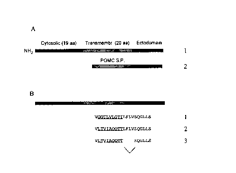
Figure 1: Construction of a soluble form of PEX. Figure 1A represents the
schematic
structure of the native membrane-bound form of the enzyme and the construct in
which
the POMC signal peptide has been fused in frame with the ectodomain of the
native
CA 02262056 1999-02-24
-4-
enzyme. Figure 1 B represents the construct where part of the sequence for the
hydrophobic transmembrane (underlined) domain has been replaced by a more
hydrophilic (C). In panel D, part of the hydrophilic sequence introduced in C
has been
deleted.
Figure 2: Amino acid sequence of human PEX. The boxed sequence represents the
hydrophobic signal peptideltransmembrane domain. The underlined sequence
represents the segment used for making the E. coli GST-fusion protein for
monoclonal
antibody production.
Figure 3: Screening of PEX monoclonal antibodies. Figure 3A: monoclonal
antibodies
were first selected for their capacity to bind the PEX,Z,_z~ fragment produced
in E. coli
as tested by using the spent medium of hybridoma cultures in ELISA assays.
Immunoglobulins from positive cultures were next tested for their ability to
bind
membrane-bound PEX from LLC-PK1 cells transfected with the PEX expression
vector. Figure 3A is the Western blot analysis of LLC-PK1 extracts stained
with the
various hybridoma supemantants. Track 12 is the staining pattern obtained with
PEX
polyclonal antibody prepared in rabbit. Figure 3B: immunoprecipitation of a
soluble
form of PEX (secPEX). LLC-PK1 cells were first transfected with a vector
encoding a
soluble form of PEX as explained in the Material and Methods section. The
spent
medium of transfected LLC-PK1 cells was then used as a source of secPEX for
immunoprecipitation experiments. The immunoprecipitation was performed by
first
saturating protein A Sepharose beads (Pharmacia) with a rabbit anti-mouse IgG
fraction and then with the mouse immunoglobulins from the hybridoma
supernatants
selected as shown in Figure 3A. After washing, these beads were incubated in
aliquots
of the spent medium of LLC-PK1 cells producing secPEX (40Ng of total protein).
The
beads were pelleted by centrifugation, washed and the presence of secPEX was
assessed by boiling the proteins bound to protein A Sepharose in the
electrophoresis
sample buffer followed by Western blot analysis with a PEX polyclonal
antibody. Track
8 shows the results of an immunoprecipitation done in the same conditions with
a
rabbit PEX polyclonal antiserum. Tracks 10 and 11 are control experiments
prepared
from mock transfected cells.
Figure 4: Expression of membrane-bound and soluble forms of recombinant PEX in
COS-1 cells. COS-1 cells were transfected with SV-40 derived expression
vectors
containing either the entire coding sequence of PEX (Figure 4A) or a construct
capable
of promoting the secretion of the PEX ectodomain (see Methods) (Figure 4B).
The
cells were kept in culture for 16 h after transfection and either a membrane
fraction
(Figure 4A) or the spent medium (Figure 4B) were prepared as explained in
Methods.
The expression of PEX was monitored in Western blots with monoclonal antibody
15D7. As seen in Panel A a band migrating with a mobility corresponding to an
apparent Mr of 105,000 was present in the membrane fraction of cells
transfected with
CA 02262056 1999-02-24
-5-
the pCDNA3IRSV-PEX-FLB vector (lane 1 ). This band was absent from the extract
of
control cells (lane 2). Panel B shows the presence of a secreted soluble form
of PEX
in the spent medium of transfected cells, but not in control mock transfected
cells.
METHODS
Expression of human PEX in transfected cells
A cDNA encoding for the full-length human PEX was obtained by Polymerase
Chain Reaction (PCR) as previously described (Beck et al., 1997). The plasmid
pCR2.1-PEX-FLB was generated by cloning this cDNA into pCR2.1 (Invitrogen). A
restriction fragment (Spel-EcoRV), which contained the entire PEX coding
sequence,
was digested, blunted, and subcloned into the mammalian expression vector
(pCDNA3/RSV). The resulting plasmid (pCDNA31RSV-PEX-FLB) contained the entire
PEX cDNA under the control of the Rous Sarcoma Virus (RSV) promoter.
This recombinant vector was then expressed transiently in COS-1 cells by
transfection. COS-1 cells were grown at 37°C under a 5% COZ atmosphere
in
Dulbecco's modified Eagle's medium (DMEM) containing 5% COSMIC (Hiclone), 100
U/ml penicillin, and 100 Nglml streptomycin. COS-1 cells were transfected
using the
calcium phosphate-DNA coprecipitation procedure. The day following
transfection, the
serum-containing medium was changed for a synthetic medium that consists of
DMEM
supplemented with 1 Nglml BSA, 2.5 Nglml insulin, 17.5 Nglml transferrin, 2
Nglml
ethanolamine, 100 Nglml soybean trypsin inhibitor and 10 Nglml aprotinin.
Finally,
sodium butyrate was added to the synthetic medium, at a concentration of 10
mM, to
enhance the expression of the plasmids carrying the RSV promoter. After 48 h,
the
cells were harvested and the membrane were prepared according to the procedure
of
(Korth et al., 1997).
The plasmid pCDNA31RSV-PEX-FLB was also transfected in LLC-PK1 cells by
the CaPO, precipitation method. Transfected cells were selected by adding 1 %
G-418
(vlv) to the medium. G-418 resistant cells were grown in rollers in medium 199
with
Earle's salts, 2mM L-glutamine, Hepes and bicarbonate buffer supplemented with
5%
fetal bovine serum (FBS), 50 unitslml penicillin, and 50 Nglml streptomycin.
Cells were
grown up to confluence, for about a week, and harvested by scraping with a
rubber
policeman.
Construction and expression of a soluble form of recombinant PEX
To obtain a soluble form of recombinant human PEX, we first attempted to fuse
in frame the cDNA encoding the signal sequence of a secreted protein (pro-
opiomelanocortin or POMC) to the cDNA sequence of the ectodomain of human PEX
(Figure 1, panel A). This strategy, which had successfully been used for other
members of this family of peptidases, namely NEP and ECE (Lemay et al., 1989;
Korth
CA 02262056 1999-02-24
-6-
et al., 1997), resulted in the production of a misfolded PEX protein that
remained
trapped in the rough endoplasmic of transfected cells: Therefore, an alternate
strategy
was developed consisting in the substitution of selected amino acids in the N-
terminal
hydrophobic membrane anchor of PEX to transform it into a cleavable signal
sequence.
Transformation of the membrane anchor into a cleavable signal sequence for
was carried out on the pCDNA31RSV/PEX-FLB plasmid. Site-directed mutations (9
codons) and deletions (4 codons) were introduced by Polymerase Chain Reaction
(PCR) amplification using oligonucleotide #5136 as the sense primer
5'CTGACAGTGATCGCTCAACAAACAACCAGTCAAGGTCTCTTAAGTCTCCAAG3'
and oligonucleotide #5134 as the antisense primer
5'GGTTGTTTGTTGAGCGATCACTGTCAGGACAAACACGACCAGGGCAATTCG3'
(Figure 1, panel B). The resulting plasmid, designated as to pCDNA31RSVIPEX-
MutE,
encoded for a secreted form of PEX (secPEX).
This recombinant vector was then expressed transiently in COS-1 cells by
transfection as described above. After 16 hours of incubation, the medium was
recovered and concentrated by ultrafiltration (MW cut-off = 30 kDa) using a
Centriprep
cartridge (Amicon). To induce the stable expression of sec PEX in LLC-PK,
cells, the
plasmid pCDNA3IRSV-PEX-MutE was transfected in LLC-PK, cells by the CaP04
precipitation method. Transfected cells were selected by adding 400 Nglml G-
418 to
the medium. 6418 resistant cells were grown in rollers in medium 199 with
Earle's
salts, 2mM L-glutamine, 1 mM sodium pyruvate, Hepes and bicarbonate buffer
supplemented with 5% fetal bovine serum (FBS), 100 Nglml G-418, 50 unitslml
penicillin, and 50 Ng/ml streptomycin. Cells were grown up to confluence, for
about a
week. The day before harvesting, the serum-containing medium was changed for a
synthetic medium that consists of DMEM supplemented with 1 Nglml BSA, 2.5
Ng/ml
insulin, 17.5 Ng/ml transferrin, 2 Nglml ethanolamine, 100 Nglml soybean
trypsin
inhibitor and 10 Nglml aprotinin. Finally, sodium butyrate was added to the
synthetic
medium, at a concentration of 10 mM, to enhance the expression of the secPEX
gene,
which is under the control of the RSV promoter. After 16 hours of incubation,
the
medium was recovered and concentrated by cross-flow filtration (MW cut-off =
30 kDa)
using a Sartocon Micro Unit (Sartorius).
Characterization of secPEX was done by immunoblotting. Briefly, proteins from
the concentrated media were resolved on 7.5% SDS-PAGE, and transferred onto
0.45
Nm nitrocellulose membranes. Membranes were incubated for one hour in TTBS
(iris
Buffered Saline containing 0.05% Tween-20) supplemented with 5% (wlv) instant
non-
fat dry milk (Carnation). Membranes were washed rapidly with TTBS and
incubated
with a 1:200 dilution of the anti-(human PEX) monoclonal antibody (13812) in
TTBS
supplemented with 1 % BSA (w/v). Membranes were washed in TTBS and incubated
CA 02262056 1999-02-24
-7-
for one hour with a HRP-labeled second antibody in TTBS supplemented with 1 %
BSA
(wlv). Membranes were washed and processed using a chemiluminescence reagent
(NEN).
Purification of the soluble form of PEX
1 ) Purification of secPEX by immunoprecipitation:
The concentrated medium containing secPEX (40 Ng/ml total protein) was
diluted in 0.5 ml immunoprecipitation buffer (20 mM Tris-HCI pH 7.4, 100 mM
NaCI,
2% sodium deoxycholate, 2%Triton X-100, 0.2% SDS, and 0.2% BSA) and potyclonal
PEX antiserum was added. The solution was mixed for 12 hour at 4°C. By
adding
swollen protein A-Sepharose (Pharmacia) and by further mixing for 2 hour at
4°C, the
immune complexes were precipitated. The beads were washed twice with immuno-
precipitation buffer and once with PBS. The antigen bound to the
immunoaffinity beads
(secPEX) was recovered by boiling in the electrophoresis sample buffer and
analyzed
by SDS-PAGE followed by immunoblotting, as described above. Aliquots of the
immunoprecipitate were kept at 4°C for enzymatic assays.
2) Purification of secPEX by ion-exchange chromatography:
The concentrated medium was supplemented with various protease inhibitors
(100 NM phenylmethanesulfonyl fluoride, 20 NM pepstatin-A, and 20 NM
leupeptin) and
clarified by centrifugation (6,OOOg, 10 min, rotor Sorvall SS-34, 4°C).
The clarified
medium was loaded on a Q-sepharose anion-exchange column (Pharmacia)
previously
equilibrated with 20 mM Tris-HCI pH 8. Whereas most of the proteins bound to
the
column, secPEX did not bind to the resin at pH 8 and was recovered in the flow-
through. The flow-through was concentrated by ultrafiltration (MW cut off = 30
kDa)
using a Centriprep cartridge (Amicon) and diluted (1110) with 50 mM
ethanolamine-HCI
pH 9.5. The flow-through was loaded on a Q-Sepharose anion-exchange column
(Pharmacia) that was equilibrated with 20 mM ethanolamine-HCI pH 9.5. SecPE'X
was
eluted with a 0 to 500 mM NaCI gradient and was analyzed by SDS-PAGE and
immunoblotting, as described above. Alternatively, it is readily conceivable
that
SecPEX can be purified on an immunoaffinity column comprising an antibody
specific
to PEX.
Preparation of PEX-containing brush border membranes
The LLC-PK1 cell line forms polarized epithelial monolayers in culture. Brush
border (apical) membranes BBMs were purified from LLC-PK1 cells homogenates as
described previously in (Blais et al., 1987). Briefly, cell membranes were
disrupted by
sonication. Non-apical membranes were precipitated at 4 °C, under
constant agitation,
by adding CaCl2 to a final concentration of 13 mM. BBMs were fractionated by
sequential centrifugation at 950 x g for 10 min and then at 35, 000 x g for 30
min. The
final pellet containing BBMs was washed twice with 50 mM Tris-HCI, pH 7.5, and
CA 02262056 1999-02-24
_$_
resuspended in the same buffer. The presence of PEX in BBMs was verified by
immunoblotting.
RESULTS
Production of monoclonal antibodies
The cDNA corresponding to amino acids 121 to 294 of the PEX amino acid
sequence (underlined segment in Figure 2) was used to construct a GST-fusion
protein
in E. coli. This fusion protein was purified from E. coli extracts by affinity
chromatography on a glutathione-Sepharose column. After thrombin cleavage, the
PEX portion of the GST fusion protein was further purified by electroelution
from a
polyacrylamide gel. This material was used to immunize 4 mice (5 injections of
=50 Ng
of PEX,2,_2~). Blood was collected from each mice after the immunization
schedule and
the presence of antibodies in mice serum was assessed by ELISA using
microtiter
plates coated with PEX,2,_Z~from E. coli extracts. Mice sera were also tested
for the
presence of PEX antibodies by Western blotting extracts of LLC-PK1 cells
transfected
with the PEX expression vector. Out of the 4 mice immunized, 3 showed good
results
both in ELISA and Western blots. One mouse selected for its high titer of PEX
specific
antibodies (as measured by ELISA) was sacrificed and its spleen cells were
collected
and immortalized by fusion with myeloma cells(strain). Hybridoma cells were
selected
for their ability to grow in HAT selection medium and cloned by several rounds
of
limiting dilution. Throughout the limiting dilution process, hybridoma were
tested for
their ability to bind to PEX,2,_2~ in the ELISA assay and to recognize
recombinant full
length PEX in Western bloting assays (Figure 3A).
Construction of an immunoaffinit~r column
Hybridoma clones secreting immunoglobulins producing a strong signal in
Western blotting (see above) were further submitted to additional screening
assays
designed to identify monoclonal antibodies capable of immunoprecipitating the
soluble
form of PEX in solution. The immunoprecipitation assay was performed by first
saturating protein A Sepharose beads (Pharmacia) with a rabbit anti-mouse IgG
fraction and then with the mouse immunoglobulins from hybridoma supernantants.
After washing, these beads were incubated in aliquots of the spent medium of
LLC-
PK1 cells producing secPEX (40 Ng of total protein). The beads were pelleted
by
centrifugation, washed and the presence of secPEX bound to the immunoaffinity
support was assessed by submitting the proteins bound to proteins A Sepharose
in a
non-covalent fashion to booting in the electrophoresis sample buffer before
immunoblot
analysis (Figure 3B). Amongst the hybridoma analyzed for their production of
specific
anti-PEX antibody, the hybridoma 15D7 has been retained. It is understood that
many
monoclonal antibodies that have an equivalent immunological profile are under
the
scope of this invention.
CA 02262056 1999-02-24
_9_
Expression of membrane-bound recombinant PEX in COS 1 cells
COS-1 cells were transfected with an SV-40 derived expression vector
containing the entire coding sequence of PEX inserted downstream from the RSV
promoter. This vector is called pCDNA3/RSV-PEX-FLB (see Methods). The cell
were
kept in culture for 16 h after the transfection and a membrane fraction was
prepared
as explained in Methods. The expression of PEX was monitored in Western blots
with
monoclonal antibody 15D7. As seen in Figure 4 a band migrating with a mobility
corresponding to an apparent Mr of 105,000 was observed in the membrane
fraction
of cells transfected with the pCDNA31RSV-PEX-FLB vector (lane 1). This band
was
absent from the extract of control cells (lane 2). The mobility of this band
was identical
to that reported previously for recombinant human and mouse PEX.
Production. purification and characterization of a soluble form of recombinant
PEX
We next wanted to determine whether it is possible to use genetic engineering
techniques to promote the secretion of a soluble and active form of PEX from
transfected eukaryotic cells. Obviously, this kind of enzyme, which can easily
be
purified from the incubation medium of cultured cells without the use of
detergent
would be very useful for further structural studies and inhibitor screening.
It could also
eventually be used as a injectable therapeutic agent or in topic applications
to increase
the rate of bone mineralization or bone healing.
PEX is a class II integral membrane protein. These membrane proteins have,
near their amino terminus, a unique hydrophobic peptide acting both as a
signal
peptide to direct the translocation of the protein through the membrane of the
rough
endoplasmic reticulum and as a transmembrane domain for anchoring the protein
in
the cell plasma membrane. Unlike class I membrane proteins which possess a
cleavable signal peptide and are anchored in the membrane by an additional
membrane-spanning hydrophobic sequence (also called Stop Transfer Sequence),
class II protein cannot be easily transformed into soluble forms by deleting
the
hydrophobic transmembrane domain. In class II proteins, deletion of the
anchoring
segment also removes the signal peptide, thereby preventing the translocation
of the
protein in the RER and its transport to the cell surface. Theoretically, there
could be
two different approaches for transforming a membrane-bound class II protein
into a
soluble form: 1 ) the extracellular domain of the protein could be fused to a
heterologous cleavable signal peptide; 2) changes in the transmembrane domain
could
be introduced to transform the combined signaUanchor into a cleavable signal
peptide.
Both strategies were successfully used to produce a soluble from of NEP (Lemay
et
al., 1989; Lemire et al., 1997).
In this work, we first constructed a PEX secretion vector by fusing in-frame-
the
sequence encoding the complete ectodomain of the human enzyme with the POMC
signal peptide (Figure 1A), these sequences being under the control of the RSV
CA 02262056 1999-02-24
-10-
promoter. Despite the fact that PEX immunoreactive material could be detected
in the
cell extract of transfected cells, expression levels were low and no enzyme
could be
found in the secretion medium. When the cell-associated PEX immunoreactive
material was digested with endoglycosidases and analyzed by Western blot, it
was
found to be essentially endo H sensitive, indicating retention of the
recombinant protein
in the RER.
Replacement of part of the transmembrane region (underlined sequence in
Figure 1 B: sequence 1 ) by the underlined sequence shown on line 2 resulted
in the
secretion of a soluble form of PEX from transfected COS-1 cells. The yield was
further
increased by deleting the sequence LFLV at the junction between the
transmembrane
and ectodomain (panel B: sequence 3). Figure 4 shows the amount of recombinant
protein secreted in the incubation medium by transfected COS-1 cells. The same
vector was also transfected in LLC-PK1 cells as described in Methods and
stable
transfectants were selected for their G-418 resistance. This pool of G-418
resistant
cells were found to secrete substantial amounts of secPEX (up to 100 NgIL) as
seen
by Western blotting. SecPEX was resistant to endo H, indicating that it has
acquired
terminal sugars most probably during its transit through the Golgi apparatus.
The
enzyme secreted by cultures of LLC-PK1 cells can then be purified either by
immunoprecipitation or by ion-exchange chromatography, as explained in
Methods.
EXAMPLES
Example I: Use of recombinant PEX to identify its natural substrate in bone
PEX is expressed at high levels in osteoblasts, and its expression is
temporally
associated with the mineralization of the extracellular matrix in cultured
osteoblasts
(Beck et al., 1997a; Du et al., 1996a; Guo and Quarles, 1997a) and during
development (Ruchon et al., 1998a). These observations suggest that bone is a
relevant site of PEX expression and that a potential relationship exists
between
mutations of PEX and aberrant osteoblast-mediated mineralization. Thus PEX may
function in osteoblasts to metabolize endogenous or exogenous factors that
regulate
the process of osteoblast-mediated mineralization. In support of this
hypothesis, a
recent report suggests that abnormal PEX from cultured osteoblasts of Hyp mice
is
associated with the accumulation of a factor or factors that inhibit
mineralization of
extracellular matrix in vitro (Xiao et al., 1998). The availability of
recombinant soluble
PEX will greatly facilitate the identification of the physiological bone
substrates) for
PEX in a series of experiments such as the one described hereunder.
Bones of Hyp mice will be dissected, freed from connective tissue and muscles
frozen in liquid nitrogen and lyophilized. The bones will then by crushed into
a powder
and extracted with a strongly acidic solution containing trifluoroacetic acid
(TFA),
formic acid and 1 M NaCI. The composition of this solution will be selected
such as to
CA 02262056 1999-02-24
-11 -
inactivate all protease activities and avoid the solubilization of large
molecular weight
proteins. The acidic extract will then be lyophilized and an aliquot
containing
approximately 100 Ng of total peptide resuspended in a physiological buffer at
pH
around 7.0, will be submitted to digestion with 1-10 Ng of PEX purified by
FPLC or by
immunoaffinity chromatography as described above. A control experiment, where
the
enzyme preparation will be inactivated by acidic or heat treatment prior to
the
incubation will be conducted in parallel. The peptide contained in the samples
will then
be are separated by reversed-phase HPLC on a C18 NBondapak column using
buffers
containing 0.1% TFA and variable concentrations of acetonitrile (i.e from 0 to
around
40%). The chromatograms of the peptide digested with active or inactivated PEX
will
be compared. The mixture of bone peptides taken from Hyp mouse and incubated
with
the inactivated PEX preparation should contain the PEX substrate. Incubation
of the
same mixture with active PEX however should allow the cleavage of the PEX
substrate
into peptide metabolites. Comparison of the chromatograms should thus allow to
identify peaks corresponding to PEX substrate and its metabolites. These peaks
will
then be collected and identified by mass spectrometry andlor automated Edman
sequence degradation.
The identification of PEX substrates could also be done using a similar
strategy
with conditioned medium taken from cultures of Hyp mouse osteoblasts.
Alternatively, an inactive soluble form of PEX immobilized on a
chromatographic
support could be used as an affinity reagent for purifying PEX substrates from
crude
extracts of tissues (such as bones) or serum. Cell surface metallopeptidases
from the
neprilysin family can me modified by the addition of a C-terminal extension
without
interfering with their enzymatic activity (Howell et al., 1995; Yang et al.,
1995). An
soluble form of PEX, extended by an additional C-terminal peptide of
approximately
20-25 amino acid residues (called here secPEX-EC) will be constructed by
fusing in
frame a synthetic oligonucleotide as explained previously for NEP (Howell et
al., 1995).
The additional sequence will be terminated by a cysteine residue such as to
allow its
efficient coupling to activated thiol-Sepharose 4B [agarose-(glutathione-2-
pyridyl
disulfide)] (Pharmacia, Fine Chemicals AB, Uppsala, Sweden). Sec-PEX-EC, will
be
produced in high yields using for example but not exclusively, a Sf-9
baculovirus
system as explained for NEP (Fossiez et al., 1992). The recombinant protein
will be
purified by column chromatography using conventional procedures. For example
the
spent medium of infected cell cultures will be concentrated and equilibrated
with 20
mM Bis-Tris buffer pH 7 by centrifugation at 1500xg on Centriprep-30
cartridges
(Amicon) at 4 °C. The concentrated culture medium will be loaded on a
Resource Q
ion-exchange chromatography column (Pharmacia) previously equilibrated with
the
same buffer. SecPEX-EC will be eluted from the column with a NaCI gradient
from 0
CA 02262056 1999-02-24
' -12-
to 0.5 M in 27.5 min at 4 mUmin. The fractions will be analyzed by SDS-PAGE
and the
purity verified by staining with Coomassie blue.
For binding the purified recombinant protein to the solid phase, the Thiol
Sepharose resin will be rehydrated to obtain approximately I ml of gel volume.
The gel
will equilibrated with a buffer A (0.1 M Bis-Tris, 0.5 M NaCI, pH 7.0) and
incubated with
approximately 3 mg of SecPEX-EC in buffer A (2-4 ml) overnight at 4 °C
under
constant agitation. The slurry will then be washed first with approximately I
ml of buffer
B (0.1 M Bis-Tris, 5 mM DTT, pH 7.0) and then extensively with buffer A. The
quantity
of proteins coupled to the support will be determined by the Bradford assay
(BioRad)
on a small amount of gel.
The immobilized SecPEX-EC will be used as a solid phase reagent for the
screening of PEX inhibitors. Enzymatically inactive variants of this material
will also be
prepared by binding a form of SecPEX-EC carrying a mutation on the catalytic
glutamic
acid residue in position 582 to change it into a valine. A similar mutation in
the coding
sequence of NEP was previously shown to result in a catalytically inactive
enzyme that
nevertheless retained its full binding activity for inhibitors and substrates
(Devault et
al., 1988). Such an affinity reagent will be used to bind and purify PEX
peptide
substrates in crude tissue extracts. Receptors, if any, can be found using the
same
approach. Screening of inhibitor components can also be performed, although an
active PEX may be preferred. Tissue extracts prepared as described above will
be
incubated under constant agitation in a buffer such as 0. IM Bis-Tris pH 7.5
with I ml
of the affinity resin at 4°C. After washing in the same binding buffer,
the bound
peptides can be eluted from the gel by either raising or lowering the pH,
andlor by
increasing the ionic strength of the buffer.
Examlhe II:II: Construction of an enzymatic assay
A peptide consisting for instance of 10 amino acid residues spanning the
cleavage site of the natural peptide identified as explained in Example I,
will be
synthesized by solid-phase peptide synthesis and used as a substrate for PEX.
This
decapeptide (10 Ng) will incubated in the presence of purified soluble PEX (1-
10 Ng
total protein), at 37°C for 60 min. in Tris-HCI pH 7.5. The reaction
will be terminated
by the addition of Iml of 0.1 % TFA . Metabolites will be analyzed using a C-
18
N-Bondapack column (Waters). For example metabolites could be resolved with a
45
min linear gradient of 0-40% acetonitrile in 0.1 % trifluoroacetic acid at a
rate of 1.0
mUmin. The eluted peptides will detected by monitoring their absorbance at 214
and
254 nm. The decapeptide should be cleaved into two shorter peptides that will
be
eluted at different retention times. The peak fractions corresponding to these
two
peptides will be collected and their molecular mass will be determined by mass
spectrometry to identify the position of the cleavage site. Once validated as
a substrate
for PEX, the synthetic peptide described here above will be modified such as
to
CA 02262056 1999-02-24
-13-
incorporate amino acid derivatives bearing either fluorescent groups,
chromogenic
groups or radioactive atoms. These peptides derivatives will then be used to
construct
fast sensitive and robust enzymatic assays for further quantifying and
characterizing
PEX in tissue extracts as described in Example III.
Example III: Screening of inhibitors
For example, the peptide identified in Example II will be used to design and
synthesize internally quenched fluorescent peptide substrates for PEX. Small
peptide
libraries are prepared with a fluorophore at one extremity and a quencher
group at the
other (Meldal, 1998). The substrate can be identified using a strategy
described in
(Apletalina et al., 1998). For each hexapeptide library, the identity of one
residue at
one position remains constant while the rest is randomized (for a total of
6*20=120
individual libraries). Each library is made-up of 3.2 million different
members and is
identified both by the position of the constant residue along the hexapeptide,
and its
identity. A purified preparation of PEX enzyme is added to each library and
the
fluorescence is recorded. The data is organized to identify the libraries
producing the
most fluorescence for each position along the hexapeptide. This arrangement
suggests
the identity of important residues at each position along the hexapeptide.
Hexapeptide
representing the best suggestions are prepared and tested in a similar
fashion. From
this set, the hexapeptide with the best fluorescence is selected. This assay
can be
useful for setting up a high throughput screening method for identifying
inhibitors in
combinatorial libraries of compounds.
Inhibitors can be identified from synthetic libraries, biota extracts and from
rationally designed inhibitors using X-ray crystallography and substituent
activity
relationships. Each molecule or extract fraction is tested for inhibitory
activity using the
enzymatic test described above. The molecule responsible for the largest
inhibition is
further tested to determine its pharmacological and toxicological properties
following
known procedures. The inhibitor with the best distribution, pharmacological
action
combined with low toxicity will be selected for drug manufacturing.
Pharmaceutically
acceptable formulation of the inhibitor or its acceptable salt will be
prepared by mixing
with known excipients to produce tablets, capsules or injectable solutions.
Between I
and 500 mg of the drug is administered to the patients;
Exama Ip a IV: Uses of recombinant PEX protein in therapeutic applications
The murine Hyp model reproduces the characteristics of human X-linked
hypophosphatemia (XLH), an inherited disease causing renal loss of phosphate
(Pi),
severe rickets and osteomalacia. The presence of renal phosphate wasting
secondary
to a mutation in the PEX gene suggests that this endopeptidase degrades a yet
unidentified phosphaturic hormone, referred to as phosphatonin (Kumar, 1997).
To test
this hypothesis directly, we will prepare primary mouse proximal tubule cell
cultures
(MPTC), expressing normal features of proximal tubule cells. The presence of
10%
CA 02262056 1999-02-24
-14-
Hyp mouse serum in HAMF12/DMEM media (1 mM Pi) for the last 48 hours of
culture
of MPTC was previously found to reduce Pi uptake by 45.7 +I- 3.9% as compared
to
normal mouse serum in a dose- and time-dependent manner (Lajeunesse et al.,
1996).
If defects in the PEX gene in Hyp mouse osteoblasts, is responsible for the
release
andlor the modification of a factor that can reach the circulation and which
inhibits
renal phosphate reabsorption, it would be possible to abolish the effect of
the Hyp
mouse serum on Pi uptake by pretreating the serum with a purified preparation
of PEX.
The effect of PEX (1-10 Ng of purified recombinant soluble PEX) on Hyp mouse
serum
will then be monitored by measuring phosphate uptake by MPTC cells. Control
experiments will include incubating the serum samples under similar conditions
but
with heat or acid inactivated PEX. If PEX treatment is found to restore normal
phosphate uptake, recombinant soluble PEX might thus be used as a therapeutic
agents for restoring normal phosphate levels first in animal models (such as
the Hyp
mouse or experimental models of chronic renal failure) and then in patients
with
pathological states characterized with chronic renal failure. These patients
develop
hyperphosphatemia that causes a number of complications such as ectopic
calcifications and secondary hyperparathyroidism. This last complication
inevitably
leads to metabolic bone diseases and increased morbidity and mortality. In
these
patients, recombinant soluble PEX given for example but not exclusively, as an
intravenously injectable drug could help lower circulating phosphate levels
and thus
alleviate the problems associated with hyperphosphatemia.
Example V: Production and use of PEX antibodies
As shown in the present work, knowledge of PEX cDNA sequences can be
used to raise specific antibodies. For example but not exclusively, regions of
less
homology between the peptidases (amino acid residues 121 to 294) can be used
to
synthesize peptides whose sequences are deduced from the translation of the
cDNAs,
andlor bacterially-expressed fragments of the cDNAs fused for example but not
exclusively to GST may be purified and injected into rabbits or mice for
polyclonal or
monoclonal antibody production. These antibodies can be used to:
- identify by immunohistochemistry the peptidergic pathways in which the
peptidases are functioning;
- study the physiopathology of PEX by immunoblotting or
immunohistochemistry on samples of biological fluids or biopsies;
- set up high through put screening assays to identify PEX inhibitors. This
can be done for example but not exclusively by using the antibodies to
attach the PEX to a solid support;
- purify PEX with said antibodies by immunoprecipitation or affinity
chromatography by identifying antibodies capable of selectively binding
to PEX in one set of conditions and releasing it in another set of
CA 02262056 1999-02-24
-15-
conditions typically involving a large pH or salt concentration change
without denaturing the PEX enzyme;
- identify antibodies that block PEX activity and use them as therapeutic
agents. Blocking antibodies can be identfied by adding antisera or
ascite fluid to an in vitro enzymatic assay as described in Example II
and looking for inhibition of NL-enzymes activities. Blocking antibodies
could then be injected to normal or disease model animals to test for in
vivo effects.
Example VI: Alternative methods for producing recombinant soluble PEX enzymes
As shown above, recombinant active PEX enzymes can be obtained by
expression of PEX cDNAs in mammalian cells. From past experience with
neprilysin,
another member of the family (Devault et al., 1988; Fossiez et al., 1992;
Ellefsen et al.,
1998), expression can also be performed in other expression systems after
cloning of
PEX cDNA in appropriate expression vectors. These expression systems may
include
but not exclusively the baculoviruslinsect cells or larvae system and the
Pichia
pastoris-based yeast system. Production of recombinant PEX enzymes includes
the
production of naturally occurring membrane bound or soluble forms of the
protein or
genetically engineered soluble forms of the enzyme. The latter can be obtained
by
substituting the cytosolic and traps-membrane domain by a cleavable signal
peptide
such as that of proopiomelanocortin, but not exclusively, as done previously
(Lemay
et al., 1989a) or by transforming by genetic manipulations the non-cleavable
signal
peptide membrane anchor domain into a cleavable signal peptide, as done
previously
(Lemire et al., 1997a) or by fusion of the ectodomain of PEX enzyme to the
amino-terminal domain (from the initiator methionine to amino acid residue
300) of
naturally occurring soluble NEP-like enzyme such as, but not exclusively, NL-
I as
done in other work.
EXAMPLE VII: Treatment of hypo-and hyper phosphatemic diseases
OHO mouse model is a hypophosphatemic disease model. This disease is
correlated with an overexpression of PEX. Therefore, the administration of an
anti-PEX
molecule would be expected to normalize the symptoms. So, the administration
of
effective amount of PEX inhibitors or neutralizing antibodies formulated in a
pharmaceutical compositions will expectedly result in treating diseases
wherein
overproduction of PEX occurs. Clinical results obtained with OHO model should
validate in humans. On the opposite, the soluble PEX enzyme will be used to
treat
hyperphosphatemic diseases.
While the invention has been described in connection with specific
embodiments thereof, it will be understood that it is capable of further
modifications
and this application is intended to cover any variations, uses, or adaptations
of the
invention following, in general, the principles of the invention and including
such
CA 02262056 1999-02-24
-16-
departures from the present disclosure as come within known or customary
practice
within the art to which the invention pertains and as may be applied to the
essential
features hereinbefore set forth, and as follows in the scope of the appended
claims.
CA 02262056 1999-02-24
' -17-
REFERENCE LIST
Apletalina, E., Appel, J., Lamango, N.S., Houghten, R.A., and Lindberg, I.
(1998).
Identification of inhibitors of prohormone convertases 1 and 2 using a peptide
combinatorial library. J.BioLChem. 273, 26589-26595.
Beck, L., Soumounou, Y., Martel, J., Krishnamurthy, G., Gauthier, C., Goodyer,
C.G.,
and Tenenhouse, H.S. (1997). PexIPEX tissue distribution and evidence for a
deletion in the 3' region of the Pex gene in X-linked hypophosphatemic mice.
J.Clin.lnvest. 99, 1200-1209.
Blais, A., Bissonnette, P., and Berteloot, A. (1987). Common characteristics
for
Na+dependent sugar transport in Caco- 2 cells and human fetal colon.
J.Membr.Biol. 99, 113-125.
Crine, P., Dion, N., and Boileau, G. (1997). Endopeptidase-24.11. In Cell-
Surface
Peptidases in Health and Disease. A.J. Kenny and C.M. Boustead, eds.
(Oxford: BIOS Scientific Publishers), pp. 79-98.
Devault, A., Lazure, C., Nault, C., Le Moual, H., Seidah, N.G., Chretien, M.,
Kahn, P.,
Powell, J., Mallet, J., Beaumont, A., Roques, B.P., Crine, P., and Boileau, G.
(1987). Amino acid sequence of rabbit kidney neutral endopeptidase 24.11
(enkephalinase) deduced from a complementary DNA. EMBO J. 6, 1317-1322.
Devault, A., Nault, C., Zollinger, M., Fournie-Zaluski, M.-C., Roques, B.P.,
Crine, P.,
and Boileau, G. (1988). Expression of neutral endopeptidase (enkephalinase)
in heterologous COS- I cells. Characterization of the recombinant enzyme and
evidence for a glutamic acid residue at the active site. J.BioLChem. 263, 4033
4040.
Du, L., Desbarats, M., Viel, J., Glorieux, F.H., Cawthorn, C., and Ecarot, B.
(1996).
cDNA cloning of the murine Pex gene implicated in X-linked hypophosphatemia
and evidence for expression in bone. Genomics 36, 22-28.
Ecarot, B., Glorieux, F.H., Desbarats, M., Travers, R., and Labelle, L.
(1992). Effect
of dietary phosphate deprivation and supplemetation of recipient mice on bone
formation by transplanted cells from normal and X-linked hypophosphatemic
mice. J.Bone Miner.Res. 7, 523-530.
Ellefsen, K., Boileau, G., and Crine, P. (1998). Immobilization of a C-
terminally
extended form of neutral endopeptidase 24.11 on a chromatographic support
by disulfide cross-linking (submitted)
Fossiez, F., Lemay, G., Labonte, N., Parmentier-Lesage, F., Boileau, G., and
Crine,
P. (1992). Secretion of a functional soluble form of neutral endopeptidase
24.11 from a baculovirus-infected insect cell line. Biochem.J. 284, 53-59.
Francis, F., Hennig, S., Kom, B., Reinhardt, R., De Jong, P., Poustka, A.,
Lehrach, H.,
Rowe, P.S.N., Goulding, J.N., Summerfield, T., Mounttord, R., Read, A.P.,
Popowska, E., Pronicka, E., Davies, K.E., O'Riordan, J.L.H., Econs, M.J.,
CA 02262056 1999-02-24
' -18-
Nesbitt, T., Drezner, M.K., Oudet, C., Pannetier, S., Hanauer, A., Strom,
T.M.,
and Meindl, A. (1995). A gene (PEX) with homologies to endopeptidases is
mutated in patients with X-linked hypophosphatemic rickets. Nature Genet. 11,
130-136.
Grieff, M., Mumm, S., Waeltz, P., Mazzarella, R., Whyte, M.P., Thakker, R.V.,
and
Schlessinger, D. (1997). Expression and cloning of the human X-linkod
hypophosphatemia gene cDNA. Biochemical & Biophysical Research
Communications 231, 635-639.
Guo, R. and Quarles, L.D. (1997). Cloning and sequencing of human PEX from a
bone
cDNA library: Evidence for its developmental stage-specific regulation in
osteoblasts. J.Bone Miner.Res. 12, 1009-1017.
Howell, S., Lanctot, C., Cailler, F., and Crine, P. (1995). Addition of a
glycosylphosphatidylinositol anchor to a soluble form of neutral endopeptidase
reestablishes its apical targeting in LLC-PKI cells. The Biochemical Society
Meeting 657, 41-41 .(Abstract)
Korth, P., Egidy, G., Parnot, C., LeMoullec, J.M., Corvol, P., and Pinet, F.
(1997).
Construction, expression and characterization of a soluble form of human
endothelin-converting-enzyme-1. FEES Lett. 417, 365-370.
Kumar, R. (1997). Phosphatonin-a new phosphaturetic hormone? (lessons from
tumourinduced osteomalacia and X-linked hypophosphataemia) [editorial].
NephroLDiaLTransplant. 12, 11-13.
Lajeunesse, D., Meyer, R.A.J., and Hamel, L. (1996). Direct demonstration of a
humorally-mediated inhibition of renal phosphate transport in the Hyp mouse.
Kidney Int. 50, 1531 - 1538.
Lemay, G., Waksman, G., Roques, B.P., Crine, P., and Boileau, G. (1989).
Fusion of
a cleavable signal peptide to the ectodomain of neutral endopeptidase (EC
3.4.24.11 ) results in the secretion of an active enzyme in COS- 1 cells.
J.BioLChem. 264, 15620- 15623.
Lemire, L, Lazure, C., Crine, P., and Boileau, G. (1997). Secretion of a type
II integral
membrane protein induced by mutation of the transmembrane segment.
Biochem.J. 322, 335-342.
Lipman, M.L., Panda, D., Bennett, H.P., Henderson, J.E., Shane, E., Shen,
Y.N.,
Goltzman, D., and Karaplis, A.C. (1998). Cloning of human PEX cDNA
Expression, subcellular localization, and endopeptidase activity. J.Biol.Chem.
273,
13729-13737.
Meldal, M. (1998). Intramolecular fluorescence-quenched substrate libraries.
Methods
MoLBiol. 87:65-74, 65-74.
CA 02262056 1999-02-24
-19-
Nelson, A.E., Mason, R.S., and Robinson, B.G. (1997). The PEX gene: not a
simple
answer for X-linked hypophosphataemic rickets and oncogenic osteomalacia.
MoLCeILEndocrinol. 132, 1-5.
Rasmussen, H. and Tenenhouse, H.S. (1995). Mendelian hypophosphatemias. In The
metabolic and Molecular Basis of Inherited disease. C.L. Scriver, A.L.
Beaudet,
W.S. Sly, and D. Valle, eds. (New York: McGraw Hill), pp. 3717-3745.
Rifas, L., Dawson, L.L., Halstead, L.R., Roberts, M., and Avioli, L.V. (1994).
Phosphate transport in osteoblasts from normal and X-linked
hypophosphatemic mice. Calcif.Tissue Int. 54, 505-510.
Ruchon, A.F., Marcinkiewicz, M., Siegfried, G., Tenenhouse, H.S.,
DesGroseillers, L.,
Crine, P., and Boileau, G. (1998). Pex mRNA is localized in developing mouse
osteoblasts and odontoblasts. J.Histochem.Cytochem. 46, 459-468.
Strom, T.M., Francis, F., Lorenz, B., Boddrich, A., Econs, M.J., Lehrach, H.,
and
Meitinger, T. (1997). Pex gene deletions in Gy and Hyp mice provide mouse
models for X-linked hypophosphatemia. Hum.MoLGenet. 6, 165- 171.
Turner, A.J. (1997a). Endothelin-converting enzymes. In Cell-surface
peptidases in
health and disease. A.J. Kenny and C.M. Boustead, eds. (Oxford, UK: BIOS
Scientific Publishers Ltd.), pp. 137-153.
Turner, A.J. and Tanzawa, K. (1997b). Mammalian membrane metallopeptidases:
NEP, ECE, KELL, and PEX. FASEB J. I I, 355-364.
Xiao, Z.S., Crenshaw, M., Guo, R., Nesbitt, T., Drezner, M.K., and Quarles,
L.D.
(1998). Intrinsic mineralization defect in Hyp mouse osteoblasts.
Am.J.PhysioLEndocrinoLMetab. 275, E700-E708
Yang, X.F., Crine, P., and Boileau, G. (1995). The nature of topogenic
sequences
determines the transport competence of topological mutants of neutral
endopeptidase-24.11. Biochem.J. 312, 99-105.