Note: Descriptions are shown in the official language in which they were submitted.
CA 02266073 1999-03-18
WO 98/14131 PCTIUS9b/15796
FLUID-ASSISTED ELECTROCAUTERY DEVICE
FIELD OF THE INVENTION
This invention relates generally to the field of medical instruments, and more
particularly relates to an electrocautery device.
BACKGROUND OF THE INVENTION
Various types of electrocautery devices for incising and cauterizing body
tissue
are known and used in the medical field. Typically, such devices include a
conductive
blade or needle which serves as one electrode in an electrical circuit which
is completed
via a grounding electrode coupled to the patient. Incision of tissue is
accomplished by
applying a source of electrical energy (most commonly, a radio-frequency
generator) to
the blade. Upon application of the blade to the tissue, a voltage gradient is
created,
thereby inducing current flow and related heat generation at the point of
contact. With
sufficiently high levels of electrical energy, the heat generated is
sufficient to cut the
tissue and, advantageously to simultaneously cauterize severed blood vessels.
It is widely recognized in the prior art that the often substantial amount of
smoke
produced by electrocauterization of tissue is at least unpleasant, and in some
cases
distracting or even hazardous to the operator and other attending medical
personnel. As
a result, it has been proposed, and is common, to provide an electrocautery
device with
smoke-aspirating capabilities, such that the smoke produced from
electrocauterization is
quickly withdrawn from the area of incision. Smoke aspiration may be
accomplished
by providing, in the handle of the electrocautery device near the
electrocautery
blade/electrode, an inlet port to be coupled to a vacuum or suction source.
Examples of
this are described in U.S. Patent No. 4,307,720 to Weber, Jr., entitled
"EIectrocautery
CA 02266073 1999-03-18
WO 98/14131 PCTIUS96/15796
2
Apparatus and Method and Means for Cleaning the Same;" in U.S. Patent No.
5,242,442 to Hirschfeld, entitled "Smoke Aspirating Electrosurgical Device;"
and in
U.S. Patent No. 5.269,781 to Hewell, entitled "Suction Assisted Electrocautery
Unit."
It has also been recognized in the prior art that the accumulation of
coagulated
blood, tissue rubble, and other debris on the electrode/blade of an
electrocautery device
can present a problem for the operator, necessitating the periodic cleaning of
the blade,
e.g., by wiping the blade over sterilized gauze or the like. This is generally
regarded as
undesirable, since the need to clean the electrode/blade tends to interrupt
the incision
procedure and increases the risks associated with contamination of the blade
or the
incision, damage to the blade, injury to the operator, and the like. To
address this
problem, it has been proposed in the prior art to provide an electrocautery
instrument in
which the electrode/blade is in slidable engagement with the instrument's
handle, such
that when the blade is retracted into the hand, any adhering debris
automatically scraped
off onto the tip of the handle. Such an instrument is proposed in the above-
referenced
Weber, Jr. '720 patent. While this arrangement may have some benefit, it still
may be
necessary to wipe off the tip of the handle once the blade is retracted. It is
believed that
a more direct and effective approach to the problem would be to reduce the
amount of
debris created during the electrocautery process, thereby eliminating or at
least reducing
the need to clean the electrode/blade.
SUMMARY OF THE INVENTION
In view of the foregoing considerations, the present invention is directed to
an
improved electrocautery instrument.
CA 02266073 2004-09-16
66742-697
3
In one embodiment of the invention, an
electrocautery instrument is configured with an
electrode/blade disposed within a retractable suction tube,
such that with the suction tube advanced, the
electrode/blade is concealed within the tube, and with the
suction tube retracted, the distal end of the
electrode/blade is exposed for performing electrocautery.
In accordance with one embodiment of the
invention, the electrocautery electrode/blade is implemented
with a hollow, conductive tube, flattened at its distal end
into a blade-like configuration. Conductive fluid is
applied to the proximal end of the hollow electrode/blade,
and expelled from the distal (blade) end thereof during
electrocautery. In accordance with another aspect of the
invention, the conductive fluid emanating from the
electrode/blade conducts the RF electrocautery energy away
from the blade, so that it is primarily the fluid, rather
than the metal blade, which actually accomplishes the
cutting of tissue. That is, the fluid serves as a "virtual"
electrocautery electrode. Since it is the fluid, rather
than the blade, which incises and cauterizes, no burns or
perforations are made to the tissue, reducing the amount of
debris in the incision. Also, the flow of fluid through the
electrode/blade tends to keep the blade clean and cool.
The invention may be summarized according to one
aspect as a medical device comprising: a non-conductive
handle defining a proximal end and a distal end; a rigid,
elongated electrode extending distal the distal end of the
handle and having an exposed porous metal element defining a
distal end of the electrode, wherein the electrode is
CA 02266073 2004-09-16
66742-697
3a
adapted to be coupled to a source of radiofrequency energy
and defines a lumen in fluid communication with the porous
metal element; and a source of conductive fluid fluidly
connected to the electrode lumen; wherein the porous metal
element is adapted to distribute fluid axially outwardly
from the lumen at the distal end of the electrode and a
distal section of the electrode is adapted to distribute
fluid in a radial fashion from the lumen, proximal the
distal end of the electrode.
According to another aspect the invention provides
a medical system comprising: a medical device including: a
non-conductive handle defining a proximal end and a distal
end; a rigid, elongated electrode extending distal the
distal end of the handle and having an exposed porous metal
element defining a distal end of the electrode, the
electrode defining a lumen in fluid communication with the
porous metal element; a non-conductive tubular element co-
axially disposed over the electrode, wherein at least a
portion of the non-conductive tubular element is exposed
immediately adjacent the handle; a conductive fluid source
fluidly connected to the electrode lumen; and a
radiofrequency energy source electrically connected to the
electrode; wherein the porous metal element is adapted to
distribute conductive fluid axially outwardly from the lumen
at the distal end of the electrode and to apply
radiofrequency energy to the distributed fluid.
BRIEF DESCRIPTION OF THE DRAWINGS
The foregoing and other aspects of the present
invention may perhaps be best appreciated with reference to
a detailed description of a specific embodiment of the
i i
CA 02266073 2004-09-16
66742-697
3b
invention, when read in conjunction with the accompanying
drawings, wherein:
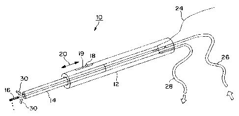
Figure 1 is a perspective view of an
electrocautery instrument in accordance with one embodiment
of the invention; and
CA 02266073 1999-03-18
WO 98/14131 PCT/US96/15796
4
Figure 2 is a enlarged perspective view of the distal end of the
electrode/blade of
the electrocautery instrument of Figure 1.
DETAILED DESCRIPTION OF A SPECIFIC EMBODIMENT OF THE
INVENTION
Referring to Figure l, there is shown a perspective view of a fluid-assisted
electrocautery instrument 10 in accordance with one embodiment of the
invention.
Electrocautery instrument 10 comprises a handle 12, a suction tube 14, and an
electrocautery electrode/blade 16. Handle 12 is preferably made of a
sterilizable, rigid,
and non-conductive material, such as nylon or the like. Suction tube 14, which
is also
preferably made of a sterilizable and non-conductive material, is slidably
disposed
partially within an internal lumen of handle 12, and projects distally out of
the end
thereof. Electrode/blade 16 is disposed within suction tube 14 and handle 12.
Suction
tube 18 is adapted to slide proximally and distally with respect to handle 12
and
electrode I6 (i.e., in the directions of arrow 20 in Figure 1) by means of a
sliding lever
18 extending out of a slot 19 in handle 12. With suction tube 14 in a
retracted position,
as shown in Figure 1, a distal portion of electrode/blade 16 projects beyond
the distal
end of tube 14, such that electrocautery can be performed. With suction tube
in an
advanced position, suction tube 14 completely conceals the tip of
electrode/blade 16.
In accordance with one aspect of the invention, electrode/blade 16 is
preferably
implemented using a hollow cylindrical tube which has been flatted at its
distal end, as
shown in the greatly enlarged perspective view of Figure 2. In addition to
being
flattened, a portion of the distal end of electrode/blade 16 is removed to
form a
longitudinal slit 22 therein.
CA 02266073 2004-09-16
66742-697
Three connections are made to electrocautery
instrument 10: One terminal (e. g., positive) of a radio-
frequency (RF) generator (not shown in Figure 1) is
electrically coupled to electrode/blade 16 via a wire 24; a
5 source of fluid to be expelled from slit 22 in
electrode/blade 16 is coupled to the proximal end of
electrode/blade 16 via a flexible tube or hose 26; and a
suction hose 28 is coupled to handle 12 so as to be in
communication with the internal lumen of handle 12 and with
suction tube 14. When suction is applied via hose 28, air
and fluid are drawn into the distal end of suction tube 14,
as indicated by arrows 30. The ability to advance or
retract suction tube 14 with respect to electrode/blade 16
enables the operator of the instrument to perform
electrocautery while simultaneously aspirating smoke and
fluid from the incision site, or to use suction tube 14
alone, without performing electrocautery.
As noted above, conductive fluid is communicated
from inflow tube 26 and communicated along the length of
electrode/blade 16 to be expelled from the distal end
thereof. This is done in order to establish a so-called
virtual electrode for performing electrocautery. The
infusion of conductive fluid simultaneously with the
application of RF energy is discussed in further detail in:
U.S. Patent No. 5,431,649, U.S. Patent No. 5,609,151 and in
U.S. Patent No. 5,876,398.
As described in the above-mentioned RF ablation
patents, the infusion of conducting fluid into the area of
application of RF energy creates a "virtual electrode", the
size and shape of which can be controllably modified, and
which can be rendered more or less conductive, thereby
CA 02266073 2004-09-16
66742-697
6
modifying the spread of RF energy. By varying such factors
as the RF energy and duration, the rate of infusion of
conductive liquid, and the conductivity of the infused
solution, the size, shape, and intensity of the "virtual
electrode" - i.e., the intensity of thermal production in
the area, can be controlled. In the case of the
electrocautery device in accordance with the present
invention, application of the conductive solution during the
application of RF energy further assists by preventing
overheating of the electrode/blade, extending the point at
which burning or charring of tissue would otherwise normally
occur. To enhance this effect, it is contemplated that the
solution being infused may first be cooled.
Conductive solutions believed to be suitable for
establishing the virtual electrode include saline, saturated
saline, and Ringer's solution, among others. Regarding the
source of conductive fluid, it is contemplated that a
conventional pump may be coupled to input line 26.
Alternatively, it is contemplated that a small, pre-
pressurized canister of conductive solution may be used,
such that no pump is required. In one embodiment, handle 12
may be configured to receive such a pressurized canister
therein, eliminating the need for input line 26.
Although in the embodiment of Figure 1, input line
26, suction line 28, and electrical connection 24 are
depicted separately, it is contemplated that these
connections
CA 02266073 1999-03-18
WO 98/14131 PCTIUS96/15796
7
to instrument 10 may be consolidated into a single line having two separate
fluid-
conducting lumens therein (one for input of conductive solution, one for
suction),
alongside an insulated electrical conductor.
Various alternate configurations of electrode/blade 16 are also contemplated.
In
one embodiment, a porous metal element is substituted for the flattened tube
configuration of Figures 1 and 2.
From the foregoing detailed description of a specific embodiment of the
invention, it should be apparent that a method and apparatus for performing
fluid-
assisted electrocautery of body tissue has been disclosed, wherein fluid
delivered out of
a hollow electrocautery electrode/blade creates a virtual electrode which
incises and
cauterizes the tissue.
Although a specific embodiment of the invention has been described herein,
this
has been done solely for the purposes of illustrating various aspects of the
invention, and
is not intended to be limiting with respect to the scope of the invention. It
is
contemplated that various substitutions, alterations, and/or modifications,
including but
not limited to those specifically discussed herein, may be made to the
disclosed
embodiment without departing from the spirit and scope of the invention as
defined in
the appended claims, which follow.