Note: Descriptions are shown in the official language in which they were submitted.
CA 02267454 1999-03-29
WO 98/14754 PCTNS9711?442
METHOD AND APPARATUS FOR DETERMINING A CONTAC'1C AREA
BETWFIEN A PROBE AND A SPECIMEN
~jELD OF THE INVEN~',~ON
This invention relates to methods and apparatus for measuring turgor
pressure of a cell or cells by determining the area of contact be 1 tween a
probe and a
specimen, and more particularly to an instrument including a transparent
mechanical
probe and its use to view the area of its contact with a specimen.
BAC'~GROUND OF THE INVENTION
A characteristic of a deformable specimen that can be related to area of
contact between a mechanical probe and the specimen is the turgor pressure of
a cell.
Growing plants are hydrostatic structures. Plant form is maintained by turgor
pressure. In most of the biomechanics of plant growth, an understanding
requires
some knowledge of turgor pressure changes to determine the physical properties
of the
plant, such as yield threshold and wall modulus. However, turgor pressure is
not
readily measured in a nondestructive, noninvasive way.
Traditional approaches for determining turgor pressure in plant cells
were conducted using either an incipient plasmolysis method, a pressure bomb
method,
or a micropipette-pressure-probe or "micropressure probe" method (see Park S.
Nobel,
Physicochemical and Environmental Plant Physiology, 103, l76-180, Academic
Press
Inc., New York, l991 ). These traditional methods are laborious and subject to
artefactual error. For example, the incipient plasmolysis method is highly
subjective,
and it radically alters the environment of the cells being measured. The
"micropressure
probe" method, in contrast, is potentially precise and accurate, but
inherently difficult
to perform. The micropressure method necessarily destroys the cells whose
turgor is
being measured. Finally, other techniques, such as the pressure-bomb method,
are
only suitable for whole organs and are generally characterized as single use,
one-shot
methods. Thus, the traditional ways of determining turgor pressure are
invasive or
disruptive to the cellular specimen, thereby interfering with the normal
dynamics of the
SUBSTITUTE SHEET (RULE 26)
CA 02267454 1999-03-29
~'~ 98~147~ PCT/US97/17442
2
cell, including cellular behavior.
In contrast, the present invention relates to a method and an apparatus
for measuring the contact area or contact patch between a specimen and a
mechanical
probe, and this can be used to determine, virtually instantaneously and
repeatably, the
turgor pressure in a cellular specimen. The method and apparatus can be non-
invasive
and non-destructive to the specimen. In cellular specimens the present
invention's
method can be repeated from point to point, for example, along a growing axis.
S_~MARY OF THE INVENTION
In accordance with this invention a method and an apparatus for
determining the area of contact between a convex probe surface and a cellular
specimen uses a transparent probe and an optical viewing path through the
probe to
the area of contact. The area of contact can be described as the contact area
or
contact patch between the objective and the specimen. In one embodiment, the
specimen is located at the working distance of a microscope objective, and the
probe is
introduced between the objective and the specimen. A known force is applied by
the
probe to a deformable specimen. The amount of deformation of the specimen will
depend on the force. If the probe's contact surface is of known geometry, for
example
spherical, and of known dimensions, the contact area between the probe's
contact
surface and the cellular specimen will be a function of the turgor pressure.
Contact area image information, i.e. the optical image or data
descriptive of the optical image, is conveyed to an image analysis system.
This
calculates the contact area and consequently, permits calculation of the
specimen
characteristic affecting contact area.
By applying a series of known forces via the probe and measuring
respective contact areas it is possible to derive data representing a plot of
area versus
force. This enables extrapolation of the specimen characteristic at zero
force. As
discussed in more detail below, when method and apparatus of the invention is
used to
determine turgor pressure in a cell, an extrapolation of this kind permits
determination
of the turgor pressure when no force is applied by the probe.
The turgor pressure of a cell is the hydrostatic pressure contained in a
SUBSTITUTE SHEET (RULE 2fi)
CA 02267454 1999-03-29
WO 98I14754 PCTIUS97/17442
3
constraining membrane of each individual cell. Given a constant force and a
spherical
probe contact surface, the greater the internal pressure of the cell, the
smaller will be
the contact area or patch between probe and cell. In measuring this pressure
in a
cellular specimen, composed of one or more cells, the method and apparatus of
the
invention have the capability of making such measurement without invasion or
destruction of the cellular specimen or any cell of the cellular specimen.
The method of nondestructively and noninvasively calculating the
turgor pressure in the cellular specimen uses an appropriate proportionality
relationship
between the turgor pressure in a supported cell that has a substantially
smooth upper
surface, and the contact area between the transparent mechanical probe, having
a
known geometry. The contact area is viewed by a microscope, which will have a
suitable support for the specimen and may have associated with it an
appropriate
means for illuminating the contact area, either by a substage light source and
condenser, by a fiber optic light guides brought in at substantially the level
of the
microscope stage and providing oblique illumination at approximately ninety
degrees
to the optical axis of the microscope, or by epi-illumination through the
objective lens
itself. The light source is manipulated until a clear image of the outline
bordering the
contact area is observed.
The term view or observe as used here is meant to include both
observation by an individual using the method and apparatus of the invention
and
retrieving of image information optically, electrically or otherwise. For
example, the
apparatus for determining the contact area may include an image capturing
system
using a CCD camera to which the image is exported. A video frame grabber and
image analysis station can be used to arrive at the actual contact area.
A force controllable mechanical probe support provides an accurately
determined contact force between the probe and the specimen. The probe is an
optically neutral element. In a preferred embodiment the probe's contact
surface was
spherical, formed by a sphere of glass, diamond, or quartz and axed to a strip
of
cover glass by a drop of ultraviolet cured optical adhesive. This arrangement
avoids
distortion at the spherical ball surface remote from the specimen.
Essentially, the
contact area is being viewed through a flat window to the far surface of the
ball. The
SUBST1'TLJTF SHEEN' (RULE ~6)
CA 02267454 1999-03-29
W4 98/14754 PCT/US97117442
4
force controllable mechanical probe may employ a jewel bearing system for
reducing
friction, e.g. one employing a sapphire or like-bearing material.
A field instrument used to measure the turgor pressure of leaves of crop
plants is one application of the turgor pressure measuring embodiment of the
invention.
Such a device can be employed to quantify water stress on plants quickly in
the field to
serve as a "go-no go" gauge for irngation, that is to say, to indicate whether
or not
irrigation is required.
DESCRIPTION OF THE DRAWINGS
The invention will be more readily understood from the description of a
preferred embodiment that follows and from the diagrammatic figures of the
drawings.
In the drawings:
Figure 1 is a cross-sectional view of a spherical force controllable
transparent mechanical probe contacting a cellular specimen;
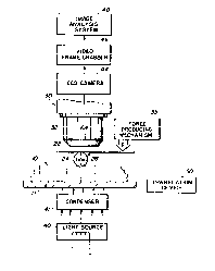
Figure 2 is a schematic view partially in section and partially in block
diagram form and illustrates a transparent mechanical probe in a system to
determine
the contact area between the probe and a specimen;
Figure 3 is a schematic view like that of Fig. 2 and diagrammatically
illustrates an alternative means of illumination of the probe-specimen contact
area; and
Figure 4 is a graphical illustration plotting a relationship between
contact area derived turgor pressure (Bars) and medium osmotic pressure
(Bars).
DESCRIPTION OF THE PREFERRED EMBODIMENTS
I. Theoretical Background for a Proportionality Relationship between the
Turgor Pressure and the Contact Area of a Spherical Force Controllable
Transparent Mechanical Probe Contacting a Cellular Specimen
In a preferred embodiment of this invention a cellular specimen 10,
shown as an individual cell, is contacted by a spherical surface of a
transparent optical
probe 20, as shown in Fig. 1. The individual cell is treated as a thin-walled
pressure
vessel to which an external load W is applied for a theoretical understanding
of the
invention. The cell has a substantially smooth upper surface 12. By
"substantially
SUBSTITUTE SHEET (RULE 26)
CA 02267454 1999-03-29
WO 98/14754 PCT/US97/17442
smooth upper surface" is meant a surface not having features, such as
epidermal hairs,
that would interfere with a force controllable transparent optical probe's
contacting
that surface of the specimen.
For the general case of a thin-walled pressure vessel to which an
5 external load is applied by means of a rigid probe, the area of the contact
patch is
related to the internal pressure, turgor pressure, of the individual cell by:
(1) W = Ap;
wherein: W is the force applied to the cell through the force controllable
transparent
mechanical probe (measured in Newtons or other units of force); A is a contact
area
(measured in square meters or other units of area); and p is a turgor pressure
(measured in Bars, Pasca.ls or other units of pressure).
Refernng to Fig. 1, where the contact surface of the probe 20 is
spherical, the probe causes an indentation in the surface 12 of the pressure
vessel or
cell 10. There is an additional supporting force which results from the stress
in the
skin acting to li$ the toad, as shown in Fig. 1. Thus, the force acting on an
indentor of
this nature will be balanced by the internal pressure, turgor pressure, of the
individual
cell according to the relationship:
(2) W = p~rz + 2~trot sin(6);
wherein: W is the force applied to the cell through the force controllable
transparent
mechanical probe causing the indentation (measured in Newtons or other units
of
force); t is the cell wall thickness (measured in meters or other units of
length); p is a
turgor pressure (measured in Bars, Pascals or other units of pressure); a is a
stress in
the cell wall (measured in Bars, Pascals or other units of pressure); R is a
radius of the
spherical, force-controllable transparent, mechanical probe (measured in
meters or
other units of length); r is a radius of the contact patch (measured in meters
or other
units of length); and 6 is a contact angle with respect to the center of the
sphere and
the outline of the contact area (measured in degrees).
SUBSTITUTE SHEET (RULE 26)
CA 02267454 1999-03-29
WO 98I14754 PCT/US97/17442
6
If one keeps the indentation of the force controllable transparent
mechanical probe into the individual cell small, sin(6) is approximated by
r/R; such that
equation (2) reduces to:
(3) W = p~rz + 2~rzat/R.
Moreover, the wall stress, Q, is related to internal pressure according to the
following
relationship:
(4) a = pD/4t;
where D is the approximate cell size (measured in meters or other units of
length).
Substitution of equation (4) into equation (3) produces the following
relationship:
(5) W = pnrz ( 1 + '/zD/R) = pA( 1 + '/zD/R).
Because D generally has a dimension near 50 pm and R in this case is 1000 um,
equation (5) can be reduced for these specific dimensions to the following
relationship:
(6) W = pA x 1.05.
If a spherical probe is applied to a surface of a cellular specimen that
comprises a rnulticellular tissue, and the probe contact area spans more that
one cell,
the additional support offered by the anticlinal walls may be considered. The
internal
support of the anticlinal walls could cause a decrease in the contact area and
an
apparent increase in the measured turgor pressures. However, if the additional
support
of the anticlinal walls within the contact area is negligible with respect to
the turgor
pressure, then the contact area is related to the average turgar pressure of
the cells in
contact with the probe. Considering the delicate nature of the cell walls and
the fact
that the experimentally measured pressures were consistently lower than would
otherwise be predicted it is presently believed that the anticlinal walls may
at this time
SUBSTITUTE SHEET (RULE 26)
CA 02267454 1999-03-29
WO 9$/14754 PCT/US97/17442
7
be safely ignored.
Additionally, there is the possibility of compartmentalization in a
cellular specimen composed of a multicellular tissue resulting in a lack of
fluid mobility
under the probe. This lack of fluid mobility could result in higher pressures
at the
center of the probe producing an apparent increase in the cell pressure.
The thin-walled model discussed above for the embodiment described
here does not incorporate any correction for either subsurface support or
fluid
compartmentalization, but corrections for these effects may be included where
necessary.
II. Method and Apparatus for the Measurement of Turgor Pressure
To determine specimen turgor pressure, an accurate measurement is
made of the contact area between the cell 10 and the force-controllable,
transparent
mechanical probe 20, of known geometry. Such probe may have, but is not
limited to,
a contacting surface that is spherical, hemispherical, or cylindrical. A
calibrated load is
1 S applied to the specimen via the probe by a suitable accurate force
producing
mechanism. The specimen 10 is supported from below by support 11. The
transparent
probe 20 may be made of any light transparent material, such as, but not
limited to,
glass, diamond, and quartz. The cellular specimen may be composed of a
plurality of
eukaryotic, either plant or animal, cells; a plurality of procaryotic cells; a
plurality of
organic micelles; a plurality of inorganic micelles; or a single cell or
micelle, provided
that the cellular specimen includes a constraining membrane 14.
The probe 20 is small enough to be inserted directly beneath an
objective lens 32 of a standard compound microscope 30 as shown in Fig. 2. The
particular probe of this embodiment includes a strip 22 of No. 2 cover glass,
acting as
a support beam, and a 1 millimeter diameter ball lens 24 cemented to the strip
22 with
a drop of ultraviolet cured optical adhesive 26. The probe is thus inserted
into the
optical path of the microscope 30. There it is manipulated into the working
distance of
the objective 32, and carefully lowered onto the cellular specimen 10,
supported by the
microscope stage 11. The adhesive prevents distortion at the spherical surface
remote
from the specimen.
SU85TiTUTE SHEET (RULE 26y
CA 02267454 1999-03-29
WO 98I14754 PCT/US97117442
8
The ball lens 24 serves as a spherical mechanical indentor, while at the
same time it provides an optically neutral, flat window at the upper surface
through
which the contact area can be observed directly. Because the image formed by
the
microscope is of the tissue in contact with the lower surface of ball lens
itself, the
optical properties of the ball lens do not contribute to total magnification
of the
system. This results in a reasonably clear observable image of the cell or
cells of
specimen 10 on which the ball lens is resting.
In an actual embodiment, the total mass of the ball lens 24 and its
support 22 was 150 milligrams. The actual controlled force applied to the
cellular
specimen 10 was 45 milligrams times gravity. For the purpose of applying the
controlled force, any accurate force producing mechanism 3 5 may be coupled to
the
probe.
As shown in part in Fig. 2, the cellular specimen 10 and the contact
patch formed with the probe 20 are transilluminated by a standard substage
condenser
41, and light source 40. Alternatively, as illustrated in Fig. 3, illumination
may be by
fiber-optic light guides 42 brought in at the substantially the level of the
microscope
stage, providing oblique illumination at approximately ninety degrees to the
optical
axis of the microscope. In still another alternative, illumination may be by
epi-
illumination through the objective lens itself. These means for illumination
are
manipulated to provide sufllcient contrast to reveal the contact area.
In the preferred embodiment, as shown in Fig. 2, the image of the
contact area is exported to an image capturing system, via, for example, a CCD
camera
44, thence to a video frame grabber 46 and finally to an image analysis
station 48
where the contact area is determined directly. The image analysis station is
suitably a
computer running OPTIIVIAST'~' or another commercially available image
analysis
program. The area may also be determined directly by the use of an eye piece
incorporating a measuring reticle.
Measurements of neighboring cells in the cellular specimen 10 can be
rapidly assessed using a translation device 50 as shown in Fig. 2. The
translation
device is movable in either one, two, or three dimensions. The translation
device
allows the probe 10 to slide over the surface of the specimen 20. Multiple
SUBSTITUTE SHEET (RULE 26)
CA 02267454 1999-03-29
WO 98l14754 PCT/US97/I7442
9
measurements can be taken as fast as the probe can be moved to another cell
and the
image captured using the image analysis system. The capturing of the image is
generally accomplished by clicking the "Freeze" button on the image analysis
system.
The turgor pressure is then directly calculated from the observed and
measured contact area using the relationships described in Equations ( 1 )
through (6).
By repeating the measurement of the contact area at a variety of different
forces of
indention, data representing a plot of turgor pressure versus force are
developed. The
turgor pressure at zero force thus may be extrapolated.
The method and apparatus described above was used to determine if the
measured areas and the calculated turgor pressures varied linearly with
cellular osmotic
pressure. In this test, peeled patches of onion leaf base adaxial epidermis
were
incubated in mannitol solutions of varying osmolality, where one osmole equals
one
mole of nonpermeating molecules plus ions per liter. These solutions
corresponded to
1 Bar increments in osmotic pressure from distilled water to - 6 Bars. A
slight
meniscus of liquid around the contact patch facilitated observation of the
contact patch
outline.
Figure 4 shows a plot of the turgor pressure, calculated by the contact
area method, against the ambient osmotic pressure of the incubating medium.
This
plot shows a basically linear relationship between the calculated turgor
pressure of the
target cells, and the osmolarity (water potential) of the incubating medium.
Sources of
scatter in the graph may include: inaccuracies in the method employed, real
differences
in turgor pressure from cell to cell in the cellular specimen, and the
presence of the
contact meniscus which inflates the area and consequently lowers the apparent
turgor
pressure.
It should be noted that several factors can contribute to real differences
in turgor pressure from cell to cell in cellular specimen. These factors
include:
deformations of the cellular specimen resulting from constraining a spherical
layer of
cells onto a flat microscope slide which can result in local strains that
could either act
to raise or lower the turgor pressure in the cells, and the geometry of
individual cells
comprising the cellular specimen can also give rise to different measured
turgor
pressures.
SUBSTITUTE SHEET (RULE Z6)
CA 02267454 1999-03-29
WO 98I14754 PCTIUS97/17442
A hand held version of the turgor pressure measuring apparatus may be
fabricated. This would include the same elements as the described embodiment.
The
addition of a portable power supply to power the means for illumination is
envisioned.
In certain applications natural light may suffice to illuminate the contact
patch.
5 Whereas a specific preferred embodiment of this invention has been
described it will be understood that variations and modifications may be made
without
departure from the spirit and scope of the invention as set forth in the
appended claims.
SUBSTITUTE SHEET (RULE Z6)