Note: Descriptions are shown in the official language in which they were submitted.
CA 02268473 1999-04-12
WO 98/16260 PCT/US97/18610
SCANNING DEVICE FOR EVALUATING CLEANLINESS AND INTEGRITY
OF MEDICAL AND DENTAL INSTRUMENTS
Backqround of the Invention
The present invention relates to the cleaning,
sanitizing, disinfecting, and sterilization arts. It finds
particular application in conjunction with the sterilization
of endoscopes and will be described with particular reference
thereto. However, it is to be appreciated that the present
invention is also applicable to a wide variety of medical,
dental, surgical, mortuary, veterinary, industrial, and other
areas in which potentially hazardous microbes are removed from
devices.
Heretofore, instruments and devices which are used
in various medical, dental, surgical, veterinary, and
industrial processes are sterilized, disinfected, or at least
sanitized before use. Typically, these instruments or devices
have been rinsed in water, such as deionized water, saline
solution, or tap water, to remove organic residue after use.
Frequently, biological materials, e.g., proteins, blood,
mucous, and polysaccarides, formed a film which remained on
the instruments or devices after rinsing. This film built-up
particularly in hard to access places such as in the channels
of an endoscope.
After rinsing, the devices were typically
disinfected or sterilized using one of a variety of
techniques. Typical techniques included steam sterilization
in which the instrument or device was heated to a high
temperature and pressure in the presence of steam to kill
harmful microorganisms. Any microorganisms on the instrument
or in the biological residue were killed during steam
sterilization. The residue film was typically baked on to the
_ _ , _ _.___ ._ ~ _ __ _...._ _. __ __.__..._ _ ..~_ ... .~_..~..,._.~~.....
.__._~..._ __.~_._._. _.. __~.~. _ . _
CA 02268473 1999-04-12
- 2 -
instrument. However, because the baked on residue was
sterilized, the residue was not generally considered to be
a concern.
Other instruments, particularly those which were
not able to take the temperature and pressure of steam
sterilization were disinfected or sterilized with other
techniques. Some were immersed in glutaraldehyde, a high
level disinfectant. The glutaraldehyde tends to blacken
the protein film. Other instruments were treated with
ethylene oxide gas, which again killed the living organisms
in the biological residue.
Instruments that are treated with a flowing
liquid sterilant or disinfectant, such as that shown in
U.S. Patent Nos. 4,892,706 and 5,217,698 normally have the
residue removed. The flowing liquid included not only a
strong oxidant such as peracetic acid, but also a wetting
agent or detergent and water which helped to dissolve and
remove the biological residue. See also, "Surface Analysis
of Clinically-Used Expanded PTFE Endoscopic Tubing Treated
By the STERIS Process", Tucker, et al. ASAIO Journal, 1995
Abstracts, 41st Annual Conference May 4-6,' 1995, p. 17, and
"Surface Analysis of Clinically-Used Expanded PTFE
Endoscopic Tubing Treated By the STERIS Process", Tucker,
et al. ASAIO Journal 1996, May 10, 1996, pp. 001-008.
The techniques which kill living microorganisms
in the biological residue left on the device have several
drawbacks. First, some people find the use of instruments
which carry even sterile biological residue from former
patients to be objectionable. Residuals may affect the
performance of the device as it was intended to be used.
Second and more importantly, it has now been found that the
sterilized residue are not necessarily safe. when the
microorganisms are killed, the membranes of the dead cells
remain. These membranes contain pyrogens and give off
endotoxins as the cell walls break down. Thus, even the
dead cells remaining after sterilization can be toxic.
CA 02268473 1999-04-12
- - 3 -
Various analytical methods are available for
detecting components of liquids and solids. For example,
EP-A-644 4l2 discloses a method for the quantitative
analysis of sample liquids. The sample is dried on a
carrier and the resulting residue is directly illuminated
with infrared or visible light from a radiation source.
U.S. Patent No. 3,747,7S5 discloses an apparatus for
classifying municipal waste in a separating plant. Samples
of the waste are passed on a pan conveyor to a plurality of
infrared sensors. However, analytical methods such as
these are not suited to detection of biological residues on
the interior channels of medical devices, and the like.
GB-A-2 l73 300 discloses an apparatus for
optically monitoring the surface finish of a ground
workpiece by measuring the diameter of the workpiece as it
rotates. The apparatus includes a sensor which is
connected by a fiber optic conductor with a light source
and a transducer. However, no means of analyzing residues
on the surface are provided.
In accordance with the present invention, a
method and apparatus are provided for checking for the
presence of biological residue in sanitized, disinfected,
or sterilized instruments and devices.
Summary of the Invention
In accordance with one aspect of the present
invention, a cleanliness assessing apparatus is provided
for non-destructively inspecting medical and dental devices
and instruments for biological residues. A light source
provides illumination to the surface. An opto-electrical
circuit receives light from the examined surface. A
transmission light path conveys the light to the surface of
the medical or dental device examined. The opto-electrical
circuit includes a spectrophotometric means for spectrally
analyzing the reflected light and producing a signal
indicative of a spectrum of the received light.
AMENDED SHEET
CA 02268473 1999-04-12
- - 3 a-
In accordance with another aspect of the present
invention, a method of non-destructively examining a
medical or dental instrument or device for biological
residue material build-up is provided. Light is directed
to a surface of an instrument or device to be examined.
Light received from the examined surface is analyzed to
determine biological characteristics of any biological
residue on the examined surface. Light is transmitted to
the surface of the medical or dental instrument or device
along a transmission light path. The method includes
spectrophotometrically analyzing a spectrum of light
received from the examined surface of the medical or dental
instrument or device.
One advantage of the present invention is that it
monitors for the removal of biological residue film.
Another advantage of the present invention is
that it is amenable for use with instruments and devices
with hard to reach regions.
Another advantage of the present invention
resides in its simplicity and ease of use.
Brief Description of the Drawings
The drawings are only for purposes of
L:\AMS\SEP\MED2161P.AMD
Substitute Page
AMENDED SHEET
_._ _~_~_. _~.______.__... __~.~~.w__ _. _
CA 02268473 1999-04-12
WO 98I16260 PCT/US97/18610
- 4 -
illustrating a preferred embodiment and are not to be
construed as limiting the invention.
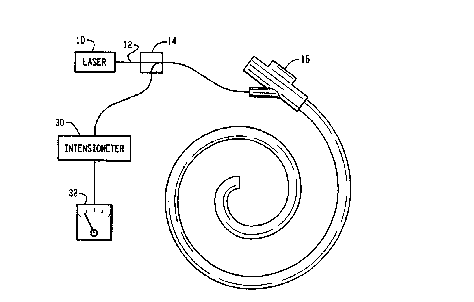
FIGURE 1 is a diagrammatic illustration of an
inspection device in accordance with the present invention;
FIGURE 2 is a detailed drawing of an end of the
optic fiber probe;
FIGURE 3 is an alternate embodiment of the
inspection device of FIGURE 1;
FIGURE 4 is another alternate embodiment of the
inspection device of FIGURE 1.
Detailed Description of the Preferred Embodiments
With reference to FIGURE 1, a source of light such
as a laser 10 outputs light such as from the infrared (IR),
near infrared, or visible region of the light spectrum along
an optical fiber 12 or other light guide. The optical fiber
extends through a beam splitter 14 and to a device or
instrument 16 to be examined. In the illustrated embodiment,
the end of the optical fiber is received within an internal
channel of an endoscope.
With reference to FIGURE 2, the optical fiber 12
which extends into the device has a lens arrangement 20
connected to its end. In the illustrated embodiment, the lens
arrangement includes an optical element 22 such as a prism,
which deflects the light from along an axis of the channel
towards adjacent walls 24 and light from adjacent walls back
along the optical fiber. The reflected light is focused to
travel along the optic fiber 12 in the opposite direction
until it reaches the beam splitter 14, where it is directed to
an opto-electric transducer 30 which converts the light to an
electrical signal. The electrical signal is related to the
interaction of the absorbed material (biological or other) on
device and light absorption. In the illustrated embodiment,
the opto-electric transducer includes an intensiometer which
converts the reflected light into an electrical signal
proportional to its intensity. An indicator, such as a gauge
32 provides a visual output proportional to the intensity of
the reflected light.
.___
CA 02268473 1999-04-12
WO 98/I6260 PCT/US97/18610
- 5 -
In one embodiment, the light source transmits light
of a wavelength, such as infrared, which is absorbed
differently by the device and the residual materials. A
change in the relative amounts of light reflected and absorbed
is indicative of the residual material build-up.
In another embodiment, the surface 24 being
inspected is constructed of or coated with a white or other
light color coating. Any biological residue remaining after
detergent washing darkens or blackens with glutaraldehyde
reducing the intensity of reflected light. In this manner,
the relative amount of reflected light indicated by the
display 32 is indicative of the presence and even the amount
of built-up biological residue.
In the embodiment of FIGURE 3, a polychromatic light
source 40 sends light along a light guide such as fiber optic
bundle 42. The light is emitted at the end of the fiber optic
bundle and is reflected off an examined surface, e.g., an
interior channel of a diagnostic instrument or device 44. A
second fiber optic bundle 46 which runs parallel to,
surrounds, or is surrounded by the fiber optic bundle 42
receives the reflected light and conveys it to a
spectrophotometer 48. Alternately, for transparent walls, the
transmission and return light guides are positioned on
opposite sides of the wall to means transmitted light. The
spectrophotometer 48 produces an output electrical signal
indicative of intensity as a function of wavelength. That is,
the spectrum of the reflected light including the relative
intensity of the reflected light at each of a plurality of
characteristic wavelengths is output by the spectrophotometer.
Typically, each molecule (i.e., protein) has a
characteristic spectra or curve of intensity versus
wavelength. A spectrum comparer 50 electronically compares
the output spectrum with each of a plurality of spectrum
indicative of known proteins or substances stored in a look-up
table 52. A memory reading circuit 54 reads out of the
look-up table 52 the name or other identification of the
protein or substance whose spectra most closely matches the
_ _ __~~ _._ ___._..~..~...~...~._
__.~~.......~...~~.....__v..___.._.w.,...~~...~.__~.. _._ . __._.._~......-
.._._..
CA 02268473 1999-04-12
WO 98I16260 PCT/US97/18610
_ 6 _
spectra received from the spectrophotometer 48. A display 56
provides a display of the protein or proteins whose spectra
are found within the instrument. An averaging circuit 58
calculates a weighted average of the spectra to provide an
indication of the intensity of the related light to provide an
indication of the relative magnitude of the output signal.
The weighted averaging circuit 58 is also connected with the
display 56 to provide an indication of a level of residue
build-up.
Optionally, rather than determining the exact nature
of the protein, a filter 60 filters the resultant spectra such
that only the intensity(ies) corresponding to one or a
selected number of wavelengths or bands is passed to a
comparator 62. The comparator 62 compares the magnitude with
a set point to determine a relative amount of residue film.
The relative amount of residue film is displayed on a display
64 to provide an indication of relative cleanliness.
Another way to analyze the surface is to monitor for
the chemical composition of the materials of which the device
is constructed. A decay in this signal versus control, not
contaminated sample of the material, is an indication of a
residue build-up. The residue can be biological, chemical,
such as a residue from detergents or washing chemicals.
Optionally, each device or instrument can be coded
with a preselected code, such as a bar code. A bar code
reader can be incorporated into the same physical housing or
another housing to read the identification of the instrument
or device. The identification of the instrument along with
the information displayed on the output display are preferably
conveyed to a memory 70 which records the identification of
the instrument, the examination results, and the date and time
of the examination.
The fiber optic probe is convenient when measuring
reflected light from within channels and deep recesses.
However, it is to be appreciated that the optical fibers may
be eliminated in favor of a lens mounted directly on the light
source and a photo pickup for reflected or transmitted ligrt
mounted behind or adjacent to the lens.
CA 02268473 1999-04-12
WO 98I16260 PCT/US97/18610
In the embodiment of FIGURE 4, a hand-held unit 80
is swept over a surface 24 for residue. An illumination
source 82, such as a light bulb 84, a reflector 86, and a
spectral filter 88, generate light with a preselected
spectrum. In the illustrated embodiment, the light passes
through a half-silvered mirror 90 and a lens 92, for striking
the surface 24. Light reflected from the surface passes back
through lens 92 and is reflected by the half-silvered mirror
90 to a lens 94 which focuses the reflected light on an
optical sensor 96. Optionally, a spectra2 filter 98 limits
the reflected light to preselected components. An analysis
circuit 100 analyzes the received light to determine the
amount of residue, the nature of the residue, or the like. In
a preferred embodiment, the analysis circuit includes a memory
which stores a reference reflected intensity. The hand-held
scanner is scanned over a clean, residue free portion of a
reference instrument to obtain a reference value for storage
in the memory. Thereafter, as the scanner is scanned over
surfaces to be analyzed, a comparitor compares the stored
reference value with the current value to determine an amount
of contamination. Optionally, any of the analysis techniques
discussed above or their equivalents can also be utilized.
Based on the analysis, a numeric or alphanumeric display 102
advises the operator of the amount and nature of the residue.
Optionally, an indicator light l04 is illuminated in response
to the amount of detected residue exceeding a preselected
residue limit.
T
__._.__...~. _.____._..~...._. ~..__~ .__ __..._.._... ~.-*...~.M.___.
