Note: Descriptions are shown in the official language in which they were submitted.
CA 02270918 2006-02-24
50927-18
- 1 -
SYSTEMS AND METHODS FOR LOCATING AND GUIDING OPERATIVE
ELEMENTS WITHIN INTERIOR BODY REGIONS
Field of the Invention
The invention generally relates to systems and
methods for guiding or locating diagnostic or therapeutic
elements in interior regions of the body.
Background of the Invention
Physicians make use of catheters today in medical
procedures to gain access into interior regions of the body
for diagnostic and therapeutic purposes. It is important
for the physician to be able to reliably and precisely
position in proximity to desired tissue locations. For
example, the need for precise control over the catheter is
especially critical during procedures that ablate myocardial
tissue from within the heart. These procedures, called
ablation therapy, are used to treat cardiac rhythm
disturbances.
Summary of the Invention
This invention has as its principal objective the
realization of safe and efficacious systems and methods for
remotely locating operative elements at precise locations
within the body.
The invention provides systems and methods for
locating an operative element within an interior body space.
The systems and methods use a locating probe, which includes
at least one transmitting element to transmit an electric
waveform output within at least a portion of the space. The
systems and methods also use a sensing element, which is
adapted to be carried by the operative element to sense a
local electric waveform within the space. A processing
CA 02270918 2006-02-24
50927-18
- 2 -
element coupled to the sensing element generates a processed
output that locates the sensing element relative to the
locating probe based, at least in part, upon a differential
comparison of the waveform output and the sensed local
waveform.
According to one aspect the invention provides a
system for locating an operative element within an interior
body space comprising a locating probe including at least
one transmitting element to transmit an electric waveform
output within at least a portion of the space, a sensing
element to be carried by the operative element to sense a
local electric waveform within the space, and a processing
element coupled to the sensing element to generate a
processed output that locates the sensing element relative
to the locating probe based, at least in part, upon a
differential comparison of the waveform output and the
sensed local waveform.
According to another aspect the invention provides
a system for locating an operative element within an
interior body space comprising a locating probe including at
least one transmitting element to generate an electric
waveform in the space, the first locating probe carrying a
return element comprising a return path for the electric
waveform, and a sensing element to be carried by the
operative element to sense spatial variations in the
electric waveform.
According to another aspect the invention provides
a system for locating an operative element within an
interior body space comprising a first locating probe
including at least one transmitting element to generate a
first electric waveform in the space, the first locating
probe including a return path for the first electric
CA 02270918 2006-02-24
50927-18
- 2a -
waveform, a second locating probe including at least one
transmitting element to generate a second electric waveform
in the space that, at least in part, intersects the first
electric waveform, the second locating probe including a
return path for the second electric waveform, and a sensing
element to be carried by the operative element to sense
spatial variations in the intersecting first and second
electric waveforms.
According to another aspect the invention provides
a system for locating an operative element within an
interior body space comprising a locating probe including at
least one transmitting element to generate an electric
waveform in the space and at least one element spaced from
the at least one transmitting element comprising a return
path for the electric waveform, a sensing element to be
carried by the operative element to sense phase of the
electric waveform within the space, and a processing element
coupled to the at least one transmitting element and the
sensing element to generate a position-indicating output
that locates the sensing element in the space relative to
the locating probe based upon analysis of the sensed phase.
According to another aspect the invention provides
a system for locating an operative element within an
interior body space comprising a first locating probe
including at least one transmitting element to generate a
first and second electric waveforms in the space and at
least one element spaced from the at least one transmitting
element comprising a return path for the electric waveform,
and second locating probes each including at least one
transmitting element to generate, respectively, first and
second electric waveforms in the space and at least one
element spaced from the at least one transmitting element
comprising a return path for the electric waveform, a
CA 02270918 2006-02-24
50927-18
- 2b -
sensing element to be carried by the operative element to
sense phase of the electric waveform within the space, and a
processing element coupled to the at least one transmitting
element and the sensing element to generate a position-
indicating output that locates the sensing element in the
space relative to the locating probe based upon analysis of
the sensed phase.
Other features and advantages of the inventions
are set forth in the following Description and Drawings, as
well as in the appended Claims.
Brief Description of the Drawings
Fig. 1 is a perspective view, somewhat
diagrammatic in form, of a system to locate the position of
an operative element within a space by generating a waveform
energy field from a single locating probe;
Fig. 2 is a diagrammatic plan view of the system
shown in Fig. 1, showing a representative position of the
operative element relative to
CA 02270918 1999-05-05
WO 98/19619 PCT/US97/21006
- 3 -
waveform phase iso-potential surfaces generated
within the space;
Fig. 3 is a schematic view of an assembly
of electrical components that the system shown in
Fig. 1 can employ in carrying out its locating
functions;
Fig. 4 is a diagrammatic plan view of a
system to locate the position of an operative
element within a space by generating a waveform
energy field from multiple locating probes,
showing a representative position of the operative
element relative to the intersecting waveform
phase iso-potential surfaces generated within the
space;
Fig. 5 is a perspective view, somewhat
diagrammatic in form, of the system shown in Fig.
4;
Fig. 6 is a side view of an assemblage of
multiple locating probes in a composite structure,
which is shown in an expanded condition ready for
use;
Fig. 7 is the composite locating probe
structure shown in Fig. 6, except shown in a
collapsed condition for deployment into a body
region;
Fig. 8 is a diagrammatic plan view of a
system to locate the position of an operative
element within a space using voltage differential
comparisons between two locating probes;
Fig. 9 is a diagrammatic view of a three-
dimensional system for locating the position and
guiding movement of an operative element within a
heart;
Fig. 10 is a diagrammatic view of a
portion of the system shown in Fig. 9, showing the
CA 02270918 1999-05-05
WO 98/19619 PCT/US97/21006
- 4 -
inputs which set the system parameters to guide
the creation of a position-identifying output;
Figs. 11 and 12 are plan views, somewhat
diagrammatic in form, showing alternative
implementations of a code to identify the geometry
of a locating probe, which code serves as one of
the inputs shown in Fig. 10;
Fig. 13 is a representative virtual image
that the system shown in Fig. 10 generates from
the position-identifying output;
Fig. 14 is a diagrammatic view of a
three-dimensional system for locating the position
and guiding movement of ablation elements within a
heart;
Fig. 15 is a plan view of a
representative continuous lesion pattern;
Fig. 16 is a plan view of an
representative interrupted lesion pattern;
Fig. 17 is a perspective and somewhat
diagrammatic view of a composite three-dimensional
basket structure of multiple locating probes
usable in association with a central processing
unit to derive a location-indicating output using
an iterative voltage distribution analysis;
Fig. 18 is a flow chart showing the steps
of an algorithm that the central processing unit
shown in Fig 17 can use to derive a location-
indicating output using an iterative voltage
distribution analysis;
Fig. 19 shows voltage distribution
patterns, one actual and the other estimated,
which the algorithm shown in Fig. 18 iteratively
matches in deriving a location-indicating output;
Fig. 20 is a diagrammatic plan view of a
system to locate the position of an operative
CA 02270918 1999-05-05
WO 98/19619 PCT/US97/21006
- 5 -
element within a space by generating multiple
frequency waveforms from multiple locating probes;
Fig. 21 is a diagrammatic plan view of a
system to locate the position of an operative
element within a space by generating multiple
frequency waveforms from a single locating probe;
and
Fig. 22 is a perspective and somewhat
diagrammatic view of a composite three-dimensional
basket structure of multiple locating probes
usable in association an operative element that
carries two electrodes for transmitting different
frequency waveforms for sensing by the locating
probes.
The invention may be embodied i-n several
forms without departing from its spirit or
essential characteristics. The scope of the
invention is defined in the appended claims,
rather than in the specific description preceding
them. All embodiments that fall within the
meaning and range of equivalency of the claims are
therefore intended to be embraced by the claims.
Description of the Preferred Embodiments
I. Differential Waveform Analysis
A. single Locating Probe
Fig. 1 shows a system 10, which locates the
position of an operative element 12 within a space
(designated S). The system 10 is well adapted for
use inside body lumens, chambers or cavities for___
either diagnostic or therapeutic purposes. For
this reason, the system 10 will be described in
the context of its use within a living body. The
system 10 particularly lends itself to
catheter-based procedures, where access to the
interior body region is obtained, for example,
CA 02270918 1999-05-05
WO 98/19619 PCT/US97/21006
- 6 -
through the vascular system or alimentary canal,
without complex, invasive surgical procedures.
For example, the system 10 can be used during
the diagnosis and treatment of arrhythmia
conditions within the heart, such as ventricular
tachycardia or atrial fibrillation. The system 10
also can be used during the diagnosis or treatment
of intravascular ailments, in association, for
example, with angioplasty or atherectomy
techniques. The system 10 also can be used during
the diagnosis or treatment of ailments in the
gastrointestinal tract, the prostrate, brain,
gall bladder, uterus, and other regions of the
body.
For deployment into an interior body space S,
the operative element 12 is carried in the
illustrated embodiment at the distal end of a
catheter tube 44. Nevertheless, the system 10 can
also be used in association with systems and
methods that are not necessarily catheter-based.
The operative element 12 can take different
forms and can be used for either therapeutic
purposes, or diagnostic purposes, or both. The
operative element 12 can comprise, for example, a
device for imaging body tissue, such as an
ultrasound transducer or an array of ultrasound
transducers, or an optic fiber element.
Alternatively, the operative element 12 can
comprise a device to deliver a drug or therapeutic
material to body tissue. Still alternatively, the
operative element 12 can comprise a device, e.g.,
an electrode, for sensing a physiological
characteristic in tissue, such as electrical
activity in heart tissue, or for transmitting
energy to stimulate or ablate tissue.
CA 02270918 1999-05-05
WO 98/19619 PCTIUS97/21006
- 7 -
The system 10 includes a locating probe 14,
which, like the operative element 12, is carried
at the distal end of a catheter tube 45 for
introduction into the body space S. In use, the
locating probe 14 establishes a localized field 20
comprising waveform energy in at least a portion
of the space S.
The system 10 provides a sensing element 16 on
the operative element 12. When located within the
energy field 20, the sensing element 16 acquires
local characteristics of the energy field 20
surrounding it. The sensing element 16 may be a
component added to the operative element 12, or it
may comprise a component already on the operative
element 12, but used for an additional purpose.
The system 10 further includes a central
processing unit 18. The central processing unit
18 receives as input the energy field
characteristic acquired by the sensing element 16.
The central processing unit 18 derives a position-
indicating output 42, which locates the position
of the sensing element 16, and thus the operative
element 12 itself, relative to the locating probe
14 within the space S.
In the illustrated embodiment, the central
processing unit 18 includes an output display
device 36 (e.g., a CRT, LED display, or a
printer). The device 36 presents the
position-indicating output 42 in a visual format
useful to the physician for remotely locating and
guiding the operative element 12 within the
localized energy field 20 generated by the
locating probe 14. Further details for processing
the position-indicating output 42 for display will
be described in greater detail later.
CA 02270918 1999-05-05
WO 98/19619 PCT/US97/21006
- 8 -
The system 10 includes an oscillator 22, which
generates the waveform comprising the energy field
20. In the illustrated embodiment, the central
processing unit 18, which is coupled to the
oscillator 22 by a control bus 24, conditions the
oscillator 22 to generate an electrical
alternating current (AC) waveform at a
predetermined amplitude and frequency.
For use within a living body space, the
selected current amplitude of the oscillator
output can vary between 0.1 mAmp to about 5 mAmp.
The frequency selected can also vary from about 5
kHz to about 100 kHz. When the space S is
adjacent heart tissue, currents substantially
above about 5 mAmp and frequencies substantially
below 5 kHz should be avoided, as they pose the
danger of inducing fibrillation. The maximum
current is a function of the frequency, as
expressed in the following equation:
I = f x 10
where I is current in Amp, and f is frequency
in kHz.
The shape of the waveform can also vary. In
the illustrated and preferred embodiment, the
waveform is sinusoidal. However, square wave
shapes or pulses can also be used, although
harmonics may be encountered if capacitive
coupling is present. Furthermore, the waveform
need not be continuous. The oscillator 22 may
generate pulsed waveforms.
The locating probe 14 carries at least one
electrode 26(1) capable of transmitting energy and
at least one energy return electrode 28 capable of
CA 02270918 1999-05-05
WO 98/19619 PCT/US97/21006
- 9 -
returning the energy to ground. These electrodes
26(1) and 28 are electrically coupled to the
oscillator 22 through an electronic switch unit
30. The locating probe 14 also carries at least
one sensing electrode (four such electrodes 26(2)
to 26(5)are shown in Fig. 1), which are located
between the transmitting electrode 26(1) and the
return electrode 28. Preferably, the sensing
electrode(s) 26(2) to 26(5) are also capable of
becoming a transmitting electrode in place of the
electrode 26(1), to change the point of energy
transmission, if desired.
For purposes of description, the illustrated
embodiment shows the one return electrode 28
carried at the distal region 32 of the locating
probe 14 and the other five electrodes 26(1) to
26(5) carried in a spaced-apart relationship along
the probe axis 34, proximal of the return
electrode 28, with the transmitting electrode
26(1) being the most proximal.
The number and placement of the electrode(s)
26 and return electrode(s) 28 on the locating
probe 14 can vary. Generally speaking, the
position-resolution capability of the system 10
improves with increased number of electrodes 26.
Also generally speaking, the position-resolution
capability of the system 10 improves as the
spacing between adjacent intermediate electrodes
26(2) to 26 (5) and the spacing between the
transmitting electrode 26(1) and the return
electrode 28 decreases.
The geometry of the locating probe 14 itself
can also vary. In the illustrated embodiment, the
locating probe 14 takes the elongated, cylindrical
form of a conventional diagnostic catheter, which
CA 02270918 1999-05-05
WO 98/19619 PCT/US97/21006
- 10 -
is well suited for deployment in interior body
regions.
In the illustrated embodiment, the central
processing unit 18 is capable of connecting the
waveform output of the oscillator 22 through the
switch unit 30 between the transmitting electrode
26(1) and the return electrode 28, which is
coupled to isolated ground or patient ground 38.
This creates an energy waveform field 20 emanating
into at least a portion of the space S.
The central processing unit 18 is also capable
of acquiring a differential voltage between
electrodes 26(1) to 26 (5) and the sensing
electrode 16 through another switch element 72 and
a data acquisition element DAQ 68. The
differential voltage measurements are taken along
iso-potential surfaces 40(1) to 40(5) in the
energy waveform field 20.
Fig.1 shows the iso-potential surfaces
associated with electrodes 26(1), 26(2), 26(3),
26(4), and 26(5) as, respectively, planes 40(1),
40(2), 40(3), 40(4), and 40(5). Fig. 2 shows the
energy field 20 and the iso-potential surfaces
40(1) to 40(5) in plan view.
For the purpose of illustration, the iso-
potential surfaces 40 are shown as planar surfaces
or planes. Actually, the iso-potential surfaces
typically will take the form of more complex,
closed curvilinear surfaces, which are orthogonal
to the probe axis 34 near the probe, but which
deviate significantly from planar with increasing
distance from the probe. The depiction of the
surfaces 40 in the drawings aids in the
understanding of the invention, as coordinate
locations in and intersections of the more complex
CA 02270918 1999-05-05
WO 98/19619 PCT/US97/21006
- 11 -
iso-potential surfaces 40 can generally be treated
equivalent to coordinate locations and
intersections of planar surfaces.
As Fig. 2 shows, the differential comparison
along the iso-potential surfaces 40(1) to 40(5)
derives either an in-phase relationship or an out-
of-phase relationship between the voltage sensed
by the element 16 (WS)and the voltage at the plane
of the sensing electrode (Wo), depending upon the
location of the sensing element 16 relative to the
iso-potential surface 40 of the electrode 26 along
which the differential measurement is acquired.
More particularly, Fig. 2 shows the sensing
element 16 to be located to the right of iso-
potential surfaces 40(1), 40(2), and 40(3) and to
the left of the iso-potential surfaces 40(4) and
40(5). In this orientation, when either surface
40(1) or 40(2) or 40(3) is the surface along
which the differential measurement is taken, the
differential comparison of WS and Wo indicates an
out-of-phase relationship between the two
waveforms. The out-of-phase relationship indicates
that the iso-potential surfaces 40(1), 40(2), or
40(3) are located in a proximal direction relative
to the sensing element 16, meaning that the
sensing element 16 is located between these iso-
potential surfaces and the return electrode 28.
Conversely, when the differential measurement
is acquired along either surface 40(4) or 40(5),----
the differential comparison of WS and W. indicates
an in-phase relationship between the two
waveforms. The in-phase relationship indicates
that the iso-potential surfaces 40(4) or 40(5) are
located in a distal direction relative to the
sensing element 16, meaning that the these iso-
CA 02270918 1999-05-05
WO 98/19619 PCT/US97/21006
- 12 -
potential surfaces are located between the sensing
element 16 and the return electrode 28.
The central processing unit 18 controls the
switch unit 72 to electronically switch the
electrodes 26(2) to 26(5) to perform a
differential comparison of the waveform WS of the
sensing electrode 16 and the waveform Wo of the
switched-on electrode 26. In Fig. 2, the
differential comparison of WS and Wo will shift
from an out-of-phase condition to an in-phase
condition when the measurement is acquired along
the iso-potential surface 40(4). The switch point
between out-of-phase and in-phase conditions marks
the longitudinal orientation of the sensing
element 16 (and thus the operative element 12)
along the axis 34 of the locating probe 14, i.e.,
between iso-potential surface 40(3) and iso-
potential surface 40(4).
The central processing unit 18 can also
perform a differential comparison between'the
signal amplitude of the acquired waveform AS and
the signal amplitude of the waveform Ap at the
switched-on sensing electrode 26. From the
differential amplitude comparison, the central
processing unit 18 derives the latitudinal
orientation of the operative element 12
perpendicular to the axis 34 of the locating probe
14, i.e., the vertical distance within the space S
between the operative element 12 and the probe
axis 34. The magnitude of the difference between
AS and A. increases as a function of increasing
distance between the sensing element 16 and the
plane of the switched-on electrode 26. The
function governing the increase of the amplitude
differential over distance can be empirically
CA 02270918 1999-05-05
WO 98/19619 PCT/US97/21006
- 13 -
determined, or be determined by finite element
analysis.
There are various electrical configurations,
analog or digital, that can be used to carry out
the above differential comparisons. Fig. 3 shows
one representative implementation.
In Fig. 3, the system 10 includes an address
bus 64, which couples the central processing unit
18 to the first-described switch unit 30. The
first switch unit 30 is also coupled to a
transmitting electrode, e.g. electrode 26(1), and
return electrode 28. The central processing unit
18 conditions the first switch unit 30 via the bus
64 to distribute the alternating current output of
the oscillator 22 in a prescribed fashion in
parallel to at least the electrodes 26 (1) for
return through the return electrode 28.
In this arrangement, the system 10 also
includes a data acquisition system (DAQ) 68. The
DAQ 68 includes a differential amplifier 70. The
sensing element 16 is coupled to the noninverting
(+) input of the amplifier 70.
The DAQ 68 further includes the second
electronic switch unit 72, which is independently
coupled to the electrodes 26(1) to 26(5). The
central processing unit 18 conditions the second
switch unit 72 via a second address bus 74 to
couple a selected one transmitting electrode 26 on
the locating probe 14 to the inverting (-) input
of the amplifier 70.
In this arrangement, the differential
amplifier 70 reads the electrical potential of the
sensing element 16 with respect to that of the
switched-on transmitting electrode 26, then
coupled to the amplifier 70 by the switch unit 72.
CA 02270918 1999-05-05
WO 98/19619 PCT/US97/21006
- 14 -
The output 71 of the amplifier 70 is an AC voltage
signal.
The DAQ 68 also includes a synchronized
rectifier 76 and peak detector 78. The rectifier
76 receives the AC signal voltage output of the
amplifier 70 and acquires its phase relative to
the phase at the output of the oscillator 22. The
detector 78 determines the peak amplitude of the
AC voltage signal output 71 of the amplifier 70.
In an alternative implementation, the rectifier 76
and detector 78 can take the form of a
synchronized phase detector, or any other element
that detects phase and amplitude (whether as an
RMS value, peak value, average rectified value, or
otherwise).
The output of the detector 78 is an analog
signal having a value corresponding to the peak
amplitude of the AC output of the amplifier 70,
and a sign (+ or -) denoting whether the AC
voltage output is in phase with the oscillator 22
(+) or out of phase with the oscillator 22 (-).
The DAQ 68 registers this analog signal in
association with the switched-on electrode 26
then-coupled to the amplifier 70 in a sample and
hold element 80. An analog to digital converter
82 converts the analog signals to digital signals
for processing by the central processing unit 18.
A suitable control bus 54 couples the sample and
hold element 80, converter 82, and differential
amplifier 70 to the central processing unit 18 for
coordination and control functions. For example,
the central processing unit 18 can set the
sampling rate of the sample and hold element 80,
the input range of the converter 82, and the
amplification of the amplifier 70.
CA 02270918 1999-05-05
WO 98/19619 PCT/US97/21006
- 15 -
In determining the longitudinal location of
the sensing element 16, the central processing
unit 18 conditions the first switch unit 30 to
connect the return electrode 28 to the isolated
ground 38 of the oscillator 22.
The central processing unit 18 also conditions
the first switch element 30 to direct AC current
flow from the oscillator 22 in parallel to the
most proximal transmitting electrode 26(1), while
also conditioning the second switch unit 72 to
couple the switched-on transmitting electrode
26(1)to the inverting input of the differential
amplifier 70. The amplifier 70 subtracts the
electrical potential measured at the switched-on
electrode 26(1) from the electrical potential
measured by the sensing element 16. The
differential potential times the gain of the
amplifier 70 constitutes the input to the
rectifier 76.
The rectifier 76 senses the synchronization of
the phase of its input voltage relative to the
phase of the oscillator 22, while the detector 78
senses the peak voltage. This signed analog value
is passed through the sample and hold element 80,
converted to a digital format by the converter 82
and registered by the central processing unit 18
in association with the identity of the switched-
on transmitting electrode 26(1).
The central processing unit 18 next conditions
the second switch unit 72 to couple the electrode
26(2) to the inverting input of the differential
amplifier 70. The central processing unit 18
processes the signal obtained for the switched-on
electrode 26(2) in the same fashion as the output
voltage signal for the first switched-on electrode
CA 02270918 1999-05-05
WO 98/19619 PCT/US97/21006
- 16 -
26(1). The central processing unit 18 proceeds in
like fashion sequentially through all the
remaining electrodes 26 (3), 26(4), and 26(5),
deriving and processing the output voltage signal
for each switched-on electrode 26. The processor
18 registers the digitally converted peak voltages
and phase synchronization for each switched-on
transmitting electrode 26(1) to 26(5).
Typically, it can be expected that the
electrical capacitances and inductances of tissue
in and about the space S are minimal. Therefore,
the synchronization of the phase of the output
voltage signal of the amplifier 70 relative to the
phase of the oscillator 22 will vary depending
upon whether the sensing element 16 is located to
the left or to the right of the transmitting
electrode 26 then-coupled to the inverting input
of the amplifier 70 (as Fig. 2 shows).
If the switched-on electrode 26 is located to
the left of the sensing element 16 (as Fig. 2
shows for electrodes 26(1), 26(2), and 26(3)), the
output voltage signal of the amplifier 70 will be
out of phase with respect to the phase of the
oscillator 22 (i.e., that analog signal received
by the sample and hold element 80 will have a(-)
sign). This is because the potential of the
sensing element 16 acquired at the noninverting
input of the amplifier 70 (during the positive
phase of oscillator output) will be more negative
than the potential acquired at the electrodes
26(1), 26(2), and 26(3), which are sensed at the
inverting input of the amplifier 70. As long as
the potential of the sensing element 16 remains
more negative under these conditions, the output
voltage signal of the amplifier 70 remains
CA 02270918 1999-05-05
WO 98/19619 PCT/US97/21006
- 17 -
negative, indicating an out of phase condition..
If the switched-on electrode.26 is located to
the right of the sensing element 16, (as Fig. 2
shows for transmitting electrode 26(4) and 26(5)),
the output voltage signal of the amplifier 70 will
be in phase with respect to the phase of the
oscillator 22. This is because the potential of
the sensing element 16 acquired at the
noninverting input of the amplifier 70 (during the
positive phase of oscillator output) will be more
positive than the potential at the electrodes
26(4) and 26(5) sensed at the inverting input of
the amplifier 70. As long as the potential of the
sensing element 16 remains more positive under
these conditions, the output voltage signal of the
amplifier 70 remains positive, indicating an in
phase condition.
The central processing unit 18 monitors the
output of the peak detector 78 to determine where
the output changes sign, by turning from-(-) to
(+) or vice versa. In Fig. 2, this transition
occurs between switched-on electrode 26(3) and
switched-on electrode 26(4). The iso-potential
surface 40(3) associated with the electrode 26(3)
sets the longitudinal coordinate of the sensing
element 16, and thus the operative element 12.
To determine the latitudinal coordinate of the
sensing element 16 using differential amplitude
sensing, the central processing unit 18 conditions
the first switch unit 30 to direct AC current flow
from the oscillator 22 to the particular switched-
on electrode 26(3) at which the phase transition
occurred. The central processing unit 18
conditions the second switch unit 72 to couple the
particular phase transition electrode 26(3) to the
CA 02270918 1999-05-05
WO 98/19619 PCTlUS97/21006
- 18 -
inverting input of the differential amplifier 70
while sensing element 16 is coupled to the
noninverting input of the amplifier 70. The
amplifier subtracts the electrical potential
measured at the phase-transition electrode 26(3)
from the electrical potential measured at the
sensing element 16. The differential potential
times the gain of the amplifier 70 constitutes the
input to the rectifier 76.
The detector 78 senses the peak voltage
amplitude of the signal. The output of the peak
detector 78 is passed through the sample and hold
element 80 and converted to digital format by the
converter 82. This digitally converted peak
voltage amplitude is registered by the central
processing unit 18. The central processing unit 18
compares the peak voltage amplitude to a voltage
amplitude variation table stored in memory, which
lists variations in peak voltage amplitude as a
function of distance from the plane of the
transmitting electrode. The voltage amplitude
variation table can be empirically determined or
based upon finite element analysis, taking into
account the physical and electrical parameters of
the space S.
In a preferred embodiment, a predetermined
threshold amplitude is established, which
corresponds to a nominal distance from the
transmitting electrode, which differentiates
between a "close condition" (i.e., equal to or
less than the nominal distance) and a "far
condition" (i.e., greater than the nominal
distance). When the sensed peak voltage amplitude
is equal to or less than the threshold amplitude,
the central processing unit 18 generates an output
CA 02270918 1999-05-05
WO 98/19619 PCT/US97/21006
- 19 -
that notifies the physician of the "close
condition" between the sensing element 16 and the
switched-on transmitting electrode 26. When the
sensed peak voltage amplitude is less than the
threshold amplitude, the central processing unit
18 generates an output that notifies the physician
of the "far condition" between the sensing element
16 and the switched-on transmitting electrode 26.
In this way, the physician has at least a
qualitative indication of the position of the
sensing element 16 relative to the switched-on
transmitting electrode 26. In one embodiment, the
physician can indicate through input to the
central processing unit 18 the magnitude of the
nominal distance, or, alternatively, establish a
range of distances that progressively indicate a
"closest", "closer" and "close" variation of
positions.
In another embodiment, the sensing of the
voltage amplitude is accomplished in a way that
also provides information regarding the
orientation of the sensing element 16 relative to
the switched-on transmitting electrode 26. More
particularly, as shown in Fig. 1, the operative
element 12 can carry a second sensing element 16'
spaced a known distance apart from the first
mentioned sensing element 16. In this
arrangement, one or more transmitting electrodes
on one probe are switched on in sequence or
simultaneously to transmit the energy field to an
indifferent patch electrode, which serves as a
return path. Sensing individually at each sensing
element 16 and 16' provides, not only a peak
voltage amplitude, but also, through a comparison
of relative phases and amplitudes at each element
CA 02270918 1999-05-05
WO 98/19619 PCT/US97/21006
- 20 -
16 and 16', information regarding the orientation
of the operative element 12 itself. For example,
the central processing unit 18 can differentially
compare the amplitude at sensing element 16' with
the amplitude at sensing element 16 to determine
that element 16 is further away from the
transmitting electrodes than element 16'. This
indicates that the orientation of the operative
element 12 is skewed within the space S.
In an alternative embodiment, the second
sensing element 16' can comprise the return path
for the transmitting electrode 26, instead of a
return path electrode 28 carried by the locating
probe 14. In yet another alternative embodiment,
the energy field can be transmitted by one of the
elements 16 or 16' and returned by the other one
of the element 16' or 16. In either of theses
arrangements, the peak voltage amplitude is sensed
by an electrode on one of the locating probes.
B. Multiple Locating Probes
Figs. 4 and 5 show a system 100 that locates
an operative element 102 within a space
(designated S) by generating an energy waveform
field 110 using two locating probes 106 and 108.
Each locating probe 106 and 108 is generally like
the locating probe 14 shown in Figs. 1 and 2,
having at least one transmitting electrode and at
least one return electrode. For purpose of
illustration, the locating probes 106 and 108 each
carry more electrodes than the probe 14. The
electrodes carried by the locating probe 106 are
designated X(1) to X(6) and the electrodes carried
by the locating probe 108 are designated Y(1) to
Y(5). Each locating probe 106 and 108 also
includes a return electrode, designated RX for
------ - -- --------
CA 02270918 1999-05-05
WO 98/19619 PCT/US97/21006
- 21 -
probe 106 and RY for_probe 108.
The locating probes 106 and 108 are positioned
relative to each other in or near the space, such
that their elongated axes, respectively 120 and
122, are not parallel, but extend at an angle.
In the illustrated embodiment, the angle is about
900, but other smaller or larger angles can be
used. Furthermore, the locating probes 106 and
108 need not lie in the same plane.
As in the Figs. 1 and 2 embodiment, the
operating element 102 carries a sensing element
104.
Like the system 10 described in Figs. 1 and 2,
the operation of the system 100 is governed by a
central processing unit 112. The central
processing unit 112 connects the waveform output
of an oscillator 114 through a switch unit 116
between the selected transmitting electrode Y(1)
and X(1) on the locating probes 106 and 108 and
the respective return electrode RY and RX, which
is also couple to isolated ground or patient
ground 118. The central processing unit 112 also
couples the sensing element 104 to the electrodes
of the probes 106 and 108 (via the switch unit 117
and DAQ 119) along the iso-potential surfaces
TX(1) to TX(6) and TY(1) to TY(5) in the energy
waveform field 110. Due to the angular placement
of the locating probes 106 and 108, the iso-
potential surfaces TX(1) to TX(6) of the probe 106
intersect the iso-potential surfaces TY(1) to
TY(5) of the probe 108. Fig.4'shows the
intersecting iso-potential surfaces TX and TY in
side view. Fig. 5 shows the intersecting iso-
potential surfaces TX and TY in perspective view.
As previously described, the central
CA 02270918 1999-05-05
WO 98/19619 PCT/US97/21006
- 22 -
processing unit 112 performs a differential
comparison of the waveform WS to the waveform
output Wo when each of the transmitting electrodes
X(1) to X(6) and Y(1) to Y(5) are switched on.
The differential comparison derives either an in-
phase or relationship an out-of-phase relationship
between Ws and Wo, depending upon the location of
the sensing element 104 relative to the iso-
potential surface TX(N) or TY(N) of the switched-
on voltage sensing electrode X(N) or Y(N).
More particularly, Fig. 4 shows the sensing
element 104 to be located to the right of (or
above, in the vertical orientation shown in Fig.
4) the iso-potential surfaces TX(1) to TX(4) and
to the left of (or below, from the vertical
orientation shown in Fig. 4) the iso-potential
surfaces TX(5) and TX(6). In this orientation,
when either plane TX(1) or TX(2) or TX(3) or
TX(4) is switched-on for sensing, the
differential comparison of WS and Wo indicates an
out-of-phase relationship between the two
waveforms. This means that the sensing element
104 is located between these planes and the return
electrode RX. Conversely, when either plane
TX(5) or TX(6) is switched-on for sensing, the
differential comparison of WS and Wo indicates an
in-phase relationship between the two waveforms.
This means that these planes are located between
the sensing electrode 104 and the return electrode
RX.
The central processing unit 112 controls the
switch unit 116 to electronically switch the
electrodes on, sequentially from most proximal to
most distal, i.e., sequentially from left to right
(or from bottom to top, in the vertical
CA 02270918 1999-05-05
WO 98/19619 PCT/US97/21006
- 23 -
orientation shown in Fig. 4) from X(1) to X(6).
This sequentially switches on differential sensing
along the iso-potential surfaces TX(1) to TX(6).
For each switched-on electrode X(1) to X(6),
the central processing unit 112 performs (via the
DAQ 119) a differential comparison of the waveform
Ws of the sensing electrode 104 and the waveform Wo
of the switched-on electrode X(N). In Fig. 4, the
differential comparison of WS and Wo will shift
from an out-of-phase condition to an in-phase
condition when measurement occurs along the iso-
potential surface TX(5). The switch point between
out-of-phase and in-phase conditions marks the
longitudinal orientation of the sensing element
104 (and thus the operative element 102)-along the
axis 120 of the locating probe 106, i.e., between
iso-potential surface TX(4) and iso-potential
surface TX(5).
The central processing unit 112 can also
perform a differential comparison between the
signal amplitude of the sensed waveform AS and the
signal amplitude of the waveform at the switched-
on transmitting electrode A0. From the
differen-tial amplitude comparison, the central
processing unit 112 derives the latitudinal
orientation of the operative element 102
perpendicular to the axis 120 of the probe 106,
i.e., the vertical distance within the space S
between the operative element 102 and the probe
axis 120.
The same methodology is repeated along the
locating probe 108. Fig. 4 shows the sensing
element 104 to be located to the right of the iso-
potential surfaces TY(1) to TY(2) and to the left
of the iso-potential surfaces TY(3), TY(4), and
CA 02270918 1999-05-05
WO 98/19619 PCT/US97/21006
- 24 -
TY(5). The central processing unit 112 controls
the switch unit 117 to electronically switch on
the transmitting electrodes, sequentially from
most proximal to most distal, i.e., sequentially
from left to right, Y(1) to Y(5). This
sequentially switches on differential sensing
along the iso-potential surfaces TY(1) to TY(5).
For each switched-on electrode Y(1) to Y(5),
the central processing unit 112 performs (via the
DAQ 119) a differential comparison of the waveform
WS of the sensing element 104 and the waveform Wo
of the switched-on transmitting electrode Y(N).
In Fig. 4, the differential comparison of WS and Wo
along the probe 108 will shift from an out-of-
phase condition to an in-phase condition when iso-
potential surface TY(3) is switched on. The
switch point between out-of-phase and in-phase
conditions marks the longitudinal orientation of
the sensing element 104 (and thus the operative
element 102) along the axis 122 of the locating
probe 108, i.e., between iso-potential surface
TY(2) and iso-potential surface TY(3).
The central processing unit 112 can also
perform a differential comparison between the
signal amplitude of the sensed waveform AS and the
signal amplitude of the waveform at the switched-
on transmitting electrode Ao to derive the
latitudinal orientation of the operative element
102 perpendicular to the axis 122 of the probe
108, i.e., the vertical distance within the space
S between the operative element 102 and the probe
axis 122. The component parts of the system 100 can
incorporate the particular electrical
configuration shown in Fig. 3, or another analog
CA 02270918 1999-05-05
WO 98/19619 PCT/US97/21006
- 25 -
or digital configuration, to carry out the above
differential comparisons.
The central processing unit 112 provides a
position-indicating output 124, which correlates
the position of the sensing element 104 (and thus
the operative element 102) within the grid of
intersecting iso-potential surfaces TX(N) and
TY(N). Preferably, the position-indicating output
124 is presented to the physician on a display
device 126.
The individual identification probes 106 and
108 shown in Figs. 4 and 5 can be assembled into a
composite structure 150, as shown in Fig. 6. In
this arrangement, the structure 150 comprises an
array of flexible spline elements 152 extending
longitudinally between a distal hub 154 and a
proximal base 156. For purpose of illustration,
the structure 150 includes four spline elements
152(1) to 152(4)(only 3 spline elements are
visible in Fig. 6). A greater or lesser number of
spline elements 152 can be present.
Each spline element 152 preferably comprises a
flexible body made from resilient, inert wire or
plastic. Elastic memory material such as nickel
titanium (commercially available as NITINOLr""
material) can be used. Resilient injection molded
plastic or stainless steel can also be used. Each
spline element 152 is preferably preformed with a
convex bias, creating a normally open three-
dimensional basket structure.
The structure 150 is carried at the end of a
catheter tube 158. An outer sheath 160 slidably
advances forward along the catheter tube 158 to
compress and collapses the structure 150 (see Fig.
7) for introduction into the body region. Rearward
CA 02270918 2006-02-24
50927-18
- 26 -
movement retracts the slidable sheath 160 away from the
structure 150, which springs open and assumes its three-
dimensional shape (as Fig. 6 shows).
In Fig. 6, the geometry of spline elements 152 is
both radially and axially symmetric. Asymmetric structures,
either radially or axially or both, can also be used.
Examples of asymmetric arrays of spline structures are shown
in U.S. Patent No. 6,216,043.
Each spline element 152 carries an array of
multiple transmitting electrodes TE and at least one return
electrode RE, as previously described. Each spline element
152 thus comprises a locating probe. The structure 150
comprises an ordered array of multiple location probes,
which, in use, create a waveform field 162 about the space
bounded by the spline elements 152.
Fig. 6 shows an operative element 172 movable
within the energy waveform field 162. The operative element
172 carries a sensing element 174.
As before described, a central processing unit 164
sequentially connects the waveform output of an oscillator
166 through a switch unit 168 to the transmitting electrodes
TE on each spline element 152 (for example, beginning with
the most proximal and moving distally), while coupling the
respective most distal return electrode RE of the spline
element 152 to isolated ground or patient ground 170. The
central processing unit 164 also sequentially couples the
electrodes TE and the
CA 02270918 1999-05-05
WO 98/19619 PCTIUS97/21006
- 27 -
sensing electrode 174 on the operative element 172
through a switch unit 169 and a DAQ 171 to acquire
a differential voltage along a grid of
intersecting iso-potential surfaces TP in the
energy waveform field 162, in the same manner
shown for the probes 106 and 108 in Figs. 4 and 5.
The differential comparison derives either an in-
phase relationship or an out-of-phase relationship
between WS and Wo,-depending upon the location of
the sensing element 174 relative to the
transmitting electrodes along each elongated
spline element 152.
The central processing unit 164 can also
perform a differential comparison between the
signal amplitude of the sensed waveform AS and the
signal amplitude of the waveform at the switched-
on electrode A. where the phase transition occurs,
to derive the latitudinal orientation of the
sensing element 174 perpendicular to each spline
element 152.
II. Differential Voltage Analysis
A. Relative Proximity Derivation
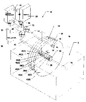
Fig. 8 shows an alternative embodiment of
system 300 that locates an operative element 302
within a space (designated S), using differential
voltage analysis instead of differential waveform
analysis. The system generates an energy waveform
field 310 between two locating probes 306 and 308.
Each locating probe 306 and 308 includes at least
one transmitting electrode, which are designated
X(1) to X(6) for probe 106 and Y(1) to Y(6) for
probe 108. The operative element 302 carries a
sensing element 304.
In the illustrated embodiment, the locating
probes 306 and 308 are positioned so that their
CA 02270918 1999-05-05
WO 98/19619 PCT/US97/21006
- 28 -
elongated axes, respectively 320 and 322, are not
parallel, but extend at some angle. In the
illustrated embodiment, the angle is about 900,
but other smaller or larger angles can be used.
Alternatively, because differential voltage
analysis is employed, the locating probes 306 and
308 in this embodiment can be located in a
parallel, mutually facing relationship.
The operation of the system 300 is governed by
a central processing unit 312. The central
processing unit 312 connects the waveform output
of an oscillator 314 through a first switch unit
316 to transmit the waveform from all transmitting
electrodes on one probe 306 to all the electrodes
on the other probe 308, which are coupled to the
isolated patient ground 318. For this reason, the
probe 306 will be called the "transmitting probe"
and the probe 308 will be called the "receiving
probe." The receiving and transmitting functions
of the probes 306 and 398 can be reversed. The
generated waveform field 310 extends between the
transmitting probe 306 and the receiving probe
308. The waveform can be generated simultaneously
between all electrodes or sequentially along the
axis of the probes 306 and 308.
As Fig. 8 shows, the waveform field 310
includes iso-potential surfaces T(1) to T(6),
which extend between the transmitting-receiving
electrode pairs X(1)-Y(1) to X(6)-Y(6).
The central processing unit 312 conditions a
second switch element 330 to couple each switched-
on electrode on the transmitting probe 306 in
succession to inverting (-) input-of a
differential amplifier 332, while coupling the
sensing element 304 to the noninverting (+) input.
CA 02270918 1999-05-05
WO 98/19619 PCT/US97/21006
- 29 -
The amplifier subtracts the electrical potential
measured by the electrode coupled to the inverting
input from the electrical potential measured by
the sensing element 304. The differential
potential times the gain of the amplifier 332
constitutes the input to a rectifier 334.
A detector 336 senses the peak voltage, and
the rectifier 334 senses the synchronization of
the phase of the voltage signal relative to the
phase of the oscillator 314. The central
processing unit 312 registers the peak voltage and
the synchronization in association.
The synchronization of the phase of the output
voltage signal of the amplifier 332 relative to
the phase of the oscillator 314 will vary
depending upon the location of the most
immediately distal iso-potential surface to the
sensing electrode 304.
More particularly, the output voltage signal
of the amplifier 332 will be in-phase with
respect to the phase of the oscillator 314 only
when the differential amplitude is measured along
the iso-potential surface which is most
immediately distal to the sensing electrode 304.
In Fig. 8, the most immediate distal iso-potential
surface to the sensing electrode 304 is T(6),
which lies between electrode pairs X(6)-Y(6). The
output voltage signal of the amplifier 332 will be
out-of-phase with respect to the phase of the
oscillator 314 for the differential amplitudes
measured along the most immediately proximal iso-
potential surface to the sensing electrode 304,
and along all other more proximal iso-potential
surfaces. In Fig. 8, the most immediate proximal
iso-potential surface is T(5), which lies between
CA 02270918 1999-05-05
WO 98/19619 PCT/US97/21006
- 30 -
electrode pairs X(5)-Y(5) and the remaining more
proximal surfaces T(4) to T(1) lie between
electrode pairs X(4)-Y(4) to X(1)-Y(1).
By way of another example, assuming another
position of the sensing element 304' (shown in
phantom lines in Fig. 8), the output voltage
signal of the amplifier 332 will be in-phase with
respect to the phase of the oscillator 314 only
when the differential amplitude is measured along
the iso-potential surface T(4), which is the most
immediately distal to the sensing electrode 304'.
The output voltage signal of the amplifier 332
will be out-of-phase with respect to the phase of
the oscillator 314 for the differential amplitudes
measured along the most immediate proximal iso-
potential surface T(3) and all other more proximal
iso-potential surfaces T(2) and T(1).
Differential voltage analysis can also be used
in association with the composite probe structure
150 shown in Fig. 6 or any of the structures shown
earlier.
III. Three-Dimensional Navigation Systems
A. Establishing a Three-Dimensional
Navigation System (Using a Waveform
Differential Analysis)
Fig. 9 shows a representative implementation
of a three-dimensional navigation system 200,
which includes three locating probes 204, 206,
and 208 positioned within a space S. In the
illustrated embodiment, the space S comprises the
interior of a heart. In use, the system 200
locates and guides an operative element 202 within
the heart. The operative element 202 can serve to
sense electrical activity in the heart to locate
potential ablation sites, or to transmit energy to
CA 02270918 1999-05-05
WO 98/19619 PCT/US97/21006
- 31 -
pace heart tissue, measure impedance, or to
ablate. Alternatively, the operative element 202
can include an imaging element to image tissue,
anatomic structures, or lesions formed within the
heart. Also, the operative element can include a
cannula to penetrate heart tissue for the purpose
of injecting an ablation media, or to inject a
drug or gene therapy agent.
For purpose of illustration, the three
locating probes 204, 206, and 208 are purposely
situated within the heart to provide spaced-apart
navigational points for locating the operative
element 202. Furthermore, the probes 204, 206,
and 208 are located at different coordinate
planes, to create a three-dimensional navigational
grid and make triangulation possible.
In the illustrated embodiment, the probes 204,
206, and 208 are individually placed at or near
known anatomic regions of the heart using, for
example, fluoroscopy or another imaging
technology, such as ultrasound. This is because
potential ablation sites within the atria are
typically identified by reference to an anatomic
landmark within the heart.
It should be appreciated that a single
locating probe or multiple locating probes may be
positioned essentially in any region within the
heart or in any tissue or vascular region
surrounding the heart for purposes of establishing
navigational points of reference to locate the
operative element 202. Any region of placement
with the body that can be imaged by fluoroscopic
or other imaging technology can be selected as a
potential navigational site. The region of
placement therefore does not have to represent a
CA 02270918 1999-05-05
WO 98/19619 PCT/US97/21006
- 32 -
particular fixed anatomic site. For example,
establishing a three-dimensional navigation system
for use within a given heart chamber, one or more
locating probes can be located within the heart
chamber, another one or more probes may be located
in a different chamber, and yet another one or
more locating probes can be located at an
epicardial location outside the interior of the
heart.
In the illustrated embodiment, the first
locating probe 204 is positioned in region of the
high right atrium; the second locating probe 206
is positioned in the region of the right
ventricular apex; and the third locating probe 208
is positioned in the region of the coronary sinus.
The three probes 204, 206, and 208 are located on
different coordinate planes, so that the probe
axes extend in mutually nonparallel relationships.
Each locating probe 204, 206, and 208 includes
multiple transmitting electrodes TE and a distal
return electrode TR, which function in the manner
previously described and shown in Fig. 1. A
transmitting electrode TE and the return electrode
TR on each probe 204, 206, and 208 are coupled via
electronic switch units 210 to an oscillator 212
to create an energy waveform field 216.
The operative element 202 carries a sensing
element 218, which can also can serve as an
ablation electrode or as sensing electrode. The
sensing element 218 is coupled to the central
processing unit 214 in the manner previously
described to sense the waveform quantity WS within
the field 216.
A DAQ 68 acquires differential waveforms along
multiple iso-potential surfaces TP, one associated
CA 02270918 1999-05-05
WO 98/19619 PCT/US97/21006
- 33 -
with each electrode TE on each probe 204, 206, and
208. As shown in Fig. 9, because the probes 204,
206, and 208 are located at different coordinate
planes, the multiple iso-potential surfaces TP
form intersection points within the field 216.
The central processing unit 214 employs the
DAQ 68 previously described (see Fig. 3) to
differentially compare WS to Wo for each switched-
on electrode TE and locate regions of phase
transitions relative to each probe 204, 206, and
208. In addition, the central processing unit 214
can also perform a differential comparison between
the signal_amplitude of the sensed waveform AS and
the signal amplitude of the waveform at the
switched-on transmitting electrode A. where the
phase transition occurs to derive the latitudinal
orientation of the sensing element 218
perpendicular to the axis of each probe 204, 206,
208.
The central processing unit 214 generates a
position-indicating output 220, which locates the
sensing element 218 (and thus the operative
element 202 itself) within the matrix of
intersecting iso-potential surfaces TP generated
by the three probes 204, 206, and 208.
B. Establishing a Three-Dimensional
Navigation system (Using an Iterative
Voltage Analysis)
Fig. 17 shows a three dimensional system 500,
which conducts an iterative differential voltage
analysis to determine the location of an operative
element 502 within a space S peripherally bounded
by multiple locating probes 504. In Fig. 17, the
multiple locating probes 504 are assembled
together by a distal hub 506 and a proximal base
CA 02270918 1999-05-05
WO 98/19619 PCT/US97/21006
- 34 -
508 into a composite,_three-dimensional basket
structure 510 of the type previously shown and
described in Fig. 6. However, it should be
appreciated that the multiple locating probes 504
need not be assembled together in a composite
structure, but exist as separate probes located
about the space S, in the manner shown in Fig. 9,
as previously described.
The composite structure 510, however, is well
suited for use within the heart and can perform
other functions in addition to navigation. For
example, the composite structure 510 can serve to
transmit electrical signals to pace heart tissue
or to characterize the electrical characteristics
of the tissue by acquiring tissue impedance
measurements. The composite structure can also
serve to sense electrical activity in myocardial
tissue to acquire electrograms for heart mapping
procedures.
The composite structure 510 shown in Fig. 17
includes eight locating probes 504, and each
probe, in turn, carries eight electrodes 505, for
a total of sixty-four electrodes 505 positioned
about the space S. Fig. 17 identifies the
electrodes 505 by the designation (A,B), where A
1 to p and B = 1 to e, where p is the total number
of probes 504 and e is the number of electrodes
505 on each probe 504 (in the illustrated
embodiment, p = 8 and e = 8).
The system 500 includes a central processing
unit 512, which couples a voltage source 514 to a
transmitting electrode 516 carried by the
operative element 502. In Fig. 17, an indifferent
electrode 518, carried as a patch on the exterior
of the patient, comprises the voltage return,
CA 02270918 1999-05-05
WO 98/19619 PCT/US97/21006
- 35 -
which is, in turn, coupled to isolated or patient
ground 520. Alternatively, another electrode
carried by the operative element 502 can serve as
the voltage return. The electrode 516 creates a
voltage field 517 within the space S, which varies
in detected amplitude at each probe electrode 505
according to its distance from the transmitting
electrode 516.
The system 500 includes a data acquisition
element 522 coupled to the central processing unit
512 and to a switch element 524. The switch
element 524 individually conditions each electrode
(A,B) to sense voltage existing at its location
within the field 517, which the data acquisition
element 522 samples and holds, in the manner
previously described, e.g., see Fig. 3.
The central processing unit 512 includes a
processing component 526 which derives a position-
indicating output 528 based upon the voltage
distribution sensed by the electrodes (A,B) on the
probes 504. Fig. 18 shows the steps of a
preferred algorithm 530 for deriving the output
528.
As Fig. 18 shows, the algorithm 530 includes,
as a first step 532, establishing an estimated
coordinate position P(x, y, Z)EST for the
transmitting electrode 516 on the operative
element 502 within the space S, where x is the x-
field coordinate, y is the y-field coordinate, and
z is the z-field coordinate.
For example, P (x, y, z)EST can be initially
arbitrarily set at P(0,0,0), which is at the
geometric center of the voltage field 517
(designated as GC in Fig. 17). Alternatively,
differential waveform analysis, or differential
CA 02270918 2006-02-24
50927-18
- 36 -
voltage analysis, or amplitude analysis, as described above,
alone or in combination, can also be used to more accurately
estimate P(x,y,z)EST. By way of another example, position
indicating methodologies disclosed in U.S. Patent
No. 5,722,402 can also be used to provide a more accurate
initial position estimate P(x,y,z)EST. To increase
processing efficiencies, multiple signals that are
orthogonal from a signal processing standpoint (for example,
waveform signals of different frequencies, waveform signals
of the same frequency but which differ by 90 in phase, and
waveforms from uncorrelated white noise sources) may be
transmitted simultaneously in the manner shown in Fig. 22
(as will be described in greater detail later).
In the next step 536, the algorithm 530 computes
the distance AD(A,B) between each probe electrode (A,B) and
the transmitting electrode 516 at P(X,Y,Z)EST. The distances
AD(A,B) can be normalized to facilitate analysis. The
algorithm then applies a preestablished, mathematical
voltage-to-distance function 534 to derive the estimated
voltage V(A,B)EST at each electrode (A,B), based upon
AD(A,B). In effect, the algorithm 530 constructs an
estimated voltage distribution matrix, which would exist,
according to the function 534, if P(x,y,z)EST was the actual
voltage transmission point. The voltage-to-distance
function 534 can be empirically determined or be based upon
finite element analysis and stored in memory accessible to
the
CA 02270918 1999-05-05
WO 98/19619 PCTIUS97/21006
- 37 -
central processing unit 512. As a next step 538,
the algorithm 530 derives an estimated or expected
voltage differential V(A,B)EST for each electrode
505.
In the next step 540, the algorithm 530
receives as input V(A, B) ACT I where V(A, B) ACT is the
measured voltage value acquired by operation of
the data acquisition element 522 at each probe
electrode (A,B). As Fig. 19 shows, the algorithm
530, in this step 540, creates a measured voltage
distribution pattern 560 based upon the values for
V (A, B)ACT, which plots (on the Y-axis) the sensed
voltage values for each electrode (numbered 1 to
64 on the X-axis). The algorithm 530 creates an
estimated voltage distribution pattern 562 based
upon the values for V (A, B)EST, which plots (on
the Y-axis) the estimated voltage values for each
electrode (again numbered 1 to 64 on the X-axis).
As a next step 542, The algorithm 530 matches
the voltage distribution pattern 560 with the
voltage distribution pattern 562 to derive a
voltage matching coef f icient VMCOEF =
The value of the voltage matching coefficient
vm COEF for a given P(x, y, Z)EST increases as P(x, y,
z)EST coincides with the actual location of the
transmitting electrode 516. That is, the value
of the voltage matching coefficient increases in
relation to the proximity of the transmitting
electrode 516 to the estimated position P
(X,y, Z)EST'
The central processing unit 512 can derive the
matching coefficient VMCOEF in various conventional
ways, for example, by employing pattern matching;
matched filtering; or cross correlation. Examples
of using these techniques to derive matching
CA 02270918 2006-02-24
50927-18
- 38 -
coefficients appear in U.S. Patent No. 5,595,183.
In the next step 544, the algorithm 530 determines
whether VMcoEF is the "best", i.e., whether it is maximized
under the processing rules applied. For the first
iteration, and for all subsequent iterations where VMcoEE is
not maximized, the algorithm 530 applies (in step 546) a
preselected incremental correction factor Lx to the x
coordinate, factor Ly to the y coordinate, and factor 4z to
the z coordinate of the estimated position of the
transmitting electrode 516 to create a new estimated
position P(x+Ax, y+py, z+Lz), which become the new
coordinates for an estimated position P(X,Y,Z)EST. The
algorithm 530 then loops through the foregoing steps 536,
538, 540, 542, and 544, to derive an iterated voltage
matching coefficient VMCOEF based upon the new estimated
location. The algorithm 530 iteratively selects Lx, Ay, and
Lz until a best (maximum value) voltage matching coefficient
VMCOEF is achieved in step 544. The coordinates P(x, y, z) EsT at
the best, maximum voltage matching coefficient VMCOEF become
the position-indicating output 528, as shown in step 548 in
Fig. 18.
There are various ways in which the iteration of
the x-, y-, and z-coordinates can be accomplished. For
example, the algorithm 530 can iterate the x-coordinate
alone (keeping the y- and z-coordinates constant) until a
best voltage
CA 02270918 1999-05-05
WO 98/19619 PCT/US97/21006
- 39 -
matching coef f icient VMcoEF is achieved, then f ix
the x-coordinate at that value and iterate the y-
coordinate alone (while also keeping the z-
coordinate constant) until another best voltage
matching coefficient VMcoEF is achieved, and then
fix the y-coordinate at that value and iterate the
z-coordinate alone (keeping the previously fixed
x- and y-coordinates constant), until another best
voltage matching coefficient VMCOEF is achieved.
The algorithm 530 then loops back through this
process, until the best voltage matching coeffi-
cient UMCOEF is obtained for each local x-, y-, and
z-coordinate, as well as for P(x, y, z)ESt overall.
Alternatively, the x-, y-, and z-coordinates
can be simultaneously incremented to maximize the
voltage matching coefficient VMCOEF for P( x, y, z) Esr,
using, for example, a conventional maximum
gradient method.
Due to its iterative nature, the algorithm 530
shown in Fig. 18 corrects for distortion of the
locating probes caused by exposure to dynamic
conditions within a body cavity, such as within a
beating heart chamber. The iterative nature of the
algorithm 530 also corrects for electrical "noise"
caused, for example, by the inherent electrical
resistance of the electrodes and associated
electrical wiring.
Furthermore, the iterative differential
voltage analysis just described also makes
possible the generation of an error signal, should
the position of the operative element 502 stray
beyond the energy field 517. Should this event
occur, the estimated voltage and the actual
voltage become mirror images. This outcome, when
sensed by the central processing unit 512, can
CA 02270918 1999-05-05
WO 98/19619 PCT/US97/21006
- 40 -
command the generation of an out-of-field error
signal.
In an alternative embodiment, the central
processing unit 512 can incorporate a neural
network 600 (see Fig. 17), which has been trained
on experimentally acquired sets of voltage
distribution data correlated with known positions
of the transmitting electrode 516. Once the
training phase is completed, the network 600 can
instantaneously output the position-indicating
output 528, based upon input from the data
acquisition element 522 of voltage distribution
data sensed by the probe electrodes 505 during
transmission of voltage by the electrode 516.
C. Displaying Three-Dimensional
Navigational Information
As Fig, 9 shows, the position-indicating
output 220 (or, in the embodiment shown in Fig.
17, the output 528) is preferably processed for
viewing on a display device 221. In a preferred
embodiment (see Fig. 10), the central processing
unit 214 includes an input 222 that receives
information pertaining to the position and
orientation of the locating probes 204, 206, and
208 within the heart. The input 222 also receives
information pertaining to the shape and size of
each locating probe 204, 206, and 208. The
central processing unit 214 includes functional
algorithms 224, which set guidance parameters
based upon the input information. These guidance
parameters are used by the central processing unit
214 to analyze the spatial variations of the
electric waveform field generated by the locating
probes 204, 206, and 208. The guidance parameters
govern the processing of differential comparison
CA 02270918 1999-05-05
WO 98/19619 PCT/US97/21006
- 41 -
data to create the position-indicating output 220
for display on the device 221. The processed
position-identifying output aids the physician in
locating and guiding the operative element 202 in
real time.
In a preferred embodiment (see Fig. 10), the
probes 204, 206, and 208 of the system 200 are
members of a family 209 of locating probes. The
various probes comprising the family 209 are
characterized by different geometries, different
densities of transmitting and return electrodes,
and other structural and functional differences.
In this embodiment, each probe 204, 206, and 208
within the family 209 includes an identification
component 270. The identification component 270
carries an assigned identification code XYZ. The
_ code XYZ identifies the shape and size of the
electrode-supporting part of the probe and the
distribution of electrodes carried thereon, in
terms of the number of electrodes and their
spatial arrangement. The structure-specific
information contained in the code XYZ aids the
central processing unit 214 in creating a
positioning matrix based upon the locating probes
when deployed.
In the illustrated embodiment (see Fig. 10),
the coded component 270 is located within the
handle 230 attached to the proximal end of the
catheter tube 232 that carries the locating probe
204, 206, and 208. However, the component 270
could be located elsewhere in relation to the
locating probe.
The coded component 270 is electrically
coupled to an external interpreter 278 when the
probe is coupled to the central processing unit
CA 02270918 2006-02-24
50927-18
- 42 -
214 for use. The interpreter 278 inputs the code XYZ that
the coded component 270 contains. The interpreter 278
electronically compares the input code XYZ to, for example,
a preestablished master table 280 of codes contained in
memory. The master table 280 lists, for each code XYZ, the
structure-specific information required to create the
positioning matrix to locate and guide the operative element
202 within the waveform field 216. The functional
algorithms 224 of the central processing unit 214 set
location and guidance parameters based upon the code XYZ.
Because knowledge of the physical characteristic
of the locating probe and the spatial relationship of the
electrodes it carries is important in setting accurate
location and guidance parameters, the algorithms 224
preferably disable the central processing unit 214 in the
absence of a recognizable code XYZ. Thus, only probes of
the family 209 possessing a coded component 270 carrying the
appropriate identification code XYZ can be used in
association with the processing element 214.
The coded component 270 can be variously
constructed. It can, for example, take the form of an
integrated circuit 284 (see Fig. 11), which expresses in
digital form the code XYZ for input in ROM chips, EPROM
chips, RAM chips, resistors, capacitors, programmed logic
devices (PLD's), or diodes. Examples of catheter
identification techniques of this type are shown in Jackson
et al. United States Patent No. 5,383,874.
Alternatively, the coded component 270 can
comprise separate electrical elements 286 (see
CA 02270918 1999-05-05
WO 98/19619 PCT/US97/21006
- 43 -
Fig. 12), each one of_which expresses an
individual characteristic. For example, the
electrical elements 286 can comprise resistors (R1
to R4), comprising different resistance values,
coupled in parallel. The interpreter 278 measures
the resistance value of each resistor Rl to R4.
The resistance value of the first resistor R1
expresses in preestablished code, for example, the
number of electrodes on the probe. The resistance
value of the second resistor R2 expresses in
preestablished code, for example, the distribution
of electrodes on the probe. The resistance value
of the third resistor R3 expresses in
preestablished code, for example, the size of the
probe. The resistance value of the fourth resistor
R4 expresses in preestablished code, for example,
the shape of the probe.
It should be appreciated that the three-
dimensional basket structure 510 shown in Fig. 17
can also carry an identification component 270
having an assigned identification code XYZ to
identify the shape and size of the multiple probe
structure 510 and the distribution of electrodes
carried thereon. In this arrangement, the
structure-specific information contained in the
code XYZ aids the position derivation component
528 and algorithm 530 in Fig. 18 to construct the
estimated voltage distribution matrix and analyze
sensed voltage differentials.
The central processing unit 512 can also
include a component 550 (see Fig. 17), which
electronically determines structure-specific
information to construct the estimated voltage
distribution matrix and analyze sensed voltage
differentials. In this arrangement, the component
CA 02270918 1999-05-05
WO 98/19619 PCTIUS97/21006
- 44 -
550 commands, in sequence, the transmission of
voltage from the source 514 through a switch unit
554 from each probe electrode (A,B) to the
indifferent electrode 518, while sensing voltage
with the remaining probe electrodes through the
switch 524 and data acquisition element 522. The
component 550 thereby acquires a first set of data
from which the voltage differential between every
electrode (A,B) can be obtained.
The component 550 includes an input 552,
through which the component 550 acquires data
relating to the linear distance between adjacent
electrodes on each probe 504. Typically, the
electrodes 505 on each probe 504 will be spaced
apart by manufacturer at the same linear-distance,
so that will typically be only a single linear
distance to input. The physician can manually
enter the linear distance information through the
input 522. Alternatively, the input 552 of linear
distance-information can be carried by a coded
component 270 as earlier described as shown in
Fig. 10, which is inputted automatically upon
coupling the probe structure 510 to the central
processing unit 512. In this arrangement, more
complex linear distance information can be readily
inputted. The linear distance information
comprises a second set of data.
Knowing the linear distance information
between adjacent electrode 505 contained in the ---
second set of data, and the sensed voltage
differentials between these electrodes 505
contained in the second set of data, the component
520 then derives using conventional estimating
techniques the distances between other,
nonadjacent electrodes 505, both along a probe 504
CA 02270918 1999-05-05
WO 98/19619 PCT/US97/21006
- 45 -
and between probes 504. The component 550
generates a geometric output 556, which, like the
code XYZ, the output 556 identifies the shape and
size of the multiple probe structure 510 and the
distribution of electrodes 505 carried thereon.
The output 556 also provides the basis for
calculating the interior volume of the structure
510. In the heart, the interior volume of the
structure 510 typically will conform to the
interior volume of the heart chamber it occupies.
The interior volume will also typically
dynamically adjust to the changing heart chamber
volumes during systole and diastole. The
component 550 therefore makes possible the
electrical analysis, for therapeutic or diagnostic
purposes, of heart chamber volumes and the changes
in heart chamber volume during systole and
diastole. The component 550 can thereby be used,
independent of or in association with a navigation
function, to characterize heart morphology and
function.
In another embodiment, the component 550 can
incorporate a neural network 558 (see Fig. 17) to
generate in situ the distance-to-voltage function
534 particular to a given structure 510, based
upon the electrically sensed geometry and
distribution of electrodes on the structure 510.
The neural network 558 is first trained on a known
set of data that have been previously acquired
experimentally. For example, using a back-
propagation model, the network 558 can be trained
to predict a voltage-to-distance function 534
based upon structure-specific information. Once
the training phase is completed, the network 558
can be used to predict the voltage-to-distance
CA 02270918 1999-05-05
WO 98/19619 PCT/US97/21006
- 46 -
function in situ.
Based upon information received by the input
222, the central processing unit 214 (or 512 in
Fig. 17) electronically constructs a three-
dimensional coordinate system representing a
virtual image 290 of the energy field 216 (or 217
in Fig. 17) and surrounding tissue mass T. Fig.
13 shows a representative virtual image 290 based
upon two locating probes. In Fig. 13, the virtual
image 290 indicates the position of the locating
probes (designated "Probe X" and "Probe Y" in Fig.
13), as well shows the geometry and location of
the iso-potential surfaces (designated "X(1) to
X(3)" and "Y(1) to Y(3)" in Fig, 13). The virtual
image 290 shows the position of the operative
element 202 (designated "Device" in Fig. 10)
within the energy field 216, as well as displays
the coordinates of the operative element
(designated "Coordinates: X(2) Y(2)" in Fig. 10).
The central processing unit 214 continuously
performs the differential comparisons and updates
the virtual image 290 to provide a real time
display for viewing by the physician.
IV. Using Multiple Waveforms
The locating and navigation systems of the
type previously described create an energy field
by applying a single waveform. Multiple waveforms
can be simultaneously applied to gain processing
efficiencies, provided the different waveforms are
orthogonal from a signal processing standpoint.
Examples of different, orthogonal processing
signals includes waveform signals of different
frequencies, waveform signals of the same
frequency but which differ by 904 in phase, and
waveforms from uncorrelated white noise sources.
CA 02270918 1999-05-05
WO 98/19619 PCT/US97/21006
- 47 -
A. Differential Waveform Analysis Using
Different Waveforms
Fig. 20 shows a system 700 that locates an
operative element 702 within a space S by
generating different waveforms using two probes
706 and 708.
In many respects, the system 700 shares common
elements with the system 100 shown in Fig. 4. The
locating probe 706 and 708 are generally like the
locating probes 106 and 108 shown in Fig. 4. The
electrodes carried by the locating probe 706 are
designated X(1) to X(6) and the electrodes carried
by the locating probe 708 are designated Y(1) to
Y(5). Each locating probe 706 and 708 also
includes a return electrode, designated RX for
probe 706 and RY for probe 708. As in Fig. 4, the
locating probes 706 and 708 are positioned
relative to each other in a non-parallel
relationship. As in the Fig. 4, the operating
element 702 carries a sensing element 704.
The system 700 includes a first waveform
source WF1, which is coupled to the probe 708.
The system also includes a second waveform source
WF2. The first waveform WF1 is different than but
orthogonal to the second waveform WF2. In the
illustrated embodiment, the waveforms WF1 and WF2
have different frequencies, and the sources
comprise separate oscillators 720 and 722.
The probe 708 is coupled via a switching unit
710 and a first filter Fl for the waveform WF1 to
the inverting (-) input of a differential
amplifier 712. The probe 706 is also coupled by a
second switching unit 714 and a second filter F2
for the WF2 is also coupled to the inverting (-)
input of the differential amplifier 712. The
CA 02270918 1999-05-05
WO 98/19619 PCT/US97/21006
- 48 -
sensing element 704 carried by the operative
element 702 is coupled to the noninverting (+)
input of the differential amplifier 712. The
output of the differential amplifier 712 is
coupled to a data acquisition element 716. The
data acquisition element 716 includes a rectifier,
peak detector, sample and hold element, and
analog-to-digital converter coupled as shown in
Fig. 3 to process the differential output in the
manner previously described, under the control of
a central processing unit 718.
Under the control of the central processing
unit 718, the multiple oscillators 720 to 722
simultaneous apply the waveform WF1 to the
electrode Y(1), for return through the return
electrode RY, and the different waveform WF2 to
the electrode X(1), for return through the return
electrode RX.
The central processing unit 718 operates the
switch units 710 and 714 to simultaneously acquire
two differential voltages, one for waveform WF1
between the sensing element 704 and the electrode
Y(1) and the other for waveform WF2 between the
sensing element 704 and the electrode X(1). The
differential amplifier 712 thus acquires phase
information for two waveforms simultaneously along
iso-potential surfaces TX(1) and TY(1).
In like fashion, the central processing unit
718 operates the switch units 710 and 714 to
simultaneously acquire two differential voltages
for the waveforms WF1 and WF2 between the sensing
element 704 and the electrodes Y(2)/X(2), then
Y(3)/X(3), and so on. In this way, the
differential amplifier 712 acquires phase
information for two waveforms simultaneously along
CA 02270918 1999-05-05
WO 98/19619 PCT/US97/21006
- 49 -
iso-potential surfaces TX(2)/TY(2), then
TX(3)/TY(3), etc. This simultaneously acquired
phase information from two waveforms WF1 and WF2
is processed by the data acquisition element 716
to provide a position-indicating output. Greater
processing efficiencies can therefore be obtained.
B. Signal Amplitude Analysis Using Different
Waveforms
Fig. 21 shows a system 800 in which multiple
oscillators 802, 804, 806, and 808 apply different
waveforms WF1, WF2, WF3, and WF4 simultaneously to
multiple electrodes, respectively E(1), E(2),
E(3), and E(4), of a single probe 810, through an
indifferent return electrode 830. As above
described, the different waveforms WF1, WF2, WF3,
and WF4 are orthogonal in a signal processing
sense possessing, for example, they possess
different frequencies. Since the waveforms are
applied simultaneously to all electrodes E(1) to
E(4), no input switching is required.
All electrodes E(1) to E(4) of the probe 810
are coupled to an output switch 812. The output
switch 810 is, in turn, coupled to filters Fl, F2,
F3, and F4 for the frequencies of, respectively,
WF1, WF2, WF3, and WF4. The output of the filters
Fl, F2, F3, and F4 are coupled to the inverting(-)
input of a differential amplifier 814. The
sensing element 816 carried by an operative
element 818 is coupled to the noninverting (+) -
input of the differential amplifier 814.
The output of the differential amplifier 814
is coupled to a data acquisition element 820. The
data acquisition element 820 includes a rectifier,
peak detector, sample and hold element, and
analog-to-digital converter coupled as shown in
CA 02270918 1999-05-05
WO 98/19619 PCT/US97/21006
- 50 -
Fig. 3 to process the differential output in the
manner previously described, under the control of
a central processing unit 818.
Under the control of the central processing
unit 818, the data acquisition element 820
simultaneously acquires the differential amplitude
of waveform WF1 between the sensing element 816
and the electrode E(1), the differential
amplitude of waveform WF2 between the sensing
element 816 and the electrode E(2), the
differential amplitude of waveform WF3 between the
sensing element 816 and the electrode E(3), and
the differential amplitude of waveform WF4 between
the sensing element 816 and the electrode E(4). As
the magnitude of the difference increases as a
function of increasing distance between the probe
electrodes and the sensing element 816, the data
acquisition element 816 is able to simultaneously
infer distance with respect to each probe
electrode E(1), E(2), E(3), and E(4).
C. Iterative Voltage Analysis Using Multiple
Waveforms
Fig. 22 shows a system 900 for conducting an
iterative voltage analysis using multiple
waveforms to determine the location of an
operative element 902 within a space S
peripherally bounded by a composite, three-
dimensional basket structure 910, like that shown
in Fig. 17.
As in Fig. 17, the composite structure 910 in
Fig. 22 includes eight locating probes 904, and
each probe, in turn, carries eight electrodes 905,
for a total of sixty-four electrodes 905
positioned about the space S. As in Fig. 17, Fig.
22 identifies the electrodes 905 by the
CA 02270918 1999-05-05
WO 98/19619 PCTIUS97/21006
- 51 -
designation (A,B), where A = 1 to p and B = 1 to
e, where p is the total number of probes 904 and e
is the number of electrodes 905 on each probe 504
(in the illustrated embodiment, p = 8 and e = 8).
Unlike Fig. 17, the operative element 902
carries two energy transmitting electrodes 912 and
914. Multiple oscillators 916 and 918 apply
different waveforms WF1 and WF2 simultaneously to
the electrodes 912 and 914. The different
waveforms WF1 and WF2 are orthogonal in a signal
processing sense possessing, for example, they
possess different frequencies. As in Fig. 21,
since the waveforms are applied simultaneously to
both electrodes 912 and 914, no input switching is
required.
In the manner described with respect to the
system 500 shown in Fig. 17, a central processing
unit 920 conditions the electrode 912 and the
electrode 914 to simultaneously transmit waveform
energy WF1 and WF2 to a patch return electrode
922. Each probe electrode (A,B) is coupled via a
switch 924 to two filters Fl and F2 for the
frequencies of the waveforms, respectively, WF1
and WF2. A data acquisition element 926 thereby
receives simultaneous inputs from two waveforms
WF1 and WF2.
For example, the input for the waveform WF1
could provide a sensed voltage, for use by the
algorithm 530 (shown in Fig. 18) in deriving the
position-indicating output 528. The input for the
waveform WF2 could provide phase and amplitude
information for comparison to the phase and
amplitude information of waveform W-Fl, from which
the_orientation of the operative element 12 can be
ascertained. By using multiple waveforms, the
CA 02270918 1999-05-05
WO 98/19619 PCT/US97/21006
- 52 -
system 900 also make possible the derivation of
both location and orientation out.
As shown in phantom lines in Fig. 22, a second
operative element 902' could be present within the
space S bounded by the basket structure 910. The
second operative element 902' carries at least one
transmitting electrode 912'. Under the control of
the central processing unit 920, the electrode 912
of the first operative element 902 transmits the
first waveform WF1, while the electrode 912' of
the second operative element 902' transmits the
second waveform WF2. A data acquisition element
926 thereby receives simultaneous inputs from two
waveforms WF1 and WF2, via the filters Fl and F2.
The input for the waveform WF1 could provides a
sensed voltage, for use by the algorithm 530
(shown in Fig. 18) in deriving the position-
indicating output 528 for the first operative
element 902, while the input for the waveform WF2
provides a sensed voltage, for use by the
algorithm 530 (shown in Fig. 18) in deriving the
position-indicating output 528 for the second
operative element 902'. Using multiple waveforms,
the system 900 is thereby able to provide locating
information for multiple operative elements.
With respect to all embodiments in this
Specification, which show a data acquisition
element coupled by a switch unit to multiple probe
electrodes, it should be appreciated that
parallel, independent data acquisition channels,
each with its own processing components and
directly coupled to a single probe electrode,
could be substituted.
V. Guiding Multiple Electrode Ablation
Arrays
CA 02270918 1999-05-05
WO 98/19619 PCT/US97/21006
- 53 -
Fig. 14 shows a multiple electrode structure
400 located in the right atrium of a heart. The
structure 400 is flexible and carries a steering
mechanism (not shown), use of which flexes the
structure 400 into curvilinear shapes. The
structure 400 carries an array of electrodes 402,
which transmit radio frequency energy to ablate
myocardial tissue.
The electrodes 402 are preferably operated in
a uni-polar mode, in which the radio frequency
ablation energy transmitted by the electrodes 402
is returned through an indifferent patch electrode
404 externally attached to the skin of the
patient. Alternatively, the electrodes 402 can be
operated in a bi-polar mode, in which ablation
energy emitted by one or more electrodes 402 is
returned an adjacent electrode 402 carried in the
structure 400.
The size and spacing of the electrodes 402 are
purposely set for creating continuous, long lesion
patterns in tissue, which are capable of treating
atrial fibrillation. Fig. 15 shows a
representative long, continuous lesion pattern 406
in tissue T. The long continuous lesion pattern
406 is created by additive heating effects between
the electrodes 402. The additive heating effects
cause the lesion pattern 406 to span adjacent,
spaced apart electrodes 402.
Additive heating effects occur either when the
spacing between the electrodes 402 is equal to or
less than about 3 times the smallest of the
diameters of the electrodes 402, or when the
spacing between the electrodes 402 is equal to or
less than about 2 times the longest of the lengths
of the electrodes 402. When the electrodes 402
CA 02270918 2006-02-24
50927-18
- 54 -
are spaced in one or both of these manners, the simultaneous
application of radio frequency energy by the electrodes 402,
in either a bipolar or unipolar mode, creates the elongated
continuous lesion pattern 406 typified in Fig. 15.
U.S. Patent No. 5,549,661 discloses further
details regarding systems and methods that create complex
long lesion patterns in myocardial tissue.
When the predetermined spacing requirements set
forth above are not met, the additive heating effects do not
occur, and a segmented, or interrupted, lesion pattern 408
is created. Fig. 16 shows a representative interrupted
lesion pattern 408 in tissue T. The interrupted lesion
pattern 408 is characterized lesion areas 412 separated by
gaps 410 of tissue free of lesions.
An interrupted lesion pattern 408 can also occur,
even with proper spacing between electrodes 402, because of
insufficient contact between electrodes 402 and tissue, or
due to other localized effects not within the immediate
control of the physician. After ablation, intracardiac
electrogram analysis or intercardiac imaging of the ablation
region, or both used in tandem, can be used to uncover the
existence of an unintended interrupted lesion pattern 408.
In this situation, the physician can deploy an auxiliary
ablation electrode 414 (shown in Fig. 14), to ablate tissue
in the gaps 410 and thereby complete the desired lesion
pattern.
Fig. 14 includes the three-dimensional
CA 02270918 1999-05-05
WO 98/19619 PCT/US97/21006
- 55 -
locating system 200, which was previously
described and is shown in greater detail in Fig.9.
Under the control of the central processing unit
214 (previously described), the system 200 locates
and helps the physician guide the multiple
electrode structure 400 within the right atrium,
both before and during the ablation procedure.
In Fig. 14, the central processing unit 214
includes a component 416, which records the
location of each ablation electrode 402 when
ablating. The position of each electrode 402 is
recorded in the same manner as the position of the
sensing element 218 of Fig. 9 is derived, using
differential comparison of waveform phases between
each ablation electrode 402 and the sequentially
switched-on transmitting electrodes carried by the
locating probes 204, 206, and 208.
When a lesion gap 410 is detected, the system
200 is operated to recall the recorded ablation
electrode coordinates from the component 416.
From the ablation electrode coordinates, the
coordinates of the gap 410 itself can be
determined. Knowing the gap coordinates, the
system 200 can be used to guide the auxiliary
ablation electrode 414 into the gap 410. This
feedback, which is preferably updated continuously
in real time as the physician moves the auxiliary
ablation electrode 414,guides the physician in
locating the ablation electrode 414 at the chosen
gap ablation site, to thereby complete the desired
lesion pattern.
Various features of the invention are set
forth in the following claims.