Note: Descriptions are shown in the official language in which they were submitted.
CA 02271544 1999-OS-27
r
-1-
METHOD TO DETECT BONE AND OTHER
CONNECTIVE TISSUE DISORDERS IN HUMANS AND ANIMALS
Technical Field
The invention relates to methods of diagnosis
in medical and veterinary contexts. More specifically,
it concerns methods to assess bone and other connective
tissue metabolism by detecting free crosslinks formed by
collagen degradation in biological fluids, such as urine.
Backcrround Art
The association of collagen as a major
structural material in a multiplicity of tissues,
including bone, cartilage, skin, tendons, dentine and
various soft tissues is well known. It is also known
that the fiber structure of collagen is stabilized by
crosslinking. The presence of the fluorescent pyridinium
ring system as a non-reducible crosslink in collagen was
reported by Fujimoto, D., et al., J Biochem (1978)
83:863-867. The Fujimoto paper reported isolation of a
fluorescent peptide from pronase digestion of bovine
Achilles tendon collagen. The isolated hydrolyzed
pyridinoline (Pyd) was thought to contain three residues
of hydroxylysine and it was recognized that, prior to
hydrolysis, peptide fragments were attached to the
pyridinoline moiety. Further work on characterization
was conducted by Gunja-Smith, Z., et al., Biochem J
(1981) 197:759-762, using hydrolyzed urine, and advantage
was taken of the presence of the pyridinoline in urine by
Robins, S.P., Biochem J (1982) 207:617-620, who linked
CA 02271544 1999-OS-27
..-
-2-
pyridinoline obtained from hydrolyzed urine to a carrier
to raise antibodies. The antibodies were then employed
in an immunoassay to determine the concentration of
pyridinoline in hydrolyzed urine. The procedure was
stated by Robins as useful to provide an index of the
degradation of certain forms of mature collagen by
analysis of physiological fluids.
In all of the foregoing, hydrolyzates were
employed to obtain total pyridinoline since much of the
crosslink retained peptide extensions of the hydroxylysyl
residues responsible for its formation. Thus, in order _
to obtain a homogenous preparation containing the
pyridinium ring, a preliminary hydrolysis step was
necessary.
By 1982, it was established that there were two
pathways of crosslink formation depending on whether
lysine or hydroxylysine residues were present in the
telopeptides from which these crosslinks were derived
(Robins, S.P., in "Collagen in Health and Disease" (1982)
Weiss, J.B., et al., eds., pages 160-178, Churchill
Livingstone, Edinburgh). This was stated to result in a
specificity of crosslinking whereby in soft tissues, such
as skin, reducible aldimine linkages are formed from
oxidized lysyl residues, whereas in cartilage and bone
these bonds, initially formed from hydroxylysine
aldehydes, undergo a spontaneous rearrangement to more
stable oxoimine crosslinks. These bonds undergo further
reaction to form 3-hydroxy-pyridinium crosslinks. The
stable crosslinking pyridinoline analog involving lysine
rather than hydroxylysine in the helix portion was
identified and quantified by Ogawa, T., et al., Biochem
Biophys Res Commune (1982) 107:1251-1257; Eyre, D.R., et
al., Anal Biochem (1984) 137:380-388, and designated
deoxypyridinoline (Dpd). This material was then believed
CA 02271544 1999-OS-27
J
i
,._
-3-
to be restricted to bone collagen, although amounts vary
between species.
Further work by Robins, S.P., Biochem J (1983)
215:167-173, provided evidence for the existence of
glycosylated pyridinoline in bone. Robins proposed a
structure which showed the derivation of the ring from
three residues of hydroxylysine and also showed that
alkali hydrolyzates of collagen provided an O-galactosyl
derivative substituted at the sidechain hydroxy group.
As this material was extremely labile to mild acid treat-
ment, this material would not have been present in
samples of hydrolyzed tissue or body fluid.
Fujimoto, D., et al., J Biochem (1983)
94:1133-1136, chromatographed unhydrolyzed urine samples
and showed that the 3-hydroxypyridinium ring portion was
present in substantial proportion as the "free" form,
i.e., the three hydroxylysyl-derived residues which
composed it did not contain further peptide extensions.
On amino acid analysis, whereas pyridinoline isolated
from an acid hydrolyzate of collagen gave an asymmetric
peak, "free" urinary pyridinoline gave a symmetric peak.
The authors concluded this to be due to isomerization by
epimerization of the hydroxylysine moiety of the
pyridinoline system during hydrolysis. In addition,
relationship of levels of total pyridinoline (after
hydrolysis) to age was determined by these workers as a
ratio to creatinine levels. It was found that the ratio
was high in the urine of children but decreased with age
until growth ceases. It was further found that this
ratio is relatively constant in adults, but increases
slightly in old age. The authors speculate that this may
correspond to the loss of bone mass observed in old age.
Attempts were also made to characterize the
above-mentioned peptide extensions. Robins, S.P., et
CA 02271544 1999-OS-27
r'
-4-
al., Biochem J (1983) 215:175-182, proposed that in
cartilage-derived type II collagen, the pyridinoline
links two C-terminal telopeptide chains with a single
chain of the helical peptide. An additional pyridinoline
crosslink, i.e., with the ring derivatized to other
peptides, was thought to link two N-terminal non-helical
peptides with a third chain in the helical portion of the
molecule. The studies were conducted by isolating the
fluorescent pyridinoline crosslinks from tissues by
specific cleavage with CNBr, thus preserving peptide
sequences as extensions of the hydroxylysyl residues
forming the ring. The crosslink was localized in the
collagen fibers by determining the amino acid sequences
of these extensions.
In a paper similar in approach to that of
Robins (supra), Wu, J.J., et al., Biochemistry (1984)
23:1850-1857, conducted CNBr cleavage of mature cartilage
and determined the sequence of the peptide extension
residues of the hydroxylysyl participants in the
pyridinium ring. Their conclusions were similar to those
of Robins.
Robins, S.P., et al., Biochim Biophys Acta
(1987) 914:233-239, used CNBr digestion of bone derived
collagen to localize the crosslinks in the type I
collagen structure. These authors concluded that the
proportions of the crosslink derived from lysine and that
derived from hydroxylysine were present in the same
proportions in each of the isolated peptide forms. They
also concluded that this showed that these two crosslink
analogs occupy the same loci in the collagen fiber and
that the form apparently derived from one lysyl
participant appears to arise through incomplete
hydroxylation of the appropriate lysine residues in the
helix. Amino acid analysis indicated that the crosslinks
CA 02271544 1999-OS-27
. ..
-5- ..
must be situated at two locations involving both the N-
and C- terminal telopeptide regions.
Henkel, W., et al., Eur J Biochem (1987)
165:427-436, determined the amino acid sequences associ-
ated with the crosslinks in type I collagen isolated from
aorta. These sequences are different from those obtained
for type II collagen. Similar results were found by
Eyre, D.R., et al., FEBS (1987) 2:337-341, who
demonstrated that the crosslinks from type IX and type II
collagens displayed distinctive peptides attached to the
pyridinoline crosslinks.
PCT application W089/04491 to Washington
Research Foundation proposes a urinary assay for
measuring bone resorption by detection in urine of the
specific crosslinks, characterized by their peptide
extensions, associated with bone collagen. The assay
relies on quantifying the concentration of peptides in a
body fluid where the peptide fragments having a
pyridinium crosslink are derived from bone collagen
resorption. Two specific entities having peptide
extensions presumed to be associated with bone collagen
are described. These are obtained from the urine of
patients suffering from Paget's disease, a disease known
to involve high rates of bone formation and destruction.
Macek, J., et al., Z Rheumatol (1987)
46:237-240, proposed an assay for osteoarthrosis which
depends upon the peptides associated with the crosslinks
from collagen breakdown. In this approach, the urine
sample was size-separated for peptides of molecular
weight greater than 10 kd, which peptides were then
separated by HPLC using a fluorescence detector to
determine those fractions containing the fluorescence due
to the pyridinium ring. The spectra obtained from
patients with osteoarthrosis were compared to those from
a
CA 02271544 1999-OS-27
1
.: l,.. _..
_6_
healthy patients, and it was easily demonstrable that the
multitude of fluorescent peaks associated with the
diseased condition was absent from the healthy
counterpart. Furthermore, urine from the same diseased
patient two weeks after total endoprosthesis of the
diseased hip, thereby decreasing the products of
osteoarthrosis, gave a spectrum of fluorescent peaks
which more closely resembled that of normals.
Furthermore, the osteoarthrosis spectrum was readily
distinguished from that obtained from patients with
rheumatoid arthritis. The closer resemblance of the
rheumatoid arthritis spectrum to that of the spectrum
from normal controls was attributed by the authors to the
higher activity of proteases in rheumatoid arthritis.
This was presumed to digest collagen structures into
smaller fragments not detectable in their system.
Study of the elevated levels of total 3-
hydroxypyridinium ring crosslinks in hydrolyzed urine of
patients with rheumatoid arthritis has also been
suggested as a method to diagnose this disease by Black,
D., et al., Annals of Rheumatic Diseases (1989) 48:641-
644. The levels of "hydrolyzed" crosslink for patients
with rheumatoid arthritis (expressed as a ratio of this
compound to creatinine) were elevated by a factor of 5 as
compared to controls. In this method, crosslinks derived
from hydroxylysine were distinguishable from those
derived from lysine; only the hydroxylysine-derived ,
crosslinks were measurably increased. In a more
extensive study using hydrolyzed urines, Seibel et al., J
Rheumatol (1989 16:964-970, showed significant increases
in the excretion of bone-specific crosslinks relative to
controls in both rheumatoid and osteoarthritis, but the
most marked increases for hydroxylysine-derived
pyridinium were in patients with rheumatoid arthritis.
CA 02271544 1999-OS-27
-7-
While measures related to the presence of
collagen-derived crosslinks have been used as indices of
the degradation of specific collagen types, including
that of bone, conversely, efforts have been made to
identify markers of bone formation. Delmas, P.D., et
al., J Bone Mineral Res (1986) 1:333-337, used the level
of GLA- protein in serum as a marker for bone formation
in children; the same group, Brown, J.P., et al., used a
similar assay to assess bone formation in post-menopausal
osteoporosis (Lancet (1984) 1091-1093.
There are many conditions in humans and animals
which are characterized by a high level of bone
resorption and by an abnormal balance between bone
formation and bone resorption. Among the best known of
these are osteoporosis and Paget's disease. However,
abnormalities in bone metabolism occur in a variety of
other conditions including the progress of benign and
malignant tumors of the bone and metastatic cancers which
have been transferred to bone cells from, for example,
prostate or breast initial tumors. Other conditions
include osteopetrosis, osteomalacial diseases, rickets,
abnormal growth in children, renal osteodystrophy, and a
drug-induced osteopenia. Irregularities in bone
metabolism are also often side effects of thyroid
treatments and thyroid conditions per se, such as primary
hypothyroidism and thyrotoxicosis as well as Cushing's
disease. It would be useful to have a diagnostic which
readily recognizes a subject's condition as an
irregularity in bone metabolism, even without defining
the precise syndrome from among the possible choices,
such as those listed here. Additional tests within the
sphere of known bone diseases can be performed once it is
established that this is the subset of problems from
which diagnosis will emerge.
CA 02271544 1999-OS-27 '
_g_
The invention provides just such a screening
test, which is general for bone metabolism abnormalities.
Disclosure of the Invention
The invention provides a straightforward, and
noninvasive, if desired, test to identify subjects who
have conditions which are characterized by abnormalities
in the formation and resorption of bone and the balance
between them. The test is based on the quantitation of
native free pyridinoline or deoxypyridinoline crosslinks
derived from collagen degradation which are present in .
biological fluids such as serum and urine. The test is
specifically directed to either or both of the forms of
crosslinks which occur in such fluids in forms
independent of additional amino acid sequence associated
with the condensed lysyl or hydroxylysyl residues which
constitute the collagen-derived crosslinks.
Accordingly, in one aspect, the invention is
directed to a method to diagnose the presence of
disorders associated with bone metabolism abnormalities,
which method comprises assessing the level of native free
crosslinks in a biological fluid of the subject. This
level is then compared with the level of the native free
crosslinks in normal subjects. Elevated levels of native
free crosslinks indicate the presence of such
abnormalities.
This method can be fine-tuned by assessing the
level of these degradation products in comparison with
indicators of bone formation. Additional information as
to the condition of the subject can be obtained if it is
found that the difference between the level of bone
resorption, as characterized by the presence of native
free crosslinks in the biological fluid, and the level of
bone formation, as characterized by the level of the
CA 02271544 1999-OS-27
r
-9-
indicator, is the same or different from that of normal
subjects. In general, those suffering from disorders
which deplete the skeletal structure are characterized by
larger differences between the resorption and formation
rates, where resorption predominates.
Thus, a further aspect of the invention is
directed to a method to diagnose the presence of the
above-mentioned metabolism abnormalities which comprises
comparing the levels of an indicator of bone formation in
a biological fluid with the level of native free
crosslinks in a biological fluid from the same individual
and comparing the difference between these levels and the
differences found for normal subjects. Elevated
differences between bone resorption and bone formation
indicate problems in maintaining skeletal integrity.
It has been found by the inventor herein that
antibodies which bind to hydrolyzed free crosslinks
obtained from tissues or biological fluids by treatment
with acids are not cross-reactive with native free
crosslinks--either those which contain a lysyl sidechain
or those with a hydroxylysyl sidechain. However,
antibodies may be prepared which are specific for these
free crosslinks. These antibodies may not be
cross-reactive with the hydrolyzed forms; for purposes of
assessing biological samples directly, this does not
matter, as the hydrolyzed forms are not present. These
antibodies may be prepared, if desired, so as to
distinguish between the lysyl and hydroxylysyl sidechain-
containing native free crosslink forms. Based on
previous experience with polyclonal antibodies against
hydrolyzed pyridinoline, the antibodies are likely to
distinguish the free forms from the native peptide-
containing forms.
CA 02271544 1999-OS-27
. i.
-10-
Accordingly, another aspect of the invention is
directed to antibodies specifically immunoreactive with
the native free crosslinks or with either the lysyl or
hydroxylysyl forms of native free crosslinks, or with the
glycosylated forms thereof.
Another aspect of the invention is a method to
identify subsets of arthritic disease by determining the
breakdown of other connective tissues, including
cartilage, which method comprises determining the ratio
of hydroxylysyl sidechain crosslinks to lysyl sidechain
crosslinks (Pyd/Dpd) in a biological fluid of a subject
and comparing said ratio to that in normal controls,
wherein an increase in said ratio in said subjects over
normal controls indicates cartilage breakdown in said
subject.
Another aspect is a method of determining the
presence of an indicator of connective tissue formation
which, in combination with free crosslink levels,
provides an assessment of the subject's metabolic state.
Still another aspect provides a kit for
immunoassay determination of the amount or concentration
of native free crosslinks in a biological fluid, said
crosslinks being determinable as total free crosslinks or
those selected from the group consisting of Pyd, Dpd,
Gal-Pyd and Glc.Ga1-Pyd. The kit includes a set of
containers at least one of which contains an antibody
composition specifically immunoreactive with native free
total crosslinks, or one or more of Pyd, Dpd, GaI-Pyd or
Glc.Ga1-Pyd, and~at least one of which contains an
additional reagent for conduct of the immunoassay such as
a label along with instructions for the conduct of the
assay. Preferably, the biological fluid is a serum or
urine. The free crosslinks may then be determined as
total native free crosslinks. However, the free
CA 02271544 1999-OS-27
..
-11- -
crosslinks can be determined individually as lysyl
sidechain crosslinks (Dpd) or as hydroxylysyl sidechain
crosslinks (Pyd), or as glycosylated Pyd, or any
combination of these.
In still another aspect, the invention is
directed to the use of the assay kits containing the
antibodies of the invention or fragments thereof as
specific reagents for the crosslinks to be detected.
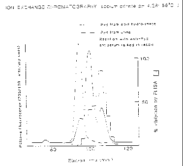
Brief Description of the Drawings
Figure 1 shows a chromatographic trace of
pyridinoline obtained from an acid hydrolyzate super-
imposed on a trace of the pyridinoline obtained without
hydrolysis from urine. The figure further compares the
elution pattern as determined by fluorescence with the
elution pattern as determined by reaction with anti-
pyridinoline antibody prepared from hydrolyzate.
Modes of Carrying Out the Invention
The invention provides an improvement over the
presently available methods to diagnose bone disorders or
other diseases characterized by abnormalities in collagen
metabolism. The invention method utilizes variations in
the levels of collagen-derived pyridinium crosslinks in
biological fluid as an index of these abnormalities.
Prior art methods have involved the hydrolysis of a
sample, typically urine, to provide analyte in the form
of hydrolyzed crosslinks, free of peptide sidechains,
which can then be quantitated in an immunoassay using
antibodies raised with respect to the hydrolyzed
crosslinks. While this method provides useful
information, the preliminary hydrolysis required prevents
the assay from becoming a simple clinical assay run
directly on an untreated biological sample.
CA 02271544 1999-OS-27
. _ f
!-
-12-
It has been found, by the inventors herein,
that antibodies raised with respect to the hydrolyzed
forms of the pyridinium crosslink do not cross-react
either with the free crosslinks present in urine or other
biological fluids, or with these crosslinks conjugated to
peptides prior to hydrolysis. Thus, the antibodies
presently available in the art cannot be used directly
with an untreated biological sample.
The present invention overcomes this
l0 disadvantage by providing reagents which can be reacted
directly with the biological sample to determine the
crosslinks present in free form as the diastereomer
present prior to hydrolysis. As shown in the examples
below, direct measurement of these free and unhydrolyzed
crosslinks provides data which are comparable to those
obtainable only through the presently available, more
complex assay.
Some background information as to the crosslink
structures involved will be useful:
Nature of the Crosslinks
The abbreviations Dpd and Pyd will be used
herein to denote the two known forms of the isolated
crosslink itself. Pyd or pyridinoline refers to
crosslinks formed wherein the ring N is the a amino
group of an hydroxylysyl residue; Dpd or
deoxypyridinoline refers to crosslinks formed wherein
the ring N is derived from the e-amino group of a
lysyl residue. (Various methods of denoting these
variations have been used; for example, HP has been used
to designate the "hydroxylysyl" form, and LP has been
used to refer to the "lysyl" form.)
Specifically, Dpd is believed to represent
compounds of the formula:
CA 02271544 1999-OS-27
-13-
CH,CHNH,COOH
HOOCH=NCHCH=CHZ \ OH
~ NJ
I
CHi
I
~2
I
~2
I
~2
I
CHNH,COOH
and Pyd is believed to describe compounds of the formula:
COO H
=IOOCH~~'CHCH,CH=
I
CHOH
I
~ HZ
2 0 CHz
CHNH,COOH
It is seen that both forms of crosslinks are
1,4,5 trisubstituted 3-hydroxypyridinium residues. Pyd
has a free hydroxyl group on the sidechain which can be
glycosylated, and it is known to be glycosylated in some
tissues. The glycosylation is labile to acid, and also
to base, but to a lesser degree. Pyd has been shown to
occur as Gal-Pyd; the inventor herein has also
demonstrated the presence of Glc.Ga1-Pyd in urine (see
PCT application WO 89/00715). These forms of free Pyd
have the acetals
CA 02271544 1999-OS-27
=:
-14-
CH.OH
HO .O
OH
CH=OH CH~_OH
HO O
O O
OH ' s"d OH
OH HO OH
conjugated to the sidechain hydroxyl, respectively.
It is seen that Dpd contains three chiral
centers--those of the three a-amino positions in the
sidechains. Pyd contains four such centers, as there is
an additional chiral center at the sidechain hydroxyl
position. Presumably, in the unhydrolyzed samples,
whether derivatized further to peptides or not, the three
a-amino groups are derived from the natively occurring
L-enantiomers, and the OH is in a configuration also
determined by the biological system.
As set forth in the Background section above, a
substantial proportion of the crosslinks present in urine
(about 40% in adults) is in the form of "free"
crosslinks--i.e., there are no peptide chains conjugated
to the Pyd, glycosylated Pyd, or Dpd structures shown
above, even before hydrolysis of the sample is conducted.
Thus, by "free" crosslink is meant compounds of the
formulas shown above.
It is noted that with respect to Pyd and Dpd,
the chirality of the chiral centers is not specified.
Thus, "free," refers to these crosslinks, whether or not
they have been subjected to hydrolysis conditions. The
present work demonstrates that these "free" crosslinks
differ in chirality when obtained in their "native" form,
as compared to their "hydrolyzed" form. As used herein,
"native free" crosslinks refers to Dpd or Pyd or its
glycosylated forms as they occur in free form in the
biological sample; "hydrolyzed free" crosslinks refers to
CA 02271544 1999-OS-27
r
-15-
these structures as they occur in hydrolysates. Of
course, as the glycosidic bond is labile to the
hydrolysis conditions, "hydrolyzed free" crosslinks will
not contain sugars.
As the native free crosslinks are the product
of the biological system, it is assumed that the
biologically favored chirality occurs at all three or
four chiral centers. Presumably the three chiral centers
represented by the a-amino groups of the sidechains are
in the L configuration, as in the naturally occurring
amino acid, and the chirality of the carbon containing
the sidechain hydroxyl in Pyd is also representative of a
single configuration. This is confirmed by the results
shown in Figure 1, in which the dotted line represents
the result of ion-exchange chromatography on sulfonated
polystyrene beads (7 ~) equilibrated with sodium citrate
performed with the previously isolated Pyd in its native
free form. As seen in Figure 1, the Pyd isolated
directly from urine elutes at a single peak. This is
consistent with the presence of only a single
diastereomer.
After hydrolysis, however, the hydrolyzed free
Pyd elutes as a mixture, shown by the solid line in
Figure 1. This is consistent with racemization at the
chiral centers to obtain a mixture of diastereomers which
no longer exhibit identical chromatographic behavior.
Similar results~are obtained comparing native free Dpd
with hydrolyzed free Dpd.
The "native free" crosslinks thus differ from
hydrolyzed free forms of crosslinks. It appears that
during conventional acid hydrolysis racemization occurs
which changes the configuration of some of the molecules.
However, enhancement of the yield of total "native free"
crosslinks in the biological sample could also be
CA 02271544 1999-OS-27
-16-
obtained by proteolytic treatment of total native Dpd and
Pyd to liberate the "native free" crosslink form. In
addition, the crosslinks per se are identical across
species, and other species besides human could be
utilized to prepare native free crosslink standards for
use in the assay system or for use as immunogens. In
particular, porcine urine contains high amounts of native
free crosslinks. Any source of the biologically
important diastereomer could be used.
It has been shown by the inventor herein that
the antibodies raised against the free Pyd which is
generated as the result of hydrolysis--i.e., wherein the
immunogen is obtained by treating the biological fluid or
tissue in concentrated acid so as to destroy peptide
linkages and separating Pyd from Dpd--show little or no
cross-reactivity with native free forms of either Dpd or
Pyd. Furthermore, antibodies raised against the Pyd
formed from the hydrolyzate cross-react only slightly
with Dpd thus formed. Antibodies raised against Pyd from
an acid hydrolyzate of bone or cartilage do cross-react
with the crosslink in urine after acid hydrolysis.
A typical set of results is shown in Table 1.
Table 1 presents the results of an ELISA assay using
antiserum obtained by immunization with the Pyd
hydrolyzate isolated from bone. The ELISA uses this
hydrolyzate as antigen, and the results are given in
terms of the ability of the candidate.crosslink to
inhibit the binding of the hydrolyzate antigen to the
antiserum. Using this criterion, antibodies which were
obtained by immunization of rabbits against Pyd isolated
from an acid hydrolyzate of cartilage or bone were only
5% cross-reactive with Pyd in its native free form from
urine (U-Pyd) although completely cross-reactive with Pyd
after hydrolysis in acid of the purified native, free
CA 02271544 1999-OS-27
._.. 1~~
t ..
-17-
crosslink isolated from urine. These antibodies,
further, were 20% cross-reactive with Dpd isolated from
the same bone hydrolyzate and were less than 1%
cross-reactive with Dpd in its native free form from
urine (U-Dpd); about 70% of the reactivity with these
antibodies was recovered after acid hydrolysis of the
native free form (Table 1).
Table 1
pmol required
for 50% % Cross
inhibition reaction
Pyd from hydrolyzate 1.6 100
of bone
Free Pyd from urine (U-Pyd) 29.6 5
U-Pyd from urine 1.5 107
hydrolyzed in acid
Dpd from hydrolyzate 8.1 20
of bone
Free Dpd from urine (U-Dpd) >260 <1
U-Dpd from urine 11.5 14
hydrolyzed in acid
This is further shown in Figure 1, which, as stated
above, presents the result of ion-exchange chromatography
on sulfonated polystyrene beads (7 u) equilibrated with
sodium citrate. The elution patterns for the free Pyd
and the acid hydrolyzate of urine were determined by
fluorescence. Antibodies raised against the acid
hydrolyzate are shown to react significantly only with
CA 02271544 1999-OS-27
.. ,:.. - .
-18- _ _-
the hydrolyzate. The discrepancy in reactivity of the
two major hydrolyzate peaks is attributable to the
differing immunogenicity of these two fractions.
Preparation of Antibodies toNativeFree Crosslinks
Antibodies are prepared to the native free
crosslink either as a total fraction or, preferably, to
each component of this fraction. Gross separation of the
pyridinium linkage in its "free" forms from the fragments
containing protein can be achieved, for example, by the
method of Fujimoto, D., J Biochem (1983) 94:1133-1136
(supra). In this preparation, a concentrate of urine is
applied to a SephadeX G-10 column and the total
pyridinium-containing fractions eluted. The eluate is
then applied to a column of phosphocellulose equilibrated
with sodium citrate, and eluted with salt. This rather
simple procedure results in the "free" crosslinks as a
single peak. As the sample is not subjected to
hydrolysis conditions this peak contains not only the Dpd
and Pyd forms, but also glycosylated Pyd including
Gal-Pyd and Glc.Ga1-Pyd as described above. Further
separation of this native free crosslink fraction is then
conveniently conducted by standard methods, for example
using ion exchange on sulfonated polystyrene beads as
described above, or using HPLC. Typical protocols for
this separation are found, for example, in Black, D., et
al., Anal Biochem (1988) 169:197-203;.Seibel, M.J., et
al., J Rheumatol (1989) 16:964-970.
Antibody preparation is by conventional techniques
including injection of the mixture or the individual
components conjugated to carrier into suitable mammalian
subjects such as rabbits or mice according to
immunological protocols generally known in the art. The
materials are conjugated to carriers such as BSA or
CA 02271544 1999-OS-27
,_
,.,_
-19-
tetanus toxoid using standard conjugation methods to
enhance immunogenicity. Sera are titrated to determine
antibody formation with respect to the immunogen. If
desired, spleen cell or peripheral blood lymphocytes may
be harvested and immortalized to produce cultures of
cells capable of continuous production of monoclonal
antibodies immunoreactive with the desired component.
These preparations have enhanced specificity with respect
to the individual components.
Thus, polyclonal antisera can be obtained which are
specifically immunoreactive with the native free form of _
the crosslinks occurring in biological fluids, in
particular in urine. By "specifically immunoreactive" is
meant that the serum is capable of forming complexes with
the native free crosslink forms in the biological fluid
with sufficiently greater affinity in comparison to other
materials in the fluid to permit determination of the
native free forms in an immunoassay. Some portion of the
polyclonal antiserum prepared either with respect to the
mixture of native free forms or with respect to the
individual components may crossreact with the native
forms having peptide chains attached; assays can be
standardized either by preparation of monoclonal
antibodies which do not thus crossreact, or by
standardizing to account for this crossreactivity.
The availability of routine techniques to obtain
monoclonal antibody preparations permits reproducible .
reproduction of antibodies of the desired specificity.
Thus, by utilizing a screening procedure which utilizes
as a criterion the ability of the immortalized cell
supernatant to immunoreact with, for example, native free
Pyd, but to fail to react either with native free Dpd or
forms of the crosslinks which are further conjugated to
peptides, a reliable source of antibodies which react
CA 02271544 1999-OS-27
f
:- v. (W
-20-
only with native free Pyd can be obtained. Conversely,
it may be advantageous to use, in assessment of
biological samples, cocktails of antibodies with these
unique specificities so that all native free forms are
determined.
Immortalized cell lines which secrete antibodies of
the desired specificity can be cultured in vitro for the
production of practical quantities of the desired
monoclonals using mammalian cell techniques known in the
art. Such culture techniques are now available on a
commercial scale. In addition, the immortalized cell
lines may be injected into mice and a somewhat cruder
preparation of the monoclonals isolated as the ascites
fluid. The antibody preparation may also be affinity
purified if desired using the immunogen as an affinity
ligand.
It should be noted that while it is clear that
antibodies prepared with respect to the hydrolyzed free
forms of the collagen-derived crosslinks failed to react
with the native free forms, it is not of importance
whether the converse is true, since hydrolyzed forms are
not present in unhydrolyzed biological samples. Thus,
screening procedures to assure the absence of this cross-
reactivity are unnecessary.
Conduct of Immunoassays
Accordingly, by utilization of an immunoassay with
the antibodies prepared as above it is possible to assay
a biological fluid sample without prior fractionation or
hydrolysis. The specificity for the desired form of
native free Pyd or Dpd or both is supplied by the
antibody preparation.
The immunoassays themselves are conducted using the
variety of standard assay protocols generally known in
' CA 02271544 1999-OS-27
-21- -
the art. As is generally understood, the assay is
constructed so as to rely on the interaction between the
specific antibody and the desired analyte for specificity
and to utilize some means to detect the complex formed by
the analyte and the antibody. The complex formation may
be between the antibody itself or an immunologically
reactive fragment thereof such as an Fab, Fab', or
F(ab')2 fragments. The antibody or immunologically
reactive fragment thereof may be complexed to solid
support and used as a capture antibody for the analyte.
This protocol may be run in a direct form, wherein the
formation of analyte/antibody complex is detected by a
fluorescent, radioactive or enzymatic label, or may be
run in a competitive format wherein a labeled standard
competes with analyte for the antibody. The format may
also be constructed as an agglutination assay or the
complex may be precipitated by addition of a suitable
precipitant to the reaction mixture. The specific design
of the immunoassay protocol is open to a wide variety of
choice, and the number of clinical assay devices and
protocols available in the art is multitudinous.
The antibodies and reagents for the conduct of an
immunoassay using standard detection protocols--i.e., for
example radioisotope labeling, fluorescent labeling or
ELISA, either in a direct or competitive format can
conveniently be supplied as kits which include the
necessary components and instructions. for the assay.
Since antibodies can be raised specifically to the
forms of the native free crosslinks which comprise the
various forms thereof, the ratios of these components can
be determined as well as their individual. levels and
their total.
Thus, the assay can be designed to include
antibodies or immunologically reactive fragments thereof
CA 02271544 1999-OS-27
( ;__
-22-
which will result in determination of total native free
crosslinks, or determination of native free Pyd, Dpd,
Gal-Pyd, or Glc.Ga1-Pyd, or any desired combination
thereof. Since the levels of the Pyd and Dpd crosslinks
in various tissues can be determined, alteration in their
relative amounts can be used as an index for degradation
of the particular tissue in question. For example, for
most normal adults, the ratio of Pyd/Dpd stays constant
throughout adulthood. As bone has a Pyd/Dpd ratio of 4/1
and appears to be the major source of liberated Dpd, an
elevation in the ratio of Dpd/Pyd may be indicative of
bone degradation. (Although aorta also contains Dpd, its
turnover rate is low.) Assessment of the level of Dpd in
biological fluids also yields a result which is
relatively bone-specific. However, it appears that in
many instances where a bone disorder is suspected, the
total free crosslink level (Dpd + Pyd) can also be used
as a measure when additional information is present.
When the symptoms do not suggest a disease of cartilage
such as rheumatoid arthritis, the majority of the excess
crosslink in free form in biological fluids will be, in
fact, due to the resorption of bone.
Since other connective tissues, such as cartilage,
for the most part contain only Pyd, not Dpd, an elevation
in the ratio of Pyd/Dpd may indicate diseases associated
with such damage.
While immunoassays using the antibodies of the
invention are convenient, the native free Pyd and Dpd
crosslinks can also be determined in a variety of ways.
Since the pyridinoline linkage is fluorescent, direct
chromatography of the sample of biological fluid as
described in the art can result in separation of Dpd from
Pyd and of the glycosylated forms of Pyd and the
~
CA 02271544 1999-OS-27
_ . , __
-23-
intensity of the fluorescence of the peaks obtained
provides an index to quantitation.
In the methods of the invention, therefore, the
native free crosslinks can be determined either as a
group or individually by determining the intensity of the
fluorescence of the chromatographed material.
As set forth in PCT application W089/04491
referenced above, the quantity of crosslinks can also be
determined using specific electrodes of appropriate redox
potential for the ring system.
In addition to the use of the native free cross-
link as an indicator of bone resorption, bone metabolic
balance is advantageously determined by combining this
determination with the determination of a marker for
formation of bone in the same or other appropriate
biological fluid from the same individual. For example,
such markers include procollagen type I, bone osteocalcin
(also known as bone GLA protein or BGP); pro bone GLA
protein, matrix GLA protein (MGP), bone specific proteins
such as bone specific sialoprotein, phosphoproteins,
alkaline phosphatase, osteonectin or other noncollagenous
bone proteins. Methods for determination of these
markers are well known in the art. Suitable methods for
determination of these markers can be found, for example,
in Delmas, P.D., et al., J Bone Min Res (1986) 1:333-337
(supra) for GLA.
The foregoing assays which provide an index to
determination of the metabolic status of tissues which
generate collagen-derived crosslinks when degradation
occurs, are useful in a variety of contexts. First, they
are a method to assess an abnormal condition of a subject
by indicating, for example, excessive bone resorption.
This may show, for example, the presence of an
osteoporotic condition or the unfortunate metastatic
CA 02271544 1999-OS-27
-24-
progress of a malignancy. Other known conditions
characterized by excessive bone resorption include
Paget's disease and hyperparathyroidism. Since the
condition of the subject can be monitored continuously,
application of these assays can also be used to monitor
the progress of therapy administered to treat these or
other conditions. The assays can also be used as a
measure of toxicity as the administration of toxic
substances often results in tissue degradation.
Thus, the assays may be applied in any situation
wherein the metabolic condition of collagen crosslink-
containing tissues can be used as an index of the
condition, treatment, or effect of substances directly
administered to the subject or to which the subject is
exposed in the environment.
The following examples are intended to illustrate
but not to limit the invention.
Examgle 1
Assay for Native Free Crosslinks in Urine
A. Isolation of U-Pyd and U-Dpd: Urine samples were
collected from patients with Paget's disease or
hyperparathyroidism (which contains elevated levels of
free crosslinks) and from growing children (in which
about 10-fold higher concentrations of crosslinks are
present compared with normal adults).. After
concentration 10-fold by rotary evaporation, batches of
the urine (20 liters) were subjected to partition
chromatography batchwise on cellulose CFl using
butanol:acetic acid: water (4:1:1 v/v/v) as mobile phase.
The pyridinium crosslink-containing fraction, eluted from
the stationary phase with water, was chromatographed on a
column (3.2 x 150 cm) of Sephadex G-10 eluted with 0.2M
CA 02271544 1999-OS-27
r
-25- ._. _
acetic acid. Pooled fractions containing the crosslinks
were then made 67 mM in Na+ and applied to a column (1.7
x 35 cm) of Dower 5oX-X8 ion-exchange resin equilibrated
with 67 mM sodium citrate buffer, pH 2.75. After raising
the column temperature to 60°C, elution with 67 mM sodium
citrate was performed with a linear pH gradient from 2.75
to 5.50 over 500 ml. The column effluent was monitored
by fluorescence (ex 325 nm/emm 400 nm) and the pooled
fractions containing U-Pyd (364-377 ml) and U-Dpd (397-
416 ml) were desalted by gel filtration on Sephadex G-10
and evaporated to dryness. The yield from 20 liters of
urine was 2.5 ~Cmoles U-Pyd and 0.6 ~Cmoles U-Dpd.
B. Results: The isolation procedure set forth in
paragraph A of this sample was applied to urine samples
from individual patients and the amounts of U-Pyd and
U-Dpd were quantitated using fluorescence measurements
relative to creatinine as is known in the art (supra).
The values obtained for normal individuals and in7
patients with bone disorders and arthritic diseases are
shown in Table 2. Values are given as the mean ~ SEM
(n=6 in each group).
30
CA 02271544 1999-OS-27
i _
-26-
Table 2
Patient GrouQ U-Pyd U-Dpd
lnmol~mmol creatinine~
Normal controls 10.3 ~ 1.0 3.27 ~ 0.57
Osteoporosis 19.6 ~ 2.3 5.90 ~ 0.68
Paget's disease 62.5 ~ 11.2 19.3 ~ 3.83
Hyperparathyroidism 55.9 ~ 14.2 16.3 ~ 4.81
Rheumatoid arthritis 38.8 ~ 8.36 8.92 ~ 2.08
Osteoarthritis 25.8 ~ 3.22 6.10 ~ 0.83
These results show dramatically elevated levels of
the free crosslinks in patients known to be suffering
from diseases characterized by excessive breakdown of
connective tissue.
Table 3 shows the proportions of U-Pyd and U-Dpd as
a percentage of the total crosslink measured after
hydrolysis in the different patient groups.
25
35
CA 02271544 1999-OS-27
_ -27-
Table 3
Patient Grouts % U-Pyd* % U-Dpd*
Normal controls 43.8 ~ 2.5 50.1 ~ 5.4
Osteoporosis 41.7 ~ 2.0 42.7 ~ 2.6
Paget's disease 46.5 ~ 2.4 47.4 ~ 4.1
Hyperparathyroidism 48.7 ~ 6.8 46.2 ~ 6.9
Rheumatoid arthritis 38.1 ~ 2.6 43.3 ~ 1.8
Osteoarthritis 43.4 ~ 3.9 47.0 ~ 2.2
* Calculated as: (U-Pyd/total Pyd) x 100 and
(U-Dpd/total Dpd) x 100. For all groups combined (n=36),
the correlation coefficient between U-Pyd and total Pyd
was 0.929 (p<0.0001) and between U-Dpd and total Dpd was
0.952 (p<0.0001).
Since, as shown in Table 3, the percentage of U-Pyd
and U-Dpd is relatively unchanged in patients with
abnormal conditions as compared to controls, concentra-
tions of the free crosslinks in urine reflect the same
increase in collagen degradation in diseases compared
with the controls as do the total crosslinks measured
after hydrolysis of the urine.
U-Pyd and U-Dpd therefore provide viable indices of
collagen degradation to facilitate diagnosis and
monitoring of diseases involving abnormalities of
connective tissue metabolism.
C. Immunoassay:' Native free crosslinks isolated by the
method described in paragraph A of this example are used
for the preparation of antigen. U-Pyd and U-Dpd are
further purified by ion-exchange chromatography with 67
mM-sodium citrate buffer, pH 4.25 using a high-resolution
CA 02271544 1999-OS-27
-28- _ .. ,
resin column of an amino acid analyzer (Locarte Co. Ltd.,
London, UK).
For immunization, the isolated crosslinks are
covalently attached to bovine serum albumin using
carbodiimide reagents and methods well known in the art.
Both monoclonal and polyclonal antibodies are
raised against the urinary crosslink components. For the
production of monoclonal antibodies, Balb/c mice are
immunized with urinary crosslink-BSA conjugates, and
hybridoma cell lines are prepared using standard
techniques after fusions of cells from the spleen or
lymph nodes with Ag8 myeloma cells. Polyclonal
antibodies are raised in rabbits. Screening of both
antisera and hybridoma cell media is performed by ELISA
using microtiter plates coated with the appropriate
urinary crosslink-gelatin conjugate prepared as described
by Robins, Biochem J (1982) 217:617-620.
Assays for each of the crosslink components present
in free form in urine are performed by an inhibition
ELISA as follows:
Urine samples (5 or 20 ~C1) or solutions containing
0.2-20 pmol of purified urinary crosslink reference
standard are diluted to 110 ~1 with phosphate buffered
,~
saline containing 0.05% Tween-20 detergent (PBS-T), and
are added to 110 ~1 of primary antibody, immunoreactive
.fragment, or antiserum diluted 1:5,000 - 1:20,000 in PBS-
T. Each sample is prepared in triplicate in round-
bottomed, 96-well microtiter plates which are then
incubated overnight at room temperature.
Portions (200 ~C1) of the samples are transferred to
flat-bottomed microtiter plates previously coated with
gelatine conjugate containing the appropriate urinary
crosslink component. After 30 minutes, the plate is
washed with PBS-T (3 times) and the bound antibodies
CA 02271544 1999-OS-27
-29-
detected by standard techniques with a biotin-labeled
antibody prepared against the species of the primary
antibody combined with a streptavidin-peroxidase and
peroxidase substrate detection system. Color development
is measured at 492 nm using an automated microtiter plate
reader. Samples containing the analyte decrease the
binding of primary antibody to the plate and thus have
reduced color concentration. The amount of free.
crosslinks in the sample is quantified with reference to
curves from standards included on each plate computed
using log-log plots.
The foregoing assay can be reformatted to be
conducted directly by coating the sample suspected of
containing antigen in the flat-bottom microtiter plate,
and adding labeled primary antibody directly to the
wells. After washing, the amount of labeled antibody
remaining in the testing solution is determined. A
decrease in levels indicates the presence of antigen.
Example 2
Sources of Native Free Crosslink
In order to determine a source for native free
crosslinks usable as standards in the assays of the
invention, the urine of a number of species of large
animals was analyzed. In bovine urine, the Pyd/Dpd ratio
is 12~2 with only about 15% as free crosslink; the values
in sheep are similar except for only about 20-25% is free
crosslink. In pig urine, the ratio of Pyd/Dpd is about
5~1 and the proportion of free crosslink relative to
total is 42~5%. The concentrations of free crosslinks
are about 380 nM for Pyd and 70 nM for Dpd.
Children's urine appears to give a better yield of
Dpd than urine from adults. Some preferential loss of
Dpd from pig urine occurs when CF-1 cellulose is used in
' CA 02271544 1999-OS-27
-30-
the purification procedure, and overall recovery of Pyd
is 40-50% for Pyd but only 20% for U-Dpd. Using
children's urine as a starting material, recovery for
both crosslinks is about 55%.
Accordingly, both children's and pig urine are
suitable sources for free crosslink standards.
As set forth above, the yield of crosslink in the
diastereomeric form characteristic of native free
crosslink could be improved by liberating total
crosslinks in these sources by enzymatic hydrolysis
procedures, such as the use of exopeptidases and
glycosidases.
Example 3
Pyd/Dpd in Human Tissues
Analyses of a range of different tissues has shown
that the crosslink content of cortical bone is slightly
higher than that of trabecular bone with a Pyd/Dpd ratio
of about 4.2. Although Dpd was not detected in
cartilage, this crosslink was present in aorta and in
ligaments. These results are summarized in Table 4.
Table 4
Tissue n Pyd Dpd
(residues/mol ecule)
Articular cartilage 15 1.47 0.23 N.D.
Cortical bone . 15 0.35 + 0.09 0.08 + 0.02
Trabecular bone 7 0.26 + 0.08 0.06 + 0.02
Aorta ~ 14 0.30 0.07 0.07 + 0.01
Invertebral disc 25 1.14 0.11 N.D.
Ligaments 10 0.47 0.35 0.05 + 0.03
CA 02271544 1999-OS-27
. . -31- . .
Both Pyd and Dpd are completely absent from the collagens
of normal skin, nor are they present in immature or newly
synthesized collagens.
Example 4
Determination of Free Crosslinks
in Osteoporosis Patient Urine
Sixty-four postmenopausal women with vertebral
fractures (type I osteoporosis) aged 53 to 74 years (mean
+ SD, 64 + 5 years) were studied. All women had lumbar
spin bone mineral density below the fracture threshold of
0.98 g/cm2 as measured by dual-photon absorptiometry and
spine radiographs showing three or more grade 1 fractures
or one or more grade 2 fractures. No other secondary
cause for the osteoporosis was identified.
As a control group, 67 postmenopausal women with
mean (~ SD) age of 65 + 6 years (range 50 to 79 years)
were studied. All women had normal spin radiographs and
had lumbar spine bone mineral densities within the normal
2o range for age as measured by dual-photon absorptiometry.
None had any illness or were taking drugs known to affect
bone metabolism.
For measurements of hydroxyproline, the subjects
were maintained on a gelatin-free diet for three days
prior to the study. Urine samples were collected and
aliquots were stored at -70°C until analyzed. The total
crosslinks were measured essentially as described
previously (Black, D., et al., Anal Biochem (1988)
169:197-203; Seibel, M.J., et al., J Rheumatol (1989)
16:964-970). For the determination of U-Pyd and U-Dpd in
unhydrolyzed urine, 0.5 ml portions were processed
directly by partition chromatography on CF1 cellulose
(which separates the free from peptide-derivatized forms
prior to the HPLC step; HPLC was conducted as for the
CA 02271544 1999-OS-27
- -32-
hydrolyzed samples). Hydroxyproline in acid hydrolysates
of urine was measured by HPLC (Dawson, C., et al., Clin
Chem (1988) 34:1572-1574).
Measurements of native free pyridinium crosslinks
and of total hydroxyproline in urine for the control and
osteoporotic groups are shown in Table 5. The results
showed that the excretion of the bone-specific crosslink,
U-Dpd, was significantly higher in the osteoporotic group
compared with the controls.
Table 5
Osteoporotic Controls
nmol/mmol nmol/24h nmol/mmol nmol/24h
creatinine creatinine
U-Pyd 21.4~6.6** 170+47 18.4~5.8 151~49
U-Dpd 5.7~2.0*** 45~14*** 4.6~1.7 37~13
U-Pyd.Gal.Glc 5.2~2.1 41~15 4.6~2.0 38~16
Hydroxyproline 21.4_+7.4** 175~66 18.2~6.7 156~77
(x 10 3)
_______________________________________________________
Statistical significance (Student's t-test) of the
difference compared with the corresponding control group
is shown: * p < 0.05; ** p < 0.01; *** p < 0.001
Differences were less marked for U-Pyd; the values for
the glycosylated derivative, U-Pyd.Gal.Glc, were not
statistically different. Linear regression analysis
showed that there were highly significant correlations
between values expressed as creatinine ratios and as the
total 24h excretion for both U-Pyd (r=0.80) and U-Dpd
(r=0.82). This observation is consistent with the
finding that there were no significant variations
diurnally in crosslink excretion for healthy male or
CA 02271544 1999-OS-27
' -33-
female volunteers (A. M. McLaren and S.P. Robins,
unpublished results).
There were significant correlations of U-
Dpd with hydroxyproline which were more marked in the
osteoporotic group (r=0.53; p<0.001) than in the controls
(r=0.21; N.S.). The relationship between U-Pyd and
hydroxyproline was similar with correlation coefficients
for the osteoporotic and control groups of r=0.45
(p<0.001) and r=0.34 (p<0.01), respectively.
For samples where both free and total
crosslinks were measured, there were highly significant
correlations between these values. For
deoxypyridinoline, the correlation coefficients for the
osteoporotic group (n=25) and control group (n=24) were
0.90 and 0.84, respectively; the corresponding
correlations for pyridinoline were r=0.96 and r=0.85.
25
35