Note: Descriptions are shown in the official language in which they were submitted.
CA 02271800 1999-OS-13
wo ~sr~e . reTi~s9~nme
INTER~~ERTEBRAL PROST~IETIC DEVTCE
This invention relates to a novel intervertebral
prosthetic device. More particularly, this invention
relates to an intervertebral prosthetic device that can
be implanted to replace a damaged intervertasbral disc.
The human spine is a flexible structure comprised
of thirty-three vertebrae. Intervertebral discs separate
and cushion adjacent vertebrae. Ths intervertebral discs
act as shock absorbers and allow bending between the
vertebrae.
An intervertebral disc comprises two major
components: the nucleus pulposus and the annulus
fibrosis. The nucleus pulposus is centrally located in
the disc and occupies 25-40% of the disc's total cross
sectional area. The nucleus pulposus usually contains
70-90% water by weight and mechanically functions like
an incompressible hydrostatic material. The annulus
fibrosis surrounds the nucleus pulposus and resists
torsional and bending forces applied to the disc. The
annulus fibrosis thus serves as the disc's main
stabilizing structure. Vertebral end-plates separate
the disc from the vertebral bodies on either side of the
disc.
Individuals with damaged or degenerated discs often
experience significant pain. They pain results in part
from instability in the intervertebral joint due to a
_
CA 02271800 1999-OS-13
WO 98122050 PCTlU5S97119610
loss of hydrostatic pressure in the nucleus pulposus.
Loss of hydrostatic pressure leads to a loss of disc
height.
A conventional treatment for degenerative disc
disease is spinal fusion. In one such surgical
procedure, a surgeon removes the damaged natural disc
and then fuses the two adjacent vertebral bones into one
piece. The surgeon fuses the vertebral bones by grafting
bone between the adjacent vertebrae and sometimes uses
metal rods, cages, or screws to hold the graft in place
until the graft heals. Other fusion procedures do not
require surgical removal of the disc.
Although spinal fusion may alleviate pain associated
with degenerative disc disease, it also results in loss
of motion at the fused vertebral joint. Lack of motion
at the fused site puts additional pressure on the discs
above and below the fusion, sometimes causing them to
degenerate and produce pain. To remedy the problems
associated with spinal fusion, prosthetic devices were
developed to replace the damaged disc with a suitable
biomechanical equivalent.
- Existing prosthetic devices have met with limited
success in reproducing the biomechanics of a natural
disc. For example, U.S. Patent No. 4,759,769 to Hedman
et. 81. discloses a synthetic disc having upper and lower
plates hinged together. Although the hinged disc allows
forward bending between adjacent vertebras, the hinged
disc does not allow axial compression or lateral flexion.
Nor does it allow axial rotation of the vertebral column
at the site of the implant. Therefore, the Hedman et.
a1. device lacks the biomechanics of a natural disc.
Likewise, the prosthetic disc device disclosed in
U.S. Patent No. 4,309,777 to Patil does not replicate
natural motion between adjacent discs. The Patil device
includes two cups, one overlapping the other and spaced
from t:~e other by springs. The cups move only in a
single axial dimension. The Patil device thus does not
CA 02271800 1999-O5-13
WA 98/220 PCT/U897/19610 . .
-3~
enable natural flexion of the spine in any direction.
In addition, the, highly constrained motion of the Patil
device can lead to high deviceitissue interface stresses
and implant loosening.
Many synthetic disc devices connect to the vertebral
bodies by conventional mechanical attachments, such as
pegs or screws, which are known to loosen under cyclic
loading conditions. Other synthetic disc devices use
plastic or elastomeric components which, over a lifetime,
l0 produce debris from wear and possible unknown side
ef facts .
The problems suggested in the preceding are not
intended to be exhaustive but rather are among many which
tend to reduce the effectiveness of known intervertebral
prosthetic devices. Other noteworthy problems may also
exist; however, those presented above should be
sufficient to demonstrate that currently known devices
are amenable to worthwhile improvement.
Accordingly, it is a general abject of the invention
to provide an intervertebral disc prosthetic and method
for implanting the same which will obviate or minimize
difficulties of the type previously described.
More particularly, it is a specific object of the
invention to provide an intervertebral prosthetic device
which replicates the mechanical properties of a natural
intervertebral disc.
It is another object of the invention to provide
an intervertebral prosthetic device which restores disc
height, defined as the axial distance between vertebrae
adj scent the damaged disc, and which duplicates the range
of motion of a natural intervertebral joint.
It is still another object of the invention to
provide an intervertebral prosthetic device which may
be implanted and maintained in stable relation to
CA 02271800 1999-OS-13
WO 9~/23A30 PCT/I1897/19610 .
-4~
adjacent vertebrae without conventional mechanical
attachments.
It is a further object of the invention to provide
an intervertebral disc prosthesis which suffers minimal
degradation of the prosthetic material and which produces
minimal wear debris under long-term cyclic loading
conditions.
It is yet a further object of the invention to
provide an intervertebral prosthetic device which axially
compresses and thus dissipates energy, may be easily
repaired or replaced, may be easily manufactured and
utilized by a surgeon, and is durable and modular.
It is yet another object of the invention to provide
a method of implanting an intervertebral prosthetic
device which stabilizes an operative intervertebral joint
and restores the mechanical properties of a degenerated
disc.
These objectives are achieved by an intervertebral
prosthetic device having a first fixation member, a
second fixation member, and a compressible member
disposed between them. The first fixation member is
- implanted within a first vertebral body, and the second
fixation member is implanted within a second vertebral
body adjacent the first vertebral body.
The first fixation member generally comprises an
adjustable member and a support member. The adjustable
member preferably has a first plate, a second plate, and
at least one adjustment element that extends between the
two plates and enables adjustment of the height of the
adjustable member along its longitudinal axis. The first
plate is operably positioned against subchondral bone
of a distant end-plate of the f first vertebral body, and
the second plate is operably positioned against the
support member.
The second fixation member may include both a
support member and an adjustable member or, in an
alternative embodiment, may include only a support
CA 02271800 1999-OS-13
w~ ~o rc~smii~sio
-5~
member. In the first embodiment; the adjustable member
is structurally similar to the adjuetabl~ member of
the
first fixation member and includes a first plate for
positioning against subchondral bone of a distant end-
s plate of the second vertebral body, a second plate for
' positioning against the support somber, and at least
one
adjustment element extending between the two plates.
In the second embodiment, the support member is operably
positioned against a near end-plate of the second
vertebral body.
One of skill in the art will recognize that, like
the second fixation member, the first fixation member
may comprise only a support member, depending on the
patient's needs. Moreover, the support members are
modular. The support members are generally wedge-shaped
and may be made in difference sizes to accommodate the
angle between adjacent vertebrae at a specific vertebral
level. The angle between adjacent vertebrae typically
ranges between 3-10 degrees, and, thus the angle created
by opposing surfaces of the wedge-shaped support member
falls within that same range.
' The compressible member has an outer periphery less
than or substantially equal to the diameter of the
nucleus pulposus of the operative intervertebral disc.
In other words, the compre~ssib3e member is sized to
replace the nucleus pulposus of an intervertebral disc
and essentially to fit within the annulus fibrosis of
the intervertebral disc. The compressible member
comprises at least one spring that can be pre-stressed
or pre-loaded to place the annulus fibrosis under tension
and to reproduce the mechanical properties of a natural
disc: Maintaining the annulus fibrosis under tension
results in an artificial intervertebral joint that is
stable.
' 35 The fixation members include a porous surface
suitable for bone ingrowth so that the fixation members
i
CA 02271800 1999-OS-13
wo 9sn~.oso rcT~s~nri~zo
-s=
fuse to the vertebrae without requiring conventional
mechanical attachments.
Additional objects and advantages of the invention
are set forth in the description which follows, and in
part will be obvious from the de~cription, or may be
learned by practice of the invention: The objects and '
advantages of the invention may be realized and obtained
by means of the instrumentalities and combinations
particularly pointed out in the specification and the
appended claims.
The accompanying drawings, which are incorporated
in and constitute a part of the specif ication, illustrate
a presently preferred embodiment of the invention, and,
together with the general description given above and
the detailed description of the preferred embodiment
given below, serve to explain the principles of the
invention.
Figure 1 is a schematic, cut-away, side view of an
- 20 intervertebral prosthetic device implanted in a spine
in accordance with a preferred embodiment of the
invention;
Figure 2 is a top perspective view of a compressible
member of the subject intervertebral prosthetic device;
Figures 3A-3C are top perspective views of different
embodiments of a spring of the compressible member of
the subject intervertebral prosthetic device;
Figure 4 is a top perspective, partially exploded
view of a fixation member of the subject intervertebral
prosthetic device and shows an adjustable member and a
support member;
Figure 5 is a top view of a second plate of the
adjustable member;
Figure 6 is a side view, in cross-section; of the
support member;
CA 02271800 1999-OS-13
VSO 98122030 PCT1U89711961fl
Figure ? is a sahamatic, cut-away, sidm view of an
intervertebral prosthetic device implanted in a spine
in accordance with another preferred embodi~tent of
the
invention;
Figure 8 is a schematic, cut-away, side view showing
subchondral bones of a superior vertebral body after
a
partial vertebrectomy;
Figure 9 is a sectional view of a vertebrae after
a partial vertebrectomy, as taken along line 9-9 of
io Figure 8;
Figure to is a schematic, cut-away, side view of
a vertebral joint area after a partial vertebrectomy
and
excision of a nucleus pulposus of a natural disc;
Figure 11 is a schematic, cut-away, side view of
a vertebral j oint and shows a f fixation member, including
an adjustable member and a support member, implanted
in
an inferior vertebral body;
Figure 12 is a schematic, cut-away, side view of
a vertebral joint and shows a compressible member
2o implanted in an intervertebral joint;
Figure 13 is a schematic, cut-away, side view of
a vertebral joint and shows a technique for adjusting
the height of an adjustable member implanted in a
superior vertebral body; and
Figure 14 is a schematic, cut-away, side view of
a vertebral joint and shows a technique for bone grafting
an adjustable member in a superior vertebral body:
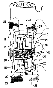
Referri~ now to the drawings, wherein like numerals
indicate like parts, and initially to Figure 1, there
will be seen an intervertebral prosthetic device,
generally indicated 10, implanted in a spine 12 in
accordance with a preferred embodiment of the present
invention: The intervertebral prosthetic device 10 is
designed to replace a damaged natural disc. The
CA 02271800 1999-OS-13
WO 98122050 PCTlt3S97/1961~
-8-_
intervertebral prosthetic device 10 has a first fixation
member 14, a second fixation member 16, and a
compressible member 18 that is positioned between the
f first f fixation member 14 and the second f fixation member
16.
The first f fixation member 14 is positioned in a
first vertebral body 20, and the second fixation member
16 is positioned within a second vertebral body 22
adjacent the first vertebral body 20. Each fixation
member 14 and 16 has an adjustable member 28 and 30,
respectively, and a support member 32 and 34,
respectively. Each fixation member also has a bone-
contacting surface, preferably porous, for positioning
against subchondral bone of an associated vertebral body.
In Figure l, a bone-contacting surface 27 of the
adjustable member 28 is positioned against the
subchondral bone of an end-plate 36 of the superior
vertebral body 20, and a bone-contacting surface 29 of
the adjustable member 30 is positioned against the
subchondral bone of an end-plate 38 of the inferior
vertebral body 22. As will be described below, the
- present intervertebral prosthetic device does not require
conventional mechanical attachments, such as Begs or
screws, to hold the prosthetic device in place. The
intravertebral (i.e., within a vertebral body)
positioning of the fixation members maintains the
prosthetic device in stable relationship at the operative
- intervertebral joint.
The adjustable member 28 of the first fixation
member 14 has an imaginary first longitudinal axis, shown
by dashed line A-A, and adjustment elements 24 that allow
adjustment of the height of the adjustable member 28
substantially along its longitudinal axis A-A. In the
embodiment shown in Figure 1, the second fixation member
16 is structurally similar to the first fixation member
14, but inverted. The adjustable member 30 of the second
fixation member 16 has a second longitudinal axis, shown
CA 02271800 1999-OS-13
WO 90 PCT/U519"lA19610 -
by dashd lin0 8-B, and adjustment elements ~6 that
allow
adjustment of the height of the adjustable member 30
substantially along its longitudinal axis B-8.
The eospressible member 18 comprises at least one
spring and, in a preferred embodiment, comprises a
plurality of springs 40. One skilled in the art,
howe~rar, will recognize that the compressible member
may
comprise other suitable configurations. For example,
the compressible member may comprise a monolithic body
made of an biocompatible material compressible in an
axial direction, that is, a direction substantially
parallel to the spine.
The compressible member i8 is implanted in the
region of an excavated nucleus pulposus of the operative
intervertebral disc. The compressible member 18 is
dimensioned so that the annulus fibrosis of the natural
disc is maintained. The present intervertebral
prosthetic device restores the mechanical properties
of
the nucleus pulposus without disrupting the annulus
fibrosis. Retention of the annulus fibrosis maintains
stability of the intervertebral joint at the implant
- site. In addition, the annulus fibrosis serves as a
boundary for the compressible member and minimizes
accidental dislodgement of the prosthetic device.
Significantly, the intervertebral prosthetic device
10 permits at least four degrees of relative motion
between the first vertebral body 20 and the second
vertebral body 22. These degrees of relative motion
include sagittal bending, coronal bending, axial
rotation, and axial compression. Moreover, the
compressible member permits small increments of
translational movement between the vertebral bodies
( i . e. , fifth and sixth degrees of relative motion,
namely
anterior-posterior translation and side-to-side, or
lateral, translation).
A pref0rred embodiment of the compressible member
18 is shown in Figure 2. The compressible member 18
has
CA 02271800 1999-OS-13
prp 9gn2psp PCT!(JS97119610
-10-
a top plate 42, a bottom plate 44, and a plurality of
coil springs 40 extending between the top plate 42 and
the bottom plate 44. The top plate 42 has a first
surface 46, which is connectable to the first fixation
member 14, and a second surface 48. The bottom plate
44 also has a first surface 50, which is connectable to
the second fixation member 16, and a second surface 52.
The springs 40 extend between the second surfaces 48 and
52.
When pre-loaded, as will be explained in more detail
below, the compressible member 18 preferably has an axial
height of approximately 1.5 cm, greatest at the L45
vertebral level and slightly less at the upper lumbar
vertebrae. The coil springs 40 are preferably designed
to have non-linear stiffness so that they become stiffer
at higher applied loads. The nonlinear stiffness
simulates physiological intervertebral stiffness.
One skilled in the art will recognize other
embodiments contemplated by the present invention. For
example, the comprsssible member 18 may comprise a
plurality of springs extending between, and directly
- connected to, support members 32 and 34. Alternatively,
the compressible member 18 may comprise a single spring
with a relatively large coil diameter (not shown)
extending between, and directly connected to, the support
members 32 and 34. Any spring arrangement may be
utilized that achieves sufficient axial compressive force
to replicate the biomechanics of the natural disc.
In each embodiment, the compressible member includes
an imaginary longitudinal axis, shown by the dashed line
C-C in Figure 2, and an outer periphery in a plane
transverse to the longitudinal axis C-C. A largest
dimension of the compressible members outer periphery
is less than or substantially egual to the diameter of
a nucleus pulposus of the natural intervertebral disc.
Put another way, the annulus fibrosis of the natural
disc, which is substantially preserved in the
CA 02271800 1999-OS-13
WO X50 PCTfUS9?fI~6i8 .
-lI=
implantation procedure, circumscribes the compressible
member i8. For example, where the compressible member
comprises a plurality of springs, the outer periphery
' of the compressible member circumscribes the springs,
and the largest dimension of that outer periphery may
extend to, but does not extend beyond, the nucleus
pulposus. In other embodiment, where the compressible
member includes a top plate and a bottom plate, and where
those plates fit within the annulus fibrosis and extend
beyond the outermost portions of the springs, the outer
periphery equals the larger of the two plate peripheries.
In quantitative terms, the outer periphery of the
. compressible member preferably ranges between 2.0 cm to
3.0 cm, which approximates the diameter of the nucleus
pulposus of a natural intervertebral disc.
Figures 3A-3C show three embodiments of a coil
spring designed to possess non-linear stiffness. In the
embodiment of Figure 3A, the coil spring 54 has a
variable, or non-uniform, cross-sectional diameter 56.
Figure 3B shows another embodiment in which a coil spring
58 has a variable pitch 60, where the pitch is defined
' as the distance between successive coils of the spring
58. Figure 3C shows a third embodiment of a coil spring
62 in which at least two of the spring coils have
different radii 64 measured from an imaginary axis D-D
extending along the central axis of the spring 62.
Figure 4 shows a preferred embodiment of the first
fixation member 14. In the embodiment shown in Figure
1, the second fixation member 1~ is structurally similar
to the first fixation member 14, but inverted. The
following discussion thus also applies to the second
fixation member 16.
The fixation member Z4 comprises an adjustable
member , genera l ly indicated 2 8 , and a support member 3 2 .
The adjustable member 28 is adjustable in an axial
direction by adjustment elements 24. The adjustment
elements 24 preferably comprise telescopic struts
a
CA 02271800 1999-OS-13
WO 901Z?,05(1 PCTlU897Ii9610 _
-12--
extending between a first plate 31 and a second plate
33. In a preferred embodiment, the first plate 31 has
a bone-contacting surface, such as 27 shown in an
operative context in Figure 1, and the second plate has
a surface 35 for positioning against the support member
32. Although the illustrative embodiment shows flat '
plates 31 and 33, it will be understood by those skilled
in the art that these structures need not be flat and
may, for example, have undulating surfaces. In fact,
in one embodiment, the bone-contacting surface 27 of the
first plate 31 is concave to match the contour of the
subchondral bone of the associated vertebral body.
The adjustment elements 24 adjust the distance
between the f first bone-contacting plate 31 and the second
plate 33, thus adjusting the height of the adjustable
member 28. A surgeon may adjust the telescopic struts
to increase the height of the adjustable member and thus
pre-load the compressible member to mechanically
reproduce the axial compression absorbed by a nucleus
pulposus of a natural disc. Pre-loading the compressible
member restores the intervertebral height at the
- operative joint and restores the function of the annulus
fibrosis. The annulus fibrosis load shares with the
compressible member which reduces implant/tissue
interface stresses.
Each telescopic strut is provided with a lock screw
63 to adjust the length of the strut 24 and hence control
the height of the adjustable member. The lock screw 63
may comprise, for example, a pin (not shown) that extends
through both the telescoping portion 65 and the housing
portion 67 of the strut 24. Each strut 24 is
independently adjustable. Figure 5 shows a top view of
the second plate 33 of the adjustable member 28. The -
ad justment elements 24 preferably are spaced equidistant
from each other to enable specific height adjustment of
various regions of the adjustable member.
CA 02271800 1999-OS-13
wo ~sn~so rc°r.~s~rro
-13-
A key feature of the present invention is that
controlling the height of the adjustable membe~cs 28 and
30, along with :electing an appropriately-sized support
member, controls the "disc" height. The diec height is
defined as the axial distance between the vertebrae above
and below the operative disc. In addition to restoring
the disc height, the compressible member 18 acts as a
shock absorber to minimize impact loading and, thus,
minimize device failure or vertebral fracture.
In a preferred embodiment, the first and second
fixation members 14 and 16 have porous portions, such
as the bone-contacting surface 27, to permit bone
ingrowth. In another embodiment, a biocompatible fabric
or suitable material may be wrapped around the f fixation
members to enable bone ingrowth. The pre8ent prosthetic
device does not require conventional mechanical
attachments, such as pegs or screws, to hold the
prosthesis permanently in place. The present prosthetic
device, however, may include mechanical or other
attachments to supplement the porous portions of the
fixation members and to temporarily fix the prosthetic
device in place until bone ingrowth has occurred.
To further promote bone ingrowth, the adjustment
elements 24 may include fins 66 extending outward from
an exterior surface of the element 24, as shown in Figure
4 . The f ins 66 increase the surface area of the fixation
member 14 to which bone may attach. Preferably, these
fins 66 ate located on the adjustment elements that are
positioned on the anterior side of the adjustable member
28. The present prosthetic device also may include
protuberances (not shown) on the bone-contacting surface
of the adjustable members to increase the surface area
of the porous portion of the fixation members and, thus,
encourage bone ingrowth.
Figure 6 shows a cross-section of support member
32. The support member 32 has a first surface 72 that
operably faces away from the compressible member 18 and
CA 02271800 1999-OS-13
W0 9A8/~030 PCT/I199'1/196H1
-ly-
a second surface 74 that operably faces towards the
compressible member 18. The first and second surfaces
72 and 74 are oblique so that a circumferential surface
77 around the support member 32 varies in width, as shown
in Figure 4. The support member 32 thus is wedge-shaped.
In other words , the support member 3 2 preferably tapers
from a maximum thickness at one side 73 to a minimum
thickness at an opposite side 75. Generally, the support
member 32 is thicker on the side of the fixation member
14 placed anteriorly in a patient ~ s spine to account for
the spines natural curvature.
The support members are constructed with various
thicknesses and with various angled surfaces, depending
upon the vertebral level of the operative intervertebral
joint. An angle a shown in Figure 6 ranges between 3-l0
degrees. The support members are shaped to maintain
sagittal alignment. Maintaining sagittal alignment
avoids nonuniform loading of the compressible member and
avoids early fatigue failure of that member.
Figure 7 shows another embodiment of the present
intervertebral prosthetic device, generally indicated
7 6 , which comprises a f first fixation member 78 , a second
fixation member 80, and a compressible member 82. The
compressible member 82 is positioned between the first
and second fixation members 78 and 80. The second
fixation member comprises a wedge-shaped support member
with an upper surface 84 that attaches to the
compressible member 82 and a lower surface 86 that rests
upon subchondral bone of a near end-plate 88 of an
inferior vertebral body. In this embodiment, adjustment
of the first fixation member 78 pre-loads the
compressible member 82 to an appropriate extent. This
embodiment is particularly suited for young patients.
Also, in this embodiment, a lower surface 86 of the
support member 80 has a slightly convex shape to match
the natural contour of the near end-plate of the inferior
CA 02271800 1999-OS-13
W0 X22030 PCT/US97l1lb~A ..
-15-
vertebral body. The surface 86 is preferably composed
of a porous material.
As evident from the embodiments of Figures 1 and
7, the present intervertebral prosthetic device has a
modular design so that the prosthesis may be sized to
the patient's anatomy and designed for the patient's
condition. The modular design also enables replacement
of individual comgonents of the prosthesis (i.e., an
adjustable member, a support member, or a compressible
member), rather than replacement of the entire prosthesis
should one component fail. The compressible member is
preferably attached to the fixation members by mechanical
attachments, such screws, rather than bone cement so that
a surgeon may easily replace damaged or worn components.
25 Moreover, because the present prosthetic device has
no ball bearings, rollers, or hinges, it produces little
wear debris. And, because the present prosthetic device
need not include plastic polymers or elastomeric
components, the present prosthetic device does not
degrade under long-term cyclic loading conditions.
The present prosthetic device comprises
biocompatible metallic materials, preferably a titanium
alloy having, for example, 4% vanadium and 6% aluminum.
. Persons of skill in the art will recognize other suitable
materials, for example, a cobalt-chromium alloy, such
as alloy number 301. Alternatively, the present
prosthetic device, with the exception of the springs of
the compressible member, may comprise a ceramic material,
such as aluminium oxide and zirconium oxide. The porous
surfaces of the bone-contacting member and support member
may be coated with hydroxyapatite or bioactive proteins
(e. g., bone morphcx~enic protein) to encourage bone
ingrowth.
A method of intervert~bral disc replaceaent now will
be described in connection with Figures 8-14. Figure
8 shows a pathological intervertebral disc 90 located
between a superior vertebral body 92 and an inferior
CA 02271800 1999-OS-13
WO 98/Z2850 PCTJU5S97I1-9610
-16-
vertebral body 94. Prior to implantation, a surgeon
performs a partial vertebrectomy to excise bone matter
from the superior vertebral body 92 for receipt of a
fixation member. The partial vertebrectomy creates a
cavity bounded by subchondral bone of a distant end-plate
96 and subchondral bone of a near end-plate 98 of the
superior vertebral body 92. Figure 9 shows a cross
sectional view of the superior vertebral body 92 after
the partial vertabrectony, as taken along line 9-9 in
Figure 8.
The surgeon next excises the nucleus pulposus of
the damaged disc to create a cavity 100, as shown in
Figure 10, for receipt of the compressible member. The
annulus fibrosis 102, seen in Figure 11, is maintained.
The surgeon may perform a partial vertebrectomy on the
inferior vertebral body or may excise cartilage matter
only to the near end-plate, depending upon whether the
surgeon implants the embodiment shown in Figure 1 or the
embodiment shown in Figure 7, respectively. The
2o following description details implantation of the
prosthesis .shown in Figure 1; however, one of skill in
' the art would understand how to modify the procedure
described below to implant the prosthesis of Figure 7.
Upon completion of the partial vertebrectomies, the
surgeon implants a fixation member 104 into the inferior
vertebral body 94, as shown in Figure 11. The surgeon
selects a support member with an appropriate thickness
to accommodate the angulation at the operative
intervertebral levels. The surgeon then inserts a
compressible member 106 into the cavity formerly
containing the nucleus pulposus of the damaged disc and
connects it to the fixation member 104, as shown in
Figure 12 . The compressible member 106 and the f fixation
member 104 nay be connected by conventional attachment
members, such as screws, or by biocompatible cement or
a suitable adhesive composition. Finally, the surgeon
i~aplants another fixation member, similar to the one
CA 02271800 1999-OS-13
wo 9sn~oso rc~rn~rs9~i:mo _
-17~
implanted in the inferior vertebral body 94, yet
inverted, in the superior vertebral body 92. Connection
of that fixation member to the compressible member 106
forms an intervertebral prosthetic device like the one
shown in Figure 1.
once the fixation menbsrs are in plat~, the surgeon
expands each adjustable member, one at a tile, by placing
a spreader device with a calibrated tensiometer between
the first and second plates of the adjustable member.
The surgeon applies distraction until the adjustable
member is seated against the subchondral bone of the
vertebral body and until the desired compression has been
applied to the compressible member. The adjustment
elements of the adjustable member are then secured.
Figure 13 shows rotation of the lock screws 112 of
individual telescopic struts 108 to secure the struts
at an appropriate height.
The surgeon next packs cancellous bone grafts 118
around the struts of each adjustable member, as shown
in Figure 14. The growth of bone around the fixation
member and into its porous surfaces secures the
intervertebrai prosthetic device in place, absent
mechanical attachments typically used in conventional
disc prostheses. The surgeon then replaces the cortical
bone from the partial vertebrectomy procedure and secures
it with a bone screw or bone cement.
Additional advantages and modifications will readily
occur to those skilled in the art. Therefore, the
invention in ita broader aspects is not limited to the
specific details, and representative devices, shown and
described herein. Accordingly, various modifications
may be made without departing from the spirit or scope
of the general inventive concept as defined by the
appended claims and their equivalents.