Note: Descriptions are shown in the official language in which they were submitted.
CA 02272365 1999-05-19
WO 98/22798 PCT/US97/21266
CONTAMINANT DETECTOR
FIELD OF THE INVENTION
The present invention generally relates to apparatus
for quantitatively determining the presence of a given
contaminant or contaminants in a given liquid. The present
invention also generally relates to apparatus for measuring
hematocrit in human blood, or'in blood products extracted or
derived from human blood, and to processes for undertaking
such measurements.
BACKGROUND OF THE INVENTION
Historically, it has often been important to
determine the amount of a given contaminant or foreign
substance present in a given product. For example,
determinations of this nature can be vitally important if a
product, during manufacture, needs to be screened in order
that unduly contaminated portions thereof can be safely
rejected and prevented from reaching the consumer public.
Some examples of products in which such determinations might
be important are, but are not limited to, the following:
clear solvents (such as alcohol, paint thinner, turpentine,
etc.); liquid pharmaceutical or medicinal products (e.g.
liquid cold/fever medicines, hydrogen peroxide, liquids for
use in vaporizers); various clear or "dye-free" products in
the market place (including, among others, liquid soaps,
detergents and waxes, shampoos, hair sprays, cosmetics,
deodorants, topical medications, beverages, ingestible and
parenteral alimentation solutions); fossil fuels, such as
petroleum (either in crude or refined form); and other liquids
which may either be essentially clear in nature or may have a
given base color.
SUBSTITUTE SHEET (rule 26 )
p Iq
CA 02272365 1999-05-19
WO 98/22798 PCTIUS97/21266
As another example, in the context of medicine and
- ~_ physiology, there has often been a need to accurately
determine the levels of certain substances, which may be
considered "contaminants", in a given portion of a patient's
bodily fluids. Such substances may be foreign to or naturally
occurring in the human body. They may be innately undesirable
or physiologically beneficial. By way of example, a brief
discussion of red blood cells as a possible "contaminant" in
certain contexts is provided herebelow.
Normally, human blood will contain a quantity of red
blood cells and a quantity of white blood cells, in addition
to other components. Historically, it has often been
important to measure, with some accuracy, the presence of
these constituent portions in a patient's blood, in order to
assist, for example, in the diagnosis of given diseases or
disorders.
One convenient parameter for assessing the relative
presence of different constituents in a sample of patient's
blood is the hematocrit parameter. Nominally, the hematocrit
parameter will indicate, with some degree of accuracy, the
degree to which the volume of the patient's blood is accounted
for by red blood cells. Generally, the hematocrit value can
be expressed as a percentage or a decimal proportion, or by
any other means for clearly expressing such a ratio or
proportion. Thus, the hematocrit of a blood sample or blood
product sample can be considered, for most purposes, as being
roughly equivalent to the percentage (by volume) of the blood
or blood product sample that is constituted by red blood
cells.
Conventionally, hematocrit measurements have often
been determined for whole-blood samples, i.e. blood samples
withdrawn directly from a patient which are not subject to
subsequent separation, treatment or other modification. In
addition, however, a tremendous value has often been placed on
-2-
SUBSTITUTE SHEET (rule 26 )
CA 02272365 1999-05-19
WO 98/22798 PCT/US97/21266
measuring hematocrit values with regard to a blood sample that ~;,,,,.
has itself already undergone some type of modification or
alteration, such as blood products, having been selectively
extracted from a whole blood sample, that contain, for
instance, a preponderance of white blood cells. In such
instances, it is often extremely vital to ensure that
hematocrit levels will not be excessively high, or, more
particularly, that they will not exceed a predetermined
threshold. It is in such instances that, for practical
purposes, the red blood cells may be viewed as a
"contaminant".
In the context of blood products containing a
preponderance of white blood cells, the need for accuracy in
hematocrit measurements has been widely recognized.
Particularly, it has been widely recognized that the
acceptable margin of error in taking hematocrit measurements
of blood products containing a preponderance of white blood
cells is tremendously smaller than in the case of measuring
whole-blood samples. Therefore, even though a margin of error
built into a given measuring apparatus or process might
arguably have a negligible effect in the context of whole
blood samples (e.g., blood samples in which the hematocrit
value is on the order of magnitude of 50% or higher), it
would, in proportion to the actual hematocrit values present,
be much more significant in the context of a blood sample
containing a preponderance of white blood cells (e.g., a blood
sample having a hematocrit value on the order of magnitude of
only a few percent or less).
The need for a high degree of accuracy at low levels
of hematocrit might be especially important in order to
properly diagnose or verify a particular disorder or disease
the patient might have in order to provide proper treatment
for the patient. For example, if a blood sample is extracted
from a patient, and then is subsequently separated in a
-3-
SUBSTITUTE SHEET (rule 26 )
~ I~
CA 02272365 1999-05-19
WO 98/22798 PCT/US97/21266
centrifuge or other cell separating device, it might be
extremely important to ensure that the hematocrit level is
sufficiently low in order for the blood sample to be able to
undergo subsequent treatment, such as irradiation in an
irradiation apparatus. In this vein, it is a distinct
possibility that an unduly high level of hematocrit in a
patient's blood sample (i.e., a blood sample containing a
preponderance of white blood cells), even on the order of
magnitude of a few tenths of a percentage point or less, could
subsequently result in relatively ineffective treatment (thus
either delaying or even jeopardizing the possibility of the
patient's recovery), or could simply represent an undesirable
waste of time and resources (in that a complete restart of the
procedures of withdrawing, centrifuging and treatment might be
necessary).
Conventionally, one method of measuring hematocrit
involves the centrifuging of a sample with a standard
centrifuge and a capillary tube. A physical measurement is
made of packed red cells in the tube, and a hematocrit
calculation is derived therefrom. However, disadvantages are
found in that the blood must first be collected and then
centrifuged, and in that results are generally not immediately
available. Further, results tend not to be highly accurate at
lower hematocrit levels, such as hematocrit levels of about
30% or less.
Another conventional method contemplates a technique
in which two LED (light-emitting diode) emitters of differing
wavelength (typically red [i.e., generally about 600nm] and
green [i.e., generally about 500nm]) are modulated through a
sampling cuvette. A photodiode and electrical circuit amplify
the light that has originated from the emitter and passed
through the cuvette. Once the LED has been switched on and
permitted to stabilize, a measurement is made of the
difference in the signal amplitude of the modulated light. A
-4-
SUBSTITUTE SHEET (rule 26 )
CA 02272365 1999-05-19
WO 98/22798 PCT/US97/21266
computer calculates the hematocrit me-asurement based
differences in the light reaching the detector. Results
obtained in connection with such systems tend not to be
accurate with respect to blood products samples having
significantly low hematocrit levels (such as about 6% or
less), and response time tends to be slow in view of the use
of modulated light and in view of the response time of the
photodiode circuit. These systems tend to be highly complex
in view of the light modulation technique and the need to
compute the difference between two detector readings.
U.S. Patent No. 5,351,686 to Steuer et al. discloses
an arrangement in which a disposable cuvette, through which
pulsatile flowing blood is to pass, has a conduit with two
opposed walls having a predetermined separation therebetween
that varies with each pulse of the flowing blood. In this
procedure, it is possible to produce a value indicative of the
change in a patient's hematocrit from one point in time to
another, as well as values indicating absolute hematocrit.
However, since this patent to Steuer et al. appears only to
contemplate the detection of hematocrit in whole blood, it
would appear that the apparatus disclosed therein may not be
as accurate as desired at relatively low levels of hematocrit
(as discussed more generally heretofore).
U.S. Patent No. 5,372,136 to Steuer et al. discloses
a system and method for hematocrit monitoring in which, for
example, a finger may be inserted into a tube-like structure
or a clip may be placed on an earlobe. In either case, a
photodiode arrangement assists in the determination of a
hematocrit value on the basis of the extinction of various
wavelengths of light that have traveled through the human body
part in question. This procedure involves what may be called
a "non-invasive" detection of hematocrit. However, it only
appears to be capable of determining a value indicative of a
change in a patient's hematocrit from one point in time to
-5-
SUBSTITUTE SHEET (rule 26 )
~ ~
CA 02272365 1999-05-19
WO 98/22798 PCT1US97/21266
another, and not absolute values of hematocrit. Further, the
- ~_ apparatus disclosed in this patent to Steuer et al. would also
appear to encompass similar disadvantages as described
immediately above and more generally heretofore (that is, it
may not be as accurate as desired at low levels of
hematocrit). Additionally, there would also appear to be a
potential distorting factor arising from the passage of light
through additional, intervening media, e.g., the patient's
skin, bone, muscle and other bodily components.
It is believed that the known devices and processes
discussed and alluded to hereinabove, for the most part, are
complex and expensive, and present results that are not as
accurate as may be desired.
In view of the foregoing, a need has arisen for the
provision of a detector or detectors that can, in the presence
of a given liquid containing an undesirable substance or
contaminant therewithin, accurately ascertain the degree of
the contaminant's presence.
SOI4IARY OF THE INVENTION
In accordance with at least one embodiment of the
present invention, an apparatus and method are contemplated in
which preferably a single light source, for emitting light of
a wavelength with peak emission generally corresponding to
that of "blue" light in the visible spectrum or to that of
light of even lower wavelength, emits light through an
arrangement containing a liquid sample, for which it is
desired to measure or detect a given contaminant. Further, a
sensing arrangement located on the other side of the liquid
sample preferably detects the amount of light passing through
the liquid sample. Appropriate circuitry will preferably
convert the measured light into a value indicative of the
relative presence of the given contaminant in the liquid
sample. With such an arrangement, it is also conceivable to
-6-
SUBSTITUTE SHEET (rule 26 )
CA 02272365 1999-05-19
WO 98/22798 PCT/US97/21266
detect instantaneous changes in the level of the contaminant
- ~._.
in question.
In this posture, it has been found that
significantly accurate measurements of the presence of a given
contaminant in a given liquid can be obtained if, as a general
rule, a principle of "color affinity" is followed in exposing
the liquid to light during a detection procedure. For
example, since "b2ue" wavelengths of light (or light of lesser
wavelengths) tend to mimic the "color", or lack of color,
present in white blood cells more closely than does light of
higher wavelengths (such as red and/or green wavelengths), it
appears that, especially in the context of a blood sample
containing a preponderance of white blood cells, the presence
of red blood cells is much more likely be distinguished by a
detector using blue light (or light of lesser wavelengths)
than if red or green light were being passed through the blood
sample in question. It will be appreciated that, consistent
with the present invention, similar principles can be applied
to measuring contaminants in liquids other than bodily fluids
including, without limitation, consumer and industrial
products.
Thus, a great deal of accuracy can be obtained by
essentially matching, or even approximately matching, the
color of the light being emitted to that portion of the liquid
sample that is not being directly measured (i.e., the non-
contaminant, "background" or "fundamental" portion of the
liquid) but whose purity may be derived through measurement of
the contaminant content therein. In this manner, it would
appear to be much easier to ascertain the presence of
contaminants that differ significantly in color from the light
being directed through the liquid sample in question.
In accordance with at least one preferred embodiment
of the present invention, a particular advantage may be found,
in the context of measuring hematocrit in a blood product
-7-
SUBSTITUTE SHEET (rule 26 )
CA 02272365 2007-01-11
sample containing a preponderance of white blood cells, and
especially in instances in which the blood produce sample is
destined for irradiation in an irradiation apparatus, in that
light having a wavelength substantially corresponding to that
of "blue" light can be considered as closely mimicking W-A
light (i.e., light having a wavelength of about 352nm), which
UV-A light itself is often used in such irradiation
procedures. Thus, by closely mimicking the physical
characteristics of light that is later to be used on the same
blood product sample during an irradiation procedure, the
likelihood that any portion of the blood product sample being
measured in a hematocrit detector will be unduly effected or
altered by the light from the LED is greatly reduced.
In summary, one aspect of the present invention
broadly contemplates a device for measuring hematocrit, the
device including:
- a light source for emitting light along a
predetermined path;
- means for disposing a portion of a human blood
sample in the path of light emitted by the light source,
wherein the light source emits light having a peak emission
wavelength no greater than that of blue light;
- means for sensing light that has originated from
the light source and that has passed through a portion of a
human blood sample disposed, by the disposing arrangement, in
the path of light emitted by the light source; and
- means for converting the light sensed by the
sensing arrangement to a hematocrit value.
-8-
CA 02272365 1999-05-19
WO 98/22798 PCTIUS97/21266
In another aspect, the present invention broadly
contemplates apparatus for measuring a contaminant present in
a liquid, the apparatus comprising:
- a light source for emitting light along a
predetermined path;
- an arrangement for temporarily disposing a portion
of a liquid sample, the sample containing a contaminant
portion and a non-contaminant portion, in the path of light
emitted by the light source, the contaminant portion of the
liquid being identifiable by emission thereof of light
predominantly comprised of a first wavelength and the non-
contaminant portion of the liquid being identifiable by
emission thereof of light predominantly comprised of a second
wavelength different from the first wavelength;
- an arrangement for sensing-light that has
originated from the light source and that has passed through a
portion of a liquid sample disposed, by the disposing
arrangement, in the path of light emitted by the light source;
and
- an arrangement for converting the light sensed by
the sensing arrangement to a value indicative of the presence
of the contaminant portion in the liquid sample;
- wherein the light source comprises an arrangement
for emitting light having a peak emission wavelength that is
substantially no greater than the second wavelength.
In yet another aspect, the present invention broadly
contemplates a method of measuring a contaminant present in a
liquid, the method including the steps of:
providing a light source for emitting light along a
predetermined path;
-9-
SUBSTITUTE SHEET (rule 26 )
~ ~
CA 02272365 1999-05-19
WO 98/22798 PCT/US97/21266
obtaining a liquid sample ccntaining a contaminant
portion and a non-contaminant portion, the contaminant portion
of the liquid being identifiable thereof by emission of light
predominantly comprised of a first wavelength and the
non-contaminant portion of the liquid being identifiable by
emission thereof of light predominantly comprised of a second
wavelength different from the first wavelength, the non-
contaminant portion having a given color;
disposing a portion of the liquid sample in the path
of light emitted by the light source;
emitting light through the liquid sample portion;
sensing light that has originated from the light
source and has passed through the liquid sample portion; and
converting the light sensed to a value indicative of
the relative presence of one of:
the contaminant portion in the liquid sample; and
the non-contaminant portion in the liquid sample;
wherein the light source emits light having a peak
emission wavelength that is substantially no greater than the
second wavelength.
BRIEF DESCRIPTION OF THE DRAWINGS
The present invention, as contemplated in accordance
with at least one preferred embodiment thereof, will be more
readily understood with reference to the accompanying
drawings, wherein:
Figure 1 illustrates a contaminant detector in
exploded view;
-io-
SUBSTITUTE SHEET (rule 26 )
CA 02272365 1999-05-19
WO 98/22798 PCTIUS97/21266
Figure 2 provides a detailed illustration of a cuvette;
Figure 3 is a front elevational view of the
contaminant detector illustrated in Figure 1, with a cover and
cuvette in place (in preparation for a detection procedure);
Figure 4 is substantially the same view as Figure 3,
but with the cuvette and cover being removed;
Figure 5 is a plan view of the contaminant detector
shown in Figures 1, 3 and 4;
Figure 6 is a cutaway view taken substantially along
the line VI-VI shown in Figure 5;
Figure 7 is a cutaway view taken substantially along
the line VII-VII shown in Figure 5;
Figure 8 is a schematic illustration of a detection
arrangement;
Figure 9 is a perspective view of an alternative
cuvette according to the present invention;
Figure 10 is a perspective view, in partial section,
of an alternative light assembly according to the invention
for receiving the cuvette of Figure 9; and
Figure 11 is a perspective view of the cuvette of
Figure 9 received within a recess on the light assembly of
Figure 10.
DESCRIPTION OF THE PREFERRED EMBODIMENTS
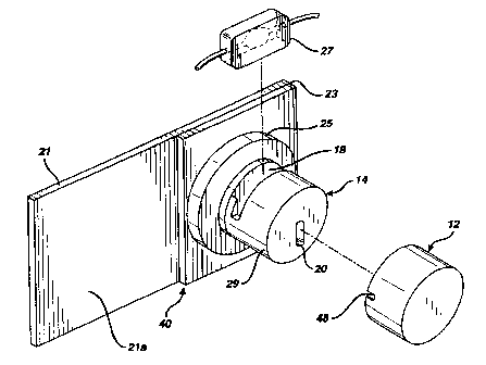
Figure 1 illustrates a contaminant detector
according to a preferred embodiment of the present invention.
Particularly, Figure 1 shows a contaminant detector 10, in
exploded view, as having cover 12 and a main body 14. Also
shown is a cuvette 27 that is selectively insertable into the
-11-
SUBSTITUTE SHEET (rule 26 )
~
CA 02272365 1999-05-19
WO 98/22798 PCT/US97/21266
main body 14 in a manner that will be_described in greater
detail hereinafter.
In accordance with at least one preferred embodiment
of the present invention, a mounting block 23 may be mounted
on a suitable mounting plate 21. In turn, mounting block 23
may preferably form a base for main body 14. As shown in
Figure 1, main body 14 could preferably be constituted by a
larger cylindrical portion 25 and a smaller cylindrical
portion 29 (i.e., "larger" and "smaller" in terms of their
relative diameters). Further, on a surface 21a of mounting
plate 21, it is conceivable to mount, in any appropriate
manner, circuitry for the purpose of processing measurements
taken by the detector 10. Alternatively, such circuitry could
be provided on that surface of mounting plate 21 disposed
opposite from surface 21a.
Preferably, smaller cylindrical portion 29 will have
a slot 18 disposed therein that is suitable for accommodating
the aforementioned cuvette 27. Also preferably provided in
cylindrical portion 29 is a light-emitting diode (LED)
arrangement or other suitable light source 20 for emitting
light during measurement procedures.
To facilitate the propagation of light through
cuvette 27 (when inserted in main body 14), the main body
further preferably comprises a first passage 22 leading from
LED 20 to slot 18 and a second passage 24 leading from the
slot 18 to a suitable sensing arrangement 26 (see Figure 5).
Preferably, slot 18 will accommodate cuvette 27 in a
manner that permits the light emitted by LED 20 to pass
through cuvette 27 and onward to sensing arrangement 26
(again, see Figure 5). Preferably, for the duration of a
detection procedure, cover 12 will be placed over main body 14
in such a manner as to significantly minimize, if not
virtually completely eliminate, the ingress of ambient light
-12-
SUBSTITUTE SHEET ( rule 26 )
CA 02272365 1999-05-19
WO 98/22798 PCT/US97/21266
(i.e., light from outside of the apparatus) towards cuvette
27.
Figure 2 more closely illustrates a cuvette 27 that
may be utilized in accordance with a preferred embodiment of
the present invention. Preferably, cuvette 27 will include an
infeed line 28, an outfeed line 32 and a main body portion 34.
Main body portion 34 will preferably be so
configured as to include therewithin a portion defining a
"flattening" chamber (which could be alternatively termed an
'% exposure", "detection" or "testing" chamber) 36 of
significantly small thickness to effectuate the provision of a
significantly thin layer of a blood product sample in the path
of light emitted from the light source 20. In one embodiment
of the present invention, the thickness of chamber 36 could be
about .030 inch (resulting in a blood film layer of similar
thickness), but slightly larger or smaller thicknesses could
also be used.
Preferably, main body portion 34 will also be so
configured as to readily accommodate infeed and.outfeed
lines 28 and 32 so that infeed and outfeed lines.28 and 32 may
respectively direct blood portions into and out of chamber 36
via suitable interior conduits 28a and 32a. Interior conduits
28a and 32a may be generally tubular in nature and may effect
a transition into chamber 36 via suitably configured
transition zones 31 and 33. Preferably, chamber 36 will be so
configured as to present a thin, and substantially laminar,
layer of liquid to light emitted from LED arrangement, or
other suitable light source 20 (see Figure 1). In accordance
with at least one preferred embodiment of the present
invention, at least chamber 36 is made of an essentially
transparent material (e.g., a clear plastic). It will be
understood that the balance of the main body portion 34, as
well as the infeed and outfeed lines 32, 34 may be made of
similar material (although materials of greater opacity may be
-13-
SUBSTITUTE SHEET ( rule 26 )
~ ~
CA 02272365 1999-05-19
WO 98/22798 PCT/US97/21266
more preferable for these components- in order to further
inhibit the ingress of ambient light into chamber 36).
Figure 3 illustrates the contaminant detector with
the cuvette 27 inserted into slot 18 (see Figure 1) and with
cover 12 in place, in preparation for a detection procedure.
Figure 4 is a front elevational view of a
contaminant detector according to the present invention, with
the aforementioned cover 12 being removed. The aforementioned
LED arrangement 20 is preferably positioned in a suitably
dimensioned slot 38.
Figure 5 is a plan view of the contaminant detector
shown in Figure 3. As illustrated, slot 18 preferably spans
at least the diameter of the smaller cylindrical portion 29 of
main body 14.
Figure 6 is a cut-away view taken substantially
along the line VI- VI shown in Figure 5. As shown, slot 18
will preferably be so configured as to fully accommodate
cuvette 27, and thus preferably includes a downward recessed
portion 42. Preferably, downward recessed portion 42 will
contain a window 43 that, upon placement of cuvette 27 in
slot 38, will be aligned with the aforementioned flattening
chamber 36 of cuvette 27 so as to direct light into second
passage 24 (see Figure 5).
Figure 7 is a cut-away view substantially taken
along the line VII-VII shown in Figure 5. As shown, this
portion of main body 14 will preferably have a hole 44
disposed therewithin configured for directing LED or other
light from first passage 22 (see Figure 5) towards flattening
chamber 36 of cuvette 27 and thence to the aforementioned
window 43.
-14-
SUBSTITUTE SHEET (rule 26 )
CA 02272365 1999-05-19
WO 98/22798 PCT/US97/21266
Figures 6 and 7 illustrate that, in accordance with
at least one preferred embodiment to the present invention,
the aforementioned cuvette-accommodating slot 18 (see Figure
5) can preferably be constituted by: downward recessed portion
42, substantially horizontal ledge portions 45 and
substantially vertical wall portions 47. Downward recessed
portion 42 itself may preferably be constituted by a first
vertical wall portion 42a (as shown in Figure 6) and a second
vertical wall portion 42b (as shown in Figure 7).
Preferably, portions 42a, 42b, 47 and 45 will be so
dimensioned and configured as to adequately accommodate
cuvette 27 when the same is inserted into slot 18 and
supported within downward recessed portion 42. In this
regard, when cuvette 27 (see Figure 2) is inserted into
downward recessed portion 42, a significant portion of main
body 34 of cuvette 27 will preferably be cradled in downward
recessed portion 42. So configured, the infeed and outfeed
lines 28 and 32 will preferably respectively rest on
corresponding horizontal ledge portions 45, whereas opposite
longitudinal ends of cuvette 27 will substantially abut
against corresponding vertical wall portions 47. Preferably,
with respect to the view shown in Figure 6, vertical wall
portion 42a will preferably be axially more recessed than
vertical wall portions 47, in order to readily accommodate the
thickness of main body 34 beyond the infeed and outfeed lines
28 and 32. With infeed line 28 and outfeed line 32 of cuvette
27 resting on horizontal ledge portions 45, the same will also
preferably be accommodated by suitably dimensioned recesses 48
in cover 12 (one of which is shown in Figure 1).
Preferably, window 43 leads to passage 24 and
terminates at suitable sensing device, or sensor, 26 (see
Figure 5). Such a sensor 26 is schematically indicated in
Figure 8, with the LED input being indicated schematically at
-15-
SUBSTITUTE SHEET ( ruie 26 )
~ ~
CA 02272365 1999-05-19
WO 98/22798 PCT/US97/21266
50. Preferably, sensor 26 will be connected to suitable
_ ~...
circuitry and/or programming 52 for the purpose of determining
the actual contaminant level in the liquid sample in question.
Figures 9 to 11 illustrate a further embodiment of
the invention. Figure 9 shows an alternative cuvette 100
molded of an opaque plastic or other suitable material. The
cuvette 100 comprises a flat elongated body 102 having an
integral light shield flange 104 molded over ends 106 and an
upper edge 108 of the body 102. Ports 110 and 112 connect to
the tubing (not shown) as in the prior embodiment.
Passageways 114 and 116 lead from ports 110 and 112
respectively into a discoidal viewing chamber 118. The
chamber 118 is defined by an annular wall 120 normal to and
penetrating the body 102. A pair of transparent windows 122
are sonically welded within the wall 120, abutting an annular
ledge 124 within the chamber 118, to enclose the chamber 118.
A longitudinal vane 126, coplanar with the body 102, extends
through an upper portion of the chamber 118 between the
windows 122 to promote laminar flow of sufficient velocity to
carry any entrained air bubbles out of the chamber 118.
Figure 10 shows an optical assembly 128 for
receiving the cuvette 100 (not shown in Figure 10). The
assembly 128 comprises a body 130 formed of an opaque material
having a recess 132 shaped to receive the cuvette 100, with an
LED 134 on one side thereof and a photodiode 136 on an
opposite side thereof. A window 138 separates the LED 134
from the recess 132. Figure 11 shows the cuvette 100 received
within the recess 132. The light shielding flange 104 and the
optical assembly body 130 shield the chamber 118 from ambient
-- light sources. The LED 134 can direct its light through its
window 138, through the chamber windows 118, and the chamber
118 to be received by the photodiode 136. The hematocrit
-16-
SUBSTITUTE SHEET (rule 26 )
CA 02272365 1999-05-19
WO 98/22798 PCT/US97/21266
level of fluids flowing through the chamber 118 can thus be
measured quickly and easily. -
It is to be understood that, in accordance with at
least one preferred embodiment of the present invention, the
contaminant detectors described and illustrated with respect
to Figures 1 to 11 provide only illustrative examples and are
in no way meant to limit the scope of the present invention.
It will be appreciated that the structural and
functional aspects of the present invention may be applicable
to a wide variety of contexts, involving a wide variety of
liquids and associated contaminants. Thus, although specific
reference has been made to the context of detecting the
presence of red blood cells in a human blood sample containing
a preponderance of white blood cells, it is to be understood
that other liquids and other contaminants can conceivably be
adopted within the scope and spirit of the present invention,
especially by employing the concept of "color affinity"
described and alluded to throughout the instant application.
Examples of such liquids include, but are not limited to:
clear solvents (such as alcohol, paint thinner, turpentine,
etc.); liquid pharmaceutical or medicinal products (e.g.
liquid cold/fever medicines, hydrogen peroxide, liquids for
use in vaporizers); various clear or "dye-free" consumer
products in the market place (including, among others, liquid
soaps, detergents and waxes, shampoos, hair sprays, cosmetics,
deodorants, topical medications, beverages, parenteral
alimentation solutions); fossil fuels, such as petroleum
(either in crude or refined form); and other liquids which may
either be essentially clear in nature or may have a given base
color.
It will be appreciated that, in accordance with at
least one preferred embodiment of the present invention, and
-17-
)
SUBSTITUTE SHEET (rule 26
~ ~
CA 02272365 1999-05-19
WO 98/22798 PCT/US97/21266
especially in the context of determining hematocrit values in
human blood or blood product samples (particularly blood
samples containing a preponderance of white blood cells), it
is desirable to utilize light that has no greater a wavelength
than that associated with "blue" light. In at least one
embodiment of the present invention, this may translate to
about 466nm or less. To date, light having a wavelength of as
low as 430 nm has been used, and it is conceivable to utilize
light of even lower wavelength. As discussed heretofore, it
would appear that such wavelengths (i.e., those associated
with "blue" light or less, such as about 466nm or less)
provide several advantages including, but not necessarily
limited to: the likelihood that the presence of red blood
cells would be distinguished more easily against the
background of white blood cells; and the compatibility of such
light with the type of light that may be used in an
irradiation procedure such as UV-A light (i.e., light having a
wavelength of about 352nm) with the resultant likelihood that
the blood product sample being measured will not be unduly
affected or altered by the light from the LED.
In accordance with at least one embodiment of the
present invention, it has been found that blue LED's
manufactured by Cree Research, Inc. of Durham, North Carolina,
are particularly effective, particularly, the "C470 Series
Silicon Carbide Blue LED's."
In accordance with at least one embodiment of the
present invention, a suitable photodiode may preferably be
used as the sensing arrangement 26 illustrated and described
herein. The "VTB Process Photodiodes" manufactured by EG&G
VACTEC of St. Louis, Missouri, have been found to be
particularly effective.
Preferably, in accordance with at least one
embodiment of the present invention, essentially any suitable
type of circuitry may be used for the purpose of converting
-1s-
SUBSTITUTE SHEET ( rule 26 )
CA 02272365 1999-05-19
WO 98/22798 PCT/iJS97/21266
the light measured by the aforementioned sensing arrangement
_ ~. .
(such as a photodiode) to a value indicative to the relative
presence of a given contaminant (such as a hematocrit value)
in the liquid sample being measured.
For example, it is conceivable to use an appropriate
amplifier for the purpose of amplifying a signal from the
sensor (e.g. photodiode) indicative of the amount of light
measured by the sensor, as well as circuitry for converting
the amplified signal into a serial bit stream. For the
purpose of calibrating the measurement apparatus, it is
conceivable to provide "on-board" memory (e.g. lookup tables
or the like). Components such as these would appear to be
well-known to those of ordinary skill in the art and will thus
not be further discussed herein. It is to be understood that
components such as these are provided here only as an example,
and that essentially any type of appropriate circuitry or
other arrangement may be utilized within the scope of the
present invention.
In view of the general considerations set forth
hereinabove, the need to measure hematocrit levels of blood
and blood products accurately, on-line (i.e. non-invasively)
and in real time has been widely recognized, particularly with
regards to the control of processes used to separate and/or
treat the blood or blood fractions. The need to measure
accurately what are considered very low hematocrit levels
(i.e. less than about 10) has been deemed particularly
important.
At least one embodiment of the present invention
contemplates an apparatus and method in which preferably a
single light source, such as one of narrowly defined
wavelength with peak emission of about 466nm (blue light), is
used to emit light through a cuvette containing a blood or
blood product sample to be measured. Further, a photodiode
located on the other side of the sample cuvette detects the
-19-
SUBSTITUTE SHEET (rule 26 )
CA 02272365 2006-10-25
amount of light passing through the thin film sample. An
amplifier and electronic circuit amplify the signal and
convert the same into a serial bit stream. The amount-of
light detected by the photodiode is inversely proportional to
the hematocrit level of the sample. As a result, it has been
found that changes in hematocrit can be detected instantly.
Preferably, calibration data will be stored in "on-board"
memory.
In accordance with at least one embodiment of the
present invention, it is contemplated that, in the context of
a blood product sample containing a preponderance of white
blood cells, hematocrit measurements can be taken prior to the
sample being irradiated in an irradiation device. Such
irradiation devices, and procedures associated therewith, are
well-known to those of ordinary skill in the art.
Several U.S. Patents disclose apparatus and
processes, as well as components and concepts associated
therewith, that may be utilized in accordance with the
embodiments of the present invention. These patents are
listed herebelow.
Some examples of irradiation' devices, and procedures
associated therewith, are to be found in the following U.S.
Patents: No. 5,459,322 to Warkentin; Nos. 4,321,919,
4,398,906 and 4,428,744 to Edelson; Nos. 4,708,715 and
4,692,138 to Troutner et al.; No. 4,737,140 to Lee et al.; and
Nos. 4,952,812 and 4,726,949 to Miropol et al.
U.S. Patent Nos. 5,416,342 and 5,027,168 disclose
examples of blue light-emitting diodes.
Blue light-emitting diodes are also discussed in
"Technology Newsletter", Electronic Design, October 24, 1995,
page 29.
-20-
CA 02272365 1999-05-19
WO 98/22798 PCTJUS97/21266
If not otherwise stated herein, it may be assumed
that all components and/or processes described heretofore may,
if appropriate, be considered to be interchangeable with
similar components and/or processes disclosed elsewhere in the
specification, unless an indication is made to the contrary.
It should be appreciated that the apparatus and
method of the present invention may be configured and
conducted as appropriate for the application. The embodiments
described above are to be considered in all respects only as
illustrative and not restrictive. The scope of the invention
is defined by the following claims rather than by the
foregoing description. All changes which come with the
meaning and range of equivalency of the claims are to be
embraced within their scope.
-21-
)
SUBSTITUTE SHEET ( ruie 26