Note: Descriptions are shown in the official language in which they were submitted.
CA 02273467 1999-06-02
CATHETER FOR INJECTING THERAPEUTIC AND DIAGNOSTIC AGENTS
Irma P. Hill
Dean M. Ponzi
FIELD OF THE INVENTION
This invention relates to a catheter for infusing therapeutic
or diagnostic agents into the tissue of organs, and more
particularly to a catheter system in which the infusion needle may
be very precisely positioned within the heart to infuse drugs into
the wall of the heart.
BACKGROUND OF THE INVENTION
Targeted delivery of therapeutic or diagnostic agents, such as
occurs in gene therapy, is very desirable but often presents a
difficult challenge. A potential benefit of targeted delivery is
that there is an increased efficiency obtained by the precise
placement of the therapeutic agent. There are several problems to
this procedure which must be overcome in order to obtain
satisfactory results from such therapy, such as the problems of
obtaining access to the delivery site, transporting the therapeutic
agent to the desired site, injecting the therapeutic agent at the
proper depth within the organ tissue, steering the distal end of
the catheter to a desired location within the organ prior to
infusing the agent, and positioning the distal tip of the catheter
at precisely the same location where prior measurements have
indicated that the drug should be infused. It is also important
1
CA 02273467 1999-06-02
for a physician to be able to monitor the position of the infusion
needle with respect to the wall of the organ. In the case of an
organ, such as the heart, in which the walls are in constant
motion, the activity of positioning and monitoring the position of
the distal tip of the catheter, or infusion needle, becomes
especially difficult.
U.S. Patent No. 3,598,119 discloses a medical device for
injecting drugs in which the injection needle is guided through an
inner lumen of a catheter for insertion of the needle under skin
tissue. A bladder at the distal end of the catheter may be
inflated through another lumen for holding the point of the needle
point in a fixed position beneath the skin.
U.S. Patent No. 4,578,061 discloses a catheter for injecting
a liquid into a vein, or artery, through an injection needle which
is longitudinally movable beyond the distal end of the catheter. A
dual chamber system is utilized within the catheter tip to provide
for movement of a plunger to extend the injection needle and also
to allow for a plunger to be used to apply a predetermined dose of
medication through the injection needle.
U.S. Patent No. 4,578,061 discloses an injection catheter
having a longitudinal movable needle which may be moved through a
lumen in order to extend out of the side wall of the catheter for
injecting a liquid into a blood vessel. The needle is normally
retracted into the device so that the needle will not penetrate
tissue as the device is moved through a body duct. Thereafter, the
2
CA 02273467 2007-11-21
needle is moved out of the side of the catheter into a
vessel wall in order to infuse a liquid into the wall of
a vessel.
U.S. Patent No. 5,244,460 is directed toward a
method for improving blood flow to the heart. More
particularly this patent is directed toward a medical
procedure for improving the growth of cardiac blood
vessels by inserting a catheter into a coronary artery
and injecting into the heart a blood vessel growth
promoting peptide through an injection port of the
catheter.
U.S. Patent No. 5,419,777 is directed toward a
catheter for injection of a fluid into body cavities such
as coronary vessels and arteries. This patent, as is the
case with the '061 patent, illustrates the use of an
injection needle which pretrude laterally through the
side walls of the distal tip of the catheter. In the case
of drug injections to be made into coronary vessels and
arteries, it is very desirable to have the needles extend
out of the side walls of the catheter and at an acute
angle to the walls of the vessel in order to penetrate
the walls of the vessel for injection of the agent.
U.S. Patent No. 5,431,168, assigned to the same
assignee as the present patent application, is directed
toward a steerable catheter which includes a puller wire
for controlling the distal end of the catheter from a
control handle which is mounted on the proximal end of
the catheter.
U.S. Patent No. 6,309,370, entitled "Intracardiac
Drug Delivery," assigned to an affiliated company of the
assignee of this application, discloses an injection
3
CA 02273467 2007-11-21
catheter system for infusing a diagnostic or therapeutic
agent into the wall of an organ which includes an
electromagnetic sensor disposed within the distal tip of
the catheter for providing very precise location
informationfor-the distal tip of the catheter.
For obvious reasons, catheters which are used to
deliver therapeutic or diagnostic agents into a targeted
region of the heart must be designed so that the
physician is able to maintain precise control over the
distal tip of the catheter. In addition, the catheter
must be designed to provide information as to these
precise location of the distal tip, or infusion needle,
of the catheter. The present invention is directed toward
an improved injection catheter which allows the physician
to have greater control over the position of the distal
tip and to also obtain accurate information as to the
position of the catheter tip.
SUMMARY OF THE INVENTION
The present invention provides for a steerable drug
injection catheter system which includes a catheter body
having an outer wall, proximal and distal ends, and at
least one lumen extending therethrough. The catheter also
includes a control handle attached to the proximal end of
the catheter for controlling the position of the distal
tip of the catheter and for controlling the extension of
the injection needle and for the injection of a
diagnostic or therapeutic agent. In addition, the
catheter includes a tip
4
CA 02273467 1999-06-02
section which is comprised of a flexible tubing having a lumen
extending therethrough, an injection needle which extends through
the lumen of the catheter and is movable from a first position in
which the distal tip of the needle is withdrawn into the distal
face of the tip section to a second position in which the distal
tip of the needle extends out of the distal face of the tip
section, a needle control means within the control handle for
moving the needle from the first position to the second position,
and control means for deflecting the distal tip of the catheter
upon manipulation of the control handle.
In accordance with another aspect of the present invention,
the drug injection catheter also includes a tip electrode mounted
at the distal end of the tip section and an electrode conductor
which is electrically connected to the tip electrode and extends
through the lumen of the tip section and into the control handle.
This tip electrode may be used for measuring electrical potentials
within the heart, for example cardiac mapping.
In accordance with still another aspect of the present
invention, the drug injection catheter includes an electrode
magnetic mapping sensor which is mounted in the distal portion of
the tip section for producing electrical signals indicative of the
location of the electrode magnetic mapping sensor in order to
provide very precise information with respect to the location of
the distal tip of the catheter.
In accordance with still another aspect of the present
invention, the drug injection catheter includes a sensor cable
5
CA 02273467 1999-06-02
electrically attached to the electrode magnetic mapping sensor
which extends through the lumen of the catheter and into the
control handle where it is then electrically attached to a circuit
board situated within the control handle.
In accordance with still another aspect of the present
invention, the drug infusion catheter includes a position sensor,
preferably an electromagnetic sensor, which is disposed within the
distal tip of the catheter for providing precise information as to
the location of the distal tip of the catheter.
In accordance with still another aspect of the present
invention, the control handle includes a first member fixedly
attached to the proximal end of the catheter body and a second
member which is movable with respect to the first member. The
deflecting means is comprised of a puller wire having a proximal
end and a distal end which extends from the control handle through
the lumen in the catheter and is fixedly secured within the tip
section. The proximal end of the puller wire is fixedly secured to
the second member of the control handle whereby manipulation of the
first member of the handle relative to the second member of the
control handle moves the puller wire relative to the catheter body
resulting in deflection of the tip section.
In accordance with still another aspect of the present
invention, the deflecting means further comprises a compression
coil situation in the catheter body in surrounding relation to the
puller wire and extending into the lumen of the tip section in
order to prevent buckling of the catheter shaft.
6
CA 02273467 1999-06-02
BRIEF DESCRIPTION OF THE DRAWINGS
These and other features and advantages of the present
invention will be better understood by reference to the following
detailed description when considered in conjunction with the
accompanying drawings wherein:
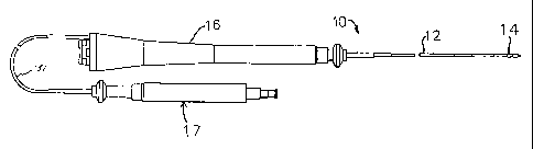
Figure 1 is a side plan view of one embodiment of the catheter
of the present invention;
Figure 2 is a side cross-sectional view of the needle control
handle for the embodiment of Figure 1;
Figure 3 is a side cross-sectional view of the catheter tip
section showing an embodiment having three lumens and showing the
position of the electromagnetic mapping sensor and the injection
needle;
Figure 4 is a side cross-sectional view of the catheter tip
section showing an embodiment having three lumens and showing the
position of the electromagnetic mapping sensor and the puller wire;
Figure 5 is a side cross-sectional view of the catheter body,
including the junction between the catheter body and the tip
section;
Figure 6 is a transverse cross-sectional view of the catheter
tip section along line 6-6 showing an embodiment having three
lumens;
Figure 7 is a transverse cross-sectional view of the catheter
body along line 7-7; and,
Figure 8 is a side cross-sectional view of the catheter
handle;
7
CA 02273467 1999-06-02
DESCRIPTION OF A PREFERRED EMBODIMENT
In a preferred embodiment of the invention, there is provided
a catheter for use for injection of a therapeutic or diagnostic
agent into the heart. As shown in Figure 1, catheter 10 is
comprised of an elongated catheter body 12 having proximal and
distal ends, a tip section 14 at the distal end of the catheter
body 12, and a deflection control handle 16 at the proximal end of
the catheter body 12 and a needle control handle 17.
As illustrated in Figure 2, the needle control handle 17 is
comprised of a proximal Luer connector 19 which is threaded into an
outer body 21. The control handle also includes a slidable control
knob 23 which is attached to a piston 23a, which is in turn
slidably mounted within a lumen 25 within the outer body 21. A
ring seal 27 is disposed between the piston 23a and the inner lumen
of the outer body 21 to prevent fluid from entering the housing.
The slidable control knob 23 and piston 23a are fixedly attached to
a catheter tubing 31 which extends into the needle control housing
17 and the deflection control housing 16.
In addition, the needle control housing 17 includes a support
tube 33 which is fixedly mounted coaxially within the outer body 21
and is preferably formed of stainless steel. Positioned within the
lumen of support tube 33 is a slidable tube 35, preferably formed
of stainless steel, which is in turn directly coupled to the
proximal end of the catheter tubing 31. An injection needle 46 is
fixedly attached to an inner lumen of the Luer connector 19 and
extends through the lumen of the slidable tube 35 and then through
8
CA 02273467 1999-06-02
a lumen in the slidable piston 23a and then into the catheter
tubing 31.
Accordingly, since the injection needle is attached to the
Luer connector 19, it may be seen that as the control knob 23 is
moved proximally to thereby cause the piston 23a to move proximally
into the outer body 21 of the needle control housing, the injection
needle 46 is caused to slide through the catheter housing 31 with
the result that the needle is caused to be extended out of the
distal end of the injection catheter system 10. Also, it may be
seen that as fluid is applied to the inner lumen of the Luer
connector, the fluid is caused to flow through the lumen of the
injection needle and to the distal tip of the injection needle 46.
With reference to Figures 5 and 7, the catheter body 12
comprises a single, central or axial lumen 18. The catheter body
12 is flexible, i.e., bendable, but substantially non-compressible
along its length. The catheter body 12 may be of any suitable
construction and made of any suitable material. A presently
preferred construction comprises an outer wall 22 made of a
polyurethane or nylon. The outer wall 22 comprises an imbedded
braided mesh of stainless steel or the like to increase torsional
stiffness of the catheter body 12 so that, when the control handle
16 is rotated, the tip section of the catheter 10 will rotate in a
corresponding manner.
The outer diameter of the catheter body 12 is not critical,
but is preferably no more than about 8 French. Likewise the
thickness of the outer wall 22 is not critical. The inner surface
9
CA 02273467 1999-06-02
of the outer wall 22 is lined with a stiffening tube 20, which can
be made of any suitable material, preferably polyimide. The
stiffening tube, along with the braided outer wall 22, provides
improved torsional stability while at the same time minimizing the
wall thickness of the catheter, thus maximizing the diameter of the
single lumen. The outer diameter of the stiffening tube 20 is
about the same as or slightly smaller than the inner diameter of
the outer wall 22. Polyimide tubing is presently preferred because
it may be very thin walled while still providing very good
stiffness. This maximizes the diameter of the central lumen 18
without sacrificing strength and stiffness. Polyimide material is
typically not used for stiffening tubes because of its tendency to
kink when bent. However, it has been found that, in combination
with an outer wall 22 of polyurethane, nylon or other similar
material, particularly having a stainless steel braided mesh, the
tendency for the polyimide stiffening tube 20 to kink when bent is
essentially eliminated with respect to the applications for which
the catheter is used.
A particularly preferred catheter has an outer wall 22 with an
outer diameter of about 0.092 inch and an inner diameter of about
0.063 inch and a polyimide stiffening tube having an outer diameter
of about 0.0615 inch and an inner diameter of about 0.052 inch.
As shown in Figures 3 and 4, the tip section 14 comprises a
short section of tubing 19 having three lumens. The tubing 19 is
made of a suitable non-toxic material which is preferably more
flexible than the catheter body 12. A presently preferred material
CA 02273467 1999-06-02
for the tubing 19 is braided polyurethane, i.e., polyurethane with
an embedded mesh of braided stainless steel or the like. The outer
diameter of the tip section 14, like that of the catheter body 12,
is preferably no greater than about 8 French. The size of the
lumens is not critical. In a particularly preferred embodiment,
the tip section has an outer diameter of about 7 French (.092 inch)
and the first lumen 30 and second lumen 32 are generally about the
same size, having a diameter of about 0.022 inch, with the third
lumen 34 having a slightly larger diameter of about 0.036 inch.
A preferred means for attaching the catheter body 12 to the
tip section 14 is illustrated in Figure 5. The proximal end of the
tip section 14 comprises an inner counter bore 24 that receives the
outer surface of the polyimide stiffener 20. The tip section 14
and catheter body 12 are attached by glue or the like.
The stiffening tube 20 is held in place relative to the outer
wall 22 at the proximal end of the catheter body 12. In a
preferred construction of the catheter body 12, a force is applied
to the proximal end of the stiffening tube 20 which causes the
distal end of the stiffening tube 20 to firmly push against the
counter bore 24. While under compression, a first glue joint is
made between the stiffening tube 20 and the outer wall 22 by a fast
drying glue, e.g. Super Glue . Thereafter a second glue joint is
formed between the proximal ends of the stiffening tube 20 and
outer wall 22 using a slower drying but stronger glue, e.g.,
polyurethane.
l1
CA 02273467 1999-06-02
Extending through the single lumen 18 of the catheter body 12
are lead wires 40, an injection needle 46, a sensor cable 74, and
a compression coil 44 through which a puller wire 42 extends. A
single lumen 18 catheter body is preferred over a multi-lumen body
because it has been found that the single lumen 18 body permits
better tip control when rotating the catheter 10. The single lumen
18 permits the lead wires 40, the injection needle 46, the sensor
cable 74, and the puller wire 42 surrounded by the compression coil
44 to float freely within the catheter body. If such wires and
cables were restricted within multiple lumens, they tend to build
up energy when the handle 16 is rotated, resulting in the catheter
body 12 having a tendency to rotate back if, for example, the
handle is released, or if bent around a curve, to flip over, either
for which are undesirable performance characteristics.
The puller wire 42 is anchored at its proximal end to the
control handle 16 and anchored at its distal end to the tip section
14. The puller wire 42 is made of any suitable metal, such as
stainless steel or Nitinol, and is preferably coated with Teflon
or the like. The coating imparts lubricity to the puller wire 42.
The puller wire 42 preferably has a diameter ranging from about
0.006 to about 0.010 inches.
The compression coil 44 extends from the proximal end of the
catheter body 12 to the proximal end of the tip section 14. The
compression coil 44 is made of any suitable metal, preferably
stainless steel. The compression coil 44 is tightly wound on
itself to provide flexibility, i.e., bending, but to resist
12
CA 02273467 1999-06-02
compression. The inner diameter of the compression coil 44 is
preferably slightly larger than the diameter of the puller wire 42.
For example, when the puller wire 42 has a diameter of about 0.007
inches, the compression coil 44 preferably has an inner diameter of
about 0.008 inches. The Teflon coating on the puller wire 42
allows it to slide freely within the compression coil 44. Along
its length, the outer surface of the compression coil 44 is covered
by a flexible, non-conductive sheath 26 to prevent contact between
the compression coil 44 and any of the lead wires 40, injection
needle 46 or sensor cable 74. A non-conductive sheath 26 made of
polyimide tubing is presently preferred.
The compression coil 44 is anchored at its proximal end to the
proximal end of the stiffening tube 20 in the catheter body 12 by
glue and at its distal end to the tip section 14. The glue may be
applied by means of a syringe or the like through a hole made
between the outer surface of the catheter body 12 and the single
lumen 18.
The puller wire 42 extends into the second lumen 32 of the tip
section 14. The puller wire 42 is anchored to a tip electrode 36
or to the side of the catheter tip section 14. With reference to
Figures 4 and 5, within the tip section 14, and distal to the glue
joint 51, the turns of the compression coil are expanded
longitudinally. Such expanded turns 47 are both bendable and
compressible and preferably extend for a length of about 0.5 inch.
The puller wire 42 extends through the expanded turns 47 then into
a plastic, preferably Teflon , sheath 81, which prevents the puller
13
CA 02273467 1999-06-02
42 from cutting into the wall of the tip section 14 when the tip
section 14 is deflected.
With reference to Figures 3 and 4, at the distal end of the
tip section.14 is a tip electrode 36. Preferably the tip electrode
36 has a diameter about the same as the outer diameter of the
tubing 19. The tip electrode 36 is connected to the tubing 19 by
means of a plastic housing 21, preferably made of
polyetheretherketone (PEEK). The proximal end of the tip electrode
36 is notched circumferentially and fits inside the distal end of
the plastic housing 21 and is bonded to the housing 21 by
polyurethane glue or the like. The proximal end of the plastic
housing 21 is bonded with polyurethane glue or the like to the
distal end of the tubing 19 of the tip section 14.
Mounted on the distal end of the plastic housing 21 is a ring
electrode 38. The ring electrode 38 is slid over the plastic
housing 21 and fixed in place by glue or the like. If desired,
additional ring electrodes may be used and can be positioned over
the plastic housing 21 or over the flexible tubing 19 of the tip
section 14.
The tip electrode 36 and ring electrode 38 are each connected
to separate lead wires 40. The lead wires 40 extend through the
third lumen 34 of tip section 14, the catheter body 12, and the
control handle 16, and terminate at their proximal end in an input
jact (not shown) that may be plugged into an appropriate monitor
(not shown). If desired, the portion of the lead wires 40
extending through the catheter body 12, control handle 16 and
14
CA 02273467 1999-06-02
proximal end of the tip section 14 may be enclosed or bundled
within a protective tube or sheath.
The lead wires 40 are attached to the tip electrode 36 and
ring electrode 38 by any conventional technique. Connection of
lead wire 40 to the tip electrode 36 is preferably accomplished by
weld 43, as shown in Figure 4.
The injection needle assembly is comprised of the injection
needle 46 which extends from the needle control handle through the
body of the catheter, through the distal tip of the catheter and
through the tip electrode 36. The injection needle 46 is formed of
nitinol, and as illustrated in Figure 3 is preferably formed with
a beveled edge at the distal tip of the needle. Also as
illustrated in Figure 3, the needle is coaxially mounted within a
polyimide tube 47a which serves to prevent the needle from buckling
and also serves to electrically insulate the needle from the distal
electrode 36. The tube 47a additionally serves to provide a fluid-
tight seal surrounding the injection needle. The injection needle
as shown in Figure 3 is in a position where the needle extends
beyond the distal tip of the electrode as it would be positioned in
order to infuse diagnostic or therapeutic fluid into the human
heart. The needle is withdrawn within the distal tip of the
catheter during the period of time that the catheter is inserted
through the vasculature of the body and also during the period of
time in which the catheter is removed from the body.
An electromagnetic sensor 72 is contained within the distal
end of the tip section 14. The electromagnetic sensor 72 is
CA 02273467 2007-11-21
connected by means of electromagnetic sensor cable 74,
which extends through the third lumen 34 of the tip
section 14 through the catheter body 12 into the control
handle 16. The electromagnetic sensor cable 74 comprises
multiple wires encased within a plastic sheath. In the
control handle 16, the wires of the sensor cable 74 are
connected to a circuit board 64. The circuit board 64
amplifies the signal received from the electromagnetic
sensor and transmits it to a computer in a form
understandable by the computer. Also, because the
catheter is designed for single use only, the circuit
board contains an EPROM chip which shuts down the circuit
board after the catheter has been used. This prevents the
catheter, or at least the electromagnetic sensor, from
being used twice. A suitable electromagnetic sensor is
described, for example, in U.S. Pat. No. 4,391,199. A
preferred electromagnetic mapping sensor 72 is
manufactured by Biosense Ltd. Israel and marketed under
the trade designation NOGA. To use the electromagnetic
sensor 72, the patient is placed in a magnetic field
generated, for example, by situating under the patient a
pad containing coils for generating a magnetic field. A
reference electromagnetic sensor is fixed relative to the
patient, e.g., taped to the patient's back, and the
injection catheter containing a second electromagnetic
sensor is advanced into the patient's heart. Each sensor
comprises three small coils which in the magnetic field
generate weak electrical signals indicative of their
position in the magnetic field. Signals generated by both
the
16
CA 02273467 1999-06-02
fixed reference sensor and the second sensor in the heart are
amplified and transmitted to a computer which analyzes the signals
and then displays the signals on a monitor. By this method, the
precise location of the sensor in the catheter relative to the
reference sensor can be ascertained and visually displayed. The
sensor can also detect displacement of the catheter that is caused
by contraction of the heart muscle.
Using this technology, the physician can visually map a heart
chamber. This mapping is done by advancing the catheter tip into
a heart chamber until contact is made with the heart wall. This
position is recorded and saved. The catheter tip is then moved to
another position in contact with the heart wall and again the
position is recorded and saved.
The electromagnetic mapping sensor 72 can be used alone or
more preferably in combination with the tip electrode 36 and ring
electrode 38. By combining the electromagnetic sensor 72 and
electrodes 36 and 38, a physician can simultaneously map the
contours or shape of the heart chamber, the electrical activity of
the heart, and the extent of displacement of the catheter and hence
identify the presence and location of the ischemic tissue.
Specifically, the electromagnetic mapping sensor 72 is used to
monitor the precise location of the tip electrode in the heart and
the extent of catheter displacement. The tip electrode 36 and ring
electrode 38 are used to monitor the strength of the electrical
signals at that location. Healthy heart tissue is identified by
strong electrical signals in combination with strong displacement.
17
CA 02273467 1999-06-02
Dead or diseased heart tissue is identified by weak electrical
signals in combination with dysfunctional displacement, i.e.,
displacement in a direction opposite that of healthy tissue.
Ischemic, or hibernating or stunned, heart tissue is identified by
strong electrical signals in combination with impaired
displacement. Hence, the combination of the electromagnetic
mapping sensor 72 and tip and ring electrodes 36 and 38 is used as
a diagnostic catheter to determine whether and where to infuse a
drug into the wall of the heart. Once the presence and location of
ischemic tissue has been identified, the injection catheter can be
deflected so that the needle is normal, i.e., at a right angle, to
the ischemic tissue, and the injection needle may then be moved out
of the distal end of the catheter and into the wall of the heart.
It is understood that, while it is preferred to include both
electrophysiology electrodes and an electromagnetic sensor in the
catheter tip, it is not necessary to include both. For example, an
injection catheter having an electromagnetic sensor but no
electrophysiology electrodes may be used in combination with a
separate mapping catheter system. A preferred mapping system
includes a catheter comprising multiple electrodes and an
electromagnetic sensor, such as the NOGA-STAR catheter marketed by
Cordis Webster, Inc., and means for monitoring and displaying the
signals received from the electrodes and electromagnetic sensor,
such as the Biosense-NOGA system, also marketed by Cordis Webster,
Inc.
18
CA 02273467 1999-06-02
The electrode lead wires 40, injection needle 46 and
electromagnetic sensor cable 74 must be allowed some longitudinal
movement within the catheter body so that they do not break when
the tip section 14 is deflected. To provide for such lengthwise
movement, there are provided tunnels through the glue joint 50,
which fixes the proximal end of the compression coil 44 inside the
catheter body 12. The tunnels are formed by transfer tubes 27,
preferably made of short segments of polyimide tubing. In the
embodiment shown in Figure 5, there are two transfer tubes 27 for
the glue joint 50. Each transfer tube is approximately 60 mm long
and has an outer diameter of about .021 inch and an inner diameter
of about .019 inch. Extending through one transfer tube 27 are the
lead wires 40 and the electromagnetic sensor cable 74. Extending
through the other transfer tube 27 is the injection needle 46.
An additional transfer tube 29 is located at the joint between
the tip section 14 and the catheter body 12. Extending through
this transfer tube is the injection needle 46. This transfer tube
29 provides a tunnel through the glue joint formed when the tip
section 14 is glued to the catheter body 12. It is understood that
the number of transfer tubes may vary as desired.
Longitudinal movement of the puller wire 42 relative to the
catheter body 12, which results in deflection of the tip section
12, is accomplished by suitable manipulation of the control handle
16. The distal end of the control handle 16 comprises a piston 54
with a thumb control 56 for manipulating the puller wire 42. The
19
CA 02273467 1999-06-02
proximal end of the catheter body 12 is connected to the piston 54
by means of a shrink sleeve 28.
The puller wire 42, lead wires 40 and electromagnetic sensor
cable 74 extend through the piston 54. The puller wire 42 is
anchored to an anchor pin 36, located proximal to the piston 54.
The lead wires 40 and electromagnetic sensor cable 74 extend
through a first tunnel 58, located near the side of the control
handle 16. The electromagnetic sensor cable 74 connects to the
circuit board 64 in the proximal end of the control handle 16.
Wires 80 connect the circuit board 64 to a computer and imaging
monitor (not shown).
Within the piston 54, the puller wire 42 is situated within a
transfer tube 27, and the electromagnetic sensor cable 74 and lead
wires 40 are situated within another transfer tube 27 to allow
longitudinal movement of the wires and cable near the glue joint
53. The guide tube 66 extends through a second tunnel 60 situated
near the side of the control handle 16 opposite the anchor pin 36.
In another preferred embodiment constructed in accordance with
the present invention, two or more puller wires (not shown) are
provided to enhance the ability to manipulate the tip section. In
such an embodiment, a second puller wire and a surrounding second
compression coil extend through the catheter body and into separate
off-axis lumens in the tip section. The lumens of the tip section
receiving the puller wires may be in adjacent quadrants. The first
puller wire is preferably anchored proximal to the anchor location
of the second puller wire. The second puller wire may be anchored
CA 02273467 1999-06-02
to the tip electrode or may be anchored to the wall of the tip
section adjacent the distal end of tip section.
The distance between the distal end of the compression coils
and the anchor sites of each puller wire in the tip section
determines the curvature of the tip section 14 in the direction of
the puller wires. For example, an arrangement wherein the two
puller wires are anchored at different distances from the distal
ends of the compression coils allows a long reach curve in a first
plane and a short reach curve in a plane 90 from the first, i.e.,
a first curve in one plane generally along the axis of the tip
section before it is deflected and a second curve distal to the
first curve in a plane transverse, and preferably normal to the
first plane. The high torque characteristic of the catheter tip
section 12 reduces the tendency for the deflection in one direction
to deform the deflection in the other direction.
As an alternative to the above described embodiment, the
puller wires (not shown) may extend into diametrically opposed off-
axis lumens in the tip section. In such an embodiment,each of the
puller wires may be anchored at the same location along the length
of the tip section, in which case the curvatures of the tip section
in opposing directions are the same and the tip section can be made
to deflect in either direction without rotation of the catheter
body.
The preceding description has been presented with reference to
presently preferred embodiments of the invention. Workers skilled
in the art and technology to which this invention pertains will
21
CA 02273467 1999-06-02
appreciate that alterations and changes in the described structure
may be practiced without meaningful departing from the principal,
spirit and scope of this invention.
Accordingly, the foregoing description should not be read as
pertaining only to the precise structures described and illustrated
in the accompanying drawings, but rather should be read consistent
with and as support to the following claims which are to have their
fullest and fair scope.
22