Note: Descriptions are shown in the official language in which they were submitted.
CA 02279474 1999-07-30
-1-
B&P File No. 7771-039/MG
Title: Novel Antibody Composition For Debulking Blood and
Bone Marrow Samples From CML Patients
FIELD OF THE INVENTION
The present invention relates to a method of depleting
normal and transformed lineage committed cells from a sample from a
patient with chronic myeloid leukemia.
BACKGROUND OF THE INVENTION
Chronic myeloid leukemia (CML) is a monoclonal expansion
of a transformed pluripotent stem cell (Fialkow et al., 633:125, 1977,
American Journal of Medicine). Myeloid cells, erythroid cells and less
frequently lymphocytes arise from the leukemic clone (Bakhshi et al., New
Eng. J. Med. 309:826, 1983). CML is characterized in more than 90% of
patients by the rearrangement between the break cluster region (BCR gene,
located on chromosome 22) and the ABL gene (located on chromosome 9)
(Bartran et al., Nature 306:277, 1983).
Although patients with CML may have a prolonged course,
the disease is invariably lethal. Bone marrow transplantation is the
treatment of choice for this patient population with a curative rate of 90%
in some centres. However, for 60% of patients this therapy may not be
available either due to the lack of a suitable donor due to differences in
human leukocyte antigens (HLA) or the age of the recipient.
For these reasons, other treatment options have been
evaluated for their ability to remove the leukemic cells from the harvested
material without depleting or damaging the co-existing benign (non-
malignant) stem cells. The methods have included various drug
regiments (Degliantoni et al., Blood 655:753, 1985) or the culture of the
patient cells using the Dexter (long term culture) system which was shown
to preferentially support the proliferation of benign stem cells as compared
to malignant cells (Barnett et al., Bone Marrow Transplant 4:345, 1985).
Clinical experience has confirmed that although the leukemic
burden has been greatly reduced using such protocols, the malignant cells
CA 02279474 1999-07-30
-2-
in most patients have not been entirely eradicated and patients relapse
with their original disease. (Coutintro et al., Progress in Clinical and
Biological Research 333:415, 1990 and Deisseroth et al., Blood 83:3068, 1994)
In addition, the high incidence of graft failure also suggests that certain
types of treatment may have had adverse effects on the non-malignant
stem cells (Talpaz et al., Blood 85:3257, 1995 and Daley and Goldman, Exp.
Hematol. 21:731, 1993).
Further analysis of this disease has focussed on dissecting out
certain populations of primitive cells in an attempt to understand at what
stage the clonal abnormality occurs (Verfaille et al., Blood 87:4770, 1996).
These studies may be limited by the low frequency of primitive cells due to
the clonal proliferation of lineage committed cells. Further studies of this
disease may be facilitated if the mature lineage committed
"contaminating" cells could be reduced or eliminated.
SUMMARY OF THE INVENTION
The present inventors have developed an antibody
composition for use in preparing cell preparations depleted of normal and
transformed lineage committed cells, for example from blood or bone
marrow samples from patients with chronic myeloid leukemia. The
antibodies in the antibody composition are specific for selected markers
associated with lineage committed cells. In particular, the present
inventors have found that using an antibody composition containing
antibodies specific for glycophorin A, CD2, CD3, CD14, CD15, CD16, CD19,
CD24, CD56, CD66b and IgE gives a cell preparation enriched for
hematopoietic stem cells and progenitor cells and depleted of committed
lineage or differentiated cells.
Accordingly, the present invention relates to an antibody
composition comprising antibodies specific for glycophorin A, CD2, CD3,
CD14, CD15, CD16, CD19, CD24, CD56, CD66b and IgE which gives a cell
preparation depleted of lineage committed cells.
The present invention also provides an antibody
composition comprising antibodies specific for glycophorin A, CD2, CD3,
CA 02279474 1999-07-30
-3-
CD14, CD16, CD19, CD24, CD56, CD66b and IgE. Such a composition can be
used in combination with antibodies to CD15 to prepare a cell preparation
depleted of lineage committed cells.
The present invention also includes a negative selection
method for depleting lineage committed cells from a sample from a
patient with chronic myeloid leukemia comprising:
(a) reacting the sample with an antibody composition
comprising antibodies specific for glycophorin A, CD2, CD3, CD14, CD15,
CD16, CD19, CD24, CD56, CD66b and IgE under conditions so that
conjugates between the antibodies and cells in the sample having the
antigens glycophorin A, CD2, CD3, CD14, CD15, CD16, CD19, CD24, CD56,
CD66b and IgE on their surfaces;
(b) removing the conjugates; and
(c) recovering a cell preparation which is depleted of lineage
committed cells.
The antibody composition of the invention may be used to
prepare cell preparations from patients with chronic myeloid leukemia
that are depleted of matured differentiated or lineage committed cells and
can withstand freezing.
In a preferred embodiment, the sample is first treated with an
antibody to CD15 and then it is treated with a cocktail or composition
comprising the remaining antibodies to glycophorin A, CD2, CD3, CD14,
CD16, CD19, CD24, CD56, CD66b and IgE.
The present invention also relates to a kit useful for
performing the processes of the invention comprising antibodies specific
for glycophorin A, CD2, CD3, CD14, CD15, CD16, CD19, CD24, CD56, CD66b
and IgE and instructions for performing the process of the invention.
Other features and advantages of the present invention will
become apparent from the following detailed description. It should be
understood, however, that the detailed description and the specific
examples while indicating preferred embodiments of the invention are
given by way of illustration only, since various changes and modifications
CA 02279474 1999-07-30
-4-
within the spirit and scope of the invention will become apparent to those
skilled in the art from this detailed description.
BRIEF DESCRIPTION OF THE DRAWINGS
The invention will now be described in relation to the
drawings in which:
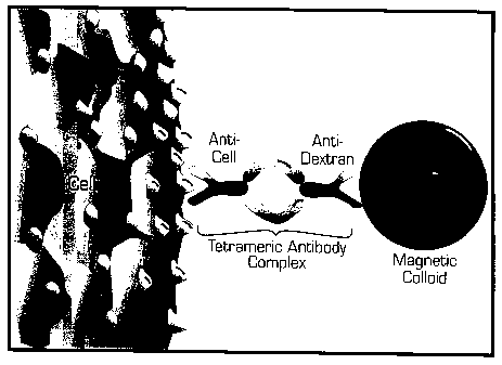
Figure 1 is a schematic drawing showing Magnetic Labelling
of Human Cells: Cells are cross-linked to magnetic particles using
tetrameric antibody complexes comprised of two murine IgG1 monoclonal
antibodies held in tetrameric array by two rat anti-mouse IgG 1 monoclonal
antibody molecules. One murine antibody molecule recognizes the cell
surface antigen and the other recognizes the dextran on the magnetic
particle.
Figure 2 is a schematic drawing showing the cell separation
procedure for CML samples.
Figure 3 shows FACS dotplots of CML bone marrow before
and after processing with the standard lineage depletion cocktail and the
CML debulking cocktail. Both the side and forward scatter of the cells are
shown (Figures 3B, 3D and 3F), and the cells were stained with anti C1334-
PE and anti CD15-FITC (Figures 3A, 3C and 3E).
DETAILED DESCRIPTION OF THE INVENTION
1. ANTIBODY COMPOSITION
In one embodiment, the present invention relates to an
antibody composition comprising antibodies specific for the antigens
glycophorin A, CD2, CD3, CD14, CD15, CD16, CD19, CD24, CD56, CD66b and
IgE which are present on the surface of differentiated or lineage committed
cells.
In another embodiment, the present invention relates to an
antibody composition comprising antibodies specific for the antigens
glycophorin A, CD2, CD3, CD14, CD16, CD19, CD24, CD56, CD66b and IgE
which are present on the surface of differentiated or lineage committed
cells.
CA 02279474 2009-06-02
-5-
Within the context of the present invention, antibodies are
understood to include monoclonal antibodies and polyclonal antibodies,
antibody fragments (e.g., Fab, and F(ab')2) and recombinantly produced
binding partners.
Polyclonal antibodies against selected antigens on the surface
of human cells may be readily generated by one of ordinary skill in the art
from a variety of warm-blooded animals such as horses, cows, various
fowl, rabbits, mice, or rats.
Preferably, monoclonal antibodies are used in the antibody
compositions of the invention. Monoclonal antibodies specific for selected
antigens
on the surface of human cells may be readily generated using conventional
techniques (see U.S. Pat. Nos. RE 32,011, 4,902,614, 4,543,439, and 4,411,993;
see
also Monoclonal Antibodies, Hybridomas: A New Dimension in Biological
Analyses, Plenum Press, Kennett, McKearn, and Bechtol (eds.), 1980, and
Antibodies: A Laboratory Manual, Harlow and Lane (eds.), Cold Spring Harbor
Laboratory Press, 1988).
Other techniques may also be utilized to construct
monoclonal antibodies (see William D. Huse et al., "Generation of a Large
Combinational Library of the Immunoglobulin Repertoire in Phage
Lambda," Science 246:1275-1281, December 1989; see also L. Sastry et al.,
"Cloning of the Immunological Repertoire in Escherichia coli for
Generation of Monoclonal Catalytic Antibodies: Construction of a Heavy
Chain Variable Region-Specific cDNA Library," Proc Natl. Acad. Sci USA
86:5728-5732, August 1989; see also Michelle Alting-Mees et al.,
"Monoclonal Antibody Expression Libraries: A Rapid Alternative to
Hybridomas," Strategies in Molecular Biology 3:1-9, January 1990; these
references describe a commercial system available from Stratacyte, La Jolla,
California, which enables the production of antibodies through
recombinant techniques).
CA 02279474 2009-06-02
-6-
Similarly, binding partners may be constructed utilizing
recombinant DNA techniques. Within one embodiment, the genes which
encode the variable region from a hybridoma producing a monoclonal
antibody of interest are amplified using nucleotide primers for the variable
region. These primers may be synthesized by one of ordinary skill in the
art, or may be purchased from commercially available sources. The
primers may be utilized to amplify heavy or light chain variable regions,
which may then be inserted into vectors such as ImmunoZAPTM H or
ImmunoZAPTM L (Stratacyte), respectively. These vectors may then be
introduced into E. coli for expression. Utilizing these techniques, large
amounts of a single-chain protein containing a fusion of the VH and VL
domains may be produced (See Bird et al., Science 242:423-426, 1988). In
addition, such techniques may be utilized to change a "murine" antibody
to a "human" antibody, without altering the binding specificity of the
antibody.
Antibodies against selected antigens on the surface of
differentiated or lineage committed cells may also be obtained from
commercial sources.
Antibodies may be selected for use in the antibody compositions of
the invention based on their ability to deplete targeted differentiated cells
and
recover non-targeted cells (i.e. progenitor and stem cells, or specific
differentiated
cells) in magnetic cell separations as more particularly described herein, and
in co-
pending U.S. patent application Serial Nos. 08/566,295 and 09/088,227, and
U.S.
Patent Nos. 5,514,340 and 5,877,299. In general, an antibody is selected that
gives
approximately a 3 log depletion of the target cell, with greater than 75%
recovery of
CD34+ cells (bone marrow, mobilized blood and cord blood) or non-targeted
lymphocytes (steady state blood), in test magnetic cell separations as
described
herein.
The anti-glycophorin A antibodies contained in the antibody
composition of the invention are used to label erythrocytes. Examples of
monoclonal antibodies specific for glycophorin A are 2B7.1 (StemCell
CA 02279474 1999-07-30
-7-
Technologies) 1OF7MN (U.S. Patent No. 4,752,582, Cell lines: ATCC
accession numbers HB-8473, HB-8474, and HB-8476), and D2.10
(Immunotech, Marseille, France). The concentration of anti-glycophorin
A antibodies used in the antibody composition are generally less than the
concentration that will cause agglutination (i.e. 3-10 g/ml). Preferably the
concentration of anti-glycophorin A antibodies used in the antibody
composition is between about 0.5 to 5 g/ml, preferably 1 to 2 g/ml.
The antibodies against CD15 are used to label mature myeloid
cells. Examples of monoclonal antibodies specific for CD15 include DU
HL60-3 (Sigma, Saint Louis, MS) MMA (Becton Dickinson, Mountain
View, California), H198 (Pharmingen, San Diego, California) and 80H5
(Immunotech, Marseille, France). The concentration of CD15 antibodies
used in the antibody composition is usually 3 gg/ml. Preferably the
concentration of CD15 antibodies used in the antibody composition is
between about 1 to 3 g/ml preferably 3 gg/ml.
The antibodies against CD2, CD3, CD19, CD24 and CD56 in the
antibody composition of the invention are used to label B and T-
lymphocytes and NK cells. Examples of monoclonal antibodies specific for
CD2, CD3, CD19, CD24 and CD56 are 6F10.3 (Immunotech, Marseille,
France) SK7 (Becton Dickinson) L1CHT1 (Immunotech, Marseille, France)
and 4G7 (Beckon Dickinson, Mountain View, California), 32D12 (Dr.
Steinar Funderud, Institute for Cancer Research, Department of
Immunology, Oslo, Norway) and ALBS (Immunotech, Marseille, France)
and T199 (Immunotech, Marseille, France) or M431 (Beckon Dickinson,
Mountain View, California). The concentration of each of the monoclonal
antibodies against CD2, CD3, CD19, CD24 and CD56 for an antibody
composition of the invention is about 1 to 3 g/ml, preferably 3 gg/ml for
each antibody concentration, the preferred concentration is 3.0 gg/ml.
The antibodies against CD14, CD16 and CD66b in the antibody
compositions of the invention are used to label monocytes and
granulocytes. Examples of monoclonal antibodies specific for CD14, CD16
CA 02279474 1999-07-30
-8-
and CD66b are MEM15 and MEM18 (Dr. Vaclav Horejsi, Institute of
Molecular Genetics Academy of Sciences of the Czech Republic, Praha,
Czech Republic; Cedarlane Laboratories, Hornby, Ontario, Canada); MEM
154 (Dr. Vaclav Horejsi, Institute of Molecular Genetics Academy of
Sciences of the Czech Republic, Praha, Czech Republic; Cedarlane
Laboratories, Hornby, Ontario, Canada); and, B13.9 (CLB, Central
Laboratory of the Netherlands, Red Cross Blood Transfusion Service) and
80H3 (Immunotech, Marseille, France), respectively. The concentration of
each of the monoclonal antibodies against CD14, CD16 and CD66b for an
antibody composition of the invention is about 1 to 3 g/ml, preferably 3
gg/ml, except 2 gg for CD16 (MEM 154)
The antibodies to IgE molecules bind IgE antibodies and mast
cells and basophils. Examples of monoclonal antibodies specific for IgE
include 47-18 (Pharmingen, San Diego, California) and E124.2.8
(Immunotech, Marseille, France). Preferably the concentration of anti-IgE
antibodies used in the antibody composition is between about 1 to 3 g/ml,
preferably 3 gg/ml.
II. PROCESSES FOR PREPARING CELL PREPARATIONS
The antibody composition of the invention may be used to
prepare cell preparations from patients with chronic myeloid leukemia
(CML) that are depleted of matured differentiated or lineage committee
cells and can withstand freezing. Preferably, the antibody composition can
be used on blood or bone marrow samples from patients with CML. The
negative selection method of the invention is advantageous because the
desired stem cells and progenitor cells that are recovered in the method
are not labelled or coated with antibodies. In addition, additional
processing steps such as positive selection protocols are not required in
order to recover a cell preparation enriched in stem cells and progenitor
cells but depleted of lineage committed or differentiated cells.
CA 02279474 1999-07-30
-9-
Accordingly, the present invention provides a negative
selection method for depleting differentiated or lineage committed cells
from a sample from a patient with chronic myeloid leukemia comprising:
(a) reacting the sample with an antibody composition
comprising antibodies specific for glycophorin A, CD2, CD3, CD14, CD15,
CD16, CD19, CD24, CD56, CD66b and IgE under conditions so that
conjugates form between the antibodies and cells in the sample having the
antigens glycophorin A, CD2, CD3, CD14, CD15, CD16, CD19, CD24, CD56,
CD66b and IgE on their surfaces;
(b) removing the conjugates; and
(c) recovering a cell preparation which is depleted of lineage
committed cells.
Preferably, the present invention provides a method for
depleting differentiated or lineage committed cells from a sample from a
patient with chronic myeloid leukemia comprising:
(a) reacting the sample with an antibody specific for CD15
under conditions so that conjugates form between the antibodies and cells
in the sample having the antigen CD15 on their surfaces;
(b) reacting the sample from step (a) with an antibody
composition comprising antibodies specific for glycophorin A, CD2, CD3,
CD14, CD16, CD19, CD24, CD56, CD66b and IgE under conditions so that
conjugates form between the antibodies and the cells in the sample having
the antigens glycophorin A, CD2, CD3, CD14, CD16, CD19, CD24, CD56,
CD66b and IgE on their surfaces;
(c) removing the conjugates; and
(d) recovering a cell preparation which is depleted of lineage
committed cells.
Prior to conducting the above described methods of the
invention the sample may be treated to obtain low density cells from the
sample, for example by density centrifugation.
Conditions which permit the formation of cell conjugates
may be selected having regard to factors such as the nature and amounts of
CA 02279474 1999-07-30
-10-
the antibodies in the antibody composition, and the estimated
concentration of targeted human cells in the sample.
The antibodies in the antibody composition may be labelled
with a marker or they may be conjugated to a matrix. Examples of markers
are biotin, which can be removed by avidin bound to a support, and
fluorochromes, e.g. fluorescein, which provide for separation using
fluorescence activated sorters. Examples of matrices are magnetic beads,
which allow for direct magnetic separation (Kemshead 1992), panning
surfaces e.g. plates, (Lebkowski, J.S, et al., (1994), J. of Cellular
Biochemistry
supple. 18b:58), dense particles for density centrifugation (Van Vlasselaer,
P., Density Adjusted Cell Sorting (DACS), A Novel Method to Remove
Tumor Cells From Peripheral Blood and Bone Marrow StemCell
Transplants. (1995) 3rd International Symposium on Recent Advances in
Hematopoietic Stem Cell Transplantation-Clinical Progress, New
Technologies and Gene Therapy, San Diego, CA), adsorption columns
(Berenson et al. 1986, Journal of Immunological Methods 91:11-19.), and
adsorption membranes (Nordon et al. 1994, Cytometry 16:25-33). The
antibodies may also be joined to a cytotoxic agent such as complement or a
cytotoxin, to lyse or kill the targeted differentiated cells.
The antibodies in the antibody composition may be directly or
indirectly coupled to a matrix. For example, the antibodies in the
composition of the invention may be chemically bound to the surface of
magnetic particles for example, using cyanogen bromide. When the
magnetic particles are reacted with a sample, conjugates will form between
the magnetic particles with bound antibodies specific for antigens on the
surfaces of the differentiated cells, and the differentiated cells having the
antigens on their surfaces.
Alternatively, the antibodies may be indirectly conjugated to
a matrix using antibodies. For example, a matrix may be coated with a
second antibody having specificity for the antibodies in the antibody
composition. By way of example, if the antibodies in the antibody
CA 02279474 2009-06-02
-11-
composition are mouse IgG antibodies, the second antibody may be rabbit
anti-mouse IgG.
The antibodies in the antibody composition may also be
incorporated in antibody reagents which indirectly conjugate to a matrix.
Examples of antibody reagents are bispecific antibodies, tetrameric antibody
complexes, and biotinylated antibodies.
Bispecific antibodies contain a variable region of an antibody
in the antibody composition of the invention, and a variable region
specific for at least one antigen on the surface of a matrix. The bispecific
antibodies may be prepared by forming hybrid hybridomas. The hybrid
hybridomas may be prepared using the procedures known in the art such
as those disclosed in Staerz & Bevan, (1986, PNAS (USA) 83: 1453) and
Staerz & Bevan, (1986, Immunology Today, 7:241). Bispecific antibodies
may also be constructed by chemical means using procedures such as those
described by Staerz et al., (1985, Nature, 314:628) and Perez et al., (1985
Nature 316:354), or by expression of recombinant immunoglobulin gene
constructs.
A tetrameric immunological complex may be prepared by
mixing a first monoclonal antibody which is capable of binding to at least
one antigen on the surface of a matrix, and a second monoclonal antibody
from the antibody composition of the invention. The first and second
monoclonal antibodies are from a first animal species. The first and second
antibodies are reacted with an about equimolar amount of monoclonal
antibodies of a second animal species which are directed against the
Fc-fragments of the antibodies of the first animal species. The first and
second antibodies may also be reacted with an about equimolar amount of
the F(ab') 2 fragments of monoclonal antibodies of a second animal species
which are directed against the Fc-fragments of the antibodies of the first
animal species. (See U.S. Patent No. 4,868,109 to Lansdorp for a description
of
tetrameric antibody complexes and methods for preparing same).
CA 02279474 2009-06-02
-12-
The antibodies of the invention may be biotinylated and
indirectly conjugated to a matrix which is labelled with (strept) avidin. For
example, biotinylated antibodies contained in the antibody composition of
the invention may be used in combination with magnetic iron-dextran
particles that are covalently labelled with (strept) avidin (Miltenyi, S. et
al.,
Cytometry 11:231, 1990). Many alternative indirect ways to specifically
cross-link the antibodies in the antibody composition and matrices would
also be apparent to those skilled in the art.
In an embodiment of the invention, the cell conjugates are
removed by magnetic separation using magnetic particles. Suitable
magnetic particles include particles in ferrofluids and other colloidal
magnetic solutions. "Ferrofluid" refers to a colloidal solution containing
particles consisting of a magnetic core, such as magnetite (Fe304) coated or
embedded in material that prevents the crystals from interacting.
Examples of such materials include proteins, such as ferritin,
polysaccharides, such as dextrans, or synthetic polymers such as sulfonated
polystyrene cross-linked with divinylbenzene. The core portion is
generally too small to hold a permanent magnetic field. The ferrofluids
become magnetized when placed in a magnetic field. Examples of
ferrofluids and methods for preparing them are described by Kemshead
J.T. (1992) in J. Hematotherapy, 1:35-44, at pages 36 to 39, and Ziolo et al.
Science (1994) 257:219. Colloidal particles of dextran-iron complex are
preferably
used in the process of the invention. (See Molday, R. S. and McKenzie, L.L.
FEBS
Lett. 170:232, 1984; Miltenyi et al., Cytometry 11:231, 1990; and Molday, R.S.
and
MacKenzie, D., J. Immunol. Methods 52:353, 1982; Thomas et al., J. Hematother.
2:297 (1993); and U.S. Patent No. 4,452,733).
Figure 1 is a schematic representation of magnetic cell
labeling using tetrameric antibody complexes and colloidal dextran iron.
Cells are cross-linked to magnetic particles using tetrameric antibody
complexes comprised of two murine IgG1 monoclonal antibodies held in
CA 02279474 2009-06-02
-13-
tetrameric array by two rat anti-mouse IgG1 monoclonal antibody
molecules. One murine antibody molecule recognizes the cell surface
antigen and the other recognizes the dextran on the magnetic particle.
In accordance with the magnetic separation method, the
sample containing the progenitor and stem cells to be recovered, is reacted
with the above described antibody reagents, preferably tetrameric antibody
complexes, so that the antibody reagents bind to the targeted differentiated
cells present in the sample to form cell conjugates of the targeted
differentiated cells and the antibody reagents. The reaction conditions are
selected to provide the desired level of binding of the targeted
differentiated cells and the antibody reagents. Preferably the sample is
incubated with the antibody reagents for a period of 5 to 60 minutes at
either 4 C or ambient room temperature. The concentration of the
antibody reagents is selected to optimize cell labeling in a sample of 2-8 x
107 nucleated cells per ml. Generally, the concentration is between about
0.1 to 50 g/ml of sample. The magnetic particles are then added and the
mixture is incubated for a period of about 5 minutes to 30 minutes at the
selected temperature. The sample is then ready to be separated over a
magnetic filter device. Preferably, the magnetic separation procedure is
carried out using the magnetic filter and methods described in co-pending
U.S. Patent No. 5,514,340 to Lansdorp and Thomas.
The sample containing the magnetically labelled cell
conjugates is passed through the magnetic filter in the presence of a
magnetic field. In a preferred embodiment of the invention, the magnet is
a permanent gap magnet with 0.5-2.0" diameter bore and having a
magnetic field of 0.5-2 Tesla. The magnetically labelled cell conjugates are
retained in the high gradient magnetic column and the materials which
are not magnetically labelled flow through the column after washing with
a buffer.
The preparation containing non-magnetically labelled cells
may be analyzed using procedures such as flow cytometry. The ability of
CA 02279474 1999-07-30
-14-
the cells in the preparation to produce colony-forming cells or long term
culture initiating cells (LTCIC) in culture may also be assessed. The
efficiency of the separation procedure may also be determined by
monitoring the recovery of CD34+ cells, CD34+ CD38- cells and colony
forming cells.
III. USES
Methods and compositions of the invention may be used in
processing samples from patients with chronic myeloid leukemia
including samples of blood or bone marrow. It has been known for over 2
decades that the maturing leukemic myeloid cells in CML are lighter than
their normal counterparts (Moore, MAS, et al., (1973), J. Natl. Cancer Inst.
50:603). Hence they are more prevalent in the low density fraction of cells
obtained using standard commercial media that efficiently separate
normal red cells and granulocytes. In addition, blood and marrow samples
from many CML patients contain elevated numbers of basophils and their
precursors as part of their increased granulopoiesis. Such myeloid cells do
not survive freezing/ thawing and debris from their lysis hampers the
recovery of other cells in the sample. When a blood or bone marrow
sample from a patient with chronic myeloid leukemia is frozen and then
thawed generally only 2% of CD34 cells are recovered. Approximately 10%
of colony forming cells (CFC) are recovered. However, when the sample is
first processed using the antibody composition of the invention, the
inventors have shown that there is 60% recovery of CD34+ cells and CFC.
This is advantageous as it permits the storage of samples from chronic
myeloid leukemia patients allowing for further study of the disease and
the cells involved in the disease.
IV. KIT
The present invention also relates to a kit containing the
antibody composition of the composition of the invention for use in
making cell preparations from patients with chronic myeloid leukemia
which are depleted of differentiated or lineage committed cells. The kit
includes instructions for performing the process of depleting cells from
CA 02279474 2009-06-02
-15-
samples from such patients as well as antibodies specific for glycophorin A,
CD2, CD3, CD14, CD15, CD16, CD19, CD24, CD56, CD66b and IgE and
reagents helpful in carrying out the process of the invention. Also
optionally included are containers and other materials appropriate for
conducting the process of the invention.
The following examples provide illustrations of the present
invention and in no way serve to narrow the scope of the claims.
EXAMPLES
Example 1
Lineage committed cells were depleted from samples of blood
(Tables 1A and 1B) and bone marrow (Tables 2A and 2B) from CML
patients by treating the sample first with antibodies to CD15 and then with
tetrameric antibody complexes recognizing CD2, CD3, CD14, CD16, CD19,
CD24, CD56, CD66b, IgE, glycophorin A and biotin. The combination of
antibodies to CD15 and the tetrameric antibody complexes is referred to
herein as "the CML debulking cocktail". Low density cells were obtained
TM
using Ficoll density centrifugation. The cells were then washed twice with
phosphate buffered saline (PBS), resuspended at 5 x 10⁷ cell/mL in PBS
plus 2% fetal calf serum (FCS) and incubated for 30 minutes on ice with 3
gg/mL biotinylated anti-CD15. After a single wash with PBS + 2% FCS the
cells were resuspended again at 5 x 10⁷/mL and incubated for 30 minutes
on ice with the remainder of the CML debulking cocktail (tetrameric
complexes recognizing CD2, CD3, CD14, CD16, CD19, CD24, CD56, CD66b,
IgE, glycophorin A and biotin). Colloidal magnetic dextran iron particles
were added to the cells, the cells incubated for an additional 30 minutes
and then passed through a magnetic column. Figure 2 is a schematic
drawing showing the cell separation procedure for CML samples. The cells
collected in the column flow through were depleted of mature lineage
committed cells.
Removal of mature cells enriches for immature CD34+ cells
and progenitors which have the potential to form hematopoietic colonies
CA 02279474 1999-07-30
-16-
in semi-solid medium (Colony Forming Cells - CFC). The purity of CD34+
cells obtained following processing CML samples with the CML debulking
cocktail ranged from 54-79% for blood samples and 36-80% for bone
marrow samples. The fold enrichment of CD34+ cells depends on the
frequency of CD34+ cells in the start cell suspension which varies greatly
(0.3%-11% for these samples). The recovery of CD34+ cells and CFC also
varies greatly. This was due to abnormal co-expression of mature lineage
markers and CD34+ on CML cells and the ability of some CML cells which
express lineage markers to form colonies in culture (see later discussion).
More primitive hematopoietic progenitors can be assayed by the potential
for colony formation in semi-solid medium after 6 weeks of culture in
liquid long-term culture medium. The frequency and recovery of these
primitive cells (week 6 CFC) following processing with the CML debulking
cocktail (method outlined above) was determined for one CML blood and
one CML bone marrow sample (Table 2C). These primitive hematopoietic
progenitors were highly enriched with essentially 100% recovery.
Example 2
Lineage committed cells were depleted from samples of blood
and bone marrow from CML patients using a standard lineage depletion
cocktail of antibodies (CD2, CD3, CD14, CD16, CD19, CD24, CD56, CD66b and
glycophorin-A) designed to enrich for hematopoietic progenitors from
normal peripheral blood and bone marrow and the CML debulking
cocktail (which further includes antibodies to CD15 and IgE) as described in
Example 1. Direct comparisons of these 2 cocktails in processing CML
samples show the CML debulking cocktail provides slightly higher purities
and enrichments of CD34+ cells (p<0.05) and did not compromise the
recovery of CD34+ cells (Table 3A). Both cocktails offered similar
enrichment of Colony Forming Cells (CFC) (Table 3B).
Example 3
The recovery of CD34+ cells and CFC from CML samples
following freezing and thawing was tested with and without processing
with the CML debulking cocktail. Samples of low density cells from CML
CA 02279474 1999-07-30
-17-
patients were divided in two. Half the cells were directly frozen and the
other half were processed with the CML debulking cocktail (method
outlined in Example 1) and then frozen. The recovery of CD34+ cells and
CFC were assessed. Percent recoveries were calculated relative to the same
original starting value in the fresh low density cell population. Thus in
the case of the cells processed with the CML debulking cocktail the
calculated recoveries include losses due to lineage depletion as well as
freezing and thawing. The recoveries of both CD34+ cells and CFC were
improved several-fold (p<0.85) (mean 36 fold increase in recovery of
CD34+ cells and mean 7 fold increase in recovery of CFC) by prior
processing with the CML debulking cocktail according to Example 1 (Table
4).
Summary of Results
Representative FACS dotplots from a CML patient sample in
which the percentage of CD34+ cells in the input fraction was only 1.7% are
shown in Figure 3A. The CD34+ population is discreet whereas the CD15+
population is large and represents granulocytes, basophils and less mature
myeloid cells which are present in CML (See Figure 3B high SSC). After
separation using the standard lineage depletion cocktail (Figure 3C) there
is enrichment of the CD34+ population, the resulting purity having
increased to 28% (a 16 fold increase from the start sample). However, a
large distinct population of CD34- cells remained which following staining
was confirmed to be partially CD15+ cells (Figure 3C). When the CML
Debulking cocktail described in Example 1 (which contains two extra
antibodies against CD15 and IgE) was used on the same start material, the
numbers of residual cells expressing CD15 diminished leading to a greater
overall purity and enrichment of CD34+ cells (Figure 3E) and to a less
heterogenous side scatter profile (Figure 3F). The frequency of CD15+ cells
in the start fraction was 87%. This decreased to 21% following separation
with the standard lineage depletion cocktail, but where the CML
Debulking cocktail of Example 1 was used, this was reduced further to
2.8%.
CA 02279474 2009-06-02
-18-
The comparative recoveries of CD34+ cells and CFC was
assessed following freezing and thawing of CML patient samples which
had either not been processed or which had been processed with the CML
Debulking cocktail of Example 1. The results are shown in Table 4. The
percent recovery data shown in this figure includes cell losses occurring in
a cell separation procedure and during the freeze/thaw cycle. Processing
with the CML debulking cocktail increases the recovery of CD34+ cells from
1.7 to 61% and recovery of CFC from 8.3 to 58%. Use of the CML debulking
cocktail has made cryopreservation of CML samples a viable option for
researchers studying the biology of CML.
While what is shown and described herein constitutes various
preferred embodiments of the subject invention, it will be understood that
various
changes can be made to such embodiments without departing from the subject
invention, the scope of which is defined in the appended claims.
CA 02279474 1999-07-30
-19-
DETAILED REFERENCES
Bakhshi A, Minowada J, Arnold A, Cossman J, Jensen JP, Whang-Peng J,
Waldmann, TA and Korsmeyer, SJ: Lymphoid blast crises of chronic
myelogenous leukemia represent stages in the development of B-cell
precursors. New England Journal of Medicine 309:826, 1983
Barnett MJ, Eaves CJ, Phillips GL, Kalousek DK, Klingemann HG,
Lansdorp TM, Reece CE, Shepherd JD, Shaw GJ, and Eaves AC: Successful
autografting in chronic myeloid leukemia after maintenance of marrow in
culture. Bone Marrow Transplant. 4:345, 1989.
Bartram CR, de Klein A, Hagemeijer A, van Agthoven T, van Kessel AG,
Bootsma D, Grosveld G, Ferguson-Smith MA, Davies T, Stone M,
Heisterkamp N, Stephenson JR and Groffen J: Translocation of c-abl
oncogene correlates with the presence of a Philadelphia chromosome in
chronic myelocytic leukemia. Nature 306:277, 1983.
Coutinho LH, Dexter TM, Harrison C, Morgenstern G, Chang J, Testa NG:
The use of cultured bone marrow cells in autologous transplantation.
Progress in Clinical and Biological Research 333:415, 1990.
Daley GQ, Goldman JM: Autologous transplant for CML revisited. Exp.
Hematol. 21:734, 1993.
Degliantoni G, Rizzoli V, Mangoni L: In vitro restoration of polyclonal
hematopoiesis in a chronic myelogenous leukemia after in vitro
treatment with 4-hydroperoxycyclophosphamide. Blood 65:753, 1985.
Deisseroth AB, Zu Z, Claxton D, Hanania EG, Fu S, Ellerson D, Goldberg L,
Thomas M, Janieck K, Anderson WF, Hester J, Korbling M, Durett A,
Moen R, Berenson R, Heinfeld S, Hamer J, Calvert L, Tibbits P, Talpaz M,
CA 02279474 1999-07-30
-20-
Kantarjian H, Champlin R and Reading C: Genetic marking shows that
Ph+ cells present in autologous transplants of chronic myelogenous
leukemia (CML) contribute to relapse after autologous bone marrow in
CML. Blood 83:3068, 1994.
Fialkow PJ, Jacobson RJ, Papayannopoulou T: Chronic myelocytic
leukemia: clonal origin in a stem cell common to the granulocyte,
erythrocyte, platelet and monocyte/macrophage. American Journal of
Medicine 63:125, 1977.
Kemshead, J.T., J. Hematotherapy, 1:35-44, 1992.
Moore MAS, Williams N and Metcalf D: In vitro colony formation by
normal and leukemic human hematopoietic cells: Characterization of the
colony forming cell. J. Natl. Cancer Inst., Vol. 50, p. 603, 1973.
Nordon RE, Milthorpe BK, Schindhelm K, and Slowiaczek PR: An
Experimental Model of Affinity Cell Separation. Cytometry 16:25-33, 1994.
Talpaz M, Kantarjian H, Liang J, Calvert L, Harter J, Tibbits P, Durett A,
Claxton D, Ciralt S, Khari I, Przepiorka D, van Besien K, Andersson B,
Mehra R, Gajewski J, Scong D, Hester J, Estay E, Korbling M, Pollicardo N,
Berenson R, Hamfeld S, Charuplin R and Deisseroth AB: Percentage of
Philadelphia chromosome (Ph) -negative and ph-positive cells found after
autologous transplantation for chronic myelogous leukemia depends on
percentage of diploid cells induced by conventional-dose chemotherapy
before collection of autologous cells. Blood 85:3257, 1995.
Verfaillie CM, Bhatia R, Miller W, Mortari F, Roy V, Burger S,
McCullough J, Stieglbauer K, Dewald G, Heimfeld S, Miller JS, McGlave
PB: BCR/ABL-negative primitive progenitors suitable for transplantation
can be selected from the marrow of most early-chronic phase but not
CA 02279474 1999-07-30
-21-
accelerated-phase chronic myelogenous leukemia patients. Blood 87:4770,
1996.
CA 02279474 1999-07-30
-22-
TABLE 1A
The Purity and Recovery of CD34+ cells in the CML blood samples before
and after processing with CML Debulking cocktail.
Sample number % CD34+ cells %CD34+ cells Fold % Recovery of
in CML blood following CML Enrichment CD34+ cells
Debulking
1 11 56 5 12
2 4.7 54 12 3.4
3 1.8 79 45 23
Mean 5.8 63 21 13
TABLE 1B
The Frequence and Recovery of CFC in the CML blood sample before and
after processing with CML Debulking cocktail.
Sample number Frequence of Frequency of CFC Fold % Recovery of
CFC in CML following CML Enrichment CFC
blood Debulking
1 1:19 1:6.8 2.9 6.7
2 1:133 1:4.4 30 8.9
3 1:127 1:1.6 79 41
Mean 1:93 1:4.2 38 19
CA 02279474 1999-07-30
-23-
TABLE 2A
The Purity and Recovery of CD34+ cells in CML bone marrow samples
before and after processing with the CML Debulking cocktail.
Sample number % CD34+ cells %CD34+ cells Fold Recovery of CD34+
in CML bone following CML Enrichment cells
marrow Debulking
1 4.6 80 17 40.7
2 1.7 36 21 63.5
3 0.3 55 161 100
Mean 2.2 57 66 68.1
TABLE 2B
The Frequency and Recovery of CFC in CML bone marrow samples before
and after processing with the CML Debulking cocktail.
Sam p 1 e Frequence of Frequency of CFC Fold Recovery of
number CFC in CML following CML Enrichment CFC
bone marrow Debulking
1 1:62 1:2.8 23 53
2 1:204 1:6.2 33 61
3 1:47 1:5.7 8.6 9.1
Mean 1:104 1:4.7 21 57
CA 02279474 1999-07-30
-24-
TABLE 2C
The Frequency and Recovery of Week 6 CFC in CML blood and bone
marrow samples before and after processing with the CML debulking
cocktail.
Week 6 CFC: Week 6 CFC: Fold Enrichment % Recovery of
Nucleated Cells Nucleated Cells of Week 6 CFC Week 6 CFC
in Start Following CML
De-Bulking
1 1:56980 1:11 1398 100
2 1:948 1:41 87 94
(Sample 1, Peripheral Blood: Sample 2, Bone Marrow)
CA 02279474 1999-07-30
-25-
TABLE 3A
Comparison of the purity, enrichment and yield of CD34+ cells obtained
from CML low density blood or marrow samples using two different
antibody cocktails to remove mature cells.
CML Purity (%) Enrichment (Fold) Recovery (%)
Sample No.
Standard CML Standard CML Standard CML
Cocktail Debulking Cocktail Debulking Cocktail Debulking
Cocktail Cocktail Cocktail
1 32 36 23 26 67 47
2 36 55 22 33 19 35
3 39 54 27 37 16 11
4 64 80 14 17 45 40
Mean SEM 43 7 56 9* 22++3 28 4* 37 12 33 8**
* 0.05>p>0.01 (paired t-test) compared to values for the standard Ab
cocktail.
** p>0.05 (paired t-test) compared to values for the standard Ab cocktail.
TABLE 3B
Comparison of the frequency of CFC obtained from CML low density blood
or bone marrow samples using two different antibody cocktails to remove
mature cells.
Sample Number Standard Cocktail CML Debulking
Cocktail
1 1:3.4 1:2.8
2 1:6.2 1:6.2
3 1:5.7 1:5.7
4 1:19.4 1:14.2
Mean 1:8.7 1:7.2
CA 02279474 1999-07-30
-26-
TABLE 4
Percent recovery of CD34+ cells and CFC after thawing ficolled low density
cells or cells processed with the CML debulking cocktail
Sample No. CD34+ CFC
Low Density CML Low Density CML
Cells Debulking Cells Debulking
Cocktail Cocktail
1 0.8 12 6 18
2 2.5 29 32 72
3 1.2 44 1.7 60
4 0.3 102 1.4 123
5 3.7 118 0.4 15
Mean SEM 1.7 0.6 61 21* 8.3 6.0 58 20*
Recovery values are expressed as a percent of the total number of cells of
the type assessed present in the correpsonding low density or lin-
population prior to cryopreservation.
* 0.05>p>0.01 (paired t-test) compared to values for low density cells.