Note: Descriptions are shown in the official language in which they were submitted.
CA 02279946 1999-08-09
MEDIATION OF CYTOKINE:S BY MELANIN
1Ø FIELD OF THE INVENTION
Methods and compositions are described for the use of purified melanin to
treat disease in
animals and man. The disclosed melanin compositions are particularly useful
for regulating
cytokine production by mammalian and human cells both in vitro and in vivo.
2Ø 13ACKGROUND OF THE INVENTION
Tumor necrosis factor-alpha (TNF-«) is a 17-kd polypeptide released primarily
by
macrophages. Generally, TNF-~« is not present in measurable quantity in sera
from healthy
individuals; but appears rapidly in response to immunostimulators (Beutler and
Cerami, Adv.
Immunol. 42:213-232, 1988). At physiological concentrations, TNF-« limits the
growth and
spread of invasive pathogens. However, excessive or uncontrolled production of
this cytokine
contributes to the pathogenesis of number of disease conditions.
TNF-«, acting alone and/or in concert with other mediators, evokes a
potentially fatal
syndrome of irreversible cardiovascular collapse (shock) and critical organ
failure (Beutler,
Science 229:869-871, 1985; Tracy et al., Nature 330:662-664, 1987; Waage et
al., Lancet 1:355-
357, 1987). Additionally, TNF-~a acting in concert or in synergy with the
interleukins IL-l and
IL-6 contributes to the developnnent of wasting syndrome (cachexia) (Grunfeld,
et al., Am. J.
Clin: Nutr. 55:455-460, 1992; Grunfeld and Feingold, N. Engl. J. Med. 327:329-
337, 1992).
For example, the experimental administration of supernatants from endotoxin-
stimulated
macrophages produced severe weight loss in rodents (Cerami et al., Immunol.
Lett. 11:173-177,
1985). Moreover, nude mice implanted with genetically engineered tumor cells
that secreted
either TNF-
1
,AMENDED SNEET
CA 02279946 1999-08-09
« (Rouzer and Cerami, ~l~fol. Biochem. I'arasitol. 2:31-38, 1980) or IL-6
(Black et al.,
Endocrinology 128:267-269, 1991) became progressively anorectic and wasted.
Administering TNF-«, IL-1, aJ-td IL-6 increases plasma triglycerides in
rodents by boosting
hepatic lipogenesis and very-low-density lipoprotein production leading to
futile cycling of fatty
acid/triglyceride -and eventually wasting (Feingold and Grunfeld, J. Clin.
Invest. 80:184-190,
1987; Grunfeld et al., Cancer Res. 50:4233-4238, 1990; Feingold, et al.,
Arterioscler-. Thromb.
Yasc. Biol. 11:495-500, 1991).
Cytokines also boost the levels of key catabolic hormones; alter glucose and
amino acid
metabolism; and have profound effect on food intake. In experimental animals,
TNF-«
decreases gastric motility and consequently leads to retention of food (Patton
et al., J. Clin.
Invest. 80:1587-1596, 1987), artd IL-1 induces continuous anorexia by
indirectly affecting the
hypothalamic appetite center (I-Iellerstein et al., J. Clin. Invest. 84:228-
235, 1989). In humans,
wasting syndrome is often associated with cancer and a variety of infectious
diseases including,
but not limited to tuberculosis and AIDS.
In addition to progressive weight loss, many patients experience anorexia
(reduced
appetite), nausea, muscle weakness, and anemia (Lawson et al., Ann. Rev. Nutr.
2:277-301
(1982); Grunfeld and Feingold, New Engl. J. Med. 327:329-337, 1992). Although
cachexia may
involve anorexia, usually the degree of lean body mass lost in cachexia
associated with cancer
and infectious disease cannot be explained by reduced caloric intake
(Spiegelman and
Hotamisligil, Ce1173:625-627, :1993).
Cachexia is considered a.s a detrimental end point because, apart from
directly effecting
patient survival, the progressive weight loss and anemia usually restrict the
ability of cachectic
patients to tolerate aggressive therapy (Decays et al., Am. J. Med. 69:491-
497, 1980).
The prevalence of cache:~cia makes this syndrome a significant
2
AMENDED SH~~,r
CA 02279946 1999-08-09
medical problem. Taken together, these results provide a mechanistic basis for
considering the
use of melanin, an agent that interferes with the synthesis/release of IL-l,
IL-6 and TVF'- «, for
managing wasting in patients.
TNF-« can also induce adult respiratory distress syndrome CARDS), a severe
consequence of gram-negative sepsis in humans (Shasby et al., In:
Pathophysiology of
Endotoxin, J.B. Hinshaw, ed. A,msterdam: Elsevier Science Publishers, pp. 105-
128, 1980.
TNF-a concentrations in excess of 12,000 pg/ml were detected in pulmonary
aspirates from
ARDS patients (Millar et al., Lancet 2: 712-714, 1989). This cytokine is also
known to increase
the adherence of polymorphonuclear leukocytes to endothelial cells (Gamble et
al., Proc. Natl.
Acad. Sci., USA 82:8667-8671, 1985). Increased adherence of activated
granulocytes in the
microvasculature of the lungs and upper respiratory tract is one of the major
causes of pulmonary
vascular injury in ARDS. Of note, expression of intracellular adhesion
molecule (ICAM), and
endothelial leukocyte adhesion molecule (ELAM) on endothelial cells is either
induced or
enhanced by cytokines -such as 'INF-« or IL-1 (Munre et al., Am. J. Pathol.
135:121-132, 1989),
a phenomenon which results in the augmentation of cell binding.
TNF-«, IL-1 and IL-6 also play a major role in the pathology of rheumatoid
arthritis
(Saklatvala., _Nature 322:547-549, 1986; Miossec. Clin. Rheumatol. 5:305-308,
1987; Lupia et
al. Eur. J. Immunol., 26: 1690-1694, 1996). Synovial fluids from patients with
rheumatoid
arthritis contain TNF-« (Same et al., Arthritis Rheumatism 31:1041-1132, 1988)
and IL-6
(Guerne et al., J. Clin. Invest. 8.~:585-592, 1989). Current evidence suggests
that immune
complexes may stimulate monoc;ytes -to secrete TNF-« (Vissers et al., Am. J.
Pathol. 134:1-6,
1989) and IL-1 (Chantry et al., I?ur. J. Irr~munol. 19:189-192, 1989). TNF-«
and IL-1 in turn
stimulates production of proteases and prostaglandins by synoviocytes and bone
resorption by
osteoclasts
3
AMENDED SHEET
CA 02279946 1999-08-09
, ~ - '.
- : _.
, . . .. . ":
(Miossec, Clin. Rheumatol. 5:305-308, 1987; Dayer et al., J. E.rp. Med.
162:2163-2168, 1985;
Saklatvala, Natacre 322:547-54!x, 1986). Moreover, the presence of TNF-« and
IL-1 in
rheumatoid joints may act together to perpetuate synovitis by stimulating IL-6
synthesis which, if
found in close proximity to plasma cells, may lead to autoantibody production.
IL-6 is
spontaneously produced by synoviocytes and high levels of IL-6 are present in
synovial fluids
from patients with inflammatory arthropathies (Guerne et al., J. Clin. Invest.
83:85-592, 1989).
Cerebral malaria is a lethal hyperacute neurological syndrome and prognosis of
some
malaria patients which has been associated with threshold levels of serum TNF-
« (Grau et al.,
Science 237:1210-1212, 1987; Mark et al. Am. J. Pathol., 129:192-199, 1987;
Grau et al., New
Engl. J. Med 320:1586-1591, 1089; Kwiatkowski et al., Lancet 336:1201-1204,
1990; McGuire
et al. Nature 371: 508-511, 1994). Similarly, in Graft versus Host Reactions,
increases in
TNF-« concentration have been associated with major complications (Holler et
al., Blood
75:1011-1016, 1990).
TNF-« alone, or in synergy with either IL-1 or IL-6, enhances replication of
HIV-1 in
latently infected T cells and monocytes (:Folks et al., Science 238:800-802,
1987; Folk et al.,
Proc. Natl. Acad. Sci. USA 86:2:365-2368, 1989; Poli et al., J. Exp. Med.
172:151-158, 1990;
Poli et al., Proc. Natl. Acad. Sci. USA 91:108-112, 1994). TNF-« is a strong
inducer of NF-e>3,
a transcriptional factor used by HIV (Nobel and Baltimore, N. Engl. J. Med.
234:308-317, 1987;
Duh et al., Proc. Natl. Acad. Sci. USA 86:5974-5978, 1989). Moreover,
synthesis of TNF-«, IL-
1, and IL- -6 are upregulated as a consequence of HIV infection (Folks et al.,
Science 238:800-
802, 1987; Nakajima et al., J. Im~munol. 142:531-536, 1989). Serum and
cerebrospinal fluid of
patients with AIDS contain increased levels of TNF-«, IL-1, and IL-6
(Lahdevirta et al., Am. J.
Med. 85:289-291, 1988; Emille Eat al., J. (:lin. Invest. 86:148-159, 1990;
Breen et
4
AMENDED SH~~T
CA 02279946 1999-08-09
al., J. Immunol. 144:480-484, 1990).
The apoptotic neuronal loss occurring in HIV-1 encephalitis is associated with
TNF-x
(DeSimone et al., Immunol. Today 17:256-258, 1996). In addition, TiVF-x has
been implicated
in AIDS associated cachexia (Wright et al., J. Immz~nol. 141:99-104, 1988).
Therefore, the
down-regulation of abnormal cytokine production by monocytes, and particularly
the down-
regulation of TNF-« is expected to retard the progression of HIV infection and
provide
supportive care for cachexic patients.
IL-6 is also an autocrinc: growth factor for cells derived from Kaposi sarcoma
(KS)
lesions of patients with AIDS (Miles et al., Proc. Natl. Acad. Sci. 87:4068-
4072, 1990). KS, a
multifocal vascular lesion, is also seen in other immunosuppressed states such
as in patients
receiving renal or cardiac transplants (Gange, R. and Jones,E. Clin. Erp.
Dermatol. 3:135-146,
1978; Greenfield et al., .l. Rheumatol. 1 x:637-640, 1986). AIDS-KS-derived
cell lines contain
and secrete substantial amounts of IL-6 and AIDS-KS growth-enhancing effects
of tat protein are
mediated by increased IL-6 production. Indeed, addition of IL-6 antisense
oligodeoxynucleotides to these cells resulted in decreased IL-6 production as
well as marked
inhibition of their growth (Miles et al., Proc. Natl. Acad. Sci. 87:4068-4072,
1990).
AIDS-KS derived cells produce other cytokines including IL-1 (Marx, Science
248:442-
443, 1990) Addition of anti-IL-1 antibody to KS cell lines also resulted in
decreased cellular
proliferation. The increased levels of serum IL-6 and polyclonal B cell
activation may be
associated with increased frequewcy of B cell malignancies seen in AIDS
patients (Akira and
Kishimoto, Immunol. Rev. 127:2 5-50, 1992).
Like KS, the existence o:f an IL-6-IL-6-receptor autocrine loop has been
implicated in the
pathogenesis of multiple myelorna (Kawano et al., Nature 332:83-85, 1988).
Elevated levels of
IL-6 have been observed in other pathological conditions such as mesangial
proliferative
glomerulonephritis
~~,~Fr~~~~ s~E~T
CA 02279946 1999-08-09
.. ,.
. ; ; ;
. . . . .. W...
.. , .. ..
(Horn et al., J. Immunol. 143: 3949-3955, 1989), and psoriasis (Grossman et
al., Proc. Natl.
Acad. Sci. 86:6367-6371, 1989).
A wide variety agents have been used to combat inflammation and life-
threatening
aspects of cytokines. Anti-TIVh-« antibody, the TNF-« receptor, anti-IL-6, and
IL-1 receptor
antagonist (IL-1Ra) therapy were shown to reduce death after acute systemic
toxicity (e.g., septic
shock) in experimental animals (Beutler et al., Science 229:869-871, 1985;
Tracy et al., Narure
330:662-664, 1987; Ohlsson et al., Nature 348:550-552, 1990; Starnes et al.,
J. Immunol.
145:4185-4191, 1990; Ashkenazi, et al., Proc. Natl. Acad. Sci. USA 88:10535-
10539, 1991;
Lesslaner et al., Eur. J. Immunol. 21:2883-2886, 1991 ). However, the response
to these cytokine
blockers depended on the prophylactic administration of the agent, or the site
of infection (Bagby
et al., .I. InJ'ect. Dis. 163:83-88, 1991). Moreover, in a number of studies,
anti-cytokine
antibodies only partially protected the animals (Feingold et al., J. Clin.
Invest. 83:1116-1121,
1989).
Data from several studies indicated that blockade of cytokines by infusion of
either anti-
TNF-« (Elliott et al., Arthritis Rlieum. 36:1681-1690, 1993; Elliott et al.,
Lancet 344:1105-1110,
1994), or anti-IL-6 (Wendling et al., J. Rheumatol. 20:259-262) monoclonal
antibody, as well as
soluble TNF-« receptors (Moreland et al., Arthritis Rheum. 37:S295, 1994), or
soluble IL-1
receptor (Drevlow et al., Arthritis Rheum. 37:5339, 1994) is effective in the
treatment of
rheumatoid arthritis. However, use of soluble cytokine receptors or antibodies
to a single factor
is constrained by the presence of multiple cytokines that participate in the
manifestation of
inflammatory conditions. Moreover, the large-scale treatment with anticytokine
antibody may
lead to production of anti-idiotypi:c antibodies.
Agents such as dexamethasone (Luce et al., Am. Rev. Respir. Dis. 13 8:62-68,
1988),
pentoxifylline (Netea et al., J. Inf act. Dis. 171:393-399, 1995), thalidomide
(Klausner et al., .I.
Acquir. Immun. Defic. Syndr. Hurrn.
6
hi~~~~~u~J ~~i~cT
CA 02279946 1999-08-09
WO 98/34602 PCT/t1S98/02971
Retrovirol. x:247-257, 1996), suramin (Strassman et al., J. Clin. Invest
x:2152-2159, 1993), or a-melanocyte-stimulating hormone (a-MSH) (Chio et
al., J. Clin. Invest. ;2:2038-2044, 1996) have also been used for limiting
synthesis of proinflammatory cytokines. With the exception of a-MSH,
these agents have :limited clinical utility because they are either
ineffective
when given after challenge {dexamethasone and pentoxifylline), do not
target multiple cytokines, or have multiple side effects.
Thalidomide and pentoxifylline inhibit production of TNF-a but not
IL-1p or IL-6 (Sampaio et al., J. Exp. Med. x:699-703, 1991). Because
multiple cytokines contribute to the pathogenesis of inflammatory
disorders, inhibition of a single cytokine may not reverse or prevent the
progression of disE~ase. Thalidomide is teratogenic and has been used in the
past as a sedative and antiemetic, and suramin has considerable toxicity
(Stein, Cancer Res.. 5:2239-2248,1993).
Corticosteroids, the mainstay anti-inflammatory agents, manifest
adverse effects such as susceptibility to infection, suppression of the
hypothalamic-pituitary-adrenal axis, and Cushingoid features. Use of
cydosporine A may result in hypertension and nephrotoxicity.
Melanin, inter alia, is a free radical scavenger that acts as a bacterial
virulence factor by protecting the organism from some host defense
mechanisms (Wang and Casadevail, Infect. lmmun. ~:30C14, 1994).
Additional studies have shown that melanin expression by bacteria may be
a virulence factor that helps bacterial pathogens avoid the afferent phase of
T cell-mediated irnmune responses in the host (Huffnagle et al., J.
1 m m a n o 1. 5~,,,~:35CI7-3516,1995).
3Ø SUMMARY OF THE INVENTION
The present invention is directed to the use of melanin as a
therapeutic agent in animals, including humans. The preferred method of
SUBSTITUTE SHEET (RULE 26)
CA 02279946 1999-08-09
WO 98/34602 PCT/US98I02971
treatment comprises the administration of purified melanin, or
biosynthetic melanin, to an animal in an amount sufficient to alleviate or
prevent an adverse symptom of disease or illness. Accordingly, an object of
the invention is a method of using purified melanin to treat or prevent
illness in a patient which comprises administering melanin to the patient
in an amount sufficient to provide a therapeutic benefit to the patient.
In a preferred embodiment of the present invention, the purified
melanin provides a therapeutic benefit by being administered in an amount
sufficient to modulate the immune response of the patient. In a
particularly preferred embodiment, the purified melanin is administered in
an amount sufficient to be associated with a decrease in host cytokine
production, and in particular TNF-a, IL-1 and IL-6. In general, the decrease
in cytokine production may be either a cause or effect of the beneficial
clinical indications associated with the administration of purified melanins.
~e purified melanins used in the presently described invention may
also be administered in combination with a wide variety of
pharmaceutically useful carriers or excipients. Accordingly, an additional
embodiment of the present invention is the use of pharmaceutical
compositions comprising purified melanin to reduce TNF-a production or
otherwise provide a therapeutic benefit to a patient.
An additional embodiment of the present invention is the use of
highly purified melanins that have a substantially homogeneous structure,
and are substantially free of incorporated contaminating amino acids or
derivatives thereof.
The presently described therapeutic use of purified melanin is
particularly deemed to be useful for the treatment of cachexia, sepsis, acute
respiratory distress syndrome, cerebral malaria, rheumatoid arthritis,
epithelial ulcers of the skin and gut (particularly inflammatory bowel
disease - ulcerative colitis, Crohn's disease, etc.), or other disorders
_g_
SUBSTITUTE SHEET (RULE 26)
CA 02279946 1999-08-09
WO 98/34602 PCT/US98/02971
associated with high levels of TNF-a or other cytokine expression or the
adverse symptoms. associated therewith.
In particular, given that graft rejection is often associated with an
inflammatory response, an additional embodiment of the present
invention is the use of purified melanin, or purified synthetic melanin, to
reduce or prevent the rejection of transplanted organs and grafts. Similarly,
the purified melansns are also deemed to be useful in the treatment and
prevention of graft-versus-host disease.
In view of the above, an additional embodiment of the present
invention is a method of modulating cytokine production by an animal cell
by administering purified melanin to said cell in an amount sufficient to
modulate cytokine production by said cell. In a preferred embodiment, the
purified melanin will have been tested in vitro to verify that compositions
comprising the purified melanin have the property of being capable of
modulating cytoki.ne expression by mammalian or other animal cells.
4Ø DESCRIPTTON OF THE FIGLmES
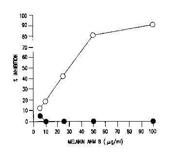
Figure 1 shows that melanin inhibits LPS-induced TNF-a
Production. Open circles depict TNF-a production/release by monocytes
(1x10⁶ cells/ml) that were incubated for QO min at 37°C with various
concentrations of :melanin AHM 8 before stimulation with 1 ng/ml LPS.
TNF-a production was measured in cell-free culture supernatants by ELISA.
The TNF-a concentration was also determined for supernatants collected
from monocytes stimulated with LPS in the absence of melanin (3,230
pg/106 ceils/ml), and from supernatants collected from monocytes
maintained in medium alone (36 pg/106 cells/mi).
Closed circiles depict the effect of melanin on the constitutive
synthesis of protean by melanin-treated cells. Monocytes (1x10⁵ per 0.2 ml
leucine-free medium supplemented with 10% dialyzed human AB serum)
-9-
SUBSTITUTE SHEET (RULE 26)
CA 02279946 1999-08-09
WO 98/34602 PCT/US98102971
seeded in 96-well plates were incubated for 5 hr at 37°C with the
indicated
concentrations of melanin AHM 8. Control cells were maintained in
medium alone for the duration of culture. At 4 hours prior to harvest, cells
were pulsed with 5 ~Ci per well of [3H]-leucine. The mean (fSEM)
incorporation of [3HJ-leucine by monocytes incubated in medium alone was
28,737 ~ 712 cpm.
Figures 2(A-D) show that melanin significantly inhibits production of
TNF-a (Fig. 2A), IL-lt3 (Fig. 2B), and IL-6 (Fig. 2C), but not GM-CSF (Fig.
2D),
bY human peripheral blood monocytes. Monocytes were pretreated with
the indicated concentrations of melanin AHM 8 (0 ~g/ml, open bar; 50
~g/ml, slashed bar; 100 ~g/ml, solid bar) before stimulation with 1 ng/ml
LPS. Controls included (1) melanin-nontreated cells stimulated with LPS,
{2) melanin-treated, LPS-nonstimulated monocytes, and (3) monocytes
incubated in complete medium in the absence of additives.
The change (O) in the amount of pg of TNF-a = (pg cytokine / 106 LPS-
stimulated monocytes/ml) - (pg cytokine/106 LPS-nonstimulated
monocytes/ml). The mean (t:SEM) cytokine contents in the supernatant
collected from 106 LPS-nonstimulated monocytes incubated in the presence
of 0, 50, or 100 ~g/ml melanin were, respectively, <_108 t5 pg/ml, for TNF-a;
575 t53 pg / ml for IL-1B; 55981238 pg / ml f or IL-6; and <_1681124 pg / ml
for
GM-CSF.
Figures 3(A & B) show duplicate experiments which indicate that the
observed reversal of melanin-mediated suppression of TNF-a production is
time-dependent. Human peripheral blood monocytes were incubated at
37°C with 100 ~g/ml melanin AHM 8. After a 1 hour incubation, cells
were
washed to remove free melanin, suspended in fresh medium, and
stimulated with 1 ng/ml LPS at the indicated time points. The
concentration of TNF-a in culture supernatants collected 24 hours after LPS
-10-
SUBSTITUTE SHEET (RULE 26)
CA 02279946 1999-08-09
WO 98/34602 PCT/LTS98/02971
addition was measured by ELISA, and the percent inhibition of TNF-a
production is indicated.
Figure 4 shows that melanin treatment suppresses TNF-a production
even when applied after L.PS stimulation. Monocytes were stimulated with
1 ng/ml LPS either 1 hour after (open box), simultaneously with (slashed
box), or 1 hour before (solid black box) the addition of the indicated amount
of melanin (50 or 100 fig, respectively).
Control monocytes were incubated without LPS in either the absence
or presence of melanin (not shown). Twenty-four hours after stimulation
with LPS, the levels of TNF-a in the culture supernatants were measured by
ELISA. At both concentrations, the amount TNF-a inhibition observed was
greatest when the cells were pretreated with melanin, followed by cells
simultaneously treated with melanin and LPS, and cells treated with after
L~ stimulation (p~<0.05 when compared to TNF-a production by
monocytes treated for 1 hour with melanin before stimulation with LPS).
Figure 5 (A--D) demonstrates that AHM 8 is able to inhibit TNF-a
production independent of stimulus. Monocytes were infected with either
Mycobacterium avium (MAC, Fig. SA) strain 101 (serovar 1), or an avirulent
(H37Ra) strain of .M. tuberculosis (MTB, Fig. 5B) by incubation with an
approximate bacte~rium:monocyte ratio of 10:1. Following a 4 hr incubation
at 37° C, monocytes were thoroughly washed by low speed centrifugation
to
remove extracellular bacteria. Alternatively, monocytes were stimulated
with either PPD (50 ltg/ml, Fig. 5C) or I1'S (1 ng/mi, Fig. 5D). After 24
hours
of incubation in the presence of 100 ~tg/ml AHM 8, the release of TNF-a by
monocytes infected with either MAC or MTB or stimulated with either PPD
or LPS was reduced by 45-55%. The levels of TNF-a in each culture was
measured by ELISA. The change (D) in the amount of pg of TNF-a/106
-11-
SUBSTITUTE SHEET (RULE 26)
CA 02279946 1999-08-09
WO 98/34602 . PCT/US98/02971
cells/ml = [pg of TNF-a/ml in the supernatant of AHM 8 treated and
stimulated monocytes] - [pg of TNF-a/ml in the supernatant of AHM 8
treated and nonstimulated monocytes].
Figure 6 shows the relative amount of TNF-a mRNA produced by
human monocytes, cultured under nonadherent conditions, after treatment
with either 0, 50 or 100 ~.g/ml AHM 8 for 1 hour before stimulation with 1
ng/ml LPS (lanes 1, 2, and 3, respectively). Total cellular RNA was extracted
by the guanidinium thiocyanate method, size fractionated by
formaldehyde/agarose gel electrophoresis (10 ~g RNA/lane, normalized
using a 28S RNA as a control), and transferred onto a nylon membrane by
capillary blotting (Chomczynski and Sacchi, Anal. Biochern., x,:156-159,
1987). The blot was hybridized to a 1.1 kb 32P-labeled cDNA fragment of
TNF-a obtained by Pst I digestion of plasmid pE4 (American Type Culture
Collection, Rockville, MD). The data show that treatment with 100 ~.g / ml
AHNi 8 was able to reduce the amount of TNF-a mRNA produced by the
cells (lane 3).
25
-12-
SUBSTITUTE SHEET (RULE 26)
CA 02279946 1999-08-09
WO 98/34602 PCT/US98/02971
Figure 7 shows a Northern blot that was performed using monocytes
treated simultaneously with 1 ng/ml LPS and 100 ~g/ml AHIvI 8. Control
monocytes were stimulated with LPS in the absence of AHM 8. Total RNA,
extracted 1 and 3 hours after LPS stimulation, was probed with cDNA
fragment of human TNF-a (Figure 7, Panel A) or IL-6 (Figure 7, Panel B).
Monocytes were incubated: for 1 hr in medium alone or in the presence of
100 ~tg/ml AHM 8 alone (lanes 1 and 2, respectively); for 2 or 3 hours in the
presence of 1 ng/ml LPS alone (lanes 3 and 4, respectively); or for 2 or 3
hours in the presence of 1 ng/ml LPS and 100 ~g/ml AHM 8 (lanes 5 and 6,
respectively). Total cellular RNA was probed with '~P-labelled cDNA probes
specific for TNF-a (panel A), or IL-6 (panel B) (using the 1.5 kb Bgl II / Bam
HI fragment from pT7.7/hhIL-6 in E. coli BL21 (DE3), ATCC 68636).
Figure 8 shows that AHM-8 does not affect the stability of TNF-a
mRNA. Monocytes were treated with 10 itg/ml actinomycin D 30 min after
stimulation with 1 ng/ml LPS (time 0). A parallel culture of monocytes was
simultaneously treated with actinomycin D and 100 ~g/ml AHM 8. The
decay of TNF-a m.RNA was analyzed by Northern blot analysis 30, 60, and
180 min after addition of actinomycin D. Open and closed circles
respectively represent cells cultured with LPS, or LPS and AHM 8.
Figure 9(A-~C) shows that melanin strongly inhibits the TNF-a
response in BALB~/c mice. Circulating plasma concentrations of TNF-a
were measured by ELISA 90 min after i.v. injection of LPS. AHM 8 (at SO
mg/kg, slashed bars) was injected either 60 min before (19 mice);
simultaneously with (40 mice); or 15 min after (18 mice) LPS injection. The
concentrations of TNF-a in the melanin treated group and nontreated
controls (open bars) were compared by the two tailed Mann-Whitney Test
using the INSTA'.C~ 2.03 program. Results are mean t SEM.
-13-
SUBSTITUTE SHEET (RULE 26)
CA 02279946 1999-08-09
WO 98/34602 PCTIUS98/02971
5Ø DETAII.Fr~ nFS(~RIPTION OF THE INVENTION
The present invention is broadly directed to the discovery that
melanin is useful for the therapeutic treatment of disease in animals,
including humans.
In one embodiment, the melanins used in the presently described
invention are substantially pure. In general, the term "substantially pure"
melanin shall refer to melanin preparations that are comprised of at least
about 75 percent of the desired melanin, specifically at least about 85
percent,
more specifically at least about 90 percent, and preferably at least about 95
weight percent.
As a consequence of normal melanin production, a wide variety of
protein and amino acid contaminants are typically incorporated into
naturally occurring melanins. Additionally, the wide variety of substrates
and contaminants that are typically available during normal melanin
production in vivo may lead to the production of melanins with
amorphous composition. Similarly, the wide variety of contaminants that
are typically found in commercially available preparations of tyrosinase, the
enzyme that makes melanin, are often incorporated into melanins
produced in vitro.
Where pharmaceutical applications of melanin are contemplated,
melanin products with defined and predictable compositions and structural
features are highly desirable, and may even be necessary. Additionally, the
contaminating proteins, and amino acids contained therein, that are often
incorporated into naturally occurring or previously described melanins may
also prove immunogenic in the host. Thus, melanin preparations that are
to be administered in vivo shall preferably be substantially free of
contaminating proteins, amino acids, and especially toxins of microbial
origin (i.e., bacterial endotoxins, etc.).
The term "biosynthetic" melanin shall refer to melanin that is
-14-
SUBSTITUTE SHEET (RULE 26)
CA 02279946 1999-08-09
WO 98/34602 PCT/US98I02971
produced by a recombinantly expressed and/or purified tyrosinase protein
that has been provided with a substrate for melanin production. By
producing melanin using high specific activity tyrosinase in conjunction
with defined substrates, melaruns are produced with substantially more
uniform structure and composition than melanins typically found in
nature. With proper methods of synthesis, the resulting "biosynthetic"
melanins may also be substantially pure, or further processed to produce
biosynthetic melaiun preparations that are substantially pure. In the
majority of instances, suitably processed biosynthetic melanin may replace
naturally occurring melanin in any of the embodiments described herein.
The purified or biosynthetic melanins used in the present invention
may optionally be characterized by being substantially free of contaminating
amino acid content. For the purposes of the present invention, the term
"substantially amino acid free" shall refer to melanin preparations that
generally contain less than about 10 percent amino acid content by weight,
preferably less than about 7.5 percent amino acid content, more preferably
less that about 5 F~ercent amino acid content, and specifically less than 2.5
percent amino acid content by weight. Moreover, compositions comprising
purified biosynthetic melanins shall generally be substantially free of
potentially toxic contaminants of bacterial origin such as, but not limited
to,
bacterial endotoxins (particularly gram negative endotoxin), and bacterial
exotoxins.
Where the therapeutic use of the presently described purified
melanins is contemplated, the purified melanin is preferably administered
in a pharmaceutically acceptable carrier, via oral, intranasal, rectal,
topical,
intraperitoneal, intravenous, intramuscular, subcutaneous, subdermal,
transdermal, intrathecal, or intracranial methods, and the like. Typically,
the preferred formulation for the purified melanin will vary depending
upon the region of the host requiring treatment.
For example, topical immune reactions are preferably treated or
-15-
SUBSTITUTE SHEET (RULE 26)
CA 02279946 1999-08-09
WO 98134602 PCT/US98/02971
prevented by melanin formulations designed for topical application,
whereas systemic reactions are preferably treated or prevented by
administration of compositions formulated for parenteral administration.
Additionally, immune-mediated disorders of the pulmonary system may be
treated both parenterally and by direct application of the therapeutic
melanin compositions to the respiratory system by inhalation therapy.
Additionally, local immune reactions, i.e., arthritic or inflamed joints,
etc.,
may be treated by localized injection purified melanin compositions into
the synovial capsule. Optionally, such local administration of purified
melanin compositions may be performed in conjunction with
corticosteroids.
Additionally, the purified melanin may be loaded into lipid-
associated structures (i.e., liposomes, or other lipidic complexes) which may
enhance the pharmaceutical characteristics of the purified melanin. The
lipid-melanin complex may subsequently be targeted to specific target cells
by the incorporation of suitable targeting agents (i.e., specific antibodies
or
receptors) into the melanin/lipid complex. Optionally, the purified
melanin may be directly complexed with a targeting agent to produce the
desired effect.
Where melanin mediated treatment of inflammatory disorders of
the digestive tract and alimentary canal are contemplated, lipid
formulations (e.g., emulsions, microemulsions, liposomes, etc.) comprising
purified melanin may significantly protect the melanin from the digestive
process. Accordingly, melanin formulations are contemplated that may be
orally administered. To the extent that additional enteric protection is
desired, for added protection, it is possible to formulate solid or liquid
formulations in accordance with the invention in an enteric-coated or
otherwise protected form. In the case of liquid formulations, they can either
be mixed or simply coadministered with a protectant, such as a liquid
mixture of medium chain triglycerides, or they can be filled into enteric
-16-
SU8ST1TUTE SHEET (RULE 26)
CA 02279946 1999-08-09
WO 98/34602 . PCT/US98/02971
capsules (for example of soft or hard gelatin, which are themselves
optionally additionally enteric coated. Alternatively, solid formulations
comprising melanin may be treated more flexibly. They may either be
coated with enteric: materials to form tablets or they can be filled into
enteric
capsules.
The thickness of enteric coating on tablets or capsules can be, for
example, from 0.5 to 4 microns in thickness, although the precise thickness
will be determined. by the skilled formulator. Enteric coated granules
(whose particle size may be, for example, from 0.5 to mm) may themselves
be coated without being compounded into a tablet for coating.
Microcapsules, sirrularly, can be enteric coated. The enteric coating may
comprise any of the enteric materials conventionally utilized in orally
admirustrable pharmaceutical formulations. Suutable enteric coating
materials are kno~nrn, for example, from "Remington's Pharmaceutical
Sciences", 15th Edition, pp. 1614-1615 (1975); 2nd Edition, pp. 116-117, 371-
374
(1976); and "Hagars Handbuch der Pharmazeutischen Praxie", 4th Edition,
Volume 7a (Springer Verlag, pages 739 to 742 and 776 to 778 (1971).
Examples o:f suitable enteric coating materials include cellulose
acetylphthalate, hyrdroxypropylmethylcellulose-phthalate (HPMC-P),
benzophenyl salicylate, cellulose acetosuccinate, copolymers of styrene and
malefic acid, formulated gelatin, keratin, stearic acid, myristic acid,
polyethylene glycol, shellac, gluten, acrylic and methacrylic resins and
copolymers of malefic acid and phthalic acid derivatives. The enteric coating
materials) may be dissolved in solvents such as dichloromethane, ethanol
and water, cellulose phthalate, or polyvinyl acetate phthalate. It is
preferred
to utilize HPMC-F', polyethylene glycol 6000 or shellac as the enteric
coating.
A proprietary preparation of HPMC-P aimed at dissolution or dissipation at
pH 5.5, which is erncountered in the human pylorus, is available under the
trade mark HP5-5,, and is particularly preferred.
Additionally, any of a variety of stabilizing agents may be utilized in
-17-
SU6STITUTE SHEET (RULE 26)
CA 02279946 1999-08-09
WO 98/34602 PCT/US98l02971
conjunction with the described melanin compositions. Although the
melanin itself may function as an antioxidant, the oxidation of melanin or
other components of the described compositions may be substantially
reduced by preparing formulations in accordance with the present
invention under an inert atmosphere, such as nitrogen, this is a somewhat
inconvenient and expensive process and so it is often preferred to add
chemical anti-oxidants. Suitable pharmaceutically acceptable antioxidants
include propyl gallate, butylated hydroxyanisole, butylated hydroxytoluene,
ascorbic acid or sodium ascorbate, DL- or D-a-tocopherol and DL- or D-a-
tocopheryl acetate. The anti-oxidant, if present, may be added singly or in
combination to the polynucleotide delivery vehicles either before, during,
or after vehicle assembly in an amount of up to, for example, 0.1% (w/v),
preferably from 0.0001 to 0.05%.
Formulations comprising purified melanin may also be stabilized for
I5 storage and shipment by any of a number of well established methods,
including but not limited to, freezing, refrigeration, and lyophilization.
Where one seeks to augment long-term stability by freezing or freeze-drying
melanin compositions, suitable excipients may be added to the melanin
comprising preparations prior to freezing. Examples of such stabilizing
excipients include, mono or disaccharides (e.g., glucose, sucrose, etc.),
polysaccharides, or any of a variety of well-known agents (e.g., glycerols,
gums, dextrans, and the like).
One of ordinary skill will appreciate that, from a medical
practitioner's or patient's perspective, virtually any alleviation or
prevention of an undesirable symptom (e.g., symptoms related to immune-
mediated disorders in the body) would be desirable. Thus, the terms
"treatment", "therapeutic use", or "medicinal use" used herein shall refer
to any and all methods of using the described purified melanin
compositions to remedy a disease state or symptoms, or otherwise prevent,
under, retard, or reverse the progression of disease or any other
-18-
SUBSTITUTE SHEET (RULE 26)
CA 02279946 1999-08-09
WO 98/34602 PCT/U598102971
undesirable symptoms in any way whatsoever. Similarly, a "therapeutically
effective amount" of melanin is an amount sufficient to remedy a disease
state or symptoms, or otherwise prevent, hinder, retard, or reverse the
progression of disease or any other undesirable symptoms in any way
whatsoever.
Given that adverse disease consequences have been linked with
excess proinflamrnatory cytokines (i.e., TNF-a, and interleukins including,
but not limited to,. IL-1 and IC.-6) production, in a particularly preferred
embodiment of tl~~e present invention, the purified melanin is used at a
dose that reduces or inhibits the excess production of TNF-a while still
allowing or facilii:ating an effective host immune response against the
underlying disorder or infection.
Preferably, animals that may be treated by the present invention
include, but are riot limited to, invertebrates, vertebrates, birds, mammals
such as pigs, goats, sheep, cows, dogs, cats, and particularly humans.
When used in the therapeutic treatment of disease, an appropriate
dosage of purified melanin, or modified form thereof, may be determined
by any of several well established methodologies. For instance, animal
studies are commonly used to determine the maximal tolerable dose, or
MTD, of bioactive agent per kilogram weight. In general, at least one of the
animal species tested is mammalian. Those skilled in the art regularly
extrapolate doses for efficacy and avoiding toxicity to other species,
including human. Before human studies of efficacy are undertaken Phase I
clinical studies in normal subjects help establish safe doses. Additionally,
therapeutic dosaf;es may also be altered depending upon factors such as the
severity of infection, and the size or species of the host.
Particularly where in vivo use is contemplated, the various
biochemical components used to formulate the present invention are
preferably of high purity and are substantially free of potentially harmful
contaminants (e.g., at feast National Food (NF) grade, generally at least
-19-
SUBSTITUTE SHEET (RULE 26)
CA 02279946 1999-08-09
WO 98/34602 PCT/ITS98/02971
analytical grade, and preferably at least pharmaceutical grade). To the extent
that a given compound must be synthesized prior to use, such synthesis or
subsequent purification shall preferably result in a product that is
substantially free of any potentially toxic agents, particularly endotoxins,
which may have been used or present during the synthesis or purification
procedures.
The presently described purified melaruns may also be complexed
with molecules that enhance their in vivo attributes. Examples of such
molecules include, but are not limited to, carbohydrates, polyamines, amino
acids, peptides, ions (i.e., sodium, potassium, calcium, magnesium,
manganese, etc.), and lipids.
Additionally, the purified melanins may be complexed with a variety
of well established compounds or structures that, for instance, further
enhance the in vivo stability of the melanin, or otherwise enhance its
pharmacological properties (e.g., increase in vivo half-life, reduce toxicity,
enhance solubility or uptake, etc.). Examples of such modifications include,
but are not limited to, the production of sulphate, gluconate, citrate,
phosphate, and the like.
Where diagnostic, therapeutic or medicinal use of purified melanin,
or derivatives thereof, is contemplated, the melanin may generally be
prepared and maintained under sterile conditions that minimize that risk
of, or avoid, microbial contamination. Because of the relatively small size
and inherent stability of purified melanin, compositions comprising
melanin may also be sterile filtered prior to use. In addition to the above
methods of sterile preparation and filter sterilization, antimicrobial agents
may also be added to the melanin compositions. Antimicrobial agents
which may be used, generally in amounts of up to about 3% w/v, preferably
from about 0.5 to 2.5%, of the total formulation, include, but are not limited
to, methylparaben, ethylparaben, propylparaben, butylparaben, phenol,
dehydroacetic acid, phenylethyl alcohol, sodium benzoate, sorbic acid,
- 20 -
SUBSTITUTE SHEET (RULE 26)
CA 02279946 1999-08-09
WO 98/34602 PCT/US98/02971
thymol, thimerosal, sodium dehydroacetate, benzyl alcohol, cresol, p-
chloro-m-cresol, chlorobutanol, phenylmercuric acetate, phenylmercuric
borate, phenylmercuric nitrate and benzylalkonium chloride. Preferably,
antimicrobial additives will either enhance the biochemical properties of
the melanin, or will be inert with respect melanin activity. To the extent
that a given antimicrobial agent may prove deleterious to melanin activity,
another agent may be substituted which effects the desired functions of
melanin to a lesser extent.
One embodiment of the presently claimed methods relates to the use
of purified melanin to modulate the immune system. Such modulation is
deemed to be a function of melanin's ability to either directly or indirectly
effect cytokine expression or activity in vivo or in vitro. In a preferred
embodiment, the 'therapeutic use of melanin will downregulate cytokine
expression. Melaiun's ability to downregulate cytokine expression may also
be exploited by using melanin in conjunction with established therapeutics
in order to reduces the sewerity of the adverse immune-related reactions
associated with a given therapeutic. For example, IL-2 treatment has been
associated with adverse systemic consequences that are often dose
dependent. Because of melanin's ability to modulate adverse immune
reactions, the use of melanin in conjunction with cytokine may allow for
the clinical use of higher systemic concentrations of cytokine. Accordingly,
an additional embodiment of the present invention is the use of purified
melanin to reduce the toxic side-effects of therapeutic agents.
Given that adverse disease consequences have been linked with
excess TNF-a production, in a particularly preferred embodiment of the
present invention, the purified melanin is used at a dose that reduces or
inhibits the excess production of TNF-a while still allowing or facilitating
an effective host immune response against the underlying disorder or
infection.
For the purposes of the present invention, the term "cytokine" or
-21-
SUBSTITUTE SHEET (RULE 26)
CA 02279946 1999-08-09
WO 98/34602 PCTlUS98/02971
grammatical equivalents thereof, shall generally refer to hormones that are
associated with the cells of the immune system, both lymphokines and
monokines, and others. The definition is meant to include, but is not
limited to, those hormones that act locally and circulate in the blood, and
S which, when used in accord with the present invention, will result in an
alteration of an individual's immune response. The term cytokine may
refer to, but is not limited to, IL-1 (a or B), IL-2, IL-3, IL-4, IL-5, IL-6,
IL-7, IL-8,
IL-9, IL-10, IL-11, IL-12, GM-CSF, M-CSF, G-CSF, LIF, LT, TGF-B, Y-IFN (or a
or B-IFN), TNF-a, and BCGF. Descriptions of the aforementioned cytokines
as well as other applicable immunomodulatory agents may be found in
"Cytokines and Cytokine Receptors", A.S. Hamblin, 1993, (D. Male, ed.,
Oxford University Press, New York, NY), or the "Guidebook to Cytokines
and Their Receptors", 1995, N.A. Nicola, ed. (Oxford University Press, New
York, NY) herein incorporated by reference.
Given that melanin is useful for treating the wasting syndrome that
is often associated with acquired immunodeficiency syndrome (AIDS), or
cancer, the presently described methods are also deemed to be broadly useful
for the treatment of AIDS and cancer.
~ additional embodiment of the present invention is the use of
purified melanin to treat allergy related hypersensitivity reactions.
Particularly contemplated is the use of purified melanin to prophylactically
treat individuals that may be susceptible to the adverse consequences of
allergic reactions such as, but not limited to, drug reactions, insect stings,
23 dermatitis, food allergies, and the like. Additionally contemplated is the
intervening use of purified melanin to alleviate or reduce the adverse
symptoms of allergic reactions.
Melanin is a virulence factor that contributes to the pathogenesis of a
variety of infectious agents. To the extent that melanins that are
characteristic of a particular pathogen may be identified, an additional
aspect
-22-
SUBSTITUTE SHEET (RULE 26)
CA 02279946 1999-08-09
WO 98/34602 PCTIt1S98/02971
of the presently claimed invention is the use of purified melanin, or
portions or analogues thereof, as vaccines to prevent progression and
spread of melanin producing pathogens.
Similarly, th.e identification and use of melanoma associated or
specific melanins i;s contemplated to provide an additional form of cancer
therapy comprising the use of tumor specific melanins, or fragments or
analogues thereof, as cancer vaccines, or tumor-specific immunostimulants.
Additionally, the identification of pathogen or tumor specific
melanins shall be useful for the identification or production of receptors,
ligands, or polyclonal or monoclonal antibodies that specifically bind to the
pathogen or tumor specific melanin. Accordingly, an additional
embodiment of the present invention are receptor, ligand, or antibody-
based diagnostics or therapeutics that target pathogen or tumor specific
melanins, or the cellular .receptors therefore.
The following examples serve to more fully describe the manner of
making and using the above-described invention, as well as to set forth the
best modes conternplated for carrying out various aspects of the invention.
It is understood that these examples in no way serve to limit the true scope
of this invention, but rather are presented for illustrative purposes.
6Ø EXi.~MPLES
6.1. Sv~nthe_~y» of Water-Soluble Melanin
Water soluble melanin was produced and prepared for use essentially
as described in U:S. Patent Nos. 5,340,734; 5,466,592; 5,486,351; 5,210,076
and
5,057,325 herein incorporated by reference. Melanins produced using the
described methods were further purified by acid precipitation by addition of
concentrated HCl ~pH 1.5. Precipitated melanin was recovered by
centrifugation.
When analyzed for purity, the resulting melanin (designated AHM 8)
was found to comprise about 96% percent of the final product by weight.
-23-
SUBSTITUTE SHEET {RULE 26)
CA 02279946 1999-08-09
WO 98/34602 . PCT/US98/02971
The amino acid content of melanin AHNi 8 was less than 4.2%. The total
amino acid was 8.4% by weight of which 4.38% was tyr + gly. The elemental
analysis yielded the following: %C=51.01; %H=3.74; %N=9.5; %S=0; and
%O=33.85.
The endotoxin content of melanin AHM 8 was estimated using the
chromogenic Limulus amebocyte lysate (CLAL) test kit (BioWhittaker, Inc.,
Walkersville, MD). To determine whether AHM 8 has a direct inactivating
effect on the CLAL test, in a preliminary experiment an aliquot of endotoxin
standard containing 0.25 endotoxin unit (EU) /ml was spiked with 50 ~tg / ml
~ 8 (a dose similar to that used for treatment of monocytes). The
endotoxin content of the "mixture" was determined using the CLAL test
according to the manufacturer's directions. Percent reduction in the
endotoxin content of melanin-containing standard preparation was
calculated as follows:
% ~bition = {1-[(Endotoxin content of standard) - (Endotoxin
content of AHM 8-containing standard)] /[Endotoxin content of standard]} X
100
Results from this experiment indicated that AHM 8 does not
markedly interfere with the CLAL assay. Addition of 50 ~tg/ml AHM 8
produced 22% reduction in the activity of the endotoxin standard. As
shown in Table 1, the endotoxin content of melanin AHM 8, at 50 ~g/ml,
was only 0.069 endotoxin unit (EU)/ml. Under our experimental
conditions, the production of TNF-a, by ~ human peripheral blood monocytes
required 1.6 EU/ml of LPS (i.e., 0.1 ng LPS/ml).
-24-
SUBSTITUTE SHEET (RULE 26)
CA 02279946 1999-08-09
WO 98/34602 PCT/iTS98/02971
Table 1
FNDC7T'OXIN COIVfTENT OF AHM 8
Endotoxin
Test Agent Concentration
(EU/ml)
PBS 0
LPS (0.05 ng / 1.50
ml)
Mel AHM 8 (50 0.069
~g/m()
°Fmdotoxin content of each agent was estimated by CLAL test, using a
commercially available kit.
When tested for the ability to induce TNF-a production, AI-iM 8 at 50
~g/ml did not induce a strong TNF-a response in human peripheral blood
monocytes (123 p~, TNF-a/106 cells/ml) (Table 2).
Peripheral blood monocytes used in this experiment, as well as those
described in the following sections, were isolated from the white cell
concentrates by sE~quential gradient centrifugation of Ficoll-Paque and
Percoll gradient (Markowicz and Engleman, J. Clin. Invest. $,x:955,1990).
The white cell concentrates were purchased from Stanford University Blood
Center (Palo Alto,, CA). The percoll gradient consisted of sequential layers
of
75%, 51.4%, 40%, and 30% dilutions of stock iso-osmotic solution of Percoll
(1.128 g/ml) (Pha.rmacia Biotech, Uppsala, Sweden) in Dulbecco's calcium-
and magnesium-free phosphate buffer saline (PBS) containing 5% heat-
inactivated human serum. For further enrichment, low-density cells,
mostly monocytes, were refloated onto a second Percoll gradient. The
monocyte enriched population was resuspended at 1x10⁶ cells/ml in
macrophage serum-free medium (GIBCO, Grand Island, NY) supplemented
-25-
SUBSTITUTE SHEET (RULE 26)
CA 02279946 1999-08-09
WO 98/34602 PCT/US98/02971
with 1% heat-inactivated human AB serum, 2 mM glutamine, 100 U/mi
penicillin, and 100 ~g/ml streptomycin [referred to hereafter as complete
medium]. Because monocytes are stimulated to produce cytokine following
adhesion to plastic, polypropylene tubes were used in these cell culture
experiments.
TABLE 2
PRODUCTION OF TNF-ac BY HUMAN PERIPHERAL BLOOD
MONOCYTES INCUBATED IN THE PRESENCE OF MELANIN'
TNF-a Content
of Culture Supernatants
Cells Incubated ipg ~F-a/106
with cells/ml)
Medium alone 44
p~,I g (50 ~g/ml) 123
LPS (0.1 ng/ml) 1,013
'Monocytes were incubated with AI-EvI 8 or LPS in polypropylene tubes.
Following 24 h incubation at 37°C, culture supernatants were
collected and
TNF-a release was measured by ELISA (Biosource International, Camarillo,
CA). Values are the mean of two measurements.
6.2. Pretreatment With Melanin Suppresses LPS-Induced
TNF-a Production
The effect of biosynthetic melanin on in vitro TNF-a production was
evaluated by comparing the levels TNF-a in the culture supernatants of
melanin-treated and nontreated monocytes following stimulation with
LPS. In these experiments, monocytes, (1 x 10⁶/ml), were incubated with
various doses of melanin at 37°C in a humidified atmosphere containing
5% C02. Following a 30-60 min incubation, monocytes were stimulated
-26-
SUBSTITUTE SHEET (RULE 26)
CA 02279946 1999-08-09
WO 98/34602 PCT/US98/02971
with LPS in the continuous presence of melanin. Controls included (1)
melanin-nontreated cells stimulated with LPS; (2) melanin-treated, LPS-
nonstimulated monocytes; and (3) monocytes incubated in complete
medium in the absence of additives. Twenty-four hours after stimulation
with LPS, the levels of TNF-a in the culture supernatants were measured,
in duplicate, by EL,ISA (Biosource International, Camarillo, CA). The
operable range for TNF-a was 15.6-1,000 pg/ml. Percent inhibition of the
TNF-a response w,as calculated as follows:
% ~bition = [1-(pg TNF-a/106 AHM 8-treated, LPS-stimulated
monocytes) - (pg TNF-a/106 AHNi 8-treated monocytes)/(pg TNF-a/106
AHM 8-nontreated, LPS-stimulated monocytes)-{pg TNF-a/ 106 monocytes
maintained in medium alone)] x 100.
As shown in Figure 1, treatment of monocytes with melanin AHM 8
resulted in a dose-dependent inhibition of LPS-induced TNF-a production.
To ensure tlhat the presence of melanin in the culture supernatants
did not interfere with the assay (ELISA), the TNF-a concentrations in the
supernatants collected from melanin-treated monocytes were determined
from two standard curves. For construction of the "control standard
curve", TNF-a standards were diluted in complete culture medium alone.
"Melanin-containing standard curves" were constructed by plotting optical
density (O.D.) values obtained from TNF-a standards (over a range from 0
to 1,000 pg/ml) float were diluted in complete medium incubated with 0-50
~g/ml melanin for 24 hours at 37°C. In parallel assays, the TNF-a
content of
culture supernatants collected from monocytes that were treated with 0-50
~,g/ml melanin before stimulation with LPS (referred to hereafter as the test
samples) were dei:ermined by reading their O.D. against each standard
curve. The TNF-a content of each test sample, determined from different
-27-
SUBSTITUTE SHEET (RULE 26)
CA 02279946 1999-08-09
WO 98/34602 PCT/US98102971
standard curves, was compared by calculating the Stimulation Index (SI) by
dividing the amount (in pg) of TNF-a produced per 106 cells per ml of
supernatant from LPS-treated monocytes by the amount (in pg) of TNF-a
produced per 106 cells per ml of supernatant from non-LPS-treated
monocytes.
As shown in Table 3, SI values of supernatants collected from
melanin-pretreated monocytes were consistently lower than the SI values
of supernatants collected from cells were not exposed to melanin. Taken
together, these data indicate that melanin suppresses LPS-induced TNF-a
synthesis/release by human monocytes and that this reduction is not the
consequence of an inhibitory effect of melanin on the assay system.
TABLE 3
INFLUENCE OF MELANIN ON TNF-a-SPECIFIC ELISA'
Melanin TNF-a
Content
of Culture
Supernatants
Content of from
Cells
Treated
with
AHM
8 (~tg/ml)
TNF-a
Standard
(~g/~) 0 10 25 50
(Stimul
ation
Index)
0 78 39 17 7
10 69 35 16 7
25 82 40 17 7
50 75 37 17 7
'For construction of standard curves, complete medium containing 0 to 50
-28-
SUBSTITUTE SHEET (RULE 2fi)
CA 02279946 1999-08-09
WO 98/34602 . PCT/US98/02971
~tg/ml AHM 8 was, used to dilute TNF-a standards over a range from 0-1,000
pg/ml. The plate reader was zeroed against a blank composed of
chromogen and stop solution.
6.3. The Effect of Melanin on Protein Synthesis by
Human Peripheral Blood Monoc~tes
To determine whether melanin selectively interferes with the
production of LPS-induced cytokines, the effect of melanin AHM 8 on
constitutive protein synthesis by human monocytes was measured. Protein
synthesis was measured by incorporation of [3H)leucine. Monocyte protein
synthesis after 5 hnurs incubation in the presence of 100 ~g/ml melanin
AHM 8 was roughly comparable to that displayed by melanin nontreated
control cells (23% lower). Under parallel experimental conditions
incubation of monocytes with 20 ~g/ml cycloheximide resulted in complete
l~bition of [3H]-leucine incorporation (1,219 151 cpm versus 28,737 ~712
cpm in control monocytes). In summary, even though pretreatment of
monocytes with 100 ~g/ml AHM 8 resulted in 90% suppression of TNF-a
synthesis, net protein synthesis was only reduced by 23%.
The levels of ('H]-leucine incorporation in monocytes incubated for
20 hours in the presence of 100~g / ml AHM 8 was also comparable to that of
the melanin nontreated cells {32,775 11,977 cpm versus 29,713 ~856 cpm).
30
-29-
SUBSTITUTE SHEET (RULE 26~
CA 02279946 1999-08-09
WO 98/34602 PCT/ITS98/02971
6.4. Melanin Selectively Suppresses Cytokine
Production by Human Monocytes
To determine whether melanin suppresses the production and
release of other LPS-induced cytokines, peripheral blood monocytes were
tested (essentially as described above) for the ability to produce TNF-a, IL-
1f3,
IL-6 and granulocyte/macrophage-colony stimulating factor (GM-CSF) after
melanin treatment. The levels of TNF-a, IL-1B, and GM-CSF in the culture
supernatants were measured, in duplicate, using ELISA kits purchased from
Biosource International (Camarillo, CA). ELISA kits used for measurement
of IL-6 were purchased from R&D Systems (Minneapolis, MN). The
operable range for the cytokines were TNF-a., 15.6-1,000 pg/ml; IL-1B, 3.9-250
pg/ml; IL-6, 3.12-300 pg/ml; and GM-CSF, 15.6-1,000 pg/ml. To ensure that
the presence of AHM 8 in the culture supernatants did not interfere with
the assay, the cytokine concentrations in the supernatants collected from
AHM 8-treated monocytes were determined from standard curves that were
constructed by plotting the O.D.'s obtained from the standards which were
diluted in complete medium which had been incubated with 50 ~g/ml
AHM 8 for 24 hours at 37°C. The cytokine content of supernatants
collected
from AHM 8-nontreated monocytes' however, was determined from the
"control standard curve". For construction of the "control standard curve",
each standard was diluted in complete medium alone.
Results from these experiments are summarized in Figure 2.
Monocytes pretreated with AI~vI 8 produced significantly (p <0.05) lower
levels of TNF-a, IL-1 p, and IL-6 than did their respective controls. Under
parallel conditions, melanin did not inhibit production or release of GM-
CSF by LPS stimulated monocytes. In contrast, monocytes pretreated with
100 ~g AHM 8/ml produced significantly higher levels of GM-CSF
-30-
SUBSTITUTE SHEET (RULE 26)
CA 02279946 1999-08-09
WO 98/34602 PCT/US98/02971
following stimulation with LPS (p <0.01 ). This indicates that AI-~vi 8 does
not inhibit LPS signalling. The finding that melanin does not suppress GM-
CSF secretion is of particular interest. GM-CSF affects the intracellular
phosphorylation of nucleoside analogues in monocytes and macrophages,
resulting in increased activity of AZT and stavudine (De Simone et al.,
Immunology Today 17:256-258, 1996).
6.5. Continuous Presence of Melanin Is Not Required
To determine whether suppression of TNF-a production requires the
continuous presence of melanin, freshly isolated human monocytes (1x10⁶
cells/ml of complete medium) were treated with inhibitory concentrations
of melanin AHM 8. Following a 1 hr incubation at 37°C, monocytes in one
set of culture were stimulated with LPS in the continuous presence of
melanin. Monocyt:es in a second set of cultures were washed once by low-
speed centrifugation before stimulation with LPS. Controls included (1)
melanin-nontreated cells stimulated with LPS; (2) melanin-treated, LPS-
nonstimulated mo~nocytes; and (3) monocytes incubated in complete
medium in the absence of additives. Twenty-four hours after stimulation
with LPS, the levels of TNF-a in the culture supernatants were measured in
duplicate, by ELIS.A. Suppression of TNF-a production did not require
that melanin be continuously present. In fact, T'NF-a production was
suppressed by 63°~° even after the melanin had been washed out
of the
culture immediately before stimulation with LPS (data not shown).
-31-
SU8ST1TUTE SHEET (RULE 26)
CA 02279946 1999-08-09
WO 98/34602 PCTIUS98/02971
6.6. Melanin-Mediated Suppression of TNF-a
Production Is Reversible
To allow time for recovery, monocytes pretreated with the inhibitory
concentrations of melanin AHM 8 were incubated for 2-18 hours in
complete medium before stimulation with LPS. For each time point, the
following cultures served as control: (1) melanin-nontreated, LPS-
stimulated; (2) melanin-nontreated, LPS-nonstimulated; and (3) melanin-
treated, LPS-nonstimulated monocytes. The concentration of TNF-a in the
culture supernatants was measured 24 hours after the addition of LPS.
Data from two experiments, shown in Figure 3(A & B), demonstrate
that melanin-mediated suppression of TNF-a persisted at least for 7 hours.
The suppressive effects of melanin were reversed upon short-term culture.
Monocytes stimulated with LPS 18 hours after removal of melanin
exhibited a higher TNF-a response (44-47% decrease in TNF-a release
versus a 74-88% reduction after a 7-hour AHM 8 washout period). These
data indicate that monocytes treated with 100 ltg melanin AI-lZvt 8/ml were
not killed under these experimental conditions and that recovery from
melanin-mediated suppression is time-dependent.
25
-32-
SUBSTITUTE SHEET (RULE 26)
CA 02279946 1999-08-09
WO 98/34602 PCT/US98/02971
6.7. Melanin Suppresses Production of TNF-a by
Activated Monocvtes
The data presented in the preceding sections are from experiments in
which monocytes v~ere pretreated with melanin AHM 8 before LPS
stimulation. To determine whether melanin suppresses production of
TNF-a even when it is administered after LPS-stimulation, monocytes were
treated with melanin either 1 hour after, simultaneously with, or 1 hour
before stimulation with LPS. As expected, melanin added 1 hour before LPS
drastically inhibited LPS-induced TNF-a production (84 t4% inhibition at
100 ltg/ml) (Figure 4). When melanin was added 1 hour after LPS
stimulation, a partial suppression of TNF-a response was observed (45
~13% inhibition at 100 itg/'ml, p=0.05). Melanin added at earlier time points
following LPS stimulation did not exert a stronger suppressive effect.
Treatment of monocytes with 100 ~tg/ml melanin either 7.5 or 60 min after
LPS stimulation reduced TNF-a production by 50% and 52%, respectively
(not shown). ThesE~ data indicate that melanin may provide a corrective
benefit as well as a~ preventative benefit, and may also indicate that at
least
~'° separate mechanisms are responsible for the net reduction in TNF-a
production seen ai~ter prior exposure to melanin.
The finding that melanin appears less effective at suppressing TNF-a
secretion by activated monocytes is of particular interest because this
cytokine is an ess<~ntial mediator in the immune response. This finding
suggests that melanin could be used to reestablish a balanced or normal
level of TNF-a in patients with wasting syndrome without destroying the
patient's ability to fight infection.
-33-
SUBSTITUTE SHEET (RULE 26)
CA 02279946 1999-08-09
WO 98/34602 PCT/US98/029~1
6.8. Effect of Melanin on TNF-a Production Induced
rv Additional Stimuli
To test whether AHM 8 has the ability to modulate TNF-a
production by cells that have been activated by stimuli other than LPS,
additional stimuli are used in variations of the experiments outlined above
with the exception that the dose of stimulating agent used is tailored as
appropriate for the individual agent. Agents that are used in these assays
include, but are not limited to, Ivlycobacteriurn, allergens (including
compounds that have been associated with hypersensitivity reactions), and
lectins.
To determine whether AHM 8 influences production of TNF-a by
human monocytes regardless of stimulus, monocytes infected in vitro with
Mycobacterium avium complex (MAC) [strain 101 (serovar 1)]; or an
avirulent (H371Za) strain of M. tuberculosis (MTB); or stimulated with the
purified protein derivative of MTB (PPD) were treated with inhibitory
concentrations of AHM 8. PPD, a known agonist of monocyte TNF-a
synthesis and release, prepared from autoclaved culture filtrates of MTB
was purchased from Connaught Laboratories Limited (Willowdale, Ontario,
Canada). The results of these experiments, summarized in Figures 5(A-D),
clearly demonstrate that the inhibitory effect of AHM 8 on TNF-a
production/release is independent of stimulus. After 24 hours incubation
in the presence of AHM 8, the release of TNF-a was reduced by 45-55% by
monocytes infected with either MAC or MTB or stimulated with either PPD
or LPS.
Unlike LPS, PPD does not stimulate TNF-a synthesis in monocytes
through interaction with the cell surface protein CD14 (Wright, et al.,
Science, x:1431-1433, 1990; Zhang et al., J. Clin. Invest. x,:2076-2083,1993).
-34-
SUBSTITUTE SHEET (RULE 26)
CA 02279946 1999-08-09
WO 98/34602 . PCT/US98/02971
The observed reduction in TNF-a production was not the consequence of
diminished viability or higher bacterial burden of the AHM 8 treated
monocytes. The exclusion. of trypan blue was comparable for cells incubated
with or without A:E-Bvi 8 (93% vs 98%). In three experiments, 48 ~17% of
monocytes incubated with 100 ~tg/ml AHM 8 contained acid fast bacilli as
compared to 51 t2.3% of the infected monocytes incubated in medium
alone. The numbE~r of viable intracellular bacteria recovered from
monocytes after 29: h incubation in the presence of 100 ~tg/ml AHM 8 was
also similar to that yielded by monocytes maintained in medium alone (24
~9 vs 24 ~4 colony forming MAC per cell, respectively, n=3).
To ensure that live mycobacteria as well as PPD, but not the
contaminating Ll'S, stimulated production of TNF-a by monocytes,
mycobacterial suspension or their product (PPD) were pretreated for 30 min
with polymyxin B (PMB) before being added to the monocyte preparations.
PMB inhibits LPS-induced cytokine production. As control, an aliquot of
monorytes were incubated with LPS that had been preincubated with PMB.
As expected, monocytes incubated with PMB-treated LPS failed to produce
TNF-a (93% inhibition in cytolcine release as compared to monocytes
stimulated with PMB-nontreated LPS). In agreement with data from a
previous study (1-iirsch et al., j. ImmunoI. x:743-753, 1994), preincubation
with either MAC, MTB, or PPD with PMB had no effect on their TNF-a-
inducing ability (data not shown).
6.9 Effect of melanin on the expression of TNF-a.
mRNA
Northern t>lot hybridization (Chomczynslci and Sacchi, Anal.
B i o c h a m . x,:156-159, 1987; W aleh et al., Cancer Res. x:838-843,1994)
was
used to investigate the effect of AHM 8 on TNF-a mRNA expression in
monocytes stimulated with LPS. Modulation of mRNA and protein levels
-35-
SUBSTITUTE SHEET (RULE 26)
CA 02279946 1999-08-09
WO 98/34602 PCT/US98l02971
occurs in tandem, however, this is not often the case. For example,
increases in mRNA production may not translate into increase amounts of
protein if the mRNA is unstable. Alternatively, translational interference
or mRNA processing impediments may prevent a corresponding increase
in protein production/release. TNF-a synthesis largely depends upon
translational derepression (Han et aI. J. Exp. Med. x:465-475,1990).
Initially, the effect of pretreatment with AHM 8 on accumulation of
TNF-a mRNA by human monocytes was examined. Human monocytes,
cultured under nonadherent conditions, were treated with either 0, 50 or
100 ~g/ml AHM 8 for 1 hour before stimulation with 1 ng/ml LPS (lanes 1,
2, and 3, respectively, in Figure 6). Pretreatment of monocytes with the
indicated noncytotoxic doses of AHM 8 results in strong (66-83%) inhibition
of LPS-induced TNF-a production/release (see Figures 1). One hour after
LPS stimulation, monocytes were collected and lysed for mRNA expression
studies. Data from previous studies (not shown) indicated that in
monocytes TNF-a mRNA expression peaks within 1 hour of LPS
stimulation and declined thereafter.
As shown in Figure 6, AHM 8 at SO ~tg/ml had no inhibitory effect on
the LPS-induced TNF-a mRNA expression (compare lanes 1 and 2).
However, TNF-a mRNA expression decreased in response to 100 ltg/ml
AHM 8 (compare lanes 1 and 3). The level of mRNA, assessed by using a
video densitometer, was 26% lower than that expressed by monocytes
stimulated with LPS in the absence of AHM 8.
In subsequent studies, Northern blot analysis was performed using
monocytes treated simultaneously with l ng/ml LPS and 100 ~g/ml AHM 8.
Control monocytes were stimulated with LPS in the absence of AHM 8.
Monocytes incubated either in medium alone or in the presence of 100
~g/ml Al~vi 8 served as additional controls. Total RNA, extracted 1 and 3
-36-
SUBSTITUTE SHEET (RULE 26)
CA 02279946 1999-08-09
WO 98/34602 PCT/US98/02971
hours after LPS stimulation, was probed with cDNA fragment of human
TNF-a (Figure 7, Panel A) or IL-6 (Figure 7, Panel B). For determination of
TNF-a release, mo:nocytes in the second set of cultures were incubated at
37°
C for 24 hours. The levels of TNF-a in the culture supernatants were
measured by ELISA.
As shown, when added simultaneously with LPS, AHM 8
diminished the accumulation of TNF-a as well as IL-6 mRNA. In this
experiment, mRNA levels for both cytokines were depressed by 25%; TNF-a
synthesis/secretion was, however, reduced by 68%. AHM 8 alone had no
stimulatory on TNF-a mRNA expression (lane 2); the amount of mRNA
present in AHM 8 treated cells was comparable to that of the background
(cells maintained :in medium alone) (lane 1).
To rule out the possibility that AHM 8 affects TNF-a mRNA stability,
degradation of mItNA in the LPS-stimulated monocytes was evaluated in
the presence of 10 ~g/ml actinomycin D. As depicted in Figure 8, TNF-a
mRNA extracted from the LPS-stimulated monocytes treated with either a
combination of ac~tinomycin D and 100 itg/ml AHM 8 or actinomycin D
alone have similar degradation profiles, indicating that AI-iM 8 does not
affect TNF-a mIUJA stability.
Taken together, these findings indicate that AHM 8 achieves its
regulatory effects through different mechanisms. At 100 ltg/ml, AHM 8
modestly inhibits TNF-a gene transcription, but exerts a strong inhibitory
effect on translational or processing events even at 50 ltg/ml.
b.10. Effecl;.~~f Melanin on TNF-a Production In Vivo
To determine whether melanin reduces cytokine production in vivo,
circulating concentrations of TNF-a were measured in mice that had been
-37-
SUBSTITUTE SHEET (RULE 26)
CA 02279946 1999-08-09
WO 98/34602 PCT/US98l02971
injected with AHM 8 either before or, concomitantly with, or after challenge
with LPS. The release of proinflammatory cytokines, including TNF-a,
triggers multiple cellular and molecular events including the expression of
adhesion molecules (including intercellular adhesion molecules-1 and E-
selectin), and the production of secondary inflammatory mediators (e.g.,
prostaglandins) in the course of inflammatory disease (Mizel et al., Proc.
Natl. Acad. Sci. LISA. 7:2474-2477, 1981; Dayer et al., J. Exp. Med. x:2163-
2168, 1985). Prostaglandin stimulates the production of intracellular
proteases (Baracos et aL, N. Engl. J. Med. ~Q$:553-555, 1983). TNF-a is one of
the earliest factors produced during acute inflammation (i.e., endotoxemia)
(Michalek, et al., J. Infect. Dis. x:55-63, 1980; Beutler et al., J.
Irnrnunol.
135:3972-3977, 1985; Freudenberg et al., Infect. lmmun. 5,"x:891-895, 1986;
and
Fong et al., J. Exp. Med. 170:1627-1633) and the level of this cytokine is a
predictive indicator of the outcome in endotoxin shock (Waage et al., Lancet
8529:355-357, 1987). Therefore, the in vivo TNF-a response to LPS was used
as a model to determine the cytokine regulatory effects of melanin.
In these studies, female BALB/c mice were injected intravenously
(i.v.) with a previously determined dose of LPS (0.625 mg/Kg) and 50 mg
~ 8/Kg body weight (Acute toxicity studies showed that a single i.v. dose
of 5 g/kg melanin was well tolerated by Sprague Dawley rats). Control mice
received either LPS, AHM 8, or PBS. A PBS sham injection was given to
each mouse in the control group to balance the total number of injections as
well as the i.v. fluid load of 10 ml/kg. After 90 min, blood was collected in
0.5 ml tubes containing 0.750 ~tg EDTA (Microtainer tubes #5973; Becton
Dickinson) and 50 ~g aprotinin (Sigma Chemical Co). Plasma was separated
within 3-5 min of blood collection by centrifugation at 4°C and stored
at -
70°C for <_7 days until assayed. Each plasma sample was thawed once.
Preliminary time course studies showed that in BALB/c mice, the peak
-38-
SUBSTITUTE SHEET (RULE 26)
CA 02279946 1999-08-09
WO 98/34602 PCT/US98/02971
TNF-a response occurred 90 min after LPS administration. The TNF-a
content of each plasma sample was measured, in duplicate, by ELISA
(BioSource International). A minimum of 10 mice were included in each
group because {1) normal mice exhibit varying sensitivity to endotoxin, and
therefore there is a high degree of variability in their TNF-a response; (2)
TNF-a has a short half-life in plasma (approximately 6-7 min in mice)
(Beutler et al., J. Immunol. y~:3972-3977, 1985); and (3) soluble TNF-a
receptors interfere with c,ytokine measurements (Aderka et al., Cancer Res.
x,:5602-5607, 1991). For confirmation, each experiment was repeated 2-4
times.
Results from two subsequent experiments showed that plasma TNF-
a levels in mice injected with 50 mg/kg melanin 60 min before challenge
with LPS was 77% lower than those injected with LPS alone (Figure 9A, p <
0,001 ). The concentration of TNF-a in the plasma of 10 mice given either
melanin alone or I'BS (the vehicle) was 33 t16 and 45 t17 pg/ml,
respectively.
The LPS-stimulated increase in TNF-a production/release reduced by
62% (n = 40) when mice were injected concomitantly with LPS and 50
mg/Kg AHNi 8 (F:igure 9B, p < 0.0001). Melanin was also inhibitory at 25
mg/Kg. The plasma TNF-a concentration in 12 mice injected
concomitantly witJh 0.625 mg/Kg LPS and 25 mg/kg AHM 8 was 62% lower
than in animals injected with LPS alone (1,7021345 pg/ml vs. 4,473 ~ 1,913
pg/ml, p = 0.05). The inhibitory effect exerted by AHM 8 was not due to
direct interaction of melanin with LPS because in these experiments mice
were first injected. in one tail vein with LPS and immediately into a second
tail vein with AHM 8.
Melanin was also effective when administered 15 min after LPS
challenge. As shown in Figure 9C, the levels of circulating TNF-a in mice
-39-
SUBSTITUTE SHEET (RULE 26)
CA 02279946 1999-08-09
WO 98/34602 PCTIUS98/02971
injected with melanin 15 min after LPS administration was significantly (p
= 0.008) lower than the corresponding controls injected with LPS alone.
However, melanin was incapable of down regulating TNF-a
production/release when injected 30 min after LPS insult (data not shown).
These results are consistent with the relatively transient (60-90 min) peak
of circulating TNF-a in mice (Garina et al. J. Exp. Med. x:1305-1310,1991 ),
and suggest that once the posttranscriptional phase of TNF-a biosynthesis
has been completed, melanin is incapable of downregulating the process.
Taken together these data indicate that melanin significantly reduces
TNF-a production/release under acute inflammatory condition and that
there is no need for pretreating the animals to achieve the protective effect
of melanin.
Although mice have been specifically exemplified in the above in
vivo studies, typically, any acceptable animal model may be used to assess
purified melanin's ability to modulate cytokine expression in vivo.
Additionally, experimental protocols and conditions will necessarily be
adjusted as applicable depending on the mitogen to be tested, and the mode
of injection. Accordingly, the following disclosure provides an example of
an in vivo study where prior treatment with melanin provides prophylactic
protection against subsequent challenge with endotoxin. The following
example is provided solely for purposes of exemplification and should not
be deemed as limiting the present invention in any way whatsoever.
Typical animal tests comprise a minimum of about 5-8 animals in
each treatment group in order to adequately demonstrate the statistical
reproducibility of a given experimental observation. By using at least about
10 test animals, one can compensate for variabilities such as differing
sensitivity of microorganisms in a given animal and any variables
introduced by the repeated handling and injection of the animals.
Purified melanin (AHM 8) is usually injected i.v. and is generally
-40-
SUBSTITUTE SHEET (RULE 26)
CA 02279946 1999-08-09
WO 98/34602 PCT/US98/02971
introduced into tesit animals at a concentrations of between about 10 and
about 200 mg/kg body weight, and preferably between about 25 and about 75
mg/kg body weight, and specifically at about 50 mg/kg body weight. Control
subjects are injected with corresponding volumes of buffered saline.
Following melanin treatment, the various control and test subjects
are injected with a variety of sub-lethal and lethal doses of mitogen (LPS, or
other agents useful for simulating the symptoms of systemic sepsis or
shock). Blood samples are drawn at suitable time' intervals after
introduction in order to quantify the amounts of purified melanin and
cytokine that are present in the bloodstream. Alternatively, where lethal
doses of mitogen are used, the extent to which melanin confers protection
to the test animals is determined.
It will be understood by those skilled in the art that various
modifications of the present invention as described in the foregoing
examples may be Employed without departing from the scope of the
invention. Many variations and modifications thereof will be apparent to
those skilled in thE~ art and can be made without departing from the spirit
and scope of the invention herein described. All patents and publications
referenced herein are hereby incorporated by reference in their entirety.
25
-41-
SUBSTITUTE SHEET (RULE 26)