Note: Descriptions are shown in the official language in which they were submitted.
CA 02280249 1999-08-12
-1-
TITLE OF THE INVENTION
Vanadium compounds as anti-angiogenics and as inhibitors of endothelin
production
FIELD OF THE INVENTION
This invention relates to the use of vanadium compounds, particularly
peroxovanadium
compounds, namely bpV(phen), which are potent phosphotyrosyl phosphatase
inhibitors, as anti-angiogenics.
BACKGROUND OF THE INVENTION
Almost all tissues and organs develop vascular network, which provides the
cells with nutrients and oxygen and enables the elimination of metabolic
waste. Once
formed, the vascular network is a stable system with a slow turn over. The
lost of
control upon the formation of new blood vessels can cause serious
physiological
complications. For example, cornea and cartilage are avascular in healthy
situation
while several diseases of these tissues are complicated by the massive arrival
of blood
vessels. Eye angiogenic diseases include neovascular glaucoma, retrolental
fibroplasia, macular degeneration and neovascularization of corneal grafts.
Joint
angiogenic diseases concern rheumatoid arthritis and arthrosis. Psoriasis, a
chronic
condition of the skin also exhibit hypenrascularization at the surface of the
skin. Finally,
solid tumor growth is another condition which depends upon the formation of
new
blood vessels to progress locally and spread all over the body (1). The
maintenance
of existing blood vessels also requires the regulation of cell replication
and/or
differentiation. For example acute and chronic pathological process, such as
atherosclerosis, post-angioplastic restenosis and hypertension, involve the
proliferation
of the different cellular components of mature blood vessels (endothelial
cells, smooth
muscle cells, myocytes and fibroblasts).
Several factors, of proteinic origin or other can induce a vascular response
in
vivo. These are endogenous substances, such as EGF/TGF-a (Epidermal Growth
Factor/Transforming Growth Factor-alpha), TGF-~i (Transforming Growth Factor-
beta),
TNF-a (Tumor Necrosis Factor-alpha), angiogenin, prostaglandine E2, and
monobutyrine (2-7). However, these factors have almost no mitogenic effect on
endothelial cells in cultures (TGF-a, EGF, angiogenine, prostaglandine E2,
monobutyrine) or, paradoxically, inhibit their growth (TGF-~, TNF-a) (2-9).
Their
angiogenic action is thus indirect and would depend for the most part upon the
provoked inflammatory response (10-11). Inflammatory cells produce some
factors,
such as aFGF (acidic Fibroblast Growth Factor), bFGF (basic Fibroblast Growth
Factor), PDGF (Platelet-Derived Growth Factor), and VEGF (Vascular Endothelial
Growth Factor), which are capable of stimulating the proliferation of
endothelial cells
in vitro and angiogenesis in vivo (12-20).
CA 02280249 1999-08-12
-2-
Endothelins (ETs), a family of secreted proteins, are important regulators of
the
physiological state of the mature blood vessels since they are the most potent
vasoconstrictor peptides identified to date, a stimulator of the proliferation
of
endothelial cells, smooth muscles, myocytes and fibroblasts, and a stimulator
of the
synthesis of various growth factors including VEGF(40-41). ETs are also
considered
as angiogenic factors involved in tumor development (42). Most tumor cells
synthetize
and secrete ETs (43-46). Patients affected by different cancers show elevated
blood
concentrations of ETs (47-49). Diminution of the expression of ET receptor
type B
(RETB) decreases the growth of tumor cell incubated in the presence of ETs
(50). In
addition, ETs promote proliferation and growth of endothelial cells (51 ).
Expression of
ET genes and of their receptors (RETA and RETB) was observed in endothelial
cells
of a plurality of tumors (52, 53). ETs act in an autocrine fashion, promoting
local
angiogenesis (54). ETs are further involved in hemodynamic changes that go
along
with metastatic development. For example, the ratio arterial hepatic blood
flow/portal
vein blood flow is abnormally high in patients having hepatic metastasis from
colorectal
tumors (55). In an animal model, one demonstrated that this increase was due
to the
presence of a humoral mediator (56). The tumoral vascular bed having no
innervation,
it does not respond to vasoconstriction drugs. However, these drugs have for
effect to
decrease the normal hepatic blood flow and to increase the blood flow in the
tumors.
ETs being potent vasoconstriction and angiogenic factors involved in tumor
development, they may be responsible for these altered hemodynamics. An ET
inhibitor may therefore be a valuable tool for controlling intratumor blood
flow and for
influencing the growth and degenerescence of tumors. These would be a great
interest
in verifying as to whether the anti-tumor effect of vanadium compounds
involves ETs.
ETs are also known to stimulate the production of extracellular matrix by
endothelial cells (57,58). This effect is particularly deleterious following
vascular
traumatism because of the restenosis formation. This is frequently observed
upon
intravascular reconstruction or angioplasty (59-61 ). Restenosis is
characterized by
reexpansion of atherosclerotic lesions in 30-50% patients. The causes of this
vascular
disorder are due to a local vascular blockage caused by cell proliferation,
cell migration
and extracellular matrix production.
Recently, ETs have been shown to have a major contribution in vascular
remodelling etiology, in conditions such as long term atherosclerosis,
development
associated with certain systemic dysfunction, cardiac hypertrophy (congestive
heart
failure), hypertension (pulmonary and other), and renal problems (62-65). ETs
are
strongly involved in coronary and brain vasospasms responsible for ischemia
and for
low survival rate (66-68).
Thus, because of the potent vasoconstrictor activity assigned to ETs,
inhibitors
of ET production would be generally useful as vasodilators, and particularly
useful
CA 02280249 1999-08-12
-3-
during vascular surgeries.
The angiogenic process, as currently understood, can be summarized as
follows: a cell activated (by a mutation, or lack of oxygen, etc...) releases
angiogenic
molecules (21-26) that attract inflammatory and endothelial cells and promote
their
proliferation. After binding of leukocytes to vascular endothelial cells, the
endothelial
cells reorganize the protein arrangement on their membrane to activate the
angiogenic
process (27-28). During the migration in the target tissue inflammatory cells
release
substances that intensify the angiogenic call (29-31). Activated vascular
endothelial
cells respond to the angiogenic call by secreting proteases, which digest the
blood
vessel walls to enable migration toward the site of the angiogenic call (32-
33). Several
protein fragments produced by the digestion of the blood-vessel walls
intensify the
proliferative and migratory activity of the endothelial cells (34-36).
Finally, the
endothelial cells reorganize the arrangement of their adhesive membrane
proteins so
as the capillary tube can be formed (36).
Angiogenesis is thus a complex process consisting of several critical cellular
events (37-39), among which those easily identifiable are:
- binding of leukocytes to endothelial cells and induction,
- migration of inflammatory cells in the target tissue,
- regression of the pericytes of the existing vascular system,
- dissolution of the blood-vessel walls by proteases,
- endothelial cell migration,
- endothelial cell proliferation,
- endothelial cell differentiation and formation into a tubular shape,
- formation of the capillary network,
- anastomosis,
- initiation of blood flow.
Vanadium compounds are insulin mimetic agents and potent inhibitors of
phosphotyrosyl phosphatases. A number of synthetic peroxanions compounds each
containing one oxo ligand, one or two peroxo groups in the inner coordination
sphere
of a transition metal, and one ancillary ligand were equally potent inhibitors
of
phosphotyrosyl phosphatases. These agents are stable in aqueous solutions at
physiologic pH. The mechanisms underlying the potency and the specificity of
these
peroxanion compounds versus other known inhibitors of phosphotyrosyl
phosphatases
such orthovanadate, molybdinate, tungstate and zinc are not well
characterized.
However, the ability of a given oxidant to irreversibly oxidize an essential
conserved
cysteine residue located in the catalytic domain of phosphotyrosyl
phosphatases, and
the possibility to manipulate the ancillary ligands are thought to be
important (69-70).
A reversible arrest of the proliferation of transformed cells by two
peroxovanadium
compounds, bpV(phen) and bpV(pic) was reported (74). Peroxovanadium compounds
CA 02280249 1999-08-12
-4-
administration to mice has cured aggressive infections induced by a protozoan
parasite
(Leishmaniasis) (72). Interestingly, there was no noticeable adverse effect of
chronic
administration of high doses of bpV(phen).
The international patent publication WO 95/19177 teaches the use of vanadate
compounds for the treatment of proliferative disorders, metastasis and drug-
resistant
tumors. Although, the vanadate compounds are taught to be anti-proliferative
and anti-
collagenolytic, there is no serious indication of any anti-angiogenic activity
assigned
thereto. This publication further shows that an anti-tumor effect is observed
at the dose
of vanadate higher than 5 mM. In that reference, it is admitted that a
concentration of
1.3 mM or lower of vanadate compound has no apparent anti-tumor effect.
Montesano et al. (73) teach, on the contrary, that vanadate compounds are
known as endothelial cell proliferative compounds, which findings indicate
that these
compounds are pro-angiogenics and not anti-angiogenics.
The US patent 5,716,981 mentions the use of vanadium compounds, namely
oxovanadate, orthovanadate and vanadyl compounds, as anti-angiogenics. This
reference however shows no experimental evidence that support such an anti
angiogenic effect. These compounds are only referred to as anti-angiogenics
which
could be equivalent to Paclitaxel (an anti-angiogenic compound described in
detail).
Since the remaining body of the prior art suggests that vanadium compounds are
pro
angiogenic, there is no enabling teaching in USP 5,716,981 to the use of these
compounds as anti-angiogenics.
Peroxovanadium compounds are more powerful anti-tumor molecules than oxo
compounds such as vanadate. The direct antiproliferative activity of
peroxovanadium
compounds on transformed cells is known (71). At concentrations found to be
non-
effective for vanadate compounds in W095/19177, the peroxovanadium compounds
are efficacious anti-tumor compounds. Although the anti-tumor activity is
known, the
anti-angiogenic activity is not known from any vanadium compounds, be it oxo
or
peroxo derivatives thereof.
SUMMARY OF THE INVENTION
Since the medical field is always in quest for new potent anti-angiogenic
molecules, the present invention provides vanadium compounds as novel anti-
angiogenic molecules. These molecules comprise a transition metal (such as
vanadium, molybdenum, tungsten, titanium, niobium or tantalum) and one or two
oxo
or peroxo groups. Molecules comprising peroxo groups are the most potent anti-
angiogenic molecules. Preferably the molecules also comprise an ancillary
ligand,
which include any molecules able to bind the transition metal atom by O and N.
Phenanthroline, picolinic acid, bipyridine, oxalic acid, 4,7,dimethyl-
phenanthroline or
peptides are examples of such ligands.
CA 02280249 1999-08-12
-5-
All these molecules can be used to inhibit the formation of new blood vessels
and/or to control the systemic and local levels of endothelins (ETs) in the
reparation
of existing blood vessels.
The molecules comprising peroxo compounds are more powerful than the oxo
counterparts. Therefore, the former can be used at much lower concentration to
reduce
toxicity that results from inappropriate exposure to transition metals (74).
Oxo transition metal complexes include oxo complexes such as vanadate,
tungstate, molybdate, and vanadyl complexes. Amongst these are found
methavanadate (V03 ), orthovanadate (V043-), salts thereof, vanadyl compounds
(VOZ+) like vanadyl acetyl acetonate and vanadyl sulfate. Similar complexes
exist for
the other transition metals. Other suitable tungsten and molybdenum complexes
included hydroxo derivatives derived from glycerol tartaric acid and sugars,
for
example. The peroxo transition metal complexes include any oxidizing agent
capable
of combining with the transition metal. As such the preferred peroxides are t-
butylhydroperoxide, benzoyl peroxide, m-chloroperoxibenzoic acid, cumene
hydroperoxide, peracetic acid, hydroperoxiloneic acid, ethyl peroxide,
pyridine peroxide
and hydrogen peroxide.
The general structure of the compounds of the present invention is the
following:
Z T Z'
L L'
wherein: T is a transition
metal selected from the group consisting of vanadium, molybdenum,
tungsten, titanium, niobium, tantalum.
Y is oxygen or hydroxyl
Z and Z' are independently selected from oxygen or peroxide
L and L' are nitrogen atom or oxygen atom of a ligand selected from
phenanthroline, picolinic acid, bipyridine, oxalic acid, a peptide and
4,7,dimethyl-phenanthroline.
In a preferred embodiment, the transition metal T is vanadium, Y is oxygen, Z
and Z' are peroxide and L and L' are the nitrogen atoms of phenanthroline or
the
nitrogen atom and the oxygen anion of picolinic acid. In the most preferred
embodiment L and L' are nitrogen atoms of phenanthroline.
The above vanadium compounds are anti-angiogenics, since they inhibit
endothelial cell proliferation; they further inhibit neovascularization and
the production
CA 02280249 1999-08-12
-6-
of endothelins.
DESCRIPTION OF THE PREFERRED EMBODIMENTS OF THE PRESENT
INVENTION
This invention will be described hereinbelow by referring to specific
embodiments and the following appended figures, which purpose is to illustrate
the
invention rather than to limit its scope.
BRIEF DESCRIPTION OF THE FIGURES
Figures 1a) and b) represent a fluorescence analysis of the inhibitory action
of various doses of bpV(phen) and bpV(pic) on the proliferation of human vein
endothelial cells in vitro (HUVECs). Figure 1 a) shows that the relationship
between the
number of HUVECs in a Petri dish and the amount of fluorescence is linear.
Figure 1 b)
illustrates the concentration-response effect of bpV(phen) and bpV(pic) on
HUVEC rate
of proliferation.
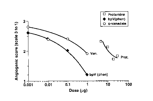
Figures 2a and b) show an anti-angiogenic response to bpV(phen),
orthovanadate (Van) and protamine (Prot) using a vascularization test on the
vitelline
membrane of chick embryos. Each point represents a minimum of 15 embryos (15
to
28). In Figure 2a), neovascularization was assessed using N/+ scoring system;
the
proportion of embryos showing anti-angiogenesis increases with dosage,
bpV(phen)
being the most potent agent. In Figure 2b), neovascularization was assessed
using the
1-2-3 scoring system; the angiogenic score decreases with dosage, bpV(phen)
being
the most potent agent.
Agents that are known to induce the proliferation of endothelial cells include
sodium orthovanadate, an inhibitor of phosphotyrosine phosphatases (83). The
mechanism by which this agent induces an invasive phenotype in capillary
endothelial
cells is not clearly understood. However, the effects of vanadate on cultured
cells are
similar, in many respects, to those elicited by PMA (74), bFGF (75), and
certain
retroviral transforming proteins, which act by inducing tyrosine-specific
protein
phosphorylation (76-79). Consistent with these observations is the finding
that both
phorbol ester and various growth factors including bFGF, VEGF and PDGF
stimulate
the phosphorylation of cellular proteins on tyrosine residues whereas vanadate
is an
inhibitor of tyrosine phosphatase, thereby producing a marked increase in
tyrosine
phosphorylation.
Other derivatives of vanadate (bpv(phen); bpv(pic)) have been synthesized and
have proved to be potent phosphotyrosine-phosphatase inhibitors (80). These
products
were expected to promote endothelial cell proliferation, like vanadate. On the
contrary,
these agents have been shown to inhibit the proliferation of several cell
types (71 ). The
cells seem to be arrested at G2/M. The simplest hypothesis to explain this
restriction
CA 02280249 1999-08-12
-7-
is that PTP(s) controlling entry of the cell into mitosis are target for the
PTP-Is. The
cdc25 protein was proposed as a target for PTP-Is (71 ).
We have tested the angiogenic potential of these PTP-Is by using both in vitro
and an ex ovo systems of analysis. The results show that PTP-Is are potent
inhibitors
of the angiogenic process (see next section), and this includes vanadate,
contrarily to
what was expected from the teachings of the prior art.
PTP-Is inhibit the proliferation of endothelial cells
Human umbilical vein endothelial cells (HUVECs) were extracted with
collagenase-controlled digestion as described in Sirois et al. (81 ). Pure
endothelial
cells were used before the fourth passage (trypsin-EDTA at each passage).
Quality of
the cells were analysed for their capacity to incorporate di-acetyl LDL and to
be
labelled with factor VIII related antigen.
Endothelial cells were plated at a density of 2500 cells/cm2 into sterile dish
coated with gelatin. Cells were cultured with complete medium (M199 + heparin
(90
~rg/ml) + L-glutamine (2mM) + bicarbonate + FBS (20%) + ECGS (100 Ng/ml))
during
24 h to insure cell adhesion. Then, cells were washed 3 times with PBS and
culture
medium was added according to experimental conditions. The last PBS wash was
considered as time 0.
Cell proliferation was evaluated with the amount of DNA present in the petri
dishes. Each experiment was performed in triplicate. Culture medium was
changed
daily. After 96 h of culture, cells were lysed with Na-Citrate-SDS solution
and
incubated with Hoescht 33358. Samples were read at 365 nm with a
spectrofluorometer.
The results show: (1 ) a linear relationship between the level of fluorescence
and
the number of cells (Fig. 1A), and (2) a dose-response inhibition of
endothelial cell
proliferation with the PTP-Is (Fig. 1 B). The approximative IC50 is 2 NM for
bpV[phen]
and 3.5 NM for bpV[pic].
Alternatively, HUVECs cultured in a fibrin matrix can form 3D tubular-like
structures in the presence of serum (82). This assay was performed in the
presence
of bpV(phen) or bpV(pic) to assess their influence on HUVECs differentiation
and
organization (Table 1 ). HUVECs were seeded on the bottom of gelatin-coated
wells
at high density to provide a confluent monolayer at 48 hours. Then 50, 000
HUVECs/ml were embedded in fibrinogen solution prior to polymerization. The
fibrin
matrix was covered by culture medium containing or not the tested substance.
Cell
behavior was observed periodically under phase contrast microscope. After 21
days
of culture, in the presence of 1 NM bpV(phen), cord-like (cords), tube-like
(tubes) and
stellates structures (Stell.struct.) were observed. At higher doses (2 and 3.5
pM),
fragmented cord- like structures were apparent. In the presence of bpV(pic),
cord and
tube-like structures were observed at 1, 2 and 3.5 NM. In the presence of
CA 02280249 1999-08-12
_$_
orthovanadate, cord and cord-like structures were still apparent at 10 NM, and
at 25
NM dead cells were sparsely distributed. These results suggest that the
peroxovanadium compounds interfere with endothelial cells organization and
terminal
differentiation. Furthermore the nature of the ancillary ligand is important
since
bpV(phen) is more potent in inhibition of tube formation than bpV(pic).
TABLE I
Doses cords tubes Stell.
(NM) struct.
0 +++ +++ +++
bpV 1 +++ +++ +++
phen 2 ++ + _
3.5 - - -
0 +++ +++ +++
bpV 1 +++ +++ +++
pic 2 +++ +++ +++
3.5 ++ + _
0 +++ +++ +++
2,5 +++ +++ +++
van
10 +++ +++ -
25
t Dead cells
The above results show that vanadate has an anti-angiogenic effect, in so far
as the stellate structures are affected (organization and terminal
differentiation).
Vanadate is at least 3 and 5 times less potent than bpV(pic) and pbV(phen),
respectively. The anti-angiogenic effect observed with vanadate is in
contradiction with
the teachings of Montesano (73).
It was indeed observed that, at low doses, all the vanadium compounds tested,
have a tendency to be pro-angiogenic, but a higher doses (almost doubling
doses), the
same become anti-angiogenic. The type of radical L, L' is important for the
potency of
the vanadium compounds, phenanthroline being twice as potent as picolinic
acid.
PTP-Is inhibit neovascularization
a. De>rnition of the test-system: The normal development of the chick embryo
involves the formation of an external vascular system located in the vitelline
membrane
CA 02280249 1999-08-12
_g_
which carries nutrients from the vitellus (yolk of an egg) to the developing
embryo.
When placed onto the vitelline membrane, anti-angiogenic substances can
inhibit the
blood vessel development that occurs in the vitelline membrane. To facilitate
access
to the vitelline membrane, chick embryos are transferred to a sterile culture
box (Petri
dish) and placed in a humidity- and temperature-controlled incubator. Embryos
can
then develop in this ex ovo condition for several days.
An aliquot of PTP-Is was mixed with a methylcellulose solution and allowed to
air-dry into thin discs. Methylcellulose forms an inert matrix from which the
PTP-Is can
diffuse slowly. Methylcellulose discs containing PTP-I were placed on the
external
border of the vascular perimeter of the vitelline membrane where the
angiogenic
process is still active.
The effect of discs-containing PTP-Is on proximal vascular development was
always compared to that of discs-containing control buffer. The discs were
placed on
the embryo's vitelline membrane on Day 0 or Day 1 of the ex ovo growth
process; at
this point, only beginnings of the main blood vessels are invading the
vitellus. The
embryos were then put in culture conditions until vascularization is assessed
(approximately 24 h). Water- and PTP-I-containing discs were always added
simultaneously on the vitelline membrane of the same embryo. Both discs were
arranged in a symmetric fashion with respect to the cephalo-caudal axis of the
embryo
in order to minimize inter-individual variations when comparing PTP-I compound
with
controls.
b. Anti-angiogenic activity: Embryonic vascularization tests (EVTs) were
performed using different concentrations of protamine (5 to 20 Ng) as a
positive control
or PTP-Is (0.001 to 10 Ng). After one day of culture, the level of
vascularization in the
area covered by the disc was graded by a pair of scientists in the usual blind
fashion.
To facilitate the location of the discs, a black O-ring was placed around it
just after its
deposition on the vitelline membrane. Evaluation scale for the EVT test is
based on
two different scoring systems.
Assessment of blood vessel formation:
Blood vessel formation was assessed in a blind fashion. The area of the
vitelline
membrane that lay beneath the methylcellulose discs was scored for the degree
of
vascularization, using two scoring systems (Ni+" or" 1-2-3"). The following
selection
criteria were used:
N/+ system:
A score of "N" for "Normal" was attributed when all the following conditions
applied:
~ Blood vessels, present in the evaluated area, grew along their path with no
abnormal deviation. Collateral branching density was normal and the growth
path of the lateral branches was also normal.
CA 02280249 1999-08-12
-10-
A score of "+" was attributed when at least one of the following conditions
applied:
~ Major blood vessels grew across the evaluated area but their path was
clearly
affected (winding).
~ Major blood vessels grew across the evaluated area but collateral branching
density was clearly diminished.
~ Major blood vessels penetrated the evaluated area but their growth path was
rapidly deviated. A kink was observed in the blood vessel.
~ Major blood vessels penetrated the evaluated area but were stunted. No
growth
was observed beyond that point.
~ A drastic deviation in the growth path occurred when major blood vessels
reached the proximal ridge of the disc.
1-2-3 grading scale system:
A score of 3 was attributed when all the following conditions applied:
~ Blood vessels, present in the evaluated area, grew along their path with no
abnormal deviation. Collateral branching density was normal and the growing
path of the lateral branches was also normal.
A score of 2 was attributed when at least one of the following conditions
applied:
~ Major blood vessels grew across the evaluated area but their path was
clearly
affected (winding).
~ Major blood vessels grew across the evaluated area but collateral branching
density was clearly diminished.
A score of 1 was attributed when at least one of the following conditions
applied:
~ Major blood vessels penetrated the evaluated area but their growing path was
rapidly deviated. A kink was observed in the blood vessel.
~ Major blood vessels penetrated the evaluated area but were stunted. No
growth
was observed beyond that point.
~ A drastic deviation in the growth path occurred when major blood vessels
reached the proximal ridge of the disc.
A score of 3 means that blood vessel development is normal whereas a score of
1
indicates the greatest degree of angiostatic activity.
CA 02280249 1999-08-12
-11-
0 M .- f~00 (O ~ O N M 'vt~ O Cfl
p ~ "''N N t-r N N N r nj N N e- N
r
C
~
O ~ f~ ~ CO~ T f~ CO tnM Wit'~ d' 00(O t-
N ;~,~-,, 6n f~Cflr N In O N N
~ ~ U
L
N N Wit'N 00 Cfl00 M ~ ~ (ON N
U N r- ~ r N N N N r r ~-N
N
f~
O M
d. O_ p M r ~ ~ 1 1 1 O 1 1 , r (V
(V(V e-r r
N ~ c
1 1 1 ~ 1 1 1
O nT
Q
C Q ~ T T T T 1 1 1 ~ 1 1 1
..
M 1 ~ 1 1 1 1 ~ ~ V O
C 0 O 1 1 N N
T N N N fa
O '
~~
~
- ~ 1 1 ~ 1 1 1 1 1 I~ N N w
t0 ~
>
(n ~
U
O
O O
C 1 1 ~ 1 1 1 1 1 ~ ~ ~ ~ ~ M
C
N
O ~ M 1 1 ~ I ~ ~ N ~ 1 1 1 1 ~ O .t/~
O N C
O U ''"' T N N N t- N
O
N O
~C O ~ i~
> 1 1 ~ 1 N ~ ~ p 1 1 1 1 ~ U N
(~ e- .C O
Q N
1 1 ~ 1 ~ ~ ~ ~ 1 1 1 1 ~ O ..
O O T
C N
p ~ M M 1 O 1 ~ M OO CO1 1 1 1 f~ N
j ~> O
O :~. "-'N N N N r r- fB
U T ~ T
~ O
fE In O O M 1 Cfl1 N r r CO1 1 1 1
_ (~
Q Q ~ ~ N ~ r M CO 00
C E
c!S C c T 1 N 1 M M ~ r'1 1 1 , O .~ N
O T !~ !~ r T r'
O U
r _~
_~
(I~ ~ .N
c0 U O r _N
lyO O O O O r r r "- C
N ~ "-' O - N M p O O O e- O
N ~ ~ ~ ~ ~ N ~ C G C O '~;O ~ :C ~
7 C_C_ C_C_ p N p N O ~
- N ~ .~~~ ~~~~ ~ ~ ~ .s=OI ~ Q. N
O ~C ~ ~ ~ ~ ~ d ~ ~ ~ f~ O o C
C Q II
a a a a ~ .n.n ~ 0 0 0 U Ic
w o m o
r r N
CA 02280249 1999-08-12
-12-
A dose-response inhibition was obtained with protamine, bpv[phen], and
orthovanadate (Figure 2). The approximative IC50 is 0.1 pg for bpv[phen], 0.6
pg for
orthovanadate, and 11 Ng for protamine (whatever the scoring system). An
overall
summary of the experimental data harvested is shown in Table II.
The results show that PTP-Is are potent inhibitors of the angiogenic process.
This is the first demonstration that the PTP-Is inhibit endothelial cell
proliferation and
angiogenesis. Prior art on the subject (vanadate) taught the opposite, that is
PTP-I
promote angiogenesis.
PTP-Is inhibit the elevation of endothelins
The data presented in Table III show the effect of bpV(phen) on the plasmatic
levels
of ETs. In this study, rats were injected intraperitoneally with bpV(phen)
(0.5 mg/100g
b.w.) 16 hours before the administration of either insulin or vehicle. Two
minutes
following the insulin administration (1.5 Ng/100g b.w., or vehicle), the
plasma levels of
ETs were determined. Insulin administration induced a strong increase of seric
ETs
concentration (40). This increase was completely abolished by bpV(phen). In
addition
bpV(phen) decreased the insulin-stimulated levels of plasmatic ETs below
control
levels. These results suggest that bpV(phen) inhibits the insulin-induced
release of ETs
that subsequently can lead to diabetic complications like vasculopathy and
neph ropathy.
TABLE III
Plasmatic endothelins
(pg/ml)
Control 113.41 t 10.91
Insulin 253.10
Insulin, bpV(phen)77.68 t 3.08
ETs (ET-1, ET-2, ET-3) were measured by RIA (Amersham kit RPA 555) after
lyophilisation and extraction on C2 columns (500 mg). Results are mean t SEM.
p<0.0209, control (n=4) vs Insulin, pV (n=3).
The above results illustrate the antiangiogenic potential of the vanadium
compounds. The administration of PTP-Is could be used to control the
progression of
several angiogeno-dependent conditions. Vanadates being anti-tumor compounds,
the
present PTP-Is should verify as anti-tumor therapeutic agents, and part of
this activity
should be due to an anti-angiogenic effect. Pharmaceutical compositions would
comprise potent PTP-Is capable of achieving about 0.1 to 100 NM, preferably 2-
40 pM,
extracellular concentration. A dose of 20 Nmoles per kilogram was successful
in rats
CA 02280249 1999-08-12
-13-
in reversing the endothelin increase after insulin administration. The
peroxovanadium
compounds which are more potent and provoke less side effects than the
vanadium
"oxo" compounds, can be administered to achieve the desired dosage.
Although the present invention has been described hereinabove by way of a
preferred embodiment thereof, this embodiment can be modified at will, within
the
scope of the appended claims, without departing from the spirit and nature of
the
subject invention.
CA 02280249 1999-08-12
-14-
6. References
1. Folkman J. (1995). Angiogenesis in cancer, vascular, rhumatoid and other
diseases.
Nature Med 1: 27-31.
2. Schreiber, A.B., Winkler, M.E., Derynck, R. (1986). Transforming growth
factor-alpha: a more potent angiogenic factor than epidermal growth factor.
Science 232: 1250-1254.
3. Roberts, A.B., Sporn, M.B., Assoian, R.K., Smith, J.M., Roche, N.J.,
Wakefield, L.M.
(1986). Transforming growth factor type beta: rapid induction of fibrosis and
angiogenesis in vivo and stimulation of collagen formation in vitro. Proc.
Natl.
Acad. Sci. USA 84: 4167-4171.
4. Frater-Schroder, M., Risau, W., Hallmann, R., Gautschi, R., Bohlen, P.
(1987).
Tumor necrosis factor type-alpha, a potent inhibitor of endothelial cell
growth
in vitro, is angiogenic in vivo. Proc. Natl. Acad. Sci. USA 84: 5277-5282.
5. Fett, J.W., Strydom, D.J., Lobb, R.R., Alderman, R.M., Riordn, J.F.,
Vallee, B.L.
(1985). Isolation and characterization of angiogenin, an angiogenic protein
from
human carcinoma cells. Biochemistry 24: 5480-5488.
6. Ziche, M., Jones, J., Gullino, P. (1982). Role of prostaglandin E1 and
copper in
angiogenesis. J. Natl. Cancer Inst. 69: 475-482.
7. Dobson, D.E., Kambe, A., Block, E., Dion, T., Lu, H., Castellot, Jr. J.J.,
Speigelman,
B.M. (1990). 1-Butyrylglycerol: a novel angiogenesis factor secreted by
differentiating adipocytes. Cell 61: 223-230.
8. Baird, A., Durkin, T. (1986). Inhibition of endothelial cell proliferation
by type-beta
transforming growth factor: interaction with acidic and basic fibroblast
growth
factors. Biochem. Biophys. Res. Commun. 138: 476-482.
9. Frater-Schroder, M., Muller, G., Birchmeier, W., Bohlen, P. (1986).
Transforming
growth factor-beta inhibits endothelial cell proliferation. Biochem. Biophys.
Res.
Commun. 137: 295-302.
10. Folkman, J., Klagsbrun, M. (1987). Angiogenic factors. Science 235: 442-
448.
11. Klagsbrun, M., D'Amore, P.A. (1991 ). Regulators of angiogenesis. Annu.
Rev.
Physiol. 53: 217-232.
12. Gospodarowicz, D., Ferrara, N., Schweigerer, L., Neufeld, G. (1987).
Structural
characterization and biological functions of fibroblast growth factor. Endocr.
Rev. 8: 95-104.
13. Thomas, K., Rios Candelore, M., Gimenez-Gallego, G., Di Salvo, J., Bennet,
C.,
Rodkey, J., Fizpatrick, S. (1985). Pure brain-derived acidic fibroblast growth
factor is a potent vascular endothelial cell mitogen with sequence homology to
interleukin-1. Proc. Natl. Acad. Sci. USA 82: 6409-6416.
14. Miyazono, K., Okabe, T., Urabe, A., Takata, F., Heldin, C.-H. (1987).
Purification
and properties of an endothelial cell mitogen from human platelets. J. Biol.
CA 02280249 1999-08-12
-15-
Chem. 262: 4098-4104.
15. Ishikawa, F., Miyazono, K., Hellman, U., Drexler, H., Westerndt, C.,
Hagiwara, K.,
Usuki, K., Takaku, F., Risau, W., Heldin, C.-H. (1989). Identification of
angiogenic activity and the cloning and expression of platelet-derived
endothelial cell growth factor. Nature 338: 557-561.
16. Leung, D.W., Cachianes, G., Kuang, W.-J., Goeddel, D.V., Ferrara, N.
(1989).
Vascular endothelial growth factor is a secreted angiogenic mitogen. Science
246: 1306-1312.
17. Houck, K.A., Ferrara, N., Winer, J., Cachianes, G., Li, B. et Leung, D.W.
(1991).
The vascular endothelial growth factor family: Identification of a fourth
molecular species and characterization of alternative splicing of RNA. Mol.
Endocrinol. 5: 1806-1814.
18. Breier, G., Albrecht, U., Sterrer, S. et Risau, W. (1992). Expression of
vascular
endothelial growth factor during embryonic angiogenesis and endothelial cell
differentiation. Development 114: 521-532.
19. Bussolino, F., Albini, A., Garnussi, G., Presta, M., Viglietto, G., Ziche,
M. and
Persico, G. (1996). Role of soluble mediators in angiogenesis. Eur. J. Cancer
32A: 2401-2412.
20. Colville-Nash, P.R. and Willoughby, D.A. (1997). Growth factors in
angiogenesis:
current interest and therapeutic potential. Mol. Med. Today. 3: 14-23.
21. Knighton, D.R., Hunt, T.K., Scheuenstuhl, H., Halliday, B.J., Werb, Z.,
Banda, M.J.
(1983). Oxygen tension regulates the expression of angiogenesis factor by
macrophages. Science 221: 1283-1285.
22. Ladoux, A. and Frelin, C. (1993). Hypoxia is a strong inducer of vascular
endothelial growth factor mRNA expression in the heart. Biochem. Biophys.
Res. Commun. 195: 1005-1010.
23. Nomura M., Yamagishi S.I., Harada S.I., Hayashi Y., Yarnashima T.,
Yamashita
J. and Yamamoto H. (1995). Possible participation of autocrine and paracrine
vascular endothelial growth factors in hypoxia-induced proliferation of
endothelial cells and pericytes. J. Biol. Chem. 270: 28316-28324.
24. Walgenbach, K.J., Gratas, C., Shestak, K.C. and Becker, D. (1995).
Ischaernia-induced expression of bFGF in normal skeletal muscle: a potential
paracrine mechanism for mediating angiogenesis in ischaemic skeletal muscle.
Nature Med. 1: 453-459.
25. Lew, A.P., Lew, N.S., Iliopoulos, O., Jiang, C., Kaeling Jr., W.G. and
Goldberg,
M.A. (1997). Regulation of vascular endothelial growth factor by hypoxia and
its modulation by the von Hippel-Lindau tumor suppressor gene. Kidney Int. 51:
575-578.
26. Sandner, P., Wolf, K., Bergmaier, U., Gess, B. and Kurtz, A. (1997).
Induction of
CA 02280249 1999-08-12
-16-
VEGF and VEGF receptor gene expression by hypoxia: Divergent regulation
in vivo and in vitro. Kidney Int. 51: 448-453.
27. Ferrara, N. (1995). Missing link in angiogenesis. Nature 376: 467.
28. Koch, A.E., Halloran, M.M., Haskell, C.J., Shaw, M.R. and Polverini, P.J.
(1995).
Angiogenesis mediated by soluble forms of E-selectin and vascular cell
adhesion molecule-1. Nature 376: 517-519.
29. Polverini, J.P., Cotran, R.S., Gimbrone Jr, M.A., Unanue, E.R. (1977).
Activated
macrophages induce vascular proliferation. Nature 269: 804-806.
30. Sunderkotter, C.. Steinbrink, K., Goebeler, M., Bhardwaj, R., Sorg, C.
(1994).
Macrophages and angiogenesis. J. Leucocyte Biol. 55: 410-422
31. Polverini, P.J. (1996). How the extracellular matrix and macrophages
contribute to
angiogenesis-dependant diseases. Eur. J. Cancer 14: 2430-2437.
32. Gross, J.L., Moscatelli, D., Jaffe, E.A., Rifkin, D.B. (1982). Plasminogen
activator
and collagenase production by cultured capillary endothelial cells. J. Cell
Biol.
95: 974-981.
33. Moscatelli, D.A., Rifkin, D.B., Jaffe, E.A. (1985). Production of latent
collagenase
by human umbilical vein endothelial cells in response to angiogenic
preparations. Exp. Cell Res. 156: 379-390.
34. Ingber, D.E. (1991). Extracellular matrix and cell shape: potential
control points for
inhibition of angiogenesis. J. Cell. Biochem. 47: 236-241.
35. Ingber, D.E. (1992). Extracellular matrix as a solid-state regulator in
angiogenesis:
identification of new targets for anti-cancer therapy. Sem. Cancer Biol. 3: 57-
63.
36. Bischoff, J. (1995). Approaches to studying cell adhesion molecules in
angiogenesis. Trends in Cell Biol. 5: 69-74.
37. Eisenstein, R. (1991). Angiogenesis in arteries: review. Pharmac. Ther.
49: 1-19.
38. Folkman, J., Ingber, D. (1992). Inhibition of angiogenesis. Cancer Biol.
3: 89-96.
39. Bicknell, R. (1994). Vascular targeting and the inhibition of
angiogenesis. Ann.
Oncol. 5: S45-S50.
40. Battistini, B., Chailler, P., D'Orleans, J., Briere, N., Sirois, P.
(1993). Growth
regulatory properties of endothelins. Peptides 14, 385-399.
41. Pedram A., Razandi M., HU R-M, Levin E.R. (1997). Vasoactive peptides
modulate vascular endothelial cell growth factor production and endothelial
cell
proliferation and invasion. J. Biol. Chem. 272: 17097 17013.
42. Asham, E.H., Loizidou, M. & Taylor, I. (1998). Endothelin-1 and tumour
development. Eur. J. Surg. Oncol. 24: 57-59.
43. Kusuhara, M., Yamaguchi, K., Nagasaki, K., Hayashi, C., Suzaki, A., Nori,
S.,
Hanada, S., Nakamura, Y. & Abe, K. (1990). Production of endothelin in human
cancer cell lines. Cancer Res. 50: 3257-3261.
44. Schichiri, M., Hirata, Y., Nakajiroa, T., Ando, K., Imal, T., Yanagisawa,
M., Mazaki,
CA 02280249 1999-08-12
-17-
T. & Marumo, F. (1991 ). Endothelin-I is an autocrine/paracrine growth factor
for
human cancer cell lines. J. Clin. Invest. 87: 1867- 1871.
45. Pagotto, U., Arzberger, T., Hopfer, U., Weidnl, A.& Stella, G. (1995).
Cellular
localization of endothelin receptor mRNA ETA and ETB in the brain tumour and
normal human brain. J. Cardiovasc. Pharmacol. 26 (Suppl. 3): S104-106.
46. Stiles, J.D., Ostrow, P.T., Balos, L.L., Greenberg, S.J., Plunkett, R.,
Grand, W. &
Heffner Jr., R.R. (1997). Correlation of endothelin-I and transforming growth
factor b1 with malignancy and vascularity in human gliomas. J. Neurobiol. 56:
435-439.
47. Ishibashi M., Fujita, M., Nagai, K., Koko, M., Furue, H., Haku, E.,
Osamura, Y.,
Yamaji, T. (1993). Production and secretion of endothelin by hepatocellular
carcinoma. J. Clin. Endocrinol. Metab. 76: 378-383.
48. Giaid, A., Hamid, Q.A., Springall, D.R., Yanagisawa, M., Shinmi, O.,
Sawamura,
T., Masaki, T., Kimura, S., Corrin, B. & Polak, J.M. (1990). Detection of
endothelin immunoreactivity and mRNA in pulmonary tumours. J. Pathol. 162:
15-22.
49. Nelson, J., Chan, K., Hedican, S., Magnuson, S., Opgenorth, T., Bova, G. &
Simons, J. (1996). Endothelin- 1 production decreased B receptor expression
in advanced prostate cancer. Cancer Res. 56: 663-668.
50. Kikuchik, K., Nakagawa, H., Kadono, T., Etoh, T., Byers, H., Mihm, M. &
Tamaki,
K. (1996). Decreased ETB receptor expression in human metastatic melanoma
cells. Biochem. Biophys. Res. Commun. 219: 734-739.
51. Ziche, M., Morbidelli, L., Donnini, S. & Ledda, F. (1995). ETB receptor
promote
proliferation and migration of endothelial cells. J. Cardiovasc. Pharmacol. 26
(Suppl. 3): S284-S286.
52. Inagaki, H., Bishop, A., Escrig, C., Wharton, J., Allen-Mersh, T. & Polak,
J. (1991).
Localization of endothelin like immunoreactivity and endothelin binding sites
in
human colon. Gastroenterology 101: 47-54.
53. Inagaki, H., Bishop, A., Eimoto, T. & Polak, J. (1992). Autoradiographic
localization
of endothelin-1 binding sites in human colonic cancer tissue. J. Pathol. 168:
263-267.
54. Eguchi, S., Hirata, Y., Imai, T. & Marumo, P. (1995). ET-1 as an autocrine
growth
factor for endothelial cells. J. Cardiovasc. Pharmacol. 26 (Suppl. 3).
S279-S283.
55. Leveson, S., Wiggins, P., Giles, G., Parkin, A. & Robinson, P. (1985).
Deranged
blood flow patterns in the detection of liver metastases. Br. J. Surg. 72:
128-130.
56. Carter, R., Anderson, J., Cooke, T., Baxter, J. & Angerson, W. (1994).
Splanchnic
blood flow changes in the presence of hepatic tumours, evidence of humoral
CA 02280249 1999-08-12
-18-
mediator. Br. J. Cancer 69: 1025-1026.
57. Nakamura, T., Ebihara, I., Fukui, M., Tomino, Y. & Koide, H. (1995).
Effect of a
specific endothelin receptor A antagonist on mRNA levels for extracellular
matrix components and growth factors in diabetic glomeruli. Diabetes 44:
895-899.
58. Lee, K.M., Tsai, K.Y., Wang, N. & Ingber, D.E. (1998). Extracellular
matrix and
pulmonary hypertension: control of vascular smooth muscle cell contractility.
Am. J. Physiol. 274: H76-82.
59. Hirshfeld, J.W. Jr, Schwartz, J.S., Jugo, R., MacDonald, R.G., Goldberg,
S.,
Savage, M.P., Bass, T.A., Vetrovec, G., Cowley, M., Taussig, A.S., et al.
(1991 ) Restenosis after coronary angioplasty: a multivariate statistical
model
to relate lesion and procedure variables to restenosis. The M-HEART
Investigators. J. Am. Coll. Cardiol. 18: 647-656
60. Maseri, A. (1996). An unresolved, persisting challenge for percutaneous
transluminal coronary angioplasty: how to identify the lesions that will not
restenose. Eur. Heart J. 17: 814-815.
61. Kirchengast, M. & Munter, K. (1998). Endothelin and restenosis.
Cardiovasc. Res.
39: 550-555.
62. Kramer, B.K., Ackermann, M., Kohler, S.M. & Riegger, G.A. (1994). Role of
endothelin in hypertension. Clin. Investig. 72: 88-93
63. Levin, E,R. (1995). Endothelins: mechanisms of disease. New Eng. J. Med.
333:
356-363.
64. Ivic, M.A. & Stefanovic, V. (1998). Endothelins in hypertension and kidney
diseases. Pathol. Biol. (Paris) 46: 723-730
65. Rohmeiss, P., Birck, R., Braun, C., Kirchengast, M. & van der Woude, F.J.
(1999).
Targets for endothelin in the diseased kidney: clues for therapeutic
intervention.
Exp. Nephrol. 7: 1-10.
66. Cardell, L.O., Uddman, R. & Edvinsson, L. (1994). Endothelins: a role in
cerebrovascular disease? Cephalalgia 14: 259-65
67. Cosentino, F. & Katusic, Z,S. (1994). Does endothelin-1 play a role in the
pathogenesis of cerebral vasospasm? Stroke 25: 904-908
68. Battistini, B. & Dussault, P. (1998). The many aspects of endothelins in
ischemia-reperfusion injury: emergence of a key mediator. J. Invest. Surg. 11:
297-313.
69. Posner, B.I., Shaver, A. and Fantus, G., 1990. Insulin Mimetic Agents:
Vanadium
and Peroxovanadium Compounds. in new antidiabetic drugs, eds: Bailey, C.J.,
Flatt, P.R., Smith-Gordon.
70. Shaver, A., Ng J.B., Hall, D.A., Posner, B.I. (1995). The chemistry of
peroxovanadium compounds relevant to insulin mimesis. Mol. Cell. Biochem.
CA 02280249 1999-08-12
-19-
153: 5-15.
71. Faure, R., Vincent, M., Dufour, M., Sghaver, A. 8~ Posner, B. (1995).
Arrest at the
G2/M transition of the cell-cycle by protein-tyrosine phosphatase inhibition:
Studies on a neuronal and a glial cell line. J. Cell. Biochem. 58, (3), 389-
401.
72. Olivier, M. Romero-Gallo B. J., St Laurent, R., Racette, N., Stevenson,
M., Jacobs,
P., Posner, B., Tremblay, M., Faure, R. (1998). Modulation of INFg induced
macrophage activation by phosphotyrosine phosphatase inhibition. J. Biol.
Chem. 273, 13944-13949.
73. Montesano, R., Pepper, M.S., Belin, D., Vassalli, J.-D. & Orci, L. (1988).
Induction
of angiogenesis in vitro by vanadate, an inhibitor of phosphotyrosine
phosphatases. J. Cell. Physiol. 134: 460466.
74. Montesano, R. and Orci, L. (1985). Tumor-promoting phorbol esters induce
angiogenesis in vitro. Cell 42: 469-477.
75. Montaseno, R., Vassalli, J.D., Baird, A., Guillemin, R. and Orci, L.
(1986). Basic
fibroblast growth factor induces angiogenesis in vitro. Proc. Natl. Acad. Sci.
USA. 83: 7297-7301.
76. Sefton, B.M. and Hunter, T. (1984). Tyrosine protein kinases. Adv. Cyclic
Nucleotide Protein Phosphorylation Res. 18: 195-203.
77. Hunter, T. and Cooper, J.A. (1985). Protein-tyrosine kinases. Annu. Rev.
Biochem.
54: 897-930.
78. Hunter, T. and Cooper, J.A. (1986). Viral oncogenes and tyrosine
phosphorylation.
In: The Enzymes, Vol. 17. P.D. Boyer and E.G. Krebs, eds. Academic Press,
Orlando, Florida, pp. 191 -246.
79. Ramachandran, J. and Ullrich, A. (1987). Hormonal regulation of protein
tyrosine
kinase activity. Trends Pharmacol. Sci. 8: 28-31.
80. Posner, B.I., Faure, R., Burgess, J.W., Bevan, A.P., Lachance, D., Zhan-
Sun, G.,
Fantus, I.G., Ng, J.B., Hall, D.A., Lum, B.S. and Shaver, A. (1994).
Peroxovanadium compounds: a new class of potent
phosphotyrosine-phosphatase inhibitors which are insulin mimetics. J. Biol.
Chem. 269: 4596-4604.
81. Sirois, E., Cote, M.F. and Doillon, C.J. (1993). Growth factors and
biological
supports for endothelial cell lining: in vitro study. Int. J. Artificial
Organs 16:
609-619.
82. Janvier R., Sourla, A., Koutsilieris, M., Doillon, C. (1997). Stromal
Fibroblasts are
required for PC-3 human prostate cancer cells to produce capillary-like
formation of endothelial cells in a three-dimensional co-culture system.
Anticancer research 17, 1551-1558.
83. Nechay, B.R., Nanninga, L.B., Nechay, P.S.E., Post, R.L., Grantham, J.J.,
Macara,
I.G., Kubena, L.F., Phillips, D.T. & Nielsen, F.H. (1986). Role of vanadium in
-20-
biology. Fed Proc 45, 123- 132.