Note: Descriptions are shown in the official language in which they were submitted.
CA 02280449 1999-08-13
P'C 10178A
-1-
STABILIZED PROTEIN COMPOSITIONS
Field Of the Invention
This invention relates to stabilized protein compositions. The stabilized
compositions
of the present invention are useful in delivering therapeutically effective
amounts of proteins,
including colony stimulating factors, such as bovine granulocyte colony
stimulating factor (bG-
CSF), to mammals, including humans, cattle, swine, horses, goats, sheep, dogs
and cats, for
sustained periods. More particularly, the stabilized compositions of the
present invention
contain stabilizing buffers such as HEPES, TES and TRICINE are capable of
maintaining a
sustained period of protein activity, both in vivo and in vitro.
Background of the Invention
Formulations of therapeutically effective proteins, such as G-CSF, remain
difficult to
formulate for extended shelf life in vitro and in vivo activity. Formulations
of such proteins
must maintain their activity and biological integrity for appropriate periods
of time for effective
treatment. In addition, formulations of such proteins must be manufacturable
as well as
capable of being administered to an animal in a pharmaceutically acceptable
manner.
Pharmaceutical compositions of proteins have been provided in frozen or
lyophilized
form and maintained in vitro under storage conditions, which maintain protein
activity for
extended periods off time. Lyophilized preparations are reconstituted prior to
use with
pharmaceutically aaxptable diluents, such as sterile water for injection.
Pharmaceutical
compositions of proteins have also been provided in liquid form. Such liquid
protein
formulations are difficult to maintain in storage due to the loss of protein
activity over time,
particularly at elevated temperatures.
Formulations of therapeutically effective proteins, whether in solid
(lyophilized) or
liquid form, are difincult to administer to animals without sudden loss of
activity after
administration, such .as by subcutaneous injection, to the animal. Rapid loss
of protein activity
at the injection site renders the protein inconvenient for treating infections
in mammals since
effective therapy requires daily doses during desired periods of coverage.
Granulocyte colony
stimulating factors (G-CSFs), such as bovine granulocyte colony stimulating
factor (bG-CSF),
are unstable at or above 40°C due to loss in secondary structure and
disulfide interchange
and subsequent loss in activity. This loss in activity occurs at the injection
site since bovine
body temperature is .around 40°C and the injection site is at
physiological pH range.
Various protein formulations for extending shelf life are known. US Patent No.
5,104,651, issued April 14, 1992 (Boone et al.), refers to a pharmaceutical
composition of G-
CSF and an acid at a pH in the range of 3.0-3.7 with a conductivity of less
than
1000wmhos/cm. U:i Patent No. 4,992,271, issued February 12, 1991 (Fernandes et
al.),
refers to a pharmaceutical composition containing a biologically active
recombinant interleukin
CA 02280449 1999-08-13
-2_
2 protein dissolved in an aqueous based carrier medium at a pH of 6.8 to 7.8
and which
further contains a si:abilizer for the protein, such as human serum albumin.
US Patent No.
4,623,717, issued November 18, 1986 (Femandes et al.), refers to pasteurized
therapeutically
active protein compositions whereby thermally sensitive, therapeutically
active proteins are
pasteurized by mixing the protein with a stabilizing amount of a sugar or
reduced sugar and
an amino acid prior to pasteurization. US Patent 4,645,830, issued February
24, 1987
(Yasushi et al.), refers to a stable interleukin 2 composition containing
interleukin 2, human
serum albumin and a reducing compound, at a pH of 3 to 6 in solution. US
Patent No.
4,647,454, issued March 3, 1987, (Cymbalista), refers to a method of
stabilizing human
fibroblast interferon with polyvinyl pyrrolidone. US Patent No. 4,675,184,
issued June 23,
1987 (Hasegawa et al.), refers to a pharmaceutical composition for treating
viral infections
containing interferon, a tri or higher polyhydric sugar alcohol, an organic
buffer and a
pharmaceutical carrier or diluent, wherein the composition has a pH of about 3
to 6.
One example of a therapeutically effective class of proteins is that of
granulocyte
colony stimulating factors (G-CSFs). Granulocyte colony stimulating factor (G-
CSF) is one of
several glycoprotein growth factors known as colony stimulating factors. Such
colony
stimulating factor$ :support the proliferation of haemopoietic progenitor
cells and stimulate
proliferation of specific bone marrow precursor cells and their
differentiation into granulocytes.
In addition, G-CSF its capable of stimulating neutrophilic granulocyte colony
formation and to
inducing terminal differentiation of murine myelomonocytic leukemic cells in
vitro. G-CSF has
also been shown to stimulate the functional activities of neutrophils
resulting in enhanced
microbiocidal activir~. G-CSF has a known amino acid sequence of 174 amino
acids.
Recombinant forms of CSFs and G-CSFs have been prepared. The cloning and
expression of DNA .encoding for human G-CSF is known (Nagata, S. et al.,
Nature, 319, 415-
418 (1986). WO-A~-8604606 and WO-A-8604506 describe a gene encoding human G-
CSF.
US Patent No. 5,606,024, issued February 25, 1997 (Boone et al.) and US Patent
No.
5,472,857 issued December 5, 1995, describe the DNA sequence encoding canine
granulocyte colony stimulating factor (cG-CSF) as well as a method for
treating or preventing
infections in canine or feline animals by administering effective amounts of
human and canine
G-CSF to such anirnals. US Patent No. 4,810,643, issued March 7, 1989 (Souza),
describes
human G-CSF like polypeptides. European Patent Application No. 719 860,
published July 3,
1996, describes the amino acid sequence of naturally occurring bovine
granulocyte colony
stimulating factor (bG-CSF), the DNA sequence encoding for bG-CSF and a method
for
treating or preventing mastitis in an animal by administering to the animal an
effective amount
of G-CSF. WO-A-8702060 describes human G-CSF like polypeptide, sequences
encoding
64680-1161
CA 02280449 1999-08-13 w
-3-
them and methods of producing them. US Patent No 4,833,127, issued May 23,
1989 (Ono et
al.), describes a novel biologically active human granulocyte colony
stimulating factor.
European Patent Application No. 612 846, published August 31, 1994, describes
certain G-
CSF analogs and compositions containing such analogs.
Granulocyte colony stimulating factors are useful as anti-infective agents
which
increases the immune competence of the animal rather than targeting a specific
microbial
target necessary for growth or virulence. There are few other commercially
available agents
used in veterinary medicine that target non-specific immune responses leading
to increased
resistance to microbial infection. Available control measures are limited to
conventional
antimicrobials and a limited number of biologicals. Economic losses associated
with milk
withdrawal periods in cattle limit the utility of conventional antimicrobials.
Current vaccines
target a limited number of species and the field efficacy of these agents vary
widely. The
most successful vaccines, (E. coli J5) are limited in their world wide use due
to safety
concerns associated with endotoxin contamination.
Mastitis is a~ major disease problem affecting dairy producers worldwide.
Economic
losses in the United States associated with mastitis exceed $1 billion
annually. These losses
are associated with mortalities, milk discard, acute and chronic decreases in
milk production,
increased early culling and drug and veterinary labor expenses. Periparturient
dairy cows
exhibit impaired immune responsiveness (neutrophil function) which increase
their
susceptibility to bacterial infections of the mammary gland. The impact of
this increased
susceptibility is exemplified by the fact that about 40% of new clinical
intramammary infections
occur within the first two weeks after calving. Mastitis is associated with a
wide variety of
bacterial pathogens including both Gram positive and Gram negative organisms.
Some of the
known pathogenic microorganisms causing mastitis are Escherichia coli,
Staphylococcus
aureus, Streptococcus agalactiae, Streptococcus uberis, Streptococcus
dysgalactiae,
Aerobacter aerogenes, Klebsiella pneumoniae and Pseudomonas aeruginosa. These
pathogens enter the udder through the teat canal and produce inflammation of
the milk
producing tissue causing the formation of scar tissue, which can result in a
permanent loss of
milk producing capacity. Various forms of mastitis include: udder infection,
chronic mastitis,
clinical mastitis and subclinical mastitis.
Current antimicrobial therapies and vaccines possess a number of deficiencies
that
limit their utility in lactating cows. Antibiotic therapy to control mastitis
has been found lacking.
There is a need for a biotherapeutic agent, which is useful in restoring
normal immune
competence resulting in the decreased incidence and severity of mastitis.
64680-1161
CA 02280449 1999-08-13
Bovine respiratory disease, also referred to as shipping fever, is another
common
disease affecting cattle. Bovine respiratory disease affects cattle after
shipment either into
feedlots or onto pasture and results from a variety of stresses affecting
cattle including
weaning, castration, dehoming, fasting, overcrowding, exposure to infectious
agents, diet
changes and temperature changes, in combination with infection by any of
several known
pathogens. Pasteurella haemolytica is one such common pathogen resulting in
damage to
the respiratory system of cattle.
Several additional infectious diseases, including various reproductive
diseases, are
also known to affect humans, swine, cattle, dogs, cats, horses, goats and
sheep. One
example of such a disease, occuring in cattle, is metritis.
There is a need for a stable protein composition which remains therapeutically
effective for sustained periods of time in vivo. In addition, there is a need
for formulations of
proteins that provide for extended in vitro shelf life and storage.
Summary of the Invention
The present invention relates to a stabilized protein composition comprising a
protein
and a stabilizing buffer, which composition is capable of maintaining
therapeutic levels of such
protein for a sustained period.
Specific embodiments of the invention include a stabilized protein composition
which
composition is at a physiological pH.
Other specific embodiments of the invention include a stabilized protein
composition
which composition is. at a physiological temperature.
Other specific embodiments of the invention include a stabilized protein
composition
wherein the stabilizing buffer is selected from the group consisting of:
HEPES, TES and
TRICINE.
Still other specific embodiments of the invention include a stabilized protein
composition wherein the sustained period is at least about three days.
Still other specific embodiments of the invention include a stabilized protein
composition wherein the protein is selected from the group consisting of:
colony stimulating
factors, somatotropins, interleukins, interferons, cytokines, antibodies and
antigens.
More specific embodiments of the invention include a stabilized protein
composition
wherein the protein is selected from the group consisting of: human G-CSF,
bovine G-CSF
and canine G-CSF.
More specific embodiments of the invention include a stabilized protein
composition
wherein the protein is G-CSF and wherein the G-CSF is present at a
concentration in the
range of 0.01 to 5 mgiml.
CA 02280449 1999-08-13
-5-
Other specific embodiments of the invention include a stabilized protein
composition
wherein the protein is G-CSF and wherein the stabilizing buffer is selected
from the group
consisting of: HEPE;i, TES and TRICINE.
Other more specific embodiments of the invention include a stabilized protein
composition wherein the protein is G-CSF and wherein the stabilizing buffer is
present in a
concentration rangirng from about 0.05M to about 2M.
Still other apecific embodiments of the invention include a stabilized protein
composition wherein the protein is G-CSF and wherein the composition is at a
physiological
pH.
Still other apecific embodiments of the invention include a stabilized protein
composition wherein the protein is G-CSF and wherein the composition is at a
physiological
temperature.
More specific embodiments of the invention include a stabilized protein
composition
wherein the protein is bovine G-CSF.
Other specific embodiments of the invention include a stabilized protein
composition
wherein the protein is bovine G-CSF and wherein the bG-CSF is present at a
concentration in
the range of 0.01 to .5 mg/ml.
Other spe~itic embodiments of the invention include a stabilized protein
composition
wherein the protein is bovine G-CSF and wherein the stabilizing buffer is
selected from the
group consisting of: HEPES, TES and TRICINE.
Still other specific embodiments of the invention include a stabilized protein
composition wherein the protein is bovine G-CSF and wherein the stabilizing
buffer is present
in a concentration ranging from about 0.05M to about 2M.
Still other specific embodiments of the invention include a stabilized protein
composition wherein the protein is bovine G-CSF and wherein the composition is
at a
physiological pH.
Still other specific embodiments of the invention include a stabilized protein
composition wherein the protein is bovine G-CSF and wherein the composition is
at a
physiological temperature.
Preferably, the stabilized protein composition of the invention is a
composition
comprising bovine G-CSF in HEPES buffer. More preferably, the HEPES buffer is
in a
concentration ranging from about 0.05M to about 2M. Such bovine G-CSF
formulations are
preferably at physiological pH, such as 7.5. Furthermore, such preferred
bovine G-CSF
formulations are capable of maintaining, for a sustained period, for from
about at least 3 days
to 7 days or more, therapeutic levels of bovine G-CSF.
CA 02280449 1999-08-13
Furthermore, the stabilized protein composition of the invention is a
composition
comprising bovine G-CSF in HEPES buffer which composition is capable of
providing for an
extended shelf life and storage. Preferably the HEPES buffer is in a
concentration ranging
from about 0.05M to about 2M. More preferably, such compositions contain are
maintained at
a pH of about 4.0 to about 7.5, preferably 4.0, and a temperature of less than
about 40°C and
preferably about 4°C;. The extended shelf life and storage is in the
range of from about 3
weeks to about 18 months, and preferably, is in the range of from about 6
weeks to about 1
year.
The present invention further relates to a pharmaceutically acceptable dosage
form of
a stabilized protein composition for parenteral administration to a mammal,
comprising a
protein and a pharmaceutically acceptable stabilizing buffer, which
composition is capable of
maintaining therapeutic levels of such protein for a sustained period, wherein
the protein is
present in an amount sufficient to provide protection to a mammal for a
sustained period of
time.
Specific embodiments of the invention include a pharmaceutically acceptable
dosage
form wherein the dosage form further comprises a component selected from the
group
consisting of viscosity modifiers and surtactant.
The present: invention further relates to a method of preparing a
pharmaceutically
acceptable dosage form of a stabilized protein composition for parenteral
administration to a
mammal, comprising the step of combining a protein and a stabilizing buffer,
which stabilized
protein composition is capable of maintaining therapeutic levels of such
protein for a
sustained period, wherein the protein is present in an amount sufficient to
provide protection
to a mammal for at IE:ast three days.
The present invention further relates to a method of treating or preventing
infections in
mammals comprising administering to the mammal a stabilized protein
composition
comprising administering to the mammal a therapeutically effective amount of a
stabilized
protein composition, wherein the stabilized protein composition comprises a
protein and a
stabilizing buffer, which composition is capable of maintaining therapeutic
levels of such
protein for a sustained period.
Specific embodiments of the invention include such a method of treating or
preventing
infections in mammals wherein the protein is G-CSF.
CA 02280449 1999-08-13
_7_
The present invention further relates to a method of treating or preventing
mastitis,
metritis or bovine respiratory disease in cattle, comprising administering to
the mammal a
stabilized G-CSF composition comprising administering to the mammal a
therapeutically
effective amount of a stabilized G-CSF composition, wherein the stabilized G-
CSF
composition comprises G-CSF and a stabilizing buffer, which composition is
capable of
maintaining therapeutic levels of such protein for a sustained period.
The present invention further relates to a method of maintaining therapeutic
levels of
a protein in a mammal for a sustained period, which comprises administering to
the mammal
a stabilized protein composition, wherein the stabilized protein composition
comprises a
protein and a stabilizing buffer, which composition is capable of maintaining
therapeutic levels
of such protein for a sustained period.
Specific embodiments of the invention include such a method of maintaining
therapeutic levels of a protein in a mammal for a sustained period, wherein
the stabilizing
buffer is selected from the group consisting of: HEPES, TES and TRICINE.
Other specific embodiments of the invention include such a method of
maintaining
therapeutic levels off a protein in a mammal for a sustained period, wherein
the sustained
period is at least about three days.
Other speGihc embodiments of the invention include such a method of
maintaining
therapeutic levels of a protein in a mammal for a sustained period, wherein
the protein is
selected from the group consisting of: colony stimulating factors,
somatotropins, cytokines,
antibodies and antigens. Specific examples of cytokines include interleukins,
such as
interleukins 1-18, and interferons, such as Interferons a, p and y.
More specific embodiments of the invention include such a method of
maintaining
therapeutic levels of a protein in a mammal for a sustained period, wherein
the protein is a
colony stimulating factor.
Still other specific embodiments of the invention include such a method of
maintaining
therapeutic levels of a protein in a mammal for a sustained period, wherein
the protein is
selected from the gn~up consisting of: human G-CSF, bovine G-CSF and canine G-
CSF.
Still other specific embodiments of the invention include such a method of
maintaining
therapeutic levels o~f G-CSF in a mammal for a sustained period, wherein the G-
CSF is
present at a concentration in the range of 0.01 to 5 mg/ml.
Still other specific embodiments of the invention include such a method of
maintaining
therapeutic levels of G-CSF in a mammal for a sustained period, wherein the
stabilizing buffer
is selected from the group consisting of: HEPES, TES and TRICINE.
CA 02280449 1999-08-13
_g_
Still other specific embodiments of the invention include such a method of
maintaining
therapeutic levels of G-CSF in a mammal for a sustained period, wherein the
stabilizing buffer
is present in a concentration ranging from about 0.05M to about 2M.
Still other specific embodiments of the invention include such a method of
maintaining
therapeutic levels of G-CSF in a mammal for a sustained period, wherein the G-
CSF is bovine
G-CSF.
Still other specific embodiments of the invention include such a method of
maintaining
therapeutic levels of bG-CSF in a mammal for a sustained period, wherein the
bG-CSF is
present at a concentration in the range of 0.01 to 5 mglml.
Still other specific embodiments of the invention include such a method of
maintaining
therapeutic levels off bG-CSF in a mammal for a sustained period, wherein the
stabilizing
buffer is selected from the group consisting of: HEPES, TES and TRICINE.
Still other specific embodiments of the invention include such a method of
maintaining
therapeutic levels of bG-CSF in a mammal for a sustained period, wherein the
stabilizing
buffer is present in a concentration ranging from about 0.05M to about 2M.
The presenl: invention further relates to a kit for administering to the
mammal a
stabilized protein composition comprising a first container having a
therapeutically effective
amount of a protein and a second container having a pharmaceutically
acceptable stabilizing
buffer, wherein the therapeutically effective amount of the protein of the
first container when
combined with the pharmaceutically acceptable stabilizing buffer of the second
container, is
capable of maintaining therapeutic levels of such protein in the mammal for a
sustained
period.
Specific embodiments of the invention include a kit of wherein the protein is
present in
an amount sufficient to provide protection to a mammal for at least three
days.
A preferred composition of the invention is a stabilizing protein composition
comprising bovine G-CSF and HEPES buffer, which composition is capable of
maintaining
therapeutic levels of bovine G-CSF in a mammal, in vivo, for at least 3 days,
wherein the
composition is at a pH of about 7.5 and wherein the composition is at a
temperature of about
physiological temperature or 40°C. Such a composition is particularly
useful wherein the
mammal is a cow. IUlore particularly, the HEPES buffer is present at a
concentration ranging
from about 0.05M to about 2M. It is particularly preferable wherein the HEPES
buffer is
present at a concentration of about 1 M. Preferably, the bovine G-CSF is
present at a
concentration in the range of about 0.01 to 5 mg/mL. Most preferably the
concentration of bG-
CSF is about 0.1 mg/mL.
Preferably, f:he invention further relates to a stabilized protein composition
comprising
bovine G-CSF and I~EPES buffer, which composition is capable of providing for
an extended
CA 02280449 1999-08-13
_g_
shelf life in the range of from about 3 weeks to about 18 months. It is
particularly prefer-ed
wherein the HEPES buffer is in a concentration ranging from about 0.05M to
about 2M.
It is also preferred wherein the composition is at a pH of about 7.5 and
wherein the
temperature of the composition is less than about 40°C, most
preferably, about 4°C. It is also
particularly preferred wherein the extended shelf life is in the range of from
about 6 months to
about 1 year. Alternatively, such stabilized composition capable of an
extended shelf life can
be maintained at a tesmperature of the composition of about 40°C.
Description of the Figures
Figure 1 shows the stability (% recovery) of 0.1 mg/ml bG-CSF solutions at pH
7.5, as
a function of time, under storage conditions of 40°C in concentrations
of 0.1 M, 1 M and 2M
HEPES buffer.
Figure 2 shows the stability (% recovery) of 0.1 mglml bG-CSF solutions at pH
7.5, as
a function of time, under storage conditions of 40°C in concentrations
of 0.1 M, 1 M and 2M
TES buffer.
Figure 3 shows the stability (% recovery) of 0.1 mg/ml bG-CSF solutions at pH
7.5, as
a function of time, under storage conditions of 40°C in concentrations
of 0.1 M, 1 M and 2M
TRICINE buffer.
Figure 4 shows the stability (% recovery) of 2 mg/ml bG-CSF solutions at pH
7.5, as a
function of time, under storage conditions of 40°C in concentrations of
0.1 M, 1 M and 2M
HEPES buffer.
Figure 5 shows the stability (%recovery) of 2 mglml bG-CSF solutions at pH
7.5, as a
function of time, under storage conditions of 40°C in concentrations of
0.1 M, 1 M and 2M TES
buffer.
Figure 6 shows the stability (%recovery) of 2 mg/ml bG-CSF solutions at pH
7.5, as a
function of time, under storage conditions of 40°C in concentrations of
0.1 M, 1 M and 2M
TRICINE buffer.
Figure 7 shows total peripheral blood PMN counts (expressed as percent
control, 0
hour value) for cattle: treated with bG-CSF formulated in water, 1 M HEPES
buffer, 1 M TES
buffer and 1 M TRICINE buffers.
Figure 8 shows the stability of bG-CSF (concentration in mglml) as a function
of time
in Neupogen~ buffer (control, pH 4.0), HEPES buffer at pH 7.4, PBS at pH 7.0,
Hanks buffer
at pH 8.5 and bicarbonate buffer at pH 8.2.
Figure 9 shows the stability of bG-CSF (concentration in mg/ml) as a function
of time
in 1000mM, 500mM, 100mM, 50mM and 20mM HEPES buffer at 40°C.
Figure 10 shows two thermograms (kcal/mole/deg) versus temperature (°C)
for two
bG-CSF solutions. The upper thermogram, with a maximum temperature of
47°C, is for bG-
CA 02280449 1999-08-13
-10-
CSF formulated in PBS at pH 7.5, and the lower thermogram, with a maximum
temperature of
59°C, is for bG-CSF formulated in 1M HEPES at pH 7.5.
Figure 11 shows a plot of % PMN (neutrophil) in tattles as a function of time,
for three
formulations: bG-CSF in water (as a control), bG-CSF in 1M HEPES and bG-CSF in
1M
HEPES + 10% polaxamer.
Figure 12 shows the solubility of bG-CSF in 1 M HEPES buffer at pH 7.5 as
measured
by absorbance at 310 nm versus bG-CSF concentration (mg/ml).
Figure 13 shows the stability (% initial concentration) of bovine G-CSF in 1 M
HEPES
buffer and in PBS at 40°C.
Figure 14 shows the stability (% initial concentration) of human G-CSF in 1 M
HEPES
buffer and in PBS at 40°C.
Figure 15 compares the stability (% initial concentration) of human G-CSF and
bovine
G-CSF in 1 M HEPES buffer at pH 7.5.
Figure 16 shows the percent initial concentration of bG-CSF in 1 M HEPES
buffer at
pH4.0 versus time (clays) at 40°C.
Figure 17 shows the percent initial concentration of bG-CSF in 1M HEPES buffer
at
pH 7.5 versus time (days) at 40°C.
Figure 18 shows the percent initial concentration of bG-CSF in 1 M TES buffer
at pH
4.0 versus time (days) at 40°C.
Figure 19 shows the percent initial concentration of bG-CSF in 1M TES buffer
at pH
7.5 versus time (days) at 40°C.
Figure 20 shows RP HPLC% initial bG-CSF concentration results for samples
stored
at 5°C versus time (weeks).
Figure 21 shows SE HPLC% initial bG-CSF concentration results for samples
stored
at 5°C versus time (weeks).
Figure 22 shows RP HPLC% initial bG-CSF concentration results for samples
stored
at 30°C versus time (weeks).
Figure 23 shows SE HPLC% initial bG-CSF concentration results for samples
stored
at 30°C versus time (weeks).
Figure 24 shows RP HPLC% initial bG-CSF concentration results for samples
stored
at 40°C.
Figure 25 shows SE HPLC% initial bG-CSF concentration results for samples
stored
at 40°C versus time (weeks).
Figure 26 is the CD (circular dichroism) spectrum of bG-CSF.
CA 02280449 1999-08-13
-11-
Figure 27 is a plot of molar ellipticity at a wave length of 222 nm as a
function of
temperature.
Figure 28 shows the percent initial concentration of bG-CSF in various
concentrations
of HEPES buffer at pH 7.5 at 40°C versus time (days).
Figure 29 is a plot of WBC versus time past injection (in hours) for bG-CSF
formulated in 1M HEPES versus a control formulation.
Detailed Description of the Invention
The present invention relates to stabilized protein compositions based on the
surprising discovery that proteins, and in particular, proteins useful in
treating infections in
mammals, such as humans, dogs, cats, goats, sheep, horses and swine, can be
stabilized by
the addition of a stabilizing buffer, such as HEPES, TES and TRICINE to the
protein such that
the stabilized protein composition is capable of maintaining a sustained
period of protein
activity both in viva and in vitro. With respect to in vivo activity, the
stabilized protein
compositions of the present invention can maintain therapeutically effective
levels of such
proteins in a mammal for a sustained period.
In particular, the present invention is a sustained release (sustained
activity)
formulation of bG-C:SF in a stabilizing buffer, such as HEPES or TES, that
provides a
prolonged therapeutic drug activity. It is known that bG-CSF denatures at
temperatures
around 40°C and is unstable at neutral pH. This is a concern since
physiological pH is close
to neutral and the body temperature of a cow is approximately 40 °C.
The proteins of the stabilized compositions of the present invention can be
naturally
occurring proteins, i solated or purified proteins, or recombinantly produced
proteins. Also
included within the invention are all proteins which have been chemically
modified chemical
modification of proteins such as methionine oxidation, cysteine S-alkylation
and disulfide
addition with beta-mercaptoethanol, alkylation of lysine amino groups etc. A
preferred protein
for use in the stabilized protein compositions of the present invention is G-
CSF, and most
preferred is the protesin bG-CSF.
By "G-CSF" is meant granulocyte colony stimulating factor, including
granulocyte
colony stimulating factor in its natural form as well as all variants and
mutants thereof,
including, for example, recombinant variants having one or more amino acid
deletions,
substitutions and/or additions. Such variants and mutants retain all or
sufficient biological
activity to provide for a therapeutic benefit in a mammal. G-CSF in its
natural form is a
glycoprotein which comprises a protein having 174 amino acids, and a form
having three
additional amino acids. Both forms have five cysteine residues, four forming
two disulfide
bonds and one in free form.
CA 02280449 1999-08-13 --
-12-
Other examples of proteins suitable for use in the stabilized protein
compositions of
the present invention include, for example, activins, adhesion molecules, such
as L-selectin,
CD-18 and ICAM-1, chemokines, chemotactic factors, erythropoietin, growth
factors, inhibins,
insulin, interferons, ouch as a, (i and y; interleukins, such as interleukins
1-18, leptin,
macrophage inflammatory proteins, macrophage migration inhibitory factor,
macrophage
stimulating protein, neurotrophins, neutrophil inhibitory factor, oncostatins,
somatostatins,
somatotrophins (all species), such as porcine, bovine or human , stem cell
factors, tumor
necrosis factors, thrombopoietins and cell associated and soluble receptors
for all of the
foregoing proteins and any and all other proteins which when administered to a
mammal are
capable of providing a beneficial or therapeutic result. Examples of
particular proteins which
can be used in the stabilized protein compositions of the present invention
are shown in
Table 1. Other proteins which can be used in the stabilized protein
compositions of the
present invention include those described in R&D Systems Catalogue, 614
McKinley Place
NE, Minneapolis MN 55413, USA.
Table 1
Proteins of Potential Therapeutic Benefit
a2-Microglobulin (p2-M)
6-Histidine
6Ckine
Amphiregulin (AR)
Angiogenin (ANG)
Annexin V
B-lymphocyte Cell Adhesion Molecule (BL-CAM)
beta Endothelial Cell Growth Factor (p-ECGF)
beta Nerve Growth Factor ((i-NGF)
beta-Actin (~i-Actin)
Betacellulin (BTC)
Brain-derived Neurotrophic Factor (BDNF)
- CD31 (PECAM-1 )
CDIO
Ciliary Neurotrophic Factor (CNTF)
Ciliary Neurotrophic Foctor Receptor alpha (CNTF Ra)
CRG-2 (IP-10)
CXCR-1 (IL-8 RA)
64680-1161
CA 02280449 1999-08-13
-13-
Proteins of Potential Therapeutic Benefit
CXCR-2 (IL-8 RB)
CXCR-3
CXCR-4 (Fusin)
Cytokine-induced Neutrophil Chemotactic Factor 1 (CINC-1])
Cytokine-induced Neutrophil Chemotactic Factor 2 beta (CINC-2p)
Cytokine-induced Neutrophil Chemototic Factor 2 alpha (CINC-2a)
Cytotoxic T-lymphocyte-associated Molecule 4 (CTLA-4)
E-Selectin
Endothelin-1 (ET-1 )
Eotaxin (Eot)
Eotaxin-2 (Eot-2)
Epidermal Growth Facor (EGF)
Epithelial-derived Neutrophil Attractant 78 (ENA-78)
Erythropoietin Receptor (Epo R)
Erythropoletin (Epo)
Fas(CD95)
Fibroblast Growth Factor 4 (FGF4)
Fibroblast Growth Factor 5 (FGF-5)
Fibroblast Growth Factor 6 (FGF-6)
Fibroblast Growth Factor 7/KGF (FGF-7)
Fibroblast Growth Factor 8 (FGF-8)
Fibroblast Growth Factor 8b (FGF-8b)
Fibroblast Growth Factor 8C (FGF-8c)
Fibroblast Growth Factor 9 (FGF-9)
Fibroblast Growth Factor acidic (FGF acidic)
Fibroblast Growth Factor basic (FGF basic)
Fibronectin (FN)
Fit-1
Fit-3 Ligand
Fractalkine
filial Cell Line-derived Neurotropic Factor (GDNF)
Glycoprotein 130 (gp 130)
Granulocyte Chemotactic Protein (GCP-2)
CA 02280449 1999-08-13
-14-
Proteins of Potential Therapeutic Benef;t
Granulocyte Colony Stimulating Factor
(G-CSF)
Granulocyte Colony Stimulating Factor Receptor (G-CSF R)
Granulocyte Macrophage Colony Stimulating Factor (GM-CSF)
Growth Related Protein (GRO)
Growth Related Protein alpha (GROa)
Growth Related Protein beta (GRO~i)
Growth Related Protein gamma (GROy)
Hemofiltrate CC Chemokine I (HCC-1 )
Heparin Binding Epidermal Growth Factor (HB-EGF)
Hepatocyte Growth Factor (HGF)
Heregulin alpha (HRG-a,)
Heregulin beta 1 (HRG-p1)
1-309
Insulin-like Growth Factor (IGF-1 )
Interferon gamma (IFN.y)
Interieukin 1 receptor antagonist (IL-Ira)
Interleukin 11 Receptor (IL-11 R)
Interleukin 12 p70 (IL-12 p70)
Interleukin 13 (IL-13)
Interleukin 16 (IL-16)
Interleukin 2 Receptor alpha (IL-2 Ra)
Interleukin 2 Receptor beta (IL-2 R(3)
Interleukin 3 (IL-3)
Interleukin 4 Receptor (IL-4 R)
Interleukin 5 (IL-5)
Interleukin 7 Receptor (IL-7 R)
Interleukin 9 (IL-9)
IP-10
J E/MC P-I
Keratinocyte Growth Factor/FGF-7 (KGF)
L-Selectin
Latency-associated Peptide (TGF-(i1) (LAP TGF-~i1)
CA 02280449 1999-08-13
-15-
Proteins of Potential Therapeutic Benefit
Latent Transforming Growth Factor beta 1 (Latent TGF-(i1 )
Lepfln (OB)
Leptin Receptor (Leptin R)
Leukemia Inhibitory Factor Receptor alpha (LIF Ra)
Leukemia Inhibitory Factor (LIF)
LFA-1
Insulin-like Growth Factor I Receptor (IGF-I R)
Insulin-like Growth Factor II (IGF-II)
Intercellular Adhesion Molecule 3 (ICAM-3)
Intercellular Adhesion Molecule I (ICAM-1)
Intereukin 11 (IL-11)
Interleukin 1 Receptor type I (IL-1 RI)
Interleukin 10 (IL-10)
Interleukin 10 Receptor (IL-10 R)
Interleukin 12 (IL-12)
Interleukin 12 p40 (IL-12 p40)
Interleukin 13 Receptor alpha (IL-13 Ra)
Interleukin 15 (IL-15)
Interleukin 17 (IL-17)
Interleukin 18/1G1F (IL-18)
Interleukin 2 (IL-2)
Interleukin 2 Receptor gamma (IL-2 RY)
Interleukin 3 Receptor alpha (IL-3 Ra)
Interleukin 4 (IL-4)
Interleukin 5 Receptor alpha (IL-5 Ra)
Interleukin 6 (IL-6)
Interleukin 6 Receptor (IL-6 R)
Interleukin 7 (IL-7)
Interleukin 8 (IL-8)
Interleukin 9 Receptor (IL-9 R)
Interleukin I alpha (IL-la)
Interleukin I beta (IL- I~)
Interleukin I Receptor type II (IL-1 RII)
CA 02280449 1999-08-13
-16-
Proteins of Potential Therapeutic Benefit
Mac-1 alpha chain
Macrophage Colony Stimulating Factor (M-CSF)
Macrophage Colony Stimulating Factor Receptor (M-CSF R)
Macrophage Inflammatory Protein 1 gamma (MIP-1y)
Macrophage Inflammatory Protein 2 (MIP-2)
Macrophage Inflammatory Protein 3 alpha (MIP-3a)
Macrophage Inflammatory Protein 3 beta (MIP-3(i)
Macrophage Inflammatory Protein I alpha (MIP-1a)
Macrophage Inflammatory Protein I beta (MIP-1 (i)
Macrophage Migration Inhibitory Factor (MIF)
Macrophage Stimulating Protein (MSP)
Macrophage-derived Chemokine (MDC/DC-CK1)
MARC/MCP-3
Midkine (MK)
MIG
Monocyte Chemotaclic Protein 1 /MCAF (MCP-1 )
Monocyte Chemotactic Protein 2 (MCP-2)
Monocyte Chemotactic Protein 3 (MCP-3)
Monocyte Chemotactic Protein 4 (MCP-4)
Monocyte Chemotactic Protein 5 (MCP-5)
Neural Cell Adhesion Molecule (NCAM))
Neurotrophin 3 (NT-3)
Neurotrophin 4 (NT-4)
of Potential Therapeutic Benefit
Oncostatin M (OSM)
P-Selectin (CD62P)
Placenta Growth Factor (PIGF)
Placenta Growth factor 2 (PIGF-2)
Plasma Selenium Glutathione Peroxidases.
Platelet GPllb/GPllla (CD41a)
Platelet-derived Endothelial Cell Growth Factor (PD-ECGF)
Platelet-derived Growth Factor (PDGF)
Platelet-derived Growth Factor A Chain (PDGF A Chain)
CA 02280449 1999-08-13
-17-
Proteins of Potential Therapeutic Benefit
Platelet-derived Growth Factor AA (PDGF-AA)
Platelet-derived Growth Factor AB (PDGF-AB)
Platelet-derived Growth Factor B Chain (PDGF B Chain)
Platelet-derived Growth Factor 8B (PBGF-BB)
Platelet-derived Growth Factor Receptor alpha (PDGF Ra)
Platelet-derived Growth Factor Receptor beta (PDGF Rp)
Pleiotrophin (PTN)
Pre-B Cell Growth Stimulating Factor/SDF-1 (PBSF)
RANTES
Secretory Leukocyte Protease Inhibitor (SLPI)
Stem Cell Factor Receptor (SCF R)
Stem Cell Factor (SCF)
:Stromal Cell-derived Factor 1 beta/PBSF (SDF-Ip)
Stromal Cell-derived Factor 11PBSF (SDF-1 )
Stromal Cell-derived Factor I alpha/PBSF (SDF- la)
Thrombopoietin (Tpo)
Thymus and Activation-regulated Chemokine (TARC)
Thymus-expressed Chemokine (TECK)
Transforming Growth Factor alpha (TGF-a)
Transforming Growth Factor beta (TGF-(i)
Transforming Growth Factor beta 1.2 (TGF-~i1.2)
Transforming Growth Factor beta 2 (TGF-p2)
Transforming Growth Factor beta Binding Protein I (TGF-~i bpl)
Transforming Growth Factor beta I (TGF-(31 )
Transforming Growth Factor beta Receptor type II (TGF-(3 RII)
Transforming Growth Factor beta Receptor type III (TGF-~i RIII)
Transforming Growth Factor/Beta 5 (YGF-p5)
Trasforming Growth Factor beta 3 (TGF-X33)
TrkB
Tumor Necrosis Factor alpha (TNF-a)
Tumor Necrosis Factor beta (TNF-(i)
Tumor Necrosis Factor Receptor type I (TNF RI)
Tumor Necrosis Factor Receptor type II (TNF RII)
CA 02280449 1999-08-13
-18-
Preferred proteins are those which are useful in treating or preventing
infections in
mammals, such as humans, dogs, cattles, swine, goats, sheep, horses and cats.
Such
infections may be bacterial infections or protozoal infections, or may be
caused by viruses.
As used herein, unless otherwise indicated, the term "infection(s)" includes
bacterial
protozoa, fungal and viral infections that occur in mammals, as well as
disorders related to
such infections that: may be treated or prevented by administering the
stabilized protein
compositions the present invention.
Infectious diseases which may be treated using the stabilized protein
compositions of
the present invention include, but are not limited to infectious diseases of
cattle, such as, for
example, bovine mastitis, associated with but not limited to Staphylococcus
aureus,
Escherichia coli, Streptococcus uberis, Streptococcus dysgalactia,
Streptococcus, agalactiae,
Klebsiella sp. CorynRbacterium sp.; bovine respiratory disease, associated
with but not limited
to infectious bovine fiinotracheitis virus (IBR), parainfluenza virus (PI3),
bovine viral diarrhea
virus (BVD), Paste~urella haemolytica, Pasteurella multocida and Haemophilus
somnus;
reproductive diseases such as metritis; and bovine diarrhea associated with
but not limited to
E. coli and Eimeria sp.
Other examples of infectious diseases which may be treated using the
stabilized
protein compositions of the present invention include, but are not limited to
infectious diseases
of dogs such as pyoderma, and respiratory disease in dogs, also referred to as
kennel cough.
The stabilized protein composition of the present invention can be used for
providing
therapeutic benefits other than in treating or preventing infections. One
example of a
therapeutic benefit or effect other than in treating or preventing infections
is the administration
of recombinant human G-CSF to dogs and cats to ameliorate chemotherapy induced
myelosuppression and to allow for more aggressive cancer treatment protocols.
As used herein, the word "stabilizing", except as otherwise indicated, refers
to
maintained therapeutic levels of the protein, for a sustained period of time.
Such maintained
therapeutic levels of the protein can occur, either after administration to a
mammal, or in vitro,
prior to use or during storage of the stabilized protein composition of the
invention. The
stability of the protein compositions of the invention can be determined, for
example, by the
initial concentration 'versus time, using the methods described herein.
After administration to a mammal, the stabilized compositions of the present
invention
provides for maintained therapeutic levels of the protein, such that the
protein is capable of
CA 02280449 1999-08-13
-19-
providing its therapeutic or beneficial effect over a sustained period. As
used herein, and
unless otherwise indicated, the term "sustained period" refers to that period
of time in which
therapeutic levels of the protein are maintained, either after administration
to a mammal, or,
alternatively, in vitro, prior to use, or during storage of the stabilized
protein composition of the
invention.
A sustained period of protein therapeutic levels provides for a beneficial or
therapeutic
effect in the mammal for a longer period of time than that which is possible
by administration
of the same protein t:o the mammal without the presence of the stabilizing
buffer, as compared
with, for example, a control solution of the protein in water or PBS.
Alternatively, under
conditions of in vitro storage, the sustained period of protein therapeutic
levels provides for
increased stability of the protein for a longer period of time than that which
is possible by
storage of the same protein under conditions without the presence of the
stabilizing buffer,
such as when compared with for example, a control solution of the protein in
water or PBS.
Preferably, the sustained period is at least three days. Most preferably, the
sustained period
is around seven days or greater.
As used herein, and unless otherwise indicated, the term "therapeutic levels"
refers to
that amount of a protein which provides therapeutic effect in various
administration regimens.
Such amounts are readily determined by those skilled in the art. The amount of
protein will
depend on the type .and severity of the infection, the route of
administration, etc.
By "stabilizing buffer'° is meant any of several buffers which, when
combined with the
protein of the stabilized composition of the present invention, provide for a
stabilized protein
composition, which composition is capable of maintaining therapeutic levels of
such protein
for a sustained period. Maintenance of therapeutic levels can be determined,
for example, by
measuring protein activity, as determined by methods known in the art.
Preferably, the
stabilizing buffer operates at physiological pH. Stabilizing buffers include,
but are not limited
to, organic buffers, such as those zwitterionic buffers, generally referred to
as "Good buffers"
which operate within the range of 6 to 8.5. Examples of such stabilizing
buffers include:
HEPES (N-2-Hydroxyethylpiperazine-N-2-ethanesulfonic acid), TES (N-
Tris(hydroxymethyl)
methyl-2 aminoethanesulfonic acid), and TRICINE (N-Tris (hydroxymethyl)
methylglycine),
cacodylic acid, Bis(2-hydroxyethyl)-imino-tris(hydroxymethyl)methane
(BISTRIS), Piperazine
N,N'bis-(2 ethane sulfonic acid) (PIPES), Imidazole and
Tris(hydroxymethyl)aminomethane
(TRIS). Examples of buffers which can be used in the stabilized protein
compositions of the
present invention are shown in
Table 2.
CA 02280449 1999-08-13
-20-
Table 2
Buffer pK,
MES 2-(N-morpholino)ethane sulfonic 5.96
acid
bis-tris bis-(2-hydroxyethyl)imino-tris
-(hydroxymethyl)methane 6.36
ADA N-2-acetamidoiminodiacetic acid 6.43
ACES N-(2-acetamido)iminodiacetic 6.54
acid
PIPS piperazine-N,N'-bis
(2-ethanesulfonic acid) 6.66
MOPSO 3-(N-morpholine) 6.75
-2-hydroxypropane sulfonic acid
bis-tris 1,3-bis[tris(hydroxymethyl)
propane methylamino]propane 6.80
BES N,N-bis-(2-hydroxyethyl)-2
-aminoethane sulfonic acid 6.88
MOPS 3-(N-morpholine)propane
sulfonic acid 7.01
TES , N-tris-(hydroxymethyl)methyl
-2-aminoethane sulfonic acid 7.16
HEPES N-2-hydroxyethylpiperazine
-N'-2-aminoethane sulfonic acid 7.31
DIPSO 3-[N-bis(hydroxyethyl)-amino]
-2-hydroxypropane sulfonic acid 7.35
TAPSO 3-[N-(tris-hydroxymethyl)
methylamino]-2-hydroxypropane
sulfonic acid 7.39
POPSO pierazine-N,N' bis
-(2-hydroxypropane sulfonic acid7.63
HEPPSO N-hydroxyethylpiperazine
-N'-2-hydroxypropane sulfonic 7.73
acid
Tricine N-tris(hydroxymethyl)
methylglycine 7.79
EPPS N-2-hydroxyethylpiperazine
-N'-2-aminopropane sulfonic acid8.00
CA 02280449 1999-08-13
-21-
Buffer PKa
bicine N,N-bis-(2-hydroxyethyl)glycine 8.04
TAPS N-tris(hydroxymethyl)
methyl-3-aminopropane
sulfonic acid 8.11
AMPSO 3-N-(a,a-dimethylhydroxuethyl)
-amino-2-hydroxypropane
sulfonic acid 9.10
CAPSO 3-N-cyclohexylamino
sulfonic acid 9.43
The pH of the stabilized protein composition of the present invention can be
in the
range of from about 4.0 to abaut 8.
As used herein, the term "physiological pH ", except as otherwise indicated,
refers to
the range in pH found in mammals, including (humans, cattle, swine, horses,
goats, sheep,
dogs and cats. The physiological pH of mammals is generally within the range
of from about
6.5 to about 8Ø
The temperature of the stabilized protein composition of the present invention
can be
in the range of from .about 20°C to about 50°C.
As used herein, the term "physiological temperature ", except as otherwise
indicated,
refers to the range in body temperatures found in mammals, including humans,
cattle, swine,
horses, goats, sheep, dogs and cats. The physiological temperature of mammals
is generally
within the range of from about 37°C to about 41°C. The
physiological temperatures for some
exemplary mammals are as follows: humans, 37°C; cattle, 39°C;
cats, 38°C; dogs, 39°C;
goats, 39°C; horses, 37°C; and pigs, 37°C.
Preferably, t:he stabilized protein compositions of the present invention
contain as the
protein, G-CSF, and more preferably, bovine G-CSF, in a stabilizing buffer,
which buffer is
selected from HEPES buffer, TES buffer and TRICINE buffer. The resulting
stabilized protein
composition is capable of maintaining the activity of bG-CSF at
therapeutically effective levels
for a sustained period of at least three days, at the physiological pH of
cattle and at the
physiological temperature for cattle of about 40°C.
A stabilized) protein composition can be prepared by combining the protein and
stabilizing buffer using known and generally available combining techniques. A
particular
method for preparing a stabilized protein composition includes using the
protein in a purified
form, prepared in accordance with protein purification techniques known to
those skilled in the
art.
CA 02280449 1999-08-13
-22-
For a particular protein of therapeutic value, one can dissolve the particular
protein
(up to its maximum solubility) in each of several buffers, such as HEPES, TES,
TRICINE or
other buffers, at varying buffer concentrations, such as from 0.05 to 2M.
Further, the pH of
the solution can be varied, typically from about pH 4.0 to about 8Ø The
maximum solubility
of the protein in a particular buffer can be determined by conventional means
known in the art.
The solution can then be stored at the physiological temperature of a mammal
that the protein
solution is intended to be administered to, and the amount of protein present
in the solution
can be determined as a function of time. The therapeutic level of protein in
the solution can
be determined by monitoring the % recovery of protein as a function of time.
The amount of
protein remaining or % recovery of protein can be compared to a known
threshold level of
protein required for therapeutic benefit. The sustained period of time can
then be determined
as the number of days during which the amount of protein remaining in the
solution is equal to
or greater than the known threshold level of protein required for therapeutic
benefit. A buffer
which is effective as a stabilizing buffer is one which, when combined with
the particular
protein, will provide 'for maintained therapeutic levels of the protein for a
sustained period, that
is, a period of time longer than that which is possible by administration of
the same protein to
the mammal without the presence of the stabilizing buffer.
The stability of a protein can be determined by measuring the activity of the
protein as
a function of time. T'he unfolding temperature (Tm) of the protein can be used
as a marker of
solution stability and in vivo stability for proteins. The unfolding
temperature of a particular
protein refers to that temperature at which the protein loses its secondary
structure and
typically, its activity and can be determined using methods known to those of
skill in the art,
such as differential scanning calorimetry.
The amounts of protein present in the stabilized protein compositions of the
present
invention can range from about 0.1 mglml to about 5 mg/ml. For G-CSF the
preferred range
is from about 0.1 mg/ml to about 3 mgiml.
One example of a stabilized protein composition in accordance with the present
invention is a composition containing bG-CSF and HEPES buffer wherein the bG-
CSF is
present in a concentration range of from about 0.1 to about 5 mg/ml and
wherein the HEPES
buffer is present in a concentration range of from about 0.1 M to about 2M.
More preferably
the bG-CSF concentration is within the range of from about 0.1 mglml to about
3 mg/ml.
Another example of a stabilized protein composition in accordance with the
present
invention is a composition containing bG-CSF and TES buffer wherein the bG-CSF
is present
in a concentration range of from about 0.1 to about 5 mgiml and wherein the
HEPES buffer is
present in a concentration range of from about 0.1 M to about 2M. More
preferably the bG
CSF concentration is within the range of from about 0.1 mg/ml to about 3
mglml.
CA 02280449 1999-08-13
-23-
Yet another example of a stabilized protein composition in accordance with the
present invention is a composition containing bG-CSF and TRICINE buffer
wherein the bG-
CSF is present in a concentration range of from about 0.1 to about 5 mg/ml and
wherein the
HEPES buffer is present in a concentration range of from about 0.1M to about
2M. More
preferably the bG-CSF concentration is within the range of from about 0.1
mg/ml to about 3
mg/ml.
The stabilized protein composition of the present invention can be prepared in
a
frozen form or a lyophilized form using conventional means known to those of
skill in the art.
Lyophilized forms of the protein can be reconstituted with the stabilizing
buffer. The solution
can, alternatively, be stored in liquid form for immediate use. Preferably,
the stabilized protein
composition of the present invention is in a liquid form which maintains its
activity in long term
storage.
The stabilized protein compositions of the present invention can be
administered
orally, parenterally (subcutaneously, intravascularly, intraperitoneally and
intramuscularly)
nasally, such as by inhalation, intraocularly or intradermally or by infusion
methods using
forms known to those of skill in the art. Parenteral administration is
preferred.
Regardless of the route of administration, the stabilized protein compositions
of the
present invention can be formulated into pharmaceutically acceptable dosage
forms by
conventional methods known or apparent to those of skill in the art.
The pharmaceutically acceptable dosage forms of the stabilized protein
compositions
of the present invention are preferably, suitable for subcutaneous
admininstration. A
pharmaceutically acceptable dosage form for subcutaneous administration is
typically, of a
volume not greater than about 20 ml (such as for admininstration to horses and
cattle), is
sterile (suitable for use in mammals), and further, is well-tolerated by the
mammal, that is,
does not induce appreciable swelling, pain or necrosis at the injection site.
In general, the pharmaceutically acceptable dosage forms of the present
invention
may contain other' pharmaceutically acceptable components, such as, for
example,
surfactants or detergents, viscosity modifying agents, sugars, or proteins,
which additional
components are present in amounts suitable for effective, safe pharmaceutical
administration.
For example, the pharmaceutically acceptable dosage form of the stabilized
protein
compositions of the present invention can be formulated following accepted
convention using
carriers, stabilizers, diluents andlor preservatives. Diluents can include
water, saline,
dextrose, ethanol, glycerol, and the like. Additives for isotonicity can
include sodium chloride,
dextrose, mannitol, sorbitol, and lactose, among others. Stabilizers can
include albumin,
among others. Suitable other vehicles and additives are known, or will be
apparent, to those
skilled in the art.
CA 02280449 1999-11-17
-24-
The stabilized protein composition of the present
invention can be provided in a kit, comprising a first container
having a therapeutically effective amount of a protein and a
second container having a pharmaceutically acceptable stabiliz-
ing buffer. The protein can be in a solid, such as frozen or
lyophilized form, or in a liquid form. The stabilizing buffer
can then be combined with the protein and administered to a
mammal, such that the therapeutically effective amount of the
protein of the first container when combined with the pharma-
ceutically acceptable stabilizing buffer of the second
container, is capable of maintaining therapeutic levels of such
protein in the mammal for a sustained period.
Specific embodiments of the invention include a kit
of wherein the protein is present in an amount sufficient to
provide protection to a mammal for at least three days.
The pharmaceutically acceptable dosage form of the
present invention can be in the range of from about 0.1 ug/kg
to about 50 ug/kg, preferably from about 1 ug/kg to about 25
ug/kg and most preferably from about 3 ug/kg to about 25 ug/kg.
The most preferred dosage form is about 24 ug/kg for use with
b-G-CSF. The dose is effective for at least about three days.
The Examples provided below illustrate specific
embodiments of the invention, but the invention is not limited
in scope to the Examples specifically exemplified.
Example 1
Sustained Stability of bG-CSF In
HEPES, TES and TRICINE Buffers
Buffer concentrations of 0.1 M, 1 M and 2 M were
prepared for each of three buffers, HEPES (N-2-Hydroxyethyl-
piperazine-N-2-ethanesulfonic acid), TES (N-Tris(hydroxymethyl)
methyl-2 aminoethanesulfonic acid), and TRICINE (N-Tris
(hydroxymethyl) methylglycine). Buffers were obtained from
Fluka Biochemica USA. The pH of each buffer was adjusted to
7.5 using sodium hydroxide (J. T. Baker, USA). The buffers
were sterile filtered using a 0.2 micron GV filter (Millipore
USA). The buffers concentrations which were prepared were
HEPES buffer: O.1M, 1M and 2M; TES buffer: O.1M, 1M and 2M;
TRICINE buffer: O.1M, 1M and 2M.
64680-1161
CA 02280449 1999-11-17
-24a-
Solutions containing 0.1 mg/ml bG-CSF were prepared
in each of the buffers, TES, TRICINE and HEPES in each of the
buffer concentrations noted above, by adding an amount of 4.69
mg of bulk bG-CSF (based on a potency of 53.30) to a 25 ml
volumetric flask, which was then brought to volume with the
appropriate buffer concentration.
Solutions containing 2 mg/ml bG-CSF were prepared in
each of the buffers, TES, TRICINE and HEPES in each of the
buffer concentrations noted in Table 1 by adding an amount of
93.8 mg of bulk bG-CSF (based on a potency of 53.3g) to a 25
ml volumetric flask, which was then brought to volume with the
appropriate buffer concentration.
64680-1161
CA 02280449 1999-08-13
-25-
The bG-CSF formulations were then filtered through a 0.22 micron low protein
binding
filter (Millipore G.V.). A volume of 1 ml of each formulation was placed in a
1 ml vial and then
placed in an oven at 40°C oven for 9 days. The bG-CSF buffer stabilized
solutions which were
prepared were 0.1 mg/ml bG-CSF in (1 ) HEPES buffer: 0.1 M, 1 M and 2M; (2)
TES buffer.
0.1 M, 1 M and 2M; and (3) TRICINE buffer: 0.1 M, 1 M and 2M. Samples removed
from each of
the vials every three days and analyzed by size exclusion HPLC (SEC-HPLC). The
results
are shown in Figures 1 to 6 and Tables 3 and 4.
Table 3
HEPES TES TRICINE
Time 0.1M 1M 2M 0.1M 1M 2M 0.1M 1M 2M
(days)
0 100% 100% 100% 100% 100% 100% 100% 100% 100%
3 15% 95% 78% 16% 85% 100% 17% 85% 94%
6 9% 96% 82% 11 97% 98% 10% 79% 88%
%
9 5% 95% 83% 9% 93% 98% 5% 70% 86%
Table 3 shows the % recovery (remaining) of 0.1 mg/ml bG-CSF solutions,
prepared
as described above, as a function of time. The solutions were stored at
40°C.
Table 4
HEPES TES TRICINE
Time 0.1M 'IM 2M 0.1M 1M 2M 0.1M 1M 2M
(days)
0 100% 100% 100% 100% 100% 100% 100% 100% 100%
3 2% 43% 62% 3% 78% 95% 2% 46% 72%
6 - 0% 37% 54% 2% 75% 89% 0% 38% 65%
9 0% 33% 49% 0% 70% 88% 0% 31 % 58%
Table 4 shows the % recovery (remaining) of 2.0 mg/ml bG-CSF solutions,
prepared
as described above., as a function of time. The solutions were stored at
40°C.
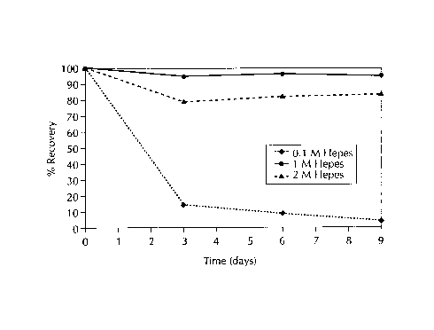
Figure 1 to 3 show the stability of 0.1 mg/ml bG-CSF solutions at pH 7.5,
under
storage conditions of 40°C in varying concentrations of 0.1 M, 1 M and
2M HEPES, TES, and
TRICINE buffers, respectively. The stability or maintenance of bG-CSF activity
improved as
the buffer concentration was increased to 1M and above, as shown in Figures 1
to 3. At
0.1 mg/ml bG-CSF there was 90% recovery of bG-CSF in IM HEPES (Figure 1 ).
CA 02280449 1999-08-13
-26-
Figure 4 to 6 show the stability of 2.0 mglml bG-CSF solutions at pH 7.5,
under
storage conditions of 40°C in varying concentrations of 0.1 M, 1 M and
2M HEPES, TES, and
TRICINE buffers, respectively. Again, the stability or maintenance of bG-CSF
activity
improved as the buffer concentration was increased to 1 M and above, as shown
in Figures 4
to 6.
The data presented in Tables 3 and 4 and Figures 1 to 6 show that the presence
of
buffers, HEPES, TES and TRICINE significantly maintain the activity of bG-CSF
for sustained
periods, from 3 to 9 days.
Example 2
In Vivo Performance of bG-CSF Formulated in Water, 1 M HEPES, 1 M TES and 1 M
TRICINE Buffers
In vivo testing of bG-CSF formulated in water, 1M HEPES, 1M TES and 1M TRICINE
Buffers was performed in calves. A 24pg/kg dose was administered to calves and
the PMN
(neutrophil) numbers monitored.
Figure 7 shows the total peripheral blood PMN counts (expressed as percent
control,
0 hour value) for cattle treated with bG-CSF formulated in water, 1 M HEPES
buffer, 1 M TES
buffer and 1M TRICINE buffer. All three buffers gave approximately 100 hours
of coverage
from a single injection. This demonstrates that all three buffers provide for
a sustained period
of protein activity in vivo at the injection site.
Example 3
Effect of HEPES Buffer on !n Vitro Stability of bG-CSF
Amounts of bG-CSF were formulated in various buffer systems, as described
below,
at a concentration of 0.1 mgJml in a pH range of about 7.0 to about 8.5. All
samples were
filtered with a 0.2 micron filter (GV Millipore, USA) prior to fill. The
samples were set up on
stability at 40°C andl were monitored for 7 to 10 days by reverse phase
HPLC (RP HPLC),
SEC HPLC and bio-assay, as described below. The unfolding temperature of bG-
CSF was
measured by a VP-DSC MicroCalorimetry system (USA).
CA 02280449 1999-08-13
_27_
Figure 8 provides a comparison of the stability of bG-CSF in various buffer
systems,
including Neupogen~ (commercially available, USA, human G-CSF), used as a
control,
HEPES at pH 7.4, PBS at pH 7.0, Hanks buffer (available commercially, USA) and
bicarbonate buffer. 'The results indicate that bG-CSF formulated in 1 M HEPES
buffer was the
most stable of all formulations tested, and exhibited similar stability to the
Neupogen~ buffer at
pH 4Ø The stability of bG-CSF in HEPES buffer, as demonstrated in Figure 8,
was
surprising and unexpected in that bG-CSF was previously known to be unstable
at neutral or
physiological pH conditions and at temperatures around 40°C or greater.
The bG-CSF
formulated in PBS at pH 7.0, Hanks buffer and bicarbonate buffer was not
stable. This was
confirmed as described in Table 5.
Table 5
Buffer Solution Positive SpecificPositive Specific
Activity Activity
(ng/ml) Initials(ng/ml) 7 days
at 40C*
Neupogen~ (control, 0.01 0.1
pH 4.0)
HEPES (pH 7.4) 0.01-0.1 0.01 to 0.1
PBS (pH 7.0) 0.1 10
Hanks (pH 6.4) 0.01-1.0 100
Bicarbonate (pH 8.2) 0.1-1.0 100-1000
*Higher values correspond to lower activity
During 7 days storage at 40°C, bG-CSF in Neupogen~ and HEPES buffer did
not lose
any activity. Also shown, bG-CSF in PBS was 10 times less active and bG-CSF in
Hanks
and Bicarbonate buffer were 100 to 1000 times less active than the initials,
respectively.
Figure 9 shcrws the effect of HEPES buffer concentration on the stability of
bG-CSF in
1000mM, 500mM, 100mM, 50mM and 20mM HEPES buffer at 40°C. As shown in
Figure 9,
as the concentration of HEPES was decreased, there was significant loss in
stability of bG-
CSF.
Figure 10 is a thermogram of two different bG-CSF solutions. The upper
thermogram,
with a maximum temperature of 47°C, is for bG-CSF formulated in PBS at
pH 7.5, and the
Power thermogram, with a maximum temperature of 59°C, is for bG-CSF
formulated in 1 M
HEPES at pH 7.5. Iln the absence of HEPES buffer the unfolding temperature of
bG-CSF at
pH 7.5 is about 40"C (onset temperature), while bG-CSF in 1 M HEPES results in
a 10°C
increase in the unfolding temperature. An increase in unfolding temperature is
indicative of
stabilization.
CA 02280449 1999-08-13
-28-
Example 4
In Vivo Performance of bG-CSF Formulated in 1M HEPES
In vivo testing of bG-CSF formulated in 1M HEPES was pertormed in calves. A
12~g/kg dose was administered to calves and the white blood cell (WBC) and PMN
(neutrophil) numbers monitored. The results are shown in Figure 11.
Figure 11, which is a plot of % PMN (neutrophil) versus time, is a comparison
of three
formulations: bG-CSF in water (as a control), bG-CSF in 1M HEPES and bG-CSF in
1M
HEPES + 10% polaxamer. As shown in Figure 11, the PMN numbers stay above the
threshold (level associated with protection) for 3 days or 72 hours. Six
cattle were tested per
formulation.
In a second study, in which a 24~glkg dose was administered to calves, using
bG-
CSF in 1M HEPES buffer + 10% polaxamer, a single injection provided
approximately 200
hours of protection, or approximately 8 days of coverage. This result
demonstrates that
HEPES buffer improves the in vivo stability of bG-CSF which in tum, provides
for a sustained
period of activity, and therefore, delivery of this protein.
Example 5
Solubility of bG-CSF In 1 M HEPES
The solubility of bG-CSF in 1M HEPES at pH 7.5 was determined. Approximately
80
mg of bG-CSF was dissolved in 30 ml of 1 M HEPES buffer (pH 7.5). The protein
solution was
filtered through a 0.2 micron GV Millipore filter and then transferred into a
50 ml ultrafiltration
cell. The cell was equipped with a low protein binding membrane with a 10,000
molecular
weight (MlIlr) cut off. The protein solution was concentrated using the
ultrafiltration cell. At
various time points samples were removed from the cell for analysis by UV-Vis
analysis at
310 nm (measure light scatter) and for concentration by RP HPLC. Absorbance at
310 nm
was plotted against concentration. Absorbance at 310 nm should increase
linearly with
concentration; at saturation there is a sudden break in the curve at 310nm and
the
absorbance at 310 nm increases dramatically. The concentration at which this
occurs is the
maximum solubility. This method is known to those of skill in the art and
typically used in
determining protein solubility. As shown in Figure 12, the maximum solubility
of bG-CSF in
1M HEPES at pH 7.5 is about 5 mg/ml. The maximum solubility of the protein is
shown at the
concentration depicting a break in the curve. At a concentration of about 5
mg/ml, there is a
sudden increase in ;absorbance at 310 nm, corresponding to the maximum
solubility of the
protein.
CA 02280449 1999-08-13
_29_
Example 6
Effect of HEPES, TES and TRICINE Buffers
on the Unfolding Temperature of bg-CSF
Solutions were prepared containing 0.5 mglml bG-CSF in 1M HEPES, 2M HEPES,
1M TES, 2M TES and 1M TRICINE; and 2 mg/ml ml bG-CSF in 1M HEPES, 2M HEPES, 1M
TES, 2M TES and 1 MI TRICINE. These solutions were prepared in the same manner
as
described in Example 'I. A control solution was prepared using PBS (Dulbecco's
Phosphate
Buffered saline, pH 7.4). The pH of the bG-CSF solutions was pH 7.5. The
unfolding
temperature of the bG-CSF was determined by differential scanning calorimetry
(Microcal Inc,
USA) using a scanning rate of 60 degrees per hour at a temperature range of
from 20 °C to
90°C. The results are shown in Table 6.
Table 6
Unfolding Temperature (°C)
PBS HEPES TES TRICINE
bG-CSF 1M 2M 1M 2m 1M
concentration
(mglml)
0.5 50..96 56.85 60.05 57.29 62.37
2 54.94 57.62 55.23 60.49 53.68
The unfolding temperature was used as a marker of solution stability and in
vivo
stability for proteins. The results in Table 6 indicate that the unfolding
temperature (T,") of bG-
CSF formulated in HEPES, TES or TRICINE buffers at concentrations 1 M and
higher was
significantly higher compared to a PBS control. The three buffers raised the
Tm by about 2 to
11 °C. The buffer concentration substantially affected the degree of
increase in Tm. The Tm of
bG-CSF increased as the buffer concentration was increased. There was about a
3°C
increase when the HEI'ES concentration was increased from 1M to 2M, and there
was about
a 5°C increase when TES concentration was increased from 1 M and 2M. As
the bG-CSF
concentration was increased, the T," of bG-CSF decreased. There was about a
2°C decrease
when the bG-CSF concentration was increased from 0.5 mg/mL to 2 mg/mL in both
HEPES
and TES buffers. The TES buffer solution increased the Tm of bG-CSF by over 11
°C at a
buffer concentration of 2M and a bG-CSF concentration of 0.5 mg/mL.
CA 02280449 1999-08-13
-30-
The results in Table 6 show that all three buffers (HEPES, TES and TRICINE)
significantly increase the Tm of bG-CSF compared to PBS. bG-CSF formulated in
2M TES
exhibits the highest solution stability relative to other buffers.
Example 7
Comparison of the Stability of Human and Bovine G-CSF in PBS and HEPES
Formulations
Formulations of 0.15 mg/mL hG-CSF and bG-CSF were prepared in Phosphate
Buffered Saline (Dulbecco's PBS, pH 7.4), and in 1 M HEPES buffer (1 M HEPES,
pH 7.5).
The formulations were placed in 1 mL vials (fill volume of 400 mL) and stored
at 40°C for 10
days. The samples were assayed every three days by Size Exclusion
Chromatography
(SEC-HPLC) and visually inspected.
Figure 13 shows that a significant improvement was observed in the stability
of
human G-CSF when formulated in 1 M HEPES buffer, when compared to PBS. Human
GCSF exhibited degradation over 10 days at 40 °C when formulated in PBS
at pH 7.4, while
a 65 % recovery was observed when it was formulated in 1 M HEPES buffer.
Figures 13 and 14 show that bovine G-CSF exhibits a somewhat better stability
in
both HEPES and PBS formulations, than human G-CSF. About an 80 % recovery of
bovine
G-CSF in the 1 M HEPES formulation was observed after 10 days at 40 °C,
white about a 65
recovery of human G-CSF was observed. Both proteins, bovine and human G-CSF,
are
substantially more stakde in 1 M HEPES buffer, than in PBS.
Example 8
Stability of Human and Bovine G-CSF in 1M HEPES Formulations
Formulations of 0.1 mg/mL hG-CSF and bG-CSF were prepared in 1 M HEPES buffer
(1 M HEPES, pH 7.5). The formulations were placed in 1 mL vials (fill volume
of 400 DL) and
stored at 40 °C for 10 days. The samples were assayed every three days
by Size Exclusion
Chromatography (SEC.-HPLC) and visually inspected.
Figure 15 shows the stability of bovine G-CSF and human G-CSF in 1 M HEPES
formulations. There was about a 90 % recovery of bovine G-CSF and about a 70%
recovery
of human G-CSF after 10 days at 40 °C.
Example 9
Effect of Formulation pH on bG-CSF Stability
Formulations of 0.1 mg/mL bG-CSF solutions were formulated in 1 M HEPES and 1
M Tes buffers at pH 4.0 & 7.5. The formulations were placed into 1 mL vials
(fill volume of
400 OL) and stored at 40 °C for 10 days. The samples were assayed every
three days by
Size Exclusion Chromatography (SEC-HPLC).
CA 02280449 1999-08-13
-31-
As shown in Figures 16 and 18, the bG-CSF recovery after 10 days at 40
°C was
about 100 % at pH 4.0, compared to Figures 17 and 19, which show about an 80-
85
recovery observed when the bG-CSF was formulated at pH 7.5.
Example 10
Six-Month Sampling of Long-Term Thermal Stability Study of bG-CSF in
1.0 M HEPES and TES Formulations
The formulations included in this study are given below. A commercial
formulation of
1.0 M HEPES was obtained from GibcoBRL (Lot # 1016436), while 1.0 M Tes was
prepared
from powder obtained from Fluka Scientific (Lot # RA12602). Buffer pH values
were adjusted
to 7.5. 8G-CSF was provided by Bioprocess (Lot # BP185-11) at a purity of 53.3
%.
Formulations were prepared of 0.1 mg/mL and 2.0 mg/mL bG-CSF In 1.OM HEPES
and 1.OM TES buffer.
Sample storage employed 3.5 mL Flint Type 1 vials (Lot # 804105-7322) with 13
mm
1888 Gray TIF stoppers (Lot # 805619-7487) using a fill volume of 1.0 mL. A
summary of
sample storage and pull points is given in Table 7. Five vials of each
formulation were stored
for each assay time point.
Table 7. Summary of Sample Storage and Potency Analysis Time Points
3 6 12 6 1 Year18
Storage InitialWeeks Weeks Weeks Months Months
5C X X X X X X X
30 C X X X X X X
40 C X X X X
The 2.0 mg/ml. bG-CSF formulations were diluted ten-fold prior to HPLC
analysis.
Three samples of each formulation were taken from each storage chamber (5, 30,
40
°C) and assayed by RF' and SE HPLC for bG-CSF potency. Each sample was
assayed three
times. Concentrations were calculated using a standard curve that was
previously
determined. Percent (%) initial bG-CSF concentrations were determined for each
analyses,
and a mean was calculated for each formulation at each time point. The mean %
initial bG-
CSF concentrations were plotted against time for each storage temperature to
graphically
depict the decrease in bG-CSF potency. Figures 20 and 21 are the results
obtained by RP
and SE HPLC, respectively, for the samples stored at 5 °C. The results
obtained for the
samples stored at 30 °C are given in Figures 22 and 23, while those
obtained for samples
stored at 40° C can be found in Figures 24 and 25.
There was little bG-CSF degradation in samples stored at 5 and 30 °C,
excluding the
0.1 mg/mL protein formulation in 1.0 M TES.
CA 02280449 1999-08-13
-32-
Example 11
Stabilizing Capabilities of HEPES Buffer on Biotherapeutic Proteins
The Tm values of the proteins of interest, discussed below, were determined in
both
phosphate and HEPES buffers using a MicroCal, Model VP-DSC, microcalorimeter.
A 25 mM
phosphate buffer solution was prepared using Na2HP04 (Lot number 08019PQ) from
Aldrich,
and the pH was adjusted to 7.5. 1.0 M HEPES buffer (pH = 7.5, Lot number
1016436) was
obtained from GibcoBRL. Buffer exchanges were accomplished via a Stirred
Ultrafiltration
Cell, Model 8010 (Arnicon, Inc.), in combination with either a YM10 or YM30
Diaflo°
Ultrafiltration Membrane (Amicon, Inc.), depending on the size of the protein.
2.0 mg of lyophilized pST (Lot number 41509-217-2) obtained from Bioprocess
was
reconstituted in 2.0 m~L of Milli-Q water. After five exchanges, 1.0 mL of the
reconstituted
protein was transferred into 25 mM phosphate buffer using a YM10 membrane. The
remaining
1.0 mL was then exchanged into 1.0 M HEPES buffer. Samples were prepared at a
1.0
mglmL protein concentration and then analyzed by microcalorimetry.
Microcalorimetry
analysis of pST was performed twice in both phosphate and HEPES buffer.
Approximately 2.0 mL of NIF (Lot number 440631-22-7) was obtained from
Bioprocess at a concentration of 2.97. The sample received from Bioprocess
were divided
into two aliquots. 1.0 ml of the protein was exchanged into 25 mM phosphate
buffer, and the
remaining NIF was exchanged into 1.0 M HEPES buffer. In each case, five
exchanges were
employed using a YM:10 membrane. Solutions were prepared at a 1.0 mglmL
concentration
in the appropriate buffer, and the Tm values were determined via
microcalorimetry.
The biotherapeutic proteins that were studied are listed in Table 8 with their
respective Tm values tfiat were determined for phosphate and HEPES buffers.
Table 8. Tm Results C>btained in the Microcalorimetry Study of Four
Biotherapeutic
Proteins in Phosphate and HEPES Buffers
PROTEIN Trt, IN PHOSPHATETm IN HEPES ~Tm
N I F 59.79 63.27 +3.48
PST ( 1 )56.14 67.90 +11.76
(2) 54.48 63.41 +8.93
Table 8 shows that HEPES buffer provides increased stability for both NIF and
pST
CA 02280449 1999-08-13
-33-
Example 12
Sustained In--Vivo Activity of Recombinant Bovine Granulocyte Colony
Stimulating
Factor (rbG-CSF) using HEPES Buffer
Bovine Granulocyte Colony Stimulating Factor (bG-CSF) was obtained from
Bioprocess Research and Development-Pfizer (Groton, CT), mannitol from E.M.
Industries
(Hawthorne, NY), 1X Dulbecco's Phosphate Buffered Saline (PBS) from GibcoBRL
(Grand
Island, NY), sodium citrate and sodium acetate from Aldrich (Milwaukee, WI),
Tween-80,
sodium chloride and hydrochloric acid from J.T. Baker (Phillipsburg, USA).
RP-HPLCISize-Exclusion Chromatography (SEC)
Solution stability was monitored by RP-HPLC and SEC. RP-HPLC was performed
using a Vydac, Protein C4 column using a mobile phase of 0.1 % TFA H20
(Solvent A) and 0.
1 % TFA CAN (Solvent B). Flow rate: I ml./min; UV detection: 220 nm;
temperature: 25 °C Size
Exclusion Chromatography was performed using a TosoHaas, TSK-GEL SWX~, 7.8 mm
ID x
30 cm column. Mobile Phase: 0.3 M NaCI in 0.05 M citrate buffer pH 5.75; flow
rate: I ml/min;
UV detection: 280 nm; temperature: 25 °C.
Microcalorimetr~r
Denaturation l'emperature (TD) was measured using a VP DSC system (MicroCal,
Inc.). Approximately 1 ml of the solution was loaded into the cell and run
against a reference
placebo formulation at a rate of about 10 °C/min.
Circular Dichroism
Secondary structure of bG-CSF was monitored using Circular Dichroism
Spectroscopy equipped with a temperature scanning measurement accessory
(CD'ORD
Model J-710/720 - Japan Spectroscopic Co., LTD).
Bioassa
in vitro activity of bG-CSF formulations were determined using a murine bone
marrow
cell proliferation assay (BMC assay). Bone marrow cells were aseptically
harvested from the
femurs of female CF1 mice (Charles River) by removing the femur and gently
flushing the
marrow out of the bore using a 3cc/23G syringe and Hanks Balanced Salt
Solution (Gibco
BRL). The cell suspension was filtered through a nylon screen to remove debris
and was then
centrifuged at 1 1 OOrpm for ten minutes at room temperature. The supernatant
was
discarded, and the pellet was resuspended in 15m1 RPM1 medium (Gibco BRL)
supplemented with 10% fetal bovine serum (Gibco BRL), 1%
penicillinstreptomycin (10,000
units/ml), 1% L-Glutamine (Gibco BRQ. Bone marrow cells were quantitated using
a Coulter
Channelyzer 256, and the cell concentration was adjusted to yield 6.67 x 10⁵
cells/ml-.
Approximately 105 cells were added to each well of a 96-welled plate. Bovine
granulocyte
colony stimulating factor formulations were then added to each well (in
triplicate) at various
CA 02280449 1999-11-17
-34-
concentrations. Following a 3 day incubation at 37°C, (5~C02)
for 3 days, 3H-thymidine (New England Nuclear, Boston, Mass)
was added to each well at a final concentration of 2uCi/ml.
Radiometric labeling was allowed to proceed for at least 18
hours at 37°C, (5gC02). The plates were frozen at -20°C,
thawed, and the cells were harvested onto glass 96-well
fibermats using a Brandel cell harvester (Biomedical Research
and Development Laboratories, Gaithersburg, Maryland).
Activity was determined using a Wallac 1205 Betaplate liquid
scintillation counter (Wallac, Gaithersburg, Maryland).
Activity of bG-CSF formulations was determined by dividing the
sample counts per minute by the media control counts per
minute (fold-over-background). Activity of 3 or more fold-over-
background was considered positive.
In-Vivo Activity of bG-CSF
In-vivo activity of bG-CSF formulations were tested
in young, crossbred beef calves ranging from approximately 100-
150 kg body weight. Calves were purchased and shipped to the
Animal Health Research Center at Terre Haute, Indiana and
acclimated to the facility for a minimum period of two days
before allotment to a study. Most calves were used for one
study, allowed to rest at least one week, and then reassigned
to a second study. Calves were not used for more than two
studies. Before allotment to a study calves were pre-screened
1-3 days prior to study initiation by assessing rectal
temperature, body weight, general health and total white blood
cell (WBC) counts and differentials. In general, calves with
rectal temperatures _>104°C and total WBC counts <4000/mm3> or
12,000/mm3"' were excluded from studies. On day 0 calves were
bled and weighed before treatment. The dose of 24 ug/kg
bG-CSF formulation was calculated at the time of treatment for
each calf based on body weight and administered via a sub-
cutaneous injection in the pre-scapular region of the neck.
Blood samples were collected in EDTA anticoagulant for
WBC/differential via venipuncture from the neck at pre-
determined times after treatment. Total WBC counts were
performed on a Nova Celltrak I hematology cell counter using
a 1:250 dilution of whole blood in isotonic diluent.
64680-1161
CA 02280449 1999-11-17
-34a-
Differential WBC counts were performed using dried blood
smears stained with a Diff-Quik stain set (Dade). A total
of 100 WBCs were counted and differentiated on a Zeiss light-
microscope with a 100x oil immersion lens and 12.5x ocular
eyepieces (total magnification = 1250x).
Effect of pH and Temperature on the Solution
Stability of bG-CSF
Table 9 shows the effect of temperature on the
stability of bG-CSF. The impact of temperature on the solution
stability of bG-CSF was followed by RP-HPLC, SEC-HPLC, bio-
assay, and visual inspection (formulation: 0.1 mg/ml bG-CSF,
5~ mannitol, 10 mM acetate buffer, 0.004 between-80, pH 4Ø
As seen from Table 9, there is a discontinuity in the
stability of bG-CSF at temperatures at or above 40 °C. By
both RPHPLC and SEC-HPLC
64680-1161
CA 02280449 1999-08-13
-35-
there is a loss of the parent protein peak at 40 °C and above. The
disappearance of the
parent peak at higher temperatures is followed by an increase in particulates
in the solution.
This was observed both visually and by monitoring light scatter at 310 nm.
Bovine G-CSF
solutions stored at 40 "C for 3 weeks are 10-100 times less active than at
5°C and
30°C. At 50°C for 3 weeks, bG-CSF is 100-1000 times less active
than solutions stored at 5
and 30 °C.
Table 9: The Stability of bG-CSF as function of temperature (3 week
stability).
TemperatureRP-HPLC SEC-HPLC Visual InspectionPositive Specific
by
(C) (% of (% of Tyndall Beam Activity (ng/ml
(Light in
initial)initial) Scatter Abs. BMC assay)*
@ 310
nm)
5 88.5 97.6 Clear (0.2 au) 0.1-1
30 67.3 76.8 Clear (0.04 au) 01.-1
40 S.Ei 5.4 Slightly cloudy 10-100
(> 0.1 au)
50 1.9 No parentCloudy (> 0.1 100-1000
au)
peak
* Activity tested in mouse BMC assay (Note: higher numbers indicate less
activity).
Circular dichroism (CD) was used to follow the impact of temperature on bG-
CSF.
The CD spectrum of bG-CSF is illustrated in Figure 26. The spectrum suggests
that the
secondary structure of bG-CSF is mostly a-helix, similar to human G-CSF, which
is
structurally very similar to bG-CSF. The CD spectra were examined at various
temperatures
in order to determine the denaturation temperature (TD). Figure 27 is a plot
of molar ellipticity
at wavelength of 222 nm (characteristic wavelength for a-helix) as a function
of temperature.
Between 40 °C and 5~0 °C, the molar ellipticity increases,
indicating the loss of secondary
structure and the denaturation of bG-CSF.
CA 02280449 1999-08-13
-36-
The effect of pH on the solution stability of bG-CSF was also assessed. In
order to
delineate only the effect of pH on stability and not the impact of
denaturation due to
temperature, the solutions were stored at 30 °C (TD of bG-CSF between
40-50 °C). Table 10
summarizes the stability of bG-CSF as a function of pH over a 2 week period at
30 °C. The
rate of protein activity loss increases as pH increases. The data suggests
that at low pH, the
cysteine in bG-CSF is protonated, and hence the formulation is more stable. At
high pH, this
free cysteine is involved in disulfide exchange reactions and are the likely
cause for instability.
Table 10: The Stability of bG-CSF as a function of pH (2 week stability at
30°C).
pH RP-HPLC SEC-HPLCVisual InspectionPositive Specific
(% of (% of by Activity
initial) initial)Tyndall Beam (ng/ml in BMC
assay)*
4.0 96.5 100 Clear solution 1-10
5.0 91.9 90.1 Clear solution 10-100
6.0 75.2 84.6 Tiny particulates100
7.0 30.5 45.7 Long gelatinous100-1000
particles
* Activity tested in mouse BMC assay (Note: higher numbers indicate less
activity).
Effect of HEPES Buffer on bG-CSF Solution Stability
We have observed that bG-CSF formulated in I M HEPES buffer at pH 7.5
exhibited
greater solution stability even when stored at 40 °C for several days.
This was unexpected
since it is previously known that bG-CSF is unstable at neutral pH and
denatures at
temperatures at or above 40 °C. Figure 28 illustrates the effect of
HEPES buffer
concentration on the solution stability of bG-CSF at pH 7.5 and storage
temperature of 40 °C.
The stability of bG-CSF decreased significantly as the concentration of HEPES
buffer was
decreased. The effect of 1 M HEPES on the denaturation temperature (TD) of bG-
CSF was
determined by microc,alorimetry. Figure 10, a thermogram, compares the two
formulations
(with and without IM HEPES) on the TD of bG-CSF. In the absence of HEPES
buffer the
onset of the endothermic transition is about 40°C, while bG-CSF
formulated in I M HEPES
buffer has a TD onset: of around 50°C. An increase in denaturation
temperature is usually
indicative of stabilization.
CA 02280449 1999-11-17
-37-
In-Vivo Activity of bG-CSF
We evaluated the in-vivo activity of this formulation
in cows. Figure 29, which is a plot of WBC count versus time,
is a comparison of bG-CSF formulated in 1 M HEPES compared to
the "control" formulation. "Control" refers to the formulation
containing 5~ mannitol, 10 mM acetate buffer, between-80, pH
4Ø
As seen in Figure 29, the VBC count stay above
threshold value of 200 baseline level (level associated with
protection against infections) for only about 24-30 hours.
However, when bG-CSF was formulated in 1 M HEPES the PMN
numbers remain above threshold for a minimum of 3 days or 72
hours (in some cases the WTC remained above threshold for
almost a week). This study was reproducible (in each study 6
cows per formulation were used). The results of this study
suggest that HEPES buffer is not only serving as a stabilizer
in vitro, but is somehow improving the in-vivo performance of
bG-CSF.
The unexpected results observed with HEPES buffer on
bG-CSF performance prompted the investigation of similar buffers
such as MOPS, HEPPS, TES, and TRICINE. These buffers also
exhibited an inproved in-vitro stability of bG-CSF similar to
the HEPES buffer. An in-vivo study was conducted where bG-CSF
was formulated in TES buffer and Tricine buffer, both formula-
tions resulted in an extended in vivo activity of bG-CSF in
cows, similar to the HEPES formulation.
Formulating bG-CSF in 1 M HEPES buffer results in
sustained activity of bG-CSF in vivo. This sustained activity
may be as a result of improved stability of bG-CSF at the
injection site. Solution stability of bG-CSF at neutral pH
and temperature of 40°C was significantly improved when bG-CSF
was formulated in 1 M HEPES. Other organic buffers, such as
MOPS, HEPPS, TES, and Tricine, also resulted in an improvement
in the stability of bG-CSF.
64680-1161