Note: Descriptions are shown in the official language in which they were submitted.
CA 02282631 1999-08-27
- 1 -
DESCRIPTION
CGI-E909
INHIBITOR OF LYMPHOCYTE ACTIVATION
Technical Field
The present invention relates to a novel inhibitor
of lymphocyte activation.
Background Art
In the environment where humans live, there are
numerous kinds of infectious microorganisms such as
viruses, bacteria, fungi, parasites and the like. When
growing in a living body each of these can cause a
disease, eventually leading even to the death of the
individual. Therefore, a defense mechanism for a living
body against exogenous microorganisms is required in
order for an individual to lead a healthy life. This
mechanism is called "immunity."
The cells that are mainly responsible for immunity
in the living body are lymphocytes. Lymphocytes are
broadly classified into the T lymphocytes (T cells) and
the B lymphocytes (B cells) by their functions. T cells
are thought to have the ability of antigen presentation,
cytotoxicity and the like, whereas B cells are believed
to have the ability of antibody production. These two
types of lymphocytes are derived from the same
hemopoietic stem cells, which undergo various kinds of
differentiation in the bone marrow or other organs or
undergo repeated differentiation by the action of growth
factors, and are finally released into the peripheral
blood.
In the case of T cells, for example, hematopoietic
stem cells differentiate into pre-T cells in the bone
marrow and subsequently migrate to the thymus, where they
undergo repeated differentiation to become mature T
cells. Thereafter, they are activated by the stimuli of
antigens to become activated T cells that have the
ability of propagation, cytotoxicity and the like. In
the case of B cells, on the other hand, hematopoietic
CA 02282631 1999-08-27
- 2 -
stem cells differentiate via pro B cells and pre B cells
into mature B cells by the stimuli of cytokines such as
IL-1, IL-2, I1-4, IL-6 and the like in the bone marrow.
They are then activated due to antigen challenge and
finally become plasma cells that have an ability to
produce antibodies.
Thus, the final activation is required for
lymphocytes to develop their respective functions. As
described above, because of its purpose of defending a
living body against an exogenous foreign substance,
immunity has a well-organized mechanism in which a
foreign substance (the non-self) and the self are
discriminatively recognized and a response is induced
-- only against the non-self as antigen. However, when this
mechanism is disrupted for some reason or other, even the
self is recognized as antigen resulting in a disease
called an autoimmune disease. When an immune response
against the non-self takes place in an excessive or
undesirable manner, tissues are impaired causing a state
called allergy.
On the other hand, a reaction (rejection) that
develops when an organ etc. of another person was
transplanted to recognize and eliminate it as a non-self
could be considered a normal biological mechanism. It is
known that individuals are genetically different from
each other, the representative of the difference being
what is called the major histocompatibility complex
(MHC). Transplantation of an organ from an individual
having a different MHC could cause a severe rejection.
Due to recent advances in the medical technologies, the
necessity of transplantation of organs etc. has become
very great in, for example, bone marrow transplantation
in the treatment of leukemia or lymphoma, kidney
transplantation in patients with terminal kidney
diseases, transplantation of the cornea and the like.
Concomitantly, the prevention of rejections has become a
major challenge.
CA 02282631 1999-08-27
- 3 -
Due to extensive experiments and researches,
pathways are being elucidated that lead from lymphocyte
activation to a disease associated with it. Accordingly,
many therapeutic drugs for diseases associated with
lymphocyte activation have been developed. Among the
autoimmune diseases caused by lymphocyte activation,
currently rheumatoid arthritis, systemic lupus
erythematosus, and scleroderma have been treated with
non-steroidal anti-inflammatory drugs such as aspirin,
steroids, or immunosuppressive agents such as
azathioprine.
Furthermore, for the purpose of suppressing
rejections associated with organ transplantation etc.,
immunosuppressive agents such as cyclosporine,
azathioprine, and mizoribine have been used mainly in
kidney transplantation and bone marrow transplantation.
In addition, as therapeutic drugs for allergy, anti-
histamine agents and inhibitors of the release of
chemical transmitters that inhibit the liberation of
chemical substances responsible for allergy have been
used. Among them, however, none of the non-steroidal
anti-inflammatory drugs, steroids, anti-histamine agents,
and the inhibitors of the release of chemical
transmitters act on the activation of lymphocytes
responsible for allergy. They only represent symptomatic
treatments of inflammation and thus do not essentially
treat the diseases.
Due to their intrinsic properties, many of the
immunosuppressive agents that are currently used have
severe side-effects, such as decrease of blood corpuscles
or shock, and hence cannot be considered adequate
therapeutic agents. Moreover, even today there are no
therapeutic regimens or drugs for many of the autoimmune
diseases.
On the other hand, Goto, T. et al. have reported a
monoclonal antibody (anti-HM1.24 antibody) that was
obtained by immunizing mice with human myeloma cells
CA 02282631 1999-08-27
- 4 -
(Blood (1994) 84, 1922-1930). When anti-HM1.24 antibody
was administered to a mouse transplanted with human
myeloma cells, the antibody accumulated in tumor tissues
in a specific manner (Masaaki Kosaka et al., Nippon
Rinsho (Japan Clinical) (1995) 53, 627-635), suggesting
that anti-HM1.24 antibody could be applied in the
diagnosis of tumor localization by radioisotopic
labeling, missile therapies such as radiotherapy, and the
like. However, it was not known that anti-HM1.24
antibody is involved in the inhibition of lymphocyte
activation.
Disclosure of the Invention
Therapeutic agents that are currently used for
diseases associated with lymphocyte activation include
various anti-inflammatory agents and immunosuppressive
agents. As mentioned above, however, they are not
completely satisfactory and therapeutic agents that can
treat these diseases and alleviate the pains of the
patient are being awaited. Thus, it is an object of the
present invention to provide an inhibitor of lymphocyte
activation.
In order to attain the above-mentioned purpose, the
inventors have conducted intensive studies on anti-HM1.24
antibody (Goto, T. et al., Blood (1994) 84, 1922-1930)
regarding its flow cytometry (FCM) analysis, its effects
on blast formation by T cells, its effects on antibody
production by B cells, and moreover on the isolation of
the antigen protein to which anti-HM1.24 antibody
specifically binds. As a result, the inventors have
found that the antigen protein recognized by anti-HM1.24
antibody is expressed on the activated lymphocytes and
that anti-HM1.24 antibody inhibits lymphocyte activation,
and thereby have completed the present invention.
Thus, the present invention provides an inhibitor of
lymphocyte activation comprising, as an active
ingredient, an antibody that specifically binds to a
protein having the amino acid sequence as set forth in
CA 02282631 1999-08-27
- 5 -
SEQ ID NO: 1.
The present invention also provides an inhibitor of
T cell- or B cell-activation comprising, as an active
ingredient, an antibody that specifically binds to a
protein having the amino acid sequence as set forth in
SEQ ID N0: 1.
The present invention also provides an inhibitor of
lymphocyte activation comprising, as an active
ingredient, a monoclonal antibody that specifically binds
to a protein having the amino acid sequence as set forth
in SEQ ID N0: 1.
The present invention also provides an inhibitor of
lymphocyte activation comprising, as an active
ingredient, an antibody that specifically binds to a
protein having the amino acid sequence as set forth in
SEQ ID N0: 1 and that has the constant region of human
antibody.
The present invention also provides an inhibitor of
lymphocyte activation comprising anti-HM1.24 antibody as
an active ingredient.
The present invention also provides an inhibitor of
lymphocyte activation comprising, as an active
ingredient, a chimeric antibody or a humanized antibody.
The present invention also provides an inhibitor of
lymphocyte activation comprising, as an active
ingredient, a chimeric anti-HM1.24 antibody or a
humanized anti-HM1.24 antibody.
The present invention also provides an inhibitor of
lymphocyte activation comprising, as an active
ingredient, an antibody that specifically binds to an
epitope recognized by anti-HM1.24 antibody.
The present invention also provides a preventive
and/or therapeutic agent for diseases associated with
lymphocyte activation, comprising, as an active
ingredient, an antibody that specifically binds to a
protein having the amino acid sequence as set forth in
SEQ ID N0: 1.
CA 02282631 1999-08-27
- 6 -
Furthermore, the present invention provides a
preventive and/or therapeutic agent for autoimmune
diseases, rejections in organ transplantation, and
allergy, comprising, as an active ingredient, an antibody
that specifically binds to a protein having the amino
acid sequence as set forth in SEQ ID NO: 1.
Brief Description of the Drawings
Fig. 1 is a scheme showing that anti-HM1.24 antibody
inhibits antibody production from B cells by SAC-
stimulation.
Fig. 2 represents a histogram of FCM analysis of
PHA-stimulated T cells with anti-HM1.24 antibody.
Fig. 3 represents a histogram of FCM analysis of
-- activated T cells with anti-HM1.24 antibody.
Fig. 4 is a scheme showing that anti-HM1.24 antibody
inhibits the blast formation reaction of PHA-stimulated T
cells.
Embodiment for Carrying Out the Invention
1. Antibody preparation
1-1. Hybridoma preparation
Hybridomas that produce antibodies for use in the
present invention can be basically constructed using a
known procedure as described below. Thus, HM1.24 antigen
protein or cells that express HM1.24 antigen may be used
as sensitizing antigens and are used for immunization in
the conventional method of immunization. The immune cells
thus obtained are fused with known parent cells in the
conventional cell fusion process, and then screened by
the conventional screening method to select cells that
produce monoclonal antibodies.
Specifically, monoclonal antibodies may be obtained
in the following manner. For example, as a HM1.24
antigen-expressing cell which is a sensitizing antigen
for obtaining antibody, there can be used a human
multiple myeloma cell line KPMM2 (Japanese Unexamined
Patent Publication (Kokai) No. 7-236475) or KPC-32 (Goto
T. et al., Jpn. J. Clin. Hematol. (1991) 32, 1400).
CA 02282631 1999-08-27
Alternatively, as the sensitizing antigen, there may be
used a protein having the amino acid sequence as set
forth in SEQ ID N0: 1 or a peptide or polypeptide
containing an epitope recognized by anti-HM1.24 antibody.
As used herein, cDNA that encodes a protein having
the amino acid sequence as set forth in SEQ ID N0: 1 has
been inserted in the Xbal cleavage site of pUCl9 vector
to construct plasmid pRS38-pUCl9. E. coli having this
plasmid has been internationally deposited under the
provisions of the Budapest Treaty as Escherichia coli
DHSa (pRS38-pUCl9) on October 5, 1993 with the National
Institute of Bioscience and Human Technology, Agency of
Industrial Science and Technology, of 1-3, Higashi 1-
chome, Tsukuba city, Ibaraki pref., Japan, as FERM BP-
4434 (see Japanese Unexamined Patent Publication (Kokai)
No. 7-196694). The cDNA fragment contained in this
plasmid pRS38-pUCl9 can be used to prepare a peptide or a
polypeptide containing an epitope recognized by anti-
HM1.24 antibody by a genetic engineering technology.
Preferably mammals to be immunized with the
sensitizing antigen are selected in consideration of
their compatibility with the parent cell for use in cell
fusion. They generally include, but are not limited to,
rodents such as mice, rats, hamsters and the like.
Immunization of animals with a sensitizing antigen
is carried out using a known method. A general method,
for example, involves the intraperitoneal or subcutaneous
administration of a sensitizing antigen to a mammal.
Specifically, a sensitizing antigen which has been
diluted and suspended in an appropriate amount of
phosphate buffered saline (PBS) or physiological saline
etc. is mixed, as desired, with an appropriate amount of
Freund's complete adjuvant. After being emulsified, it is
preferably administered to a mammal several times every 4
to 21 days. Alternatively a suitable carrier may be used
at the time of immunization with the sensitizing antigen.
After immunization and the confirmation of the
CA 02282631 1999-08-27
- g _
increase in the desired antibody level in the serum, the
immune cells are taken out from the mammal and are
subjected to cell fusion, in which the preferred immune
cells include, in particular, the spleen cells.
The mammalian myeloma cells as the other parent
cells which are subjected to cell fusion with the above-
mentioned immune cells preferably include various known
cell lines such as P3X63Ag8.653) (J. Immunol. (1979) 123:
1548-1550), P3X63Ag8U.1 (Current Topics in Microbiology
and Immunology (1978) 81: 1-7), NS-1 (Kohler, G. and
Milstein, C., Eur. J. Immunol. (1976) 6: 511-519), MPC-11
(Margulies, D.H. et al., Cell (1976) 8: 405-415), SP2/0
(Shulman, M. et al., Nature (1978) 276: 269-270), FO (de
-- St. Groth, S.F. et al., J. Immunol. Methods (1980) 35: 1-
21), S194 (Trowbridge, I.S., J. Exp. Med. (1978) 148:
313-323), 8210 (Galfre, G. et al., Nature (1979) 277:
131-133) and the like.
Cell fusion between the above immune cells and the
myeloma cells may be essentially conducted in accordance
with a known method such as is described in Milstein et
al. (Kohler, G. and Milstein, C., Methods Enzymol. (1981)
73: 3-46) and the like.
More specifically, the above cell fusion is carried
out in the conventional nutrient broth in the presence
of, for example, a cell fusion accelerator. As the cell
fusion accelerator, for example, polyethylene glycol
(PEG), Sendai virus (HVJ) and the like may be used, and,
in addition, an adjuvant such as dimethyl sulfoxide etc.
may be added as desired to enhance efficiency of the
fusion.
The preferred ratio of the immune cells and the
myeloma cells to be used is, for example, 1 to 10 times
more immune cells than the myeloma cells. Examples of
culture media to be used for the above cell fusion
include RPMI1640 medium and MEM culture medium suitable
for the growth of the above myeloma cell lines, and the
conventional culture medium used for this type of cell
CA 02282631 1999-08-27
- g -
culture, and besides a serum supplement such as fetal
calf serum (FCS) may be added.
In cell fusion, predetermined amounts of the above
immune cells and the myeloma cells are thoroughly mixed
in the above culture medium, to which a PEG solution
previously heated to about 37 °C, for example a PEG
solution with a mean molecular weight of about 1000 to
6000, is added at a concentration of 30 to 60~ (w/v) and
mixed to obtain desired fusion cells (hybridomas). Then
by repeating the sequential addition of a suitable
culture medium and centrifugation to remove the
supernatant, cell fusion agents etc., which are
undesirable for the growth of the hybridoma, can be
-. removed.
Said hybridoma is selected by culturing in a
conventional selection medium, for example, the HAT
culture medium (a culture liquid containing hypoxanthine,
aminopterin, and thymidine). Culturing in said HAT
culture medium is continued generally for a period of
time sufficient to effect killing of the cells other than
the desired hybridoma (non-fusion cells), generally
several days to several weeks. The conventional limiting
dilution method is conducted in which the hybridomas that
produce the desired antibody are selected and monclonally
cloned.
In addition to obtaining the above hybridoma by
immunizing an animal other than a human with an antigen,
it is also possible to sensitize human lymphocytes in
vitro with HM1.24 antigen or HM1.24 antigen-expressing
cells, and the resulting sensitized lymphocytes are fused
with human myeloma cell, for example U266, to obtain the
desired human antibody having the activity of binding to
HM1.24 antigen or HM1.24 antigen-expressing cells (see
Japanese Post-examined Patent Publication (Kokoku) No. 1-
59878). Furthermore, a transgenic animal having a
repertoire of all human antibody genes is immunized with
the antigen, i.e., HM1.24 antigen or HM1.24 antigen-
CA 02282631 1999-08-27
- 10 -
expressing cells, to obtain the desired humanized
antibody, in the method described above (see
International Patent Application WO 93/12227, WO
92/03918, WO 94/02602, w0 94/25585, WO 96/34096 and WO
96/33735).
The monoclonal antibody-producing hybridomas thus
constructed can be subcultured in a conventional culture
medium, or can be stored for a prolonged period of time
in liquid nitrogen.
In order to obtain monoclonal antibody from said
hybridoma, there can be mentioned a method in which said
hybridoma is cultured in a conventional method and the
antibodies are obtained in supernatant, or a method in
-. which the hybridoma is administered to and grown in a
mammal compatible with said hybridoma and the antibodies
are obtained in the ascites. The former method is
suitable for obtaining high-purity antibodies, whereas
the latter is suitable for a large scale production of
antibodies.
Specifically the anti-HM1.24 antibody-producing
hybridoma can be constructed using: the method of Goto,
T. et al. (Blood (1994) 84: 1922-1930). It can be
conducted by a method in which the anti-HM1.24 antibody-
producing hybridoma that was internationally deposited
under the provisions of the Budapest Treaty as FERM BP-
5233 on September 14, 1995 with the National Institute of
Bioscience and Human-Technology, Agency of Industrial
Science and Technology, of 1-3, Higashi 1-chome, Tsukuba
city, Ibaraki pref., Japan, is intraperitoneally injected
to BALB/c mice (manufactured by CLEA Japan) to obtain the
ascites from which the anti-HM1.24 antibody is purified,
or: a method in which said hybridoma is cultured in a
suitable culture medium such as the RPMI1640 medium
containing 10~ fetal bovine serum and 5~ BM-Condimed Hl
(manufactured by Boehringer Mannheim), the hybridoma SFM
medium (manufactured by GIBCO-BRL), the PFHM-II medium
(manufactured by GIBCO-BRL) and the like, and the anti-
CA 02282631 1999-08-27
- 11 -
HM1.24 antibody can be purified from the supernatant.
1-2. Recombinant antibody
A recombinant antibody which was produced by the
recombinant gene technology in which an antibody gene was
cloned from the hybridoma and integrated into a suitable
vector which was then introduced into a host can be used
in the present invention as monoclonal antibody (see, for
example, Carl, A.K., Borrebaeck, and James, W. Larrick,
THERAPEUTIC MONOCLONAL ANTIBODIES, published in the
United Kingdom by MACMILLAN PUBLISHERS LTD. 1990).
Specifically, mRNA encoding the variable region (V
region) of the desired antibody is isolated from the
hybridoma producing the antibody. The isolation of mRNA
-. is conducted by preparing total RNA using, for example, a
known method such as the guanidine ultracentrifuge method
(Chirgwin, J.M. et al., Biochemistry (1979) 18, 5294-
5299), the AGPC method (Chomczynski, P. et al.,
Analytical Biochemistry (1987) 162, 156-159), and then
mRNA is purified from the total RNA using the mRNA
Purification kit (manufactured by Pharmacia) and the
like. Alternatively, mRNA can be directly prepared using
the Quick Prep mRNA Purification Kit (manufactured by
Pharmacia).
cDNA of the V region of antibody may be synthesized
from the mRNA thus obtained using a reverse
transcriptase. cDNA may be synthesized using the AMV
Reverse Transcriptase First-strand cDNA Synthesis Kit and
the like. Alternatively, for the synthesis and
amplification of cDNA, the 5'-Ampli FINDER RACE Kit
(manufactured by Clontech) and the 5'-RACE method
(Frohman, M.A. et al., Proc. Natl. Acad. Sci. U.S.A.
(1988) 85, 8998-9002; Belyavsky, A. et al., Nucleic Acids
Res. (1989) 17, 2919-2932) that employs polymerase chain
reaction (PCR) may be used. The desired DNA fragment is
purified from the PCR product obtained and may be ligated
to vector DNA. Moreover, a recombinant vector is
constructed therefrom and then is introduced into E. coli
CA 02282631 1999-08-27
- 12 -
etc., from which colonies are selected to prepare a
desired recombinant vector. The nucleotide sequence of
the desired DNA may be confirmed by a known method such
as the dideoxy method.
Once the DNA encoding the v region of the desired
antibody has been obtained, it may be ligated to DNA
encoding the constant region (C region) of the desired
antibody, which is then integrated into an expression
vector. Alternatively, the DNA encoding the V region of
the antibody may be integrated into an expression vector
which already contains DNA encoding the C region of the
antibody.
In order to produce the antibody for use in the
present invention, the antibody gene is integrated as
described below into an expression vector so as to be
expressed under the control of the expression regulatory
region, for example an enhancer and/or a promoter.
Subsequently, the expression vector may be transformed
into a host cell and the antibody can then be expressed
therein.
1-3. Altered antibody
In accordance with the present invention,
artificially altered recombinant antibodies such as
chimeric antibody and humanized antibody can be used for
the purpose of lowering heterologous antigenicity against
humans. These altered antibodies can be produced using
known methods.
Chimeric antibody can be obtained by ligating the
thus obtained DNA encoding a V region of antibody to DNA
encoding a C region of human antibody, which is then
inserted into an expression vector and introduced into a
host for production of the antibody therein (see European
Patent Application EP 125023, and International Patent
Application WO 96/02576). Using this known method,
chimeric antibody useful for the present invention can be
obtained.
For example, E. coli having the plasmid that
CA 02282631 1999-08-27
- 13 -
contains DNA encoding an L chain V region or an H chain V
region of chimeric anti-HM1.24 antibody has been
internationally deposited under the provisions of the
Budapest Treaty as Escherichia coli DHSa (pUCl9-1.24L-gK)
and Escherichia coli DHSa (pUCl9-1.24H-gyl),
respectively, on August 29, 1996 with the National
Institute of Bioscience and Human-Technology, Agency of
Industrial Science and Technology, of 1-3, Higashi 1-
chome, Tsukuba city, Ibaraki pref., Japan, as FERM BP-
5646 and FERM BP-5644, respectively (see Japanese Patent
Application No. 9-271536).
Humanized antibody which is also called reshaped
human antibody has been made by grafting the
complementarity determining region (CDR) of an antibody
of a mammal other than the human, for example mouse
antibody, into the CDR of human antibody. The general
recombinant DNA technology for preparation of such
antibodies is also known (see European Patent Application
EP 125023 and International Patent Application WO
96/02576).
Specifically, a DNA sequence which was designed to
ligate the CDR of mouse antibody with the framework
region (FR) of human antibody is synthesized by PCR
method from several divided oligonucleotides having
sections overlapping with one another at the ends
thereof. The DNA thus obtained is ligated to the DNA
encoding the C region of human antibody and then is
integrated into an expression vector, which is then
introduced into a host for antibody production (see
European Patent Application EP 239400 and International
Patent Application WO 96/02576).
FRs of human antibody linked through CDRs are
selected so that the complementarity determining regions
form a favorable antigen binding site. When desired,
amino acids in the framework regions of the antibody
variable region may be substituted so that the
CA 02282631 1999-08-27
- 14 -
complementarity determining region of reshaped human
antibody may form an appropriate antigen biding site
(Sato, K. et al., Cancer Res. (1993) 53, 851-856).
For example, E. coli having plasmid that contains a
DNA encoding the version a (SEQ ID N0: 2) of the L chain
V region and that for the version r (SEQ ID NO: 3) of the
H chain V region of humanized anti-HM1.24 antibody has
been internationally deposited under the provisions of
the Budapest Treaty as Escherichia coli DHSa (pUCl9-
RVLa-AHM-gK) and Escherichia coli DHSa (pUCl9-RVHr-AHM-
gyl), respectively, on August 29, 1996 with the National
Institute of Bioscience and Human-Technology, Agency of
_. Industrial Science and Technology, of 1-3, Higashi 1-
chome, Tsukuba city, Ibaraki pref., Japan, as FERM BP-
5645 and FERM BP-5643, respectively (Japanese Patent
Application No. 9-271536). Furthermore, E. coli having
plasmid containing a DNA encoding the version s (SEQ ID
N0: 4) of the H chain V region of humanized anti-HM1.24
antibody has been internationally deposited under the
provisions of the Budapest Treaty as Escherichia coli
DHSa (pUCl9-RVHs-AHM-gyl) on September 29, 1997 with the
National Institute of Bioscience and Human Technology,
Agency of Industrial Science and Technology, of 1-3,
Higashi 1-chome, Tsukuba city, Ibaraki pref., Japan, as
FERM BP-6127 (Japanese Patent Application No. 9-271536).
For chimeric antibody or humanized antibody, the C region
of human antibody is used, and most preferably human CY
can be used as the constant region of human antibody.
Chimeric antibody comprises the variable region of
antibody derived from a mammal other than the human and
the C region derived from human antibody, whereas
humanized antibody comprises the complementarity
determining regions of an antibody derived from a mammal
other than the human and the framework regions (FRs) and
the C region of antibody derived from human antibody.
CA 02282631 1999-08-27
- 15 -
Accordingly, antigenicity thereof in the human body has
been reduced so that they are useful as the active
ingredient of the therapeutic agents of the present
invention.
A preferred embodiment of a humanized antibody for
use in the present invention includes humanized anti-
HM1.24 antibody (see Japanese Patent Application No. 9-
271536). A preferred embodiment of an L chain V region
of humanized anti-HM1.24 antibody includes one which has
the amino acid sequence encoded by the nucleotide
sequence as set forth in SEQ ID NO: 2. A preferred
embodiment of the H chain V region of humanized anti-
HM1.24 antibody includes one which has the amino acid
-. sequence encoded by the base sequence as set forth in SEQ
ID N0: 3 or 4.
1-4. Expression and production
Antibody genes constructed as described above may be
expressed and the antibody can be obtained in a known
method. In the case of mammalian cells, expression may be
accomplished using an expression vector containing a
commonly used useful promoter, an antibody gene to be
expressed, and DNA in which the poly A signal has been
operably linked at 3' downstream thereof or a vector
containing said DNA. Examples of the promoter/enhancer
include human cytomegalovirus immediate early
promoter/enhancer.
Additionally, as the promoter/enhancer which can be
used for expression of antibody for use in the present
invention, there can be used viral promoters/enhancers
such as retrovirus, polyoma virus, adenovirus, and simian
virus 40 (SV40), and promoters/enhancers derived from
mammalian cells such as human elongation factor la
(HEFla).
For example, expression may be readily accomplished
by the method of Mulligan et al. (Nature (1979) 277, 108)
when SV40 promoter/enhancer is used, or by the method of
CA 02282631 1999-08-27
- 16 -
Mizushima et al. (Nucleic Acids Res. (1990) 18, 5322)
when HEFla promoter/enhancer is used.
In the case of E. coli, expression may be conducted
by operably linking a commonly used useful promoter, a
signal sequence for antibody secretion, and the antibody
gene to be expressed, followed by expression thereof. As
the promoter, for example, there can be mentioned lacz
promoter and araB promoter. The method of Ward et al.
(Nature (1998) 341, 544-546; FASEB J. (1992) 6, 2422-
2427) may be used when lacz promoter is used, and the
method of Better et al. (Science (1988) 240, 1041-1043)
may be used when araB promoter is used.
As the signal sequence for antibody secretion, when
produced in the periplasm of E. coli, the pelB signal
sequence (Lei, S.P. et al., J. Bacteriol. (1987) 169,
4379) can be used. After separating the antibody produced
in the periplasm, the structure of the antibody is
appropriately refolded before use (see, for example, WO
96/30394).
As the origin of replication, there can be used
those derived from SV40, polyoma virus, adenovirus,
bovine papilloma virus (BPV) and the like. Furthermore,
for the amplification of the gene copy number in the host
cell system, expression vector can include as a
selectable marker an aminoglycoside transferase (APH)
gene, a thymidine kinase (TK) gene, an E. coli xanthine
guaninephosphoribosyl transferase (Ecogpt) gene, the
dihydrofolate reductase (dhfr) gene and the like.
For the production of antibody for use in the
present invention, any production system can be used.
The production system of antibody preparation comprises
the in vitro or the in vivo production system. As the in
vitro production system, there can be mentioned a
production system which employs eukaryotic cells and the
production system which employs prokaryotic cells.
When the eukaryotic cells are used, there are the
production systems which employ animal cells, plant
CA 02282631 1999-08-27
- 17 -
cells, and fungal cells. Known animal cells include (1)
mammalian cells such as CHO cells, COS cells, myeloma
cells, baby hamster kidney (BHK) cells, HeLa cells, and
Vero cells, (2) amphibian cells such as Xenopus oosytes,
or (3) insect cells such as sf9, sf2l, and Tn5. Known
plant cells include, for example, those derived from the
genus Nicotiana, more specifically cells derived from
Nicotiana tabacum, which is subjected to callus culture.
Known fungal cells include yeasts such as the genus
Saccharomyces, more specifically Saccharomyces
cereviceae, or filamentous fungi such as the genus
Asperaillus, more specifically Aspergillus niger.
When the prokaryotic cells are used, there are the
-. production systems which employ bacterial cells. Known
bacterial cells include Escherichia coli (E. coli), and
Bacillus subtilis.
By introducing via transformation the gene of the
desired antibody into these cells and culturing the
transformed cells in vitro, the antibody can be obtained.
Culturing is conducted in the known methods. For example,
as the culture media, DMEM, MEM, RPMI1640, and IMDM can
be used, and serum supplements such as fetal calf serum
(FCS) may be used in combination. In addition, antibodies
may be produced in vivo by implanting cells, into which
the antibody gene has been introduced, into the abdominal
cavity of an animal and the like.
Further, in vivo production systems, there can be
mentioned those which employ animals and those which
employ plants. When animals are used, there are the
production systems which employ mammals and insects.
As mammals, goats, pigs, sheep, mice, and cattle can
be used (Vicki Glaser, SPECTRUM Biotechnology
Applications, 1993). Also, as insects, silkworms can be
used.
When plants are used, tabacco, for example, can be
used.
Antibody genes are introduced into these animals or
CA 02282631 1999-08-27
- 18 -
plants, and the antibodies are produced in such animals
or plants, and recovered. For example, an antibody gene
is inserted into the middle of the gene encoding protein
which is inherently produced in the milk such as goat ~3
casein to prepare fusion genes. DNA fragments containing
the fusion gene into which the antibody gene has been
inserted are injected into a goat embryo, and the embryo
is introduced into a female goat. The desired antibody is
obtained from the milk produced by the transgenic goat
born to the goat who received the embryo or the offspring
thereof.
In order to increase the amount of milk containing
the desired antibody produced by the transgenic goat,
hormones may be given to the transgenic goat as
appropriate. (Ebert, K.M. et al., Bio/Technology (1994)
12, 699-702). When silkworms are used, baculovirus, into
which a desired antibody gene has been inserted, is
infected to the silkworm, and the desired antibody can be
obtained from the body fluid of the silkworm (Susumu, M.
et al., Nature (1985) 315, 592-594).
Moreover, when tabacco is used, a desired antibody
gene is inserted into an expression vector for plants,
for example pMON 530, and then the vector is introduced
into a bacterium such as Aarobacterium tumefaciens. The
bacterium is then infected to tabacco such as Nicotiana
tabacum to obtain the desired antibody from the leaves of
the tabacco (Julian, K.-C. Ma et al., Eur. J. Immunol.
(1994) 24, 131-138).
When antibody is produced in vitro or in vivo
production systems, as described above, DNA encoding the
heavy chain (H chain) or the light chain (L chain) of
antibody may be separately inserted into an expression
vector and the hosts are transformed simultaneously, or
DNA encoding the H chain and the L chain may be
integrated into a single expression vector and the host
is transformed therewith (see International Patent
Application WO 94-11523).
CA 02282631 1999-08-27
- 19 -
The antibody produced as described above can be
bound to various molecules such as polyethylene glycol
(PEG) for use as a modified antibody. "Antibody" as used
herein includes these modified antibodies. In order to
obtain such a modified antibody, the antibody obtained
may be chemically modified. These methods have already
been established in the field of the art.
2. Separation of antibody and replication
2-1. Separation of antibody and replication
Antibodies produced and expressed as described above
can be separated from the inside or outside of the cell
or from the host and then may be purified to homogeneity.
Separation and purification of the,antibody for use in
-. the present invention may be accomplished by affinity
chromatography. As the column used for such affinity
chromatography, there can be mentioned Protein A column
and Protein G column. Examples of the carriers for
Protein A column are Hyper D, POROS, Sepharose F.F. and
the like.
Alternatively, methods for separation and
purification conventionally used for proteins can be used
without any limitation. Separation and purification of an
antibody for use in the present invention may be
accomplished by combining, as appropriate, chromatography
other than the above-mentioned affinity chromatography,
filtration, ultrafiltration, salting-out, dialysis and
the like. Chromatography includes, for example, ion
exchange chromatography, hydrophobic chromatography, gel-
filtration and the like. These chromatographies can be
applied into HPLC. Alternatively, reverse-phase
chromatography can be used.
2-2. Determination of antibody concentration
The concentration of antibody obtained in the above
2-1 can be determined by the measurement of absorbance or
by the enzyme-linked immunosorbent assay (ELISA) and the
like. Thus, when absorbance measurement is employed, the
antibody for use in the present invention or a sample
CA 02282631 1999-08-27
- 20 -
containing the antibody is appropriately diluted with
PBS(-) and then the absorbance is measured at 280 nm,
followed by calculation using the absorption coefficient
of 1.35 OD at 1 mg/ml. when the ELISA method is used,
measurement is conducted as follows. Thus, 100 ul of goat
anti-human IgG (manufactured by BIO SOURCE) diluted to 1
~g/ml in 0.1 M bicarbonate buffer, pH 9.6, is added to a
96-well plate (manufactured by Nunc), and is incubated
overnight at 4 °C to immobilize the antibody.
After blocking, 100 ~1 each of appropriately diluted
antibody of the present invention or a sample containing
the antibody, or 100 yl of human IgG of a known
concentration as the standard is added, and incubated at
room temperature for 1 hour. After washing, 100 ~1 of
5000-fold diluted alkaline phosphatase-labeled anti-human
IgG antibody (manufactured by BIO SOURCE) is added, and
incubated at room temperature for 1 hour. After washing,
the substrate solution is added and incubated, followed
by the measurement of absorbance at 405 nm using the
MICROPLATE READER Model 3550 (manufactured by Bio-Rad) to
calculate the concentration of the desired antibody.
3. Preparation of cells
The cells to be used in the present invention can be
prepared according to the following methods.
3-1. Preparation of human peripheral blood
lymphocyte fraction
Peripheral blood, collected from healthy human
donors and diluted 1/2 in PBS(-), is layered onto Ficoll-
paque (manufactured by Pharmacia) in a 50 ml centrifuge
tube (manufactured by Becton Dickinson). After
centrifuge at 450 x g for 40 minutes at room temperature,
the mononuclear cell fraction in the interface is
isolated. After the fraction is prepared at a suitable
density in a RPMI1640 medium (manufactured by GIBCO-BRL)
containing 10~ fetal bovine serum (manufactured by
CA 02282631 1999-08-27
- 21 -
Moregate), it is incubated under the condition of 37°C
and 5$ COZ for 1 hour in a plastic petri dish. The
procedure is repeated twice to remove the cells attached
to the dish. The remaining cells may be used in the
following experiments as the human peripheral blood
lymphocyte fraction.
3-2. Activation of human peripheral blood B cells by
SAC
B cells in the peripheral blood lymphocytes can be
activated by incubating the human peripheral blood
lymphocytes prepared as above at a density of 5 x 10⁶
cells/ml with 0.01 SAC (Pansorbin cells, manufactured by
Calbiochem.) in the presence or the absence of 1 ng/ml of
IL-6 in a polypropylene tube under the condition of 37°C
and 5~ COz for 2 days.
3-3. Purification of human peripheral blood T cells
Human peripheral T cells can be purified from the
human peripheral blood lymphocytes prepared in the
section 3-1 using the Cellect Humm T cell Kit
(manufactured by Biotex) according to the attached
procedures.
3-4. Activation of human peripheral blood T cells by
PHA
T cells in the peripheral blood lymphocytes can be
activated by suspending the T cells prepared in the above
section 3-3 in a RPMI1640 medium containing 2~ fetal
bovine serum (manufactured by Moregate) and then
incubating the suspension at a density of 1 x 10⁶
cells/ml/well with the addition of 1 or 10 ug/ml PHA
(Phytohemagglutinin, manufactured by Sigma) in a 24-well
culture plate under the condition of 37°C and 5~ COZ for
4 days.
4. FCM analysis
Reactivity of the antibody of the present invention
with lymphocytes may be examined by flow cytometry (FCM)
analysis. The cells used may be freshly isolated cells
or the cultures thereof. As the freshly isolated cells,
CA 02282631 1999-08-27
- 22 -
there can be used, for example, peripheral blood
mononuclear cells, peripheral blood lymphocytes,
peripheral blood T cells, peripheral blood B cells and
the like.
After washing the above cells in PBS(-), 100 ul of
anti-HM1.24 antibody or a control antibody diluted to 25
~g/ml in the FRCS buffer (PBS(-) containing 2~ fetal
bovine serum and 0.1~ sodium azide) is added thereto,
which is then incubated on ice for 30 minutes. After
washing with the FRCS buffer, 100 ~1 of 25 ~g/ml FITC-
labeled goat anti-mouse antibody (GAM, manufactured by
Becton Dickinson) is added thereto, which is then
_. incubated on ice for 30 minutes. After washing with the
FRCS buffer, the cells are suspended in 600 ~1 of the
FRCS buffer, and each cell may be measured for its
fluorescence intensity using the FACScan (manufactured by
Becton Dickinson).
5. Confirmation of effects
Lymphocyte activation is accompanied by blast
formation in T cells and antibody production in B cells.
Furthermore, with the activation of both cells,
appearance or disappearance of various antigen markers on
the cell surface is observed. Confirmation of the
effects of inhibition of lymphocyte activation can be
accomplished by inhibiting blast formation after adding
the antibody for use in the present invention to the T
cells, inhibiting antibody production after adding the
antibody for use in the present invention to the B cells,
or by evaluating changes in the expression of antigen
markers on the cell surface after adding the antibody for
use in the present invention to the lymphocytes.
5-1. Effects of anti-HM1.24 antibody on blast
formation by T cells
Effects of anti-HM1.24 antibody on blast formation
by T cells can be evaluated by suspending the human
peripheral T cells purified as described above in a
CA 02282631 1999-08-27
- 23 -
RPMI1640 medium containing 2~ fetal bovine serum
(manufactured by Moregate) and then incubating the
suspension at a cell density of 1 x 10⁵ cells/200 ~.1/well
with 1 ~g/ml of PHA (Phytohemagglutinin, manufactured by
Sigma) and 20 ~g/ml of anti-HM1.24 antibody or a control
mouse IgG2a in a 96-well culture plate under the
condition of 37°C and 5~ COZ for 4 days. 3H-tymidine
(manufactured by Amersham) is added at 1 ~,Ci/well and the
incorporation of radioactivity after 4 hours may be
measured using a (3-counter (manufactured by Pharmacia).
5-2. Effects of anti-HM1.24 antibody on antibody
production by B cells
Human peripheral blood lymphocytes prepared as
described above are incubated at a density of 5 x 10⁶
cells/ml in a polypropylene tube with 0.01 SAC
(Pansorbin cells, manufactured by Calbiochem.) under the
condition of 37°C and 5$ CO2 for 2 days to activate the B
cells in the peripheral lymphocytes. The SAC-treated
peripheral blood lymphocytes are suspended in a RPMI1640
medium containing 10~ fetal bovine serum (manufactured by
Moregate), and the suspension at a cell density of 1 x
105 cells/200 ul/well is cultured with 20 ~.g/ml of anti-
HM1.24 antibody or a control mouse IgG2a in a 96-well
culture plate (manufactured by Becton Dickinson) under
the condition of 37°C and 5~ COZ for 6 days, followed by
collection of the culture supernatant.
The concentration of IgG in the culture supernatant
can be measured by a human IgG-specific ELISA. Thus, 100
ul of goat anti-human IgG (manufactured by TAGO) diluted
to 1 ~ug/ml in 0.1 M bicarbonate buffer (pH 9.6) is added
to a 96-well immunoplate (manufactured by Nunc), and then
incubated at 4 °C overnight to immobilize the antibody.
After blocking, 100 ul of appropriately diluted culture
supernatant or human IgG (manufactured by CAPPEL) as a
CA 02282631 1999-08-27
- 24 -
standard is added thereto, and then incubated at room
temperature for 1 hour.
After washing the plate, 100 ~1 of 2,000-fold
diluted alkaline phosphatase-labeled anti-human IgG
(manufactured by CAPPEL) is added to the plate, and then
incubated at room temperature for 1 hour. After washing
the plate and adding the substrate solution thereto, it
is incubated. Subsequently absorbance at 405 nm may be
determined using the MICROPLATE READER Model 3550
(manufactured by Bio-Rad).
5-3. Analysis of antigen markers on the cell surface
Human peripheral blood lymphocytes or human
peripheral T cells prepared as described above are
cultured with PHA or SAC and anti-HM1.24 antibody or
control mouse IgG2a as described in the section 5-1 or 5-
2. These cells are reacted with antibodies capable of
recognizing cell surface antigen markers that show
changes in expression before and after activation such as
CD10, CD25, CD38, CD40, CD47, CD54, CD98, PCA-1, HM1.24
antigens and the like. These can be subjected to FCM
analysis as described in the above section 4.
5-4. Confirmation of effects and related diseases
It was revealed, as shown in the examples that
follow, that the HM1.24 antigen is being expressed on the
activated lymphocytes and that the addition of anti-
HM1.24 antibody inhibited blast formation by T
lymphocytes and furthermore antibody production by B
lymphocytes. These facts indicated that anti-HM1.24
antibody has the effects of inhibiting the activation of
lymphocytes.
On the other hand, as the diseases in which
lymphocyte activation is involved, there can be mentioned
autoimmune diseases, rejections associated with organ
transplantation, and allergy. Specifically, autoimmune
diseases include, for example, Hashimoto thyroiditis,
primary myxedema, thyrotoxicosis, pernicious anemia,
autoimmune atrophic gastritis, Addison disease, premature
~.,.-
CA 02282631 1999-08-27
- 25 -
menopause, insulin-dependent diabetes mellitus,
Goodpature syndrome, myasthenia gravis, male infertility,
pemphigus vulgaris, pemphigus, ophthalmia sympathica,
lens induced uveitis, multiple sclerosis, autoimmune
hemolytic anemia, idiopathic thrombocytopenic purpura,
primary biliary cirrhosis, active chronic hepatitis,
idiopathic cirrhosis, ulcerative colitis, Sjogren's
syndrome, rheumatoid arthritis, dermatomyositis,
scleroderma, mixed connective tissue diseases, discoid
erythematosus, systemic lupus erythematosus and the like
(translation supervised by Shunnichi Hirose et al.,
Rinsho Mennekigaku Illustrated (Clinical Immunology
Illustrated) (1994), Nankodo).
As rejections associated with organ transplantation,
there are mentioned rejections associated with the
transplantation of kidney, liver, and heart, epithelial
or endothelial rejections associated with cornea
transplantation, HVD, GVHD or the like associated with
bone marrow transplantation (translation supervised by
Shunnichi Hirose et al., Rinsho Men-ekigaku Illustrated
(Clinical Immunology Illustrated) (1994), Nannkodo).
Allergy includes, for example, type I allergy represented
by atopic diseases, type II allergy observed in drug-
related allergies, type III allergy that causes various
nephritises, and type IV allergy represented by
dermatitis caused by cosmetics or metals (Taken Azeyanagi
et al., Shin-Men-ekigaku Sosho (7) Men-eki to Allergy
(New Immunology Series 7, Immunology and Allergy (1981),
Igaku Shoin). Accordingly, the therapeutic agents of the
present invention are useful as agents for treating
diseases in which lymphocyte activation is involved.
6. Route of administration and pharmaceutical
preparation
The inhibitors of lymphocyte activation of the
present invention may be administered, either
systemically or locally, by a parenteral route, for
example intravenous injection such as drip infusion,
CA 02282631 1999-08-27
- 26 -
intramuscular injection, intraperitoneal injection, and
subcutaneous injection. The method of administration may
be chosen, as appropriate, depending on the age and the
condition of the patient. The effective dosage is chosen
from the range of 0.01 mg to 100 mg per kg of body weight
per administration. Alternatively, the dosage in the
range of 1 to 1000 mg, preferably 5 to 50 mg per patient
may be chosen. The inhibitors of lymphocyte activation of
the present invention may contain pharmaceutically
acceptable carriers or additives depending on the route
of administration.
Examples of such carriers or additives include
water, a pharmaceutical acceptable organic solvent,
collagen, polyvinyl alcohol, polyvinylpyrrolidone, a
carboxyvinyl polymer, carboxymethylcellulose sodium,
polyacrylic sodium, sodium alginate, water-soluble
dextran, carboxymethyl starch sodium, pectin, methyl
cellulose, ethyl cellulose, xanthan gum, gum Arabic,
casein, gelatin, agar, diglycerin, propylene glycol,
polyethylene glycol, Vaseline, paraffin, stearyl alcohol,
stearic acid, human serum albumin (HSA), mannitol,
sorbitol, lactose, a pharmaceutically acceptable
surfactant and the like. Additives used are chosen from,
but not limited to, the above or combinations thereof
depending on the dosage form.
Examples
The present invention will now be explained
hereinbelow in more detail with reference to the
following examples. It is to be noted that the present
invention is not limited to these examples in any way.
Example 1. Construction of anti-HM1.24 antibodv
1. Preparation of mouse ascites containing anti-
HM1.24 antibody
Hybridomas producing anti-HM1.24 antibody were
obtained according to the method of Goto, T. et al.
(Blood (1994) 84, 1922-1930).
To a BALB/c mouse (manufactured by CLEA Japan) that
CA 02282631 1999-08-27
- 27 -
previously received intraperitoneal administration of 500
~~1 each of 2,6,10,14-tetramethyl pentadecane
(manufactured by Wako Pure Chemical Industries, Ltd.) 11
and 3 days before, 5 x 10⁶ hybridoma cells were
intraperitoneally injected. From day 10 after the
injection of hybridoma cells, the ascites that
accumulated in the abdominal cavity of the mouse was
collected via a 19-gauge indwelling needle Happycas
(manufactured by Medikit). The collected ascites was
centrifuged twice at a revolving speed of 1000 and 3000
rpm using a low-speed centrifuge RLX-131 (manufactured by
Tomy Seiko) to remove the hybridoma, contaminants such as
blood cells and the like.
-- 2. Purification of anti-HM1.24 antibody from mouse
ascites
Purification of anti-HM1.24 antibody from the above
mouse ascites was conducted in the following method.
After adding an equal amount of PBS(-) to the mouse
ascites, the mixture was filtered using a hollow fiber
filter Mediaprep (manufactured by MILLIPORE) and then was
affinity purified using a high speed antibody
purification instrument ConSep LC100 (manufactured by
MILLIPORE) and the Hyper D Protein A column (column
volume 20 ml, manufactured by Nihon Gaisi), and PBS(-) as
the adsorption buffer and 0.1 M sodium citrate buffer (pH
4) as the elution buffer according to the attached
instructions. The eluted fractions were immediately
adjusted to about pH 7.4 by adding 1 M Tris-HCl (pH 8.0),
and then were subjected to concentration and buffer
replacement to PBS(-) using a centrifuge ultrafiltration
concentrator Centriprep 10, which was then filter-
sterilized with a membrane filter MILLEX-GV (manufactured
by MILLIPORE) having a pore size of 0.22 ~m to obtain the
purified anti-HM1.24 antibody.
3. Purification of control mouse IgG2a
Control mouse IgG2a was purified in the following
CA 02282631 1999-08-27
- 28 -
method. Commercially available IgG2a (KAPPA) (UPC 10)
ascites (manufactured by CAPPEL) was dissolved in
purified water and PBS(-). The solution was filtered
using a membrane filter Acrodisc (manufactured by Gelman)
having a pore size of 0.2 Vim, and then was affinity-
purified using a high speed antibody purification
instrument ConSep LC100 (manufactured by MILLIPORE) and
the Hyper D Protein A column (column volume 20 ml,
manufactured by Nihon Gaisi), and PBS(-) as the
adsorption buffer and 0.1 M sodium citrate buffer (pH 4)
as the elution buffer according to the attached
instructions.
The eluted fractions were immediately adjusted to
about pH 7.4 by adding 1 M Tris-HC1 (pH 8.0), and then
were subjected to concentration and buffer replacement to
PBS(-) using a centrifuge ultrafiltration concentrator
Centriprep 10, which was then filter-sterilized with a
membrane filter MILLEX-GV (manufactured by MILLIPORE)
having a pore size of 0.22 um to obtain the purified
control mouse IgG2a.
4. Determination of antibody concentration
The concentration of the purified antibody was
determined by the measurement of absorbance. Thus, the
purified antibody was diluted in PBS(-), the absorbance
at 280 nm was measured, and the concentration was
calculated using 1.35 OD at 1 mg/ml.
Example 2. Effects of anti-HM1.24 antibody on antibody
production by, human peripheral blood B cells
stimulated by SAC
1. Preparation of the human peripheral blood
lymphocyte fraction
Peripheral blood, collected from healthy human
donors and diluted 1/2 in PBS(-), was layered onto
Ficoll-paque (manufactured by Pharmacia) in a 50 ml
centrifuge tube (manufactured by Becton Dickinson).
After centrifuge at 450 x g for 40 minutes at room
CA 02282631 1999-08-27
- 29 _
temperature, the mononuclear cell fraction in the
interface was isolated. After the fraction was prepared
at a suitable density in a RPMI1640 medium (manufactured
by GIBCO) containing 10~ fetal bovine serum (manufactured
by Moregate), it was incubated under the condition of
37°C and 5~ COz for 1 hour in a plastic petri dish. The
procedure was repeated twice to remove the cells attached
to the dish. The remaining non-adhering cells were used
in the following experiments as the human peripheral
blood lymphocyte fraction.
2. Activation of human peripheral blood B cells by
SAC
B cells in the peripheral blood lymphocytes were
activated by incubating the human peripheral blood
lymphocytes prepared as above at a density of 5 x 10⁶
cells/ml with 0.01 SAC (Pansorbin cells, manufactured by
Calbiochem.) in the presence or the absence of 1 ng/ml of
IL-6 in a polypropylene tube under the condition of 37°C
and 5~ C02 for 2 days. The SAC-treated lymphocytes were
suspended in a RPMI1640 medium containing 10$ fetal
bovine serum (manufactured by Moregate), and the
suspension at a cell density of 1 x 10⁵ cells/200 ~1/well
was cultured with 20 yg/ml anti-HM1.24 antibody or a
control mouse IgG2a in a 96-well culture plate
(manufactured by Becton Dickinson) under the condition of
37°C and 5$ C02 for 6 days, followed by collection of the
culture supernatant.
3. Quantitation of human IgG
The concentration of IgG in the culture supernatant
was measured by a human IgG-specific ELISA. Thus, 100 ail
of goat anti-human IgG (manufactured by TAGO) diluted to
1 ~g/ml in 0.1 M bicarbonate buffer (pH 9.6) was added
to a 96-well immunoplate (manufactured by Nunc), and then
incubated at 4 °C overnight to immobilize the antibody.
After blocking, 100 ~ul of appropriately diluted culture
CA 02282631 1999-08-27
- 30 -
supernatant or human IgG (manufactured by CAPPEL) as a
standard was added thereto, and then incubated at room
temperature for 1 hour.
After washing the plate, 100 ~.1 of 2,000-fold
diluted alkaline phosphatase-labeled anti-human IgG
(manufactured by CAPPEL) was added to the plate, and then
incubated at room temperature for 1 hour. After washing
the plate and adding the substrate solution thereto, it
was incubated. Absorbance at 405 nm was determined using
the MICROPLATE READER Model 3550 (manufactured by Bio-
Rad).
4. Effects of anti-HM1.24 antibody on antibody
production by human peripheral blood B cells
stimulated by SAC
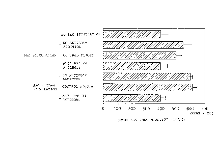
It was revealed, as shown in Fig. 1, that
stimulation by SAC resulted in enhanced IgG production
and that the addition of control mouse IgG2a thereto did
not cause any changes. However, the addition of 20 ug/ml
of anti-HM1.24 antibody completely inhibited IgG
production. It was indicated, therefore, that anti-
HM1.24 antibody inhibited the activation of B cells.
Example 3. Effects of anti-HM1.24 antibody on blast
formation by PHA-stimulated human T cells
1. Preparation of human peripheral blood T cells
Human peripheral T cells were purified from human
peripheral blood lymphocytes prepared in the above
example 2 using the Cellect Humm T cell Kit (manufactured
by Biotex) according to the attached procedures.
2. FCM analysis
Purified T cells were suspended in a RPMI1640 medium
containing 2~ fetal bovine serum (manufactured by
Moregale), and the suspension at a cell density of 1 x
106 cells/ml/well was cultured with 0, 1, and 10 ug/ml
PHA (Phytohemagglutinin, manufactured by Sigma) in a 24-
well culture plate under the condition of 37°C and 5~ CO2
for 4 days. Some of these after 4 days of culturing were
CA 02282631 1999-08-27
- 31 -
resuspended in a RPMI1640 medium containing 2~ fetal
bovine serum (manufactured by Moregale) but containing no
PHA, and were cultured for 3 more days. After washing
the cells in PBS(-), 100 ul of anti-HM1.24 antibody or
control antibody that were diluted to 25 ~g/ml in the
FACS buffer (containing 2~ fetal bovine serum and 0.1~
sodium azide) and were incubated on ice for 30 minutes.
After washing with the FRCS buffer, 100 ~1 of 25
~,g/ml FITC-labeled goat anti-mouse antibody (GAM,
manufactured by Becton Dickinson) was added thereto,
which was then incubated on ice for 30 minutes. After
washing with the FRCS buffer, the cells were suspended in
600 ~1 of the FRCS buffer, and each cell was measured for
its fluorescence intensity using the FACScan
(manufactured by Becton Dickinson). The result as shown
in Fig. 2 revealed that T cell activation by PHA is
accompanied by the expression of HM1.24 antigen on the
cells. Furthermore, as shown in Fig. 3, it was indicated
that the HM1.24 antigen that was once activated and
expressed did not disappear in the following culturing.
3. Effects of anti-HM1.24 antibody on blast
formation by PHA-stimulated human T cells
The purified T cells were suspended in a RPMI1640
medium containing 2~ fetal bovine serum (manufactured by
Moregate) and then cultured at a cell density of 1 x 10⁵
cells/200 ul/well with 1 ~g/ml PHA (Phytohemagglutinin,
manufactured by Sigma) and anti-HM1.24 antibody or a
control mouse IgG2a in a 96-well culture plate under the
condition of 37°C and 5~ COZ for 4 days. 3H-tymidine
(manufactured by Amersham) was then added at 1 uCi/well
and the incorporation of radioactivity after 4 hours was
measured using a (3-counter (manufactured by Pharmacia).
The result as shown in Fig. 4 revealed that blast
formation by PHA-stimulated T cells caused an increase in
the incorporation of 3H-thymidine, and that the addition
CA 02282631 1999-08-27
- 32 -
thereto of control mouse IgG2a at 20 ~ug/ml caused no
changes while that of anti-HM1.24 antibody at 20 ~g/ml
inhibited the incorporation of 3H-thymidine. It was
hence indicated that anti-HM1.24 antibody inhibits the
activation of T cells.
Reference Example 1. Preparation of hybridomas that
produce mouse anti-HM1.24
monoclonal antibody
In accordance with the method of Goto, T. et al.,
Blood (1994) 84, 1992-1930, hybridomas that produce mouse
anti-HM1.24 monoclonal antibody were prepared.
A plasma cell line KPC-32 (1 x 10') derived from the
bone marrow of a patient with human multiple myeloma
(Goto, T. et al., Jpn. J. Clin. Hematol. (1991) 32, 1400)
was injected twice to the abdominal cavity of a BALB/c
mouse (manufactured by Charles River) every six weeks.
Three days prior to sacrificing the animal, 1.5 x
106 KPC-32 were injected to the spleen of the mouse in
order to further enhance the antibody-producing ability
of the mouse (Goto, T. et al., Tokushima J. Exp. Med.
(1990) 37, 89). After sacrificing the animal the spleen
was extracted and the extracted organ was subjected to
cell fusion with the myeloma cell SP2/0 according to the
method of Groth, de St. & Schreidegger (Cancer Research
(1981) 41, 3465).
By the Cell ELISA (Posner, M.R. et al., J. Immunol.
Methods (1982) 48, 23) using KPC-32, the culture
supernatant of the hybridoma was screened for antibody.
5 x 10' KPC-32 were suspended in 50 ml of PBS and then
was aliquoted to a 96-well plate (U-bottomed, Corning,
manufactured by Iwaki), which was then air-dried at 37°C
overnight. After blocking with PBS containing 1~ bovine
serum albumin (BSA), the culture supernatant of the
hybridoma was added thereto and incubated at 4°C for 2
hours. Then, peroxidase-labeled anti-mouse IgG goat
antibody (manufactured by Zymed) was reacted at 4 °C for
CA 02282631 1999-08-27
- 33 -
1 hour. After washing, o-phenylene diamine solution
(manufactured by Sumitomo Bakelite) was reacted at room
temperature for 30 minutes.
Reaction was stopped by adding 2 N sulfuric acid and
the absorbance was measured at 492 nm using the ELISA
reader (manufactured by Bio-Rad). In order to remove the
hybridoma that produces antibodies against human
immunoglobulin, the culture supernatant of the positive
hybridoma had previously been adsorbed to human serum and
the reactivity to other cell lines was screened by ELISA.
Positive hybridomas were selected, and their reactivity
to various cells were investigated by flow cytometry.
The last selected hybridoma clone was cloned twice, which
-. was injected to the abdominal cavity of a pristane-
treated BALB/c mice and ascites was obtained therefrom.
Monoclonal antibodies were purified from the ascites
of the mouse by ammonium sulfate precipitation and a
Protein A affinity chromatography kit (Ampure PA,
manufactured by Amersham). The purified antibodies were
labeled with FITC using the Quick Tag FITC biding kit
(manufactured by Boehringer Mannheim).
As a result, monoclonal antibodies produced by 30
hybridoma clones reacted with KPC-32 and RPMI 8226.
After cloning, the reactivity of the culture supernatant
of these hybridomas with other cell lines or peripheral
blood mononuclear cells was investigated.
Of them, 3 clones produced monoclonal antibodies
that specifically reacted with the plasma cell. From
among the 3 clones, a hybridoma clone that was most
useful for flow cytometry analysis and had a CDC activity
to RPMI 8226 was selected and designated as HM1.24. The
subclass of the monoclonal antibody produced by this
hybridoma was determined by an ELISA using a subclass-
specific anti-mouse rabbit antibody (manufactured by
Zymed). Anti-HM1.24 antibody had a subclass of IgG2a K.
The hybridoma HM1.24 that produces anti-HM1.24 antibody
was internationally deposited under the provisions of the
CA 02282631 1999-08-27
- 34 -
Budapest Treaty as FERM BP-5233 on September 14, 1995
with the National Institute of Bioscience and Human-
Technology, Agency of Industrial Science and Technology,
of 1-3, Higashi 1-chome, Tsukuba city, Ibaraki pref.,
Japan.
Reference Example 2. Preparation of humanized anti-
HM1.24 antibody
Humanized anti-HM1.24 antibody was obtained in the
following method.
From the hybridoma HM1.24 prepared in Reference
example 1, total RNA was prepared by the conventional
method. From this, cDNA encoding the V region of mouse
antibody was synthesized and amplified by a polymerase
chain reaction (PCR) method and the 5'-RACE method. A
DNA fragment containing the gene encoding a mouse V
region was obtained, which was ligated to each plasmid
pUC cloning vector and then introduced into competent E.
coli cells to obtain an E. coli transformant. The above
plasmid was obtained from the transformant. The
nucleotide sequence of the cDNA coding region in the
plasmid was determined in the conventional method, and
the complementarity determining region (CDR) of each V
region was determined.
In order to construct a vector expressing chimeric
anti-HM1.24 antibody, cDNA encoding a V region of each of
L chain and H chain of a mouse anti-HM1.24 antibody was
inserted to the HEF vector. Furthermore, in order to
construct humanized anti-HM1.24 antibody, a V region CDR
of a mouse anti-HM1.24 antibody was grafted to a human
antibody by the CDR grafting method. The L chain of
human antibody REI was used as the L chain of human
antibody, FRs 1 to 3 of the human antibody HG3 was used
for the framework regions (FRs) 1 to 3 as the H chain of
human antibody, and FR4 of the human antibody JH6 was
used for FR4. The amino acid in the FR of the H chain V
region was replaced so that the CDR-transplanted antibody
could form a suitable antigen-binding site.
CA 02282631 1999-08-27
- 35 -
In order to express the gene of the L chain and the
H chain of the thus constructed humanized anti-HM1.24
antibody in a mammalian cell, each gene was separately
introduced into the HEF vector to construct a vector that
expresses the L chain or the H chain of the humanized
anti-HM1.24 antibody, respectively.
By simultaneously introducing these two expression
vectors into the CHO cells, a cell line that produces
humanized anti-HM1.24 antibody was established. The
antigen binding activity and the binding inhibition
activity of humanized anti-HM1.24 antibody obtained by
culturing this cell line was investigated by the Cell
ELISA using human amnion membrane cell line WISH. The
-. result indicated that the humanized anti-HM1.24 antibody
has an antigen binding activity equal to chimeric
antibody, and for the binding inhibition activity using a
biotinated mouse anti-HM1.24 antibody as well, it had an
activity equal to chimeric antibody or mouse antibody.
Incidentally, E. coli having the plasmid that
contains the DNA encoding the L chain V region or the H
chain V region of chimeric anti-HM1.24 antibody has been
internationally deposited under the provisions of the
Budapest Treaty as Escherichia coli DHSa (pUCl9-1.24L-gx)
and Escherichia coli DHSa (pUCl9-1.24H-gyl) on August 29,
1996 with the National Institute of Bioscience and Human-
Technology, Agency of Industrial Science and Technology,
of 1-3, Higashi 1-chome, Tsukuba city, Ibaraki pref.,
Japan, as FERM BP-5646 and FERM BP-5644, respectively.
Furthermore, E. coli having the plasmid that
contains the DNA encoding the version a (SEQ ID NO: 2) of
the L chain V region or the version r (SEQ ID N0: 3) of
the H chain V region of humanized anti-HM1.24 antibody
has been internationally deposited under the provisions
of the Budapest Treaty as Escherichia coli DHSa (pUCl9-
RVLa-AHM-gK) and Escherichia coli DHSa (pUCl9-RVHr-AHM-
gyl), respectively, on August 29, 1996 with the National
CA 02282631 1999-08-27
- 36 -
Institute of Bioscience and Human-Technology, Agency of
Industrial Science and Technology, of 1-3, Higashi 1-
chome, Tsukuba city, Ibaraki pref., Japan, as FERM BP-
5645 and FERM BP-5643, respectively.
Furthermore, E. coli having the plasmid that
contains the DNA encoding the version s (SEQ ID N0: 4) of
the H chain V region of humanized anti-HM1.24 antibody
has been internationally deposited under the provisions
of the Budapest Treaty as Escherichia coli DHSa (pUCl9-
RVHs-AHM-gyl) on September 29, 1997 with the National
Institute of Bioscience and Human Technology, Agency of
Industrial Science and Technology, of 1-3, Higashi 1-
chome, Tsukuba city, Ibaraki pref., Japan, as FERM BP-
6127.
Reference Example 3. Clonincr of cDNA encoding
HM1.24 antigen protein
cDNA encoding HM1.24 antigen protein specifically
recognized by anti-HM1.24 antibody was cloned.
1. Construction of cDNA library
1) Preparation of total RNA
From the human multiple myeloma cell line KPMM2,
total RNA was prepared according to the method of
Chirgwin et al. (Biochemistry, 18, 5294 (1970)). Thus,
2.2 x 10⁸ KPMM2 was completely homogenized in 20 ml of 4
M guanidine thiocyanate (manufactured by Nacalai Tesque
Inc.). The homogenate was layered on a 5.3 M cesium
chloride solution in a centrifuge tube, which was then
centrifuged in a Beckman Sw40 rotor at 31,000 rpm at 20
°C for 24 hours to precipitate RNA.
The RNA precipitate was washed in 70~ ethanol and
then dissolved in 300 ~,1 of 10 mM Tris-HC1 (pH 7.4)
containing 1 mM EDTA and 0.5~ SDS. Pronase (manufactured
by Boehringer) was added thereto to a concentration of
0.5 mg/ml and then was incubated at 37°C for 30 minutes.
The mixture was extracted with phenol and chloroform, and
RNA was precipitated with ethanol. The RNA precipitate
CA 02282631 1999-08-27
- 37 -
was then dissolved in 200 ~1 of 10 mM Tris-HC1 (pH 7.4)
containing 1 mM EDTA.
2) Preparation of poly(A)+RNA
Poly(A)+RNA was purified using as material 500 ~,g of
the total RNA prepared as described above by the Fast
Track 2.0 mRNA Isolation Kit (manufactured by Invitrogen)
according to the regimen attached to the kit.
3) Construction of cDNA library
Double stranded cDNA was synthesized using, as
material, 10 ~,g of the above poly(A)+RNA prepared by the
cDNA synthesis kit TimeSaver cDNA Synthesis Kit
(manufactured by Pharmacia) according to the regimen
attached to the kit, and was further ligated to the EcoRI
adapter supplied in the kit using the Directional Cloning
Toolbox (manufactured by Pharmacia) according to the
regimen attached to the kit. The kination and the
restriction enzyme NotI treatment of the EcoRI adapter
were carried out according to the regimen attached to the
kit. Furthermore, the adapter-added double stranded cDNA
having a size of about 500 by or greater was separated
and purified using a 1.5~ low boiling point agarose gel
(manufactured by Sigma) to obtain about 40 ~1 of adapter-
added double stranded cDNA.
The adapter-added double stranded cDNA thus
constructed was ligated using pCOSl vector (Japanese
Patent Application (Kokai) 8-255196) and T4 DNA ligase
(manufactured by GIBCO-BRL) that had previously been
treated with restriction enzymes EcoRI and NotI and
alkaline phosphatase (manufactured by Takara Shuzo) to
construct a cDNA library. The constructed cDNA library
was transduced to an E. coli strain DHSa (manufactured
by GIBCO-BRL) and consequently it was estimated to be an
independent clone having a total size of about 2.5 x 10⁶.
2. Cloning by the direct expression method
1) Transfection to COS-7 cells
CA 02282631 1999-08-27
- 38 -
About 5 x 10⁵ clones of the above transduced E. coli
were cultured in a 2-YT medium (Molecular Cloning: A
Laboratory Manual, Sambrook et al., Cold Spring Harbor
Laboratory Press (1989)) containing 50 ~g/ml ampicillin
to amplify cDNA, which was subjected to the alkali method
(Molecular Cloning: A Laboratory Manual, Sambrook et al.,
Cold Spring Harbor Laboratory Press (1989)) to recover
plasmid DNA from the E. coli. The plasmid DNA thus
obtained was transfected to COS-7 cells by the
electroporation method using the Gene Pulser instrument
(manufactured by Bio-Rad).
Thus, 10 ~g of the purified plasmid DNA was added to
0.8 ml of the COS-7 cell solution in which the cells had
been suspended in PBS at 1 x 10' cells/ml, and the
mixture was subjected to pulses of 1500 V and 25 OFD
capacity. After 10 minutes of a recovery period at room
temperature, the electroporated cells were cultured in a
DMEM culture medium (manufactured by GIBCO-BRL)
containing 10~ fetal bovine serum (manufactured by GIBCO-
BRL) under the condition of 37°C and 5~ COZ for 3 days.
2) Preparation of a panning dish
A panning dish on which mouse anti-HM1.24 antibody
were coated was prepared by the method of B. Seed et al.
(Proc. Natl. Acad. Sci. U.S.A., 84, 3365-3369 (1987)).
Thus, mouse anti-HM1.24 antibody was added to 50 mM Tris-
HCl (pH 9.5) to a concentration of 10 ~g/ml. Three
milliliters of the antibody solution thus prepared was
added to a cell culture dish with a diameter of 60 mm and
was incubated at room temperature for 2 hours. After
washing three times in 0.15 M NaCl solution, PBS
containing 5~ fetal bovine serum, 1 mM EDTA, and 0.02
NaN3 was added to the dish. After blocking, it was used
for the following cloning.
3) Cloning of cDNA library
The COS-7 cells transfected as described above were
peeled off with PBS containing 5 mM EDTA. After washing
CA 02282631 1999-08-27
- 39 -
the cells once in PBS containing 5~ fetal bovine serum,
they were suspended in PBS containing 5~ fetal bovine
serum and 0.02 NaN3 to a concentration of about 1 x 10⁶
cells/ml. The suspension was added to the panning dish
prepared as described above and was incubated at room
temperature for 2 hours. After gently washing three
times in PBS containing 5~ fetal bovine serum and 0.02
NaN3, plasmid DNA was recovered from the cells bound to
the panning dish using a solution containing 0.6~ SDS and
10 mM EDTA.
The recovered plasmid DNA was transduced into E.
coli DHSa. After amplifying as described above, the
plasmid DNA was recovered by the alkali method. The
recovered plasmid DNA was transfected into COS-7 cells by
the electroporation method and plasmid DNA recovered from
the cells bound as described above. A similar procedure
was repeated once, and the recovered plasmid DNA was
digested with restriction enzymes EcoRI and NotI, thereby
confirming the concentration of an insert having a size
of about 0.9 kbp.
Furthermore, E. coli cells in which a portion of the
recovered plasmid DNA had been transduced were inoculated
to a 2-YT agar plate containing 50 ug/ml of ampicillin.
After culturing overnight, plasmid DNA was recovered from
a single colony. It was digested with restriction
enzymes EcoRI and Notl to obtain a clone p3.19 in which
the size of the insert is about 0.9 kbp.
This clone was reacted using the PRISM, Dye
Terminater Cycle Sequencing kit (manufactured by Perkin
Elmer) according to the regimen attached to the kit, and
the base sequence was determined using the ABI 373A DNA
Sequencer (manufactured by Perkin Elmer). This base
sequence and the corresponding amino acid sequence are
shown in SEQ ID N0: 1.
Industrial Applicability
It was observed that the treatment of anti-HM1.24
CA 02282631 1999-08-27
- 40 -
antibody caused the inhibition of antibody production by
B cells and the inhibition of blast formation by T cells.
These facts indicate that anti-HM1.24 antibody has the
effect of inhibiting the activation of lymphocytes.
Reference to the microorganisms deposited under the
Patent Cooperation Treaty, Rule 13-2, and the name of
the Depository Institute
Depository Institute
Name: the National Institute of Bioscience and Human
Technology, Agency of Industrial Science and
Technology
Address: 1-3, Higashi 1-chome, Tsukuba city, Ibaraki
pref., Japan
-- Microorganism (1)
Name: Escherichia coli DHSa (pRS38-pUCl9)
Accession number: FERM BP-4434
Date deposited: October 5, 1993
Microorganism (2)
Name: hybridoma HM1.24
Accession number: FERM BP-5233
Date deposited: September 14, 1995
Microorganism (3)
Name: Escherichia coli DHSa (pUCl9-RVHr-AHM-gyl)
Accession number: FERM BP-5643
Date deposited: August 29, 1996
Microorganism (4)
Name: Escherichia coli DHSa (pUCl9-1.24H-gyl)
Accession number: FERM BP-5644
Date deposited: August 29, 1996
Microorganism (5)
Name: Escherichia coli DHSa (pUCl9-RVLa-AHM-gx)
Accession number: FERM BP-5645
Date deposited: August 29, 1996
Microorganism (6)
Name: Escherichia coli DHSa (pUCl9-1.24L-gx)
CA 02282631 1999-08-27
- 41 -
Accession number: FERM BP-5646
Date deposited: August 29, 1996
Microorganism (7)
Name: Escherichia coli DHSa (pUCl9-RVHs-AHM-gyl)
Accession number: FERM BP-6127
Date deposited: September 29, 1997
CA 02282631 1999-08-27
- 42 -
SEQUENCE LISTING
SEQ ID N0: 1
Sequence Length: 1013
Sequence Type: Nucleic acid
Stranded nos: Single
Topology: Linear
Molecular Type: cDNA
GAATTCGGCA 49
CGAGGGATCT
GG ATG GCA
TCT ACT TCG
TAT GAC TAT
TGC AGA
Met
Ala
Ser
Thr
Ser
Tyr
Asp
Tyr
Cys
Arg
5 10
GTG CCC ATG GAAGACGGG GATAAG CGCTGTAAGCTT CTGCTGGGG ATA 97
Val Pro Met GluAspGly AspLys ArgCysLysLeu LeuLeuGly Ile
15 20 25
_, GGA ATT GTGCTCCTG ATCATC GTGATTCTGGGG GTGCCCTTG ATT 145
CTG
Gly Ile Leu ValLeuLeu IleIle ValIleLeuGly ValProLeu Ile
30 35 40
ATC TTC ACC ATCAAGGCC AACAGC GAGGCCTGCCGG GACGGCCTT CGG 193
Ile Phe Thr IleLysAla AsnSer GluAlaCysArg AspGlyLeu Arg
45 50 55
GCA GTG ATG GAGTGTCGC AATGTC ACCCATCTCCTG CAACAAGAG CTG 241
Ala Val Met GluCysArg AsnVal ThrHisLeuLeu GlnGlnGlu Leu
60 65 70
ACC GAG GCC CAGAAGGGC TTTCAG GATGTGGAGGCC CAGGCCGCC ACC 289
Thr Glu Ala GlnLysGly PheGln AspValGluAla GlnAlaAla Thr
75 80 85 90
TGC AAC CAC ACTGTGATG GCCCTA ATGGCTTCCCTG GATGCAGAG AAG 337
Cys Asn His ThrValMet AlaLeu MetAlaSerLeu AspAlaGlu Lys
95 100 105
GCC CAA GGA CAAAAGAAA GTGGAG GAGCTTGAGGGA GAGATCACT ACA 385
Ala Gln Gly GlnLysLys ValGlu GluLeuGluGly GluIleThr Thr
110 115 120
TTA AAC CAT AAGCTTCAG GACGCG TCTGCAGAGGTG GAGCGACTG AGA 433
Leu Asn His LysLeuGln AspAla SerAlaGluVal GluArgLeu Arg
125 130 135
AGA GAA AAC CAGGTCTTA AGCGTG AGAATCGCGGAC AAGAAGTAC TAC 481
Arg Glu Asn GlnValLeu SerVal ArgIleAlaAsp LysLysTyr Tyr
140 145 150
CCC AGC TCC CAGGACTCC AGCTCC GCTGCGGCGCCC CAGCTGCTG ATT 529
Pro Ser Ser GlnAspSer SerSer AlaAlaAlaPro GlnLeuLeu Ile
155 160 165 170
GTG CTG CTG GGCCTCAGC GCTCTG CTGCAGTGAGATCCCAGGA 582
AGCTGGCACA
Val Leu Leu GlyLeuSer AlaLeu LeuGln***
175 180 185
TCTTGGAAGG GCTTGAACAT TCCCTTGATC 642
TCCGTCCTGC TCATCAGTTC
TCGGCTTTTC
TGAGCGGGTC GGGAGAGCAC GGGGTAGCCG 702
ATGGGGCAAC GAGAAGGGCC
ACGGTTAGCG
TCTGGAGCAG AGTCCTGGGT GTGGGGACAC 762
GTCTGGAGGG AGTCGGGTTG
GCCATGGGGC
ACCCAGGGCT TCCGGACAAT GAGTCCCCCC 822
GTCTCCCTCC TCTTGTCTCC
AGAGCCTCCC
CACCCTGAGA GGGGGGCATG TGCTGCCTGT 882
TTGGGCATGG TGTTATGGGT
GGTGCGGTGT
TTTTTTTGCG GGGTCTTTGA GCTCCAAAAA 942
GGGGGGGTTG AATAAACACT
CTTTTTTCTG
TCCTTTGAGG P~~A,AAAAAAAAA.AAAAAAAA 1002
GAGAGCACCA AAAAAAATTC
CACCTTAAAA
GGGCGGCCGC 1013
C
CA 02282631 1999-08-27
- 43 -
SEQ ID N0: 2
Sequence Length: 379
Sequence Type: Nucleic acid
Topology: Linear
Molecular Type: cDNA
Sequence
ATG GGA TGGAGCTGTATC ATCCTCTCC TTGGTAGCA GCTACA GGT 48
ACA
Met Gly TrpSerCysIle IleLeuSer LeuValAla ThrAlaThr Gly
-15 -10 -5
GTC CAC TCCGACATCCAG ATGACCCAG AGCCCAAGC AGCCTGAGC GCC 96
Val His SerAspIleGln MetThrGln SerProSer SerLeuSer Ala
-1 1 5 10
AGC GTG GGTGACAGAGTG ACCATCACC TGTAAGGCT AGTCAGGAT GTG 144
Ser Val GlyAspArgVal ThrIleThr CysLysAla SerGlnAsp Val
15 20 25
AAT ACT GCTGTAGCCTGG TACCAGCAG AAGCCAGGA AAGGCTCCA AAG 192
Asn Thr AlaValAlaTrp TyrGlnGln LysProGly LysAlaPro Lys
30 35 40 45
CTG CTG ATCTACTCGGCA TCCAACCGG TACACTGGT GTGCCAAGC AGA 240
Leu Leu IleTyrSerAla SerAsnArg TyrThrGly ValProSer Arg
50 55 60
TTC AGC GGTAGCGGTAGC GGTACCGAC TTCACCTTC ACCATCAGC AGC 288
Phe Ser GlySerGlySer GlyThrAsp PheThrPhe ThrIleSer Ser
65 70 75
CTC CAG CCAGAGGACATC GCTACCTAC TACTGCCAG CAACATTAT AGT 336
Leu Gln ProGluAspIle A1aThrTyr TyrCysGln GlnHisTyr Ser
80 85 90
ACT CCA TTCACGTTCGGC CAAGGGACC AAGGTGGAA ATCAAAC 379
Thr Pro PheThrPheGly GlnGlyThr LysValGlu IleLys
95 100 105
SEQ ID N0: 3
Sequence Length: 418
Sequence Type: Nucleic acid
Topology: Linear
Molecular Type: cDNA
Sequence
ATG GAC TGG ACC TGG AGG GTC TTC TTC TTG CTG GCT GTA GCT CCA GGT 48
Met Asp Trp Thr Trp Arg Val Phe Phe Leu Leu Ala Val Ala Pro Gly
-15 -10 -5
GCT CAC TCC CAG GTG CAG CTG GTG CAG TCT GGG GCT GAG GTG AAG AAG 96
Ala His Ser Gln Val Gln Leu Val Gln Ser Gly Ala Glu Val Lys Lys
-1 1 5 10
CCT GGG GCC TCA GTG AAG GTT TCC TGC AAG GCA TCT GGA TAC ACC TTC 144
Pro Gly Ala Ser Val Lys Val Ser Cys Lys Ala Ser Gly Tyr Thr Phe
15 20 25
CA 02282631 1999-08-27
- 44 -
ACTCCCTAC TGGATGCAG TGGGTGCGA CAGGCCCCT GGACAAGGG CTT 192
ThrProTyr TrpMetGln TrpValArg GlnAlaPro GlyGlnGly Leu
30 35 40 45
GAGTGGATG GGATCTATT TTTCCTGGA GATGGTGAT ACTAGGTAC AGT 240
GluTrpMet GlySerIle PheProGly AspGlyAsp ThrArgTyr Ser
50 55 60
CAGAAGTTC AAGGGCAGA GTCACCATG ACCGCAGAC AAGTCCACG AGC 288
GlnLysPhe LysGlyArg ValThrMet ThrAlaAsp LysSerThr Ser
65 70 75
ACAGCCTAC ATGGAGCTG AGCAGCCTG AGATCTGAG GACACGGCC GTG 336
ThrAlaTyr MetGluLeu SerSerLeu ArgSerGlu AspThrAla Val
80 85 90
TATTACTGT GCGAGAGGA TTACGACGA GGGGGGTAC TACTTTGAC TAC 384
TyrTyrCys AlaArgGly LeuArgArg GlyGlyTyr TyrPheAsp Tyr
95 100 105
TGGGGGCAA GGGACCACG GTCACCGTC TCCTCAG 418
TrpGlyGln GlyThrThr ValThrVal SerSer
110 115 120
SEQ ID N0: 4
Sequence Length: 418
Sequence Type: Nucleic acid
Topology: Linear
Molecular Type: cDNA
Sequence
ATGGACTGG ACCTGGAGG GTCTTCTTC TTGCTGGCT GTAGCTCCA GGT 48
MetAspTrp ThrTrpArg ValPhePhe LeuLeuAla ValAlaPro Gly
-15 -10 -5
GCTCACTCC CAGGTGCAG CTGGTGCAG TCTGGGGCT GAGGTGAAG AAG 96
AlaHisSer GlnValGln LeuValGln SerGlyAla GluValLys Lys
-1 1 5 10
CCTGGGGCC TCAGTGAAG GTTTCCTGC AAGGCATCT GGATACACC TTC 144
ProGlyAla SerValLys ValSerCys LysAlaSer GlyTyrThr Phe
15 20 25
ACTCCCTAC TGGATGCAG TGGGTGCGA CAGGCCCCT GGACAAGGG CTT 192
ThrProTyr TrpMetGln TrpValArg GlnAlaPro GlyGlnGly Leu
30 35 40 45
GAGTGGATG GGATCTATT TTTCCTGGA GATGGTGAT ACTAGGTAC AGT 240
GluTrpMet GlySerIle PheProGly AspGlyAsp ThrArgTyr Ser
50 55 60
CAGAAGTTC AAGGGCAGA GTCACCATC ACCGCAGAC AAGTCCACG AGC 288
GlnLysPhe LysGlyArg ValThrIle ThrAlaAsp LysSerThr Ser
65 70 75
ACAGCCTAC ATGGAGCTG AGCAGCCTG AGATCTGAG GACACGGCC GTG 336
ThrAlaTyr MetGluLeu SerSerLeu ArgSerGlu AspThrAla Val
80 85 90
TATTACTGT GCGAGAGGA TTACGACGA GGGGGGTAC TACTTTGAC TAC 384
TyrTyrCys AlaArgGly LeuArgArg GlyGlyTyr TyrPheAsp Tyr
95 100 105
TGGGGGCAA GGGACCACG GTCACCGTC TCCTCAG 418
TrpGlyGln GlyThrThr ValThrVal SerSer
110 115 120