Note: Descriptions are shown in the official language in which they were submitted.
CA 02282738 1999-08-27
WO 98/37837 PCT/US98/04433
PHYSIOLOGI TRAT VALV TMPT A1TA TON HOLI~TNCt ~Y~TFx~r
Field of the invention
This invention relates to the implantation of physiological mitral valves in
the
human heart, and more particularly to a holding system for holding the valve
in a
proper position during the implantation surgery.
Background of the invention
My copending application Serial No. 08/566,229 filed O1 December 1995 and
entitled "Physiological Mitral Valve Bioprosthesis" describes the selection,
preparation and positioning of a xenograft such as a porcine mitral valve for
valve
replacement surgery in the human heart.
As will be readily appreciated from that discussion, the orientation of the
annulus and papillary heads of a xenograft mitral valve are three-dimensional
in
nature. This also holds true for a stentless physiological mitral valve (PMV).
Both
the PMV's annulus and papillary heads are free to distort when the valve is
held in
one's hand or when the valve is implanted in the mitral position of a
recipient. If the
valve is implanted in a distorted geometry, both hemodynamic performance and
long
term durability could suffer. It is therefore desirable to hold the prosthesis
temporarily in its optimum geometric configuration so that the valve may be
implanted properly. Once the valve's implantation orientation is set relative
to the
patient's left ventricular geometry, the temporary holder is removed. The
implantation
position of the valve in the left ventricle then provides the necessary
rigidity and
structure for valve function.
Temporary holding systems for holding implants have previously been used in
annuloplasty. They have not, however, been used in stentless mitral xenografts
because that type of surgery poses more complex problems than does
annuloplasty.
Consequently, a need exists for a temporary holder system suitable for the
placing of
non-stented chordally supported mitral xenograft valves, or pericardial and
synthetic
CA 02282738 1999-08-27
WO 98/37837 PCT/US98/04433
2
derivatives of the natural mitral valve in which temporary geometric
positioning is
necessary to facilitate implantation.
The invention fills the above-described need by providing a holding system
composed of three components: an inner annulus holder, an outer annulus clamp,
and
papillary head holders. The inner annulus holder and outer annulus clamp form
the
mechanism to hold the valve annulus while the papillary head holders fix the
valve's
papillary heads in a rigid anatomic geometry relative to the valve's annulus.
Preferably, the holding system of this invention is constructed of a clear
plastic such
that the valve's inflow annulus and any positioning markers thereon may be
clearly
seen when the valve is mounted on the holder.
Brief description of the drawings
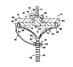
Fig. 1 is an exploded perspective view of the holding system of this
invention;
Figs. 2a and 2b are side and plan views, respectively, of the inner annulus
holder;
Figs. 3a and 3b are a plan view and a section along line 3b-3b, respectively,
of
the outer annulus clamp;
Figs. 4a, 4b and 4c are a general perspective view, an enlarged perspective
view, and a rear perspective view, respectively, of the papillary head
holders;
Fig. 5 is a perspective view of a physiologic mitral valve xenograft;
Fig. 6 is a vertical section illustrating the positioning of the valve on the
holder
in the clamping position;
Fig. 7 is a perspective view of the locking key;
Fig. 8 is a vertical section similar to Fig. 6 but showing the holder in the
release position;
Fig. 9 is a bottom plan view of an alternative embodiment of the invention;
Fig. 10 is a vertical section along line 10-10 of Fig. 9; and
Fig. 11 is a fragmentary enlarged bottom view along line 11-11 of Fig. 10.
CA 02282738 1999-08-27
WO 98/37837 PCTNS98/04433
3
Description of the,~referred embodiment
In the exploded view of Fig. l, the holding system 10 of this invention is
seen
to consist of am-inner annulus holder 12 with a detachable obturator handle
14, an
outer annulus clamping assembly 16 composed of an anterior clamp 16a and a
posterior clamp 16b, and a pair of papillary head holders 18, spaced from each
other
and from the posterior clamp 16b by a support structure 20.
The inner annulus holder 12 is depicted in more detail in Figs. 2a and 2b. It
consists essentially of a plate 22 which is attached through a tapered release
shaft 24
to a threaded handle socket 26. The plate 22 is shaped and dimensioned to
contact the
inner diameter of the PMV's annulus at the band formed by the PMV's annular
sewing
cuff 28 (Fig. 5). The plate 22 thus provides a form for the PMV's annular
inside
diameter against which the annulus can be clamped against in order to hold the
annulus in place during implantation. The PMV should require slight
stretching to fit over the inner annulus holder in order to facilitate
mounting. The
edges of the plate 22 are rounded to prevent tissue damage, and the shape of
the plate
22 is so chosen as to approximate the shape of the mitral annulus during end
systole.
An alignment tab 30 is formed on the release shaft 24 to assure proper
alignment of
the holder 12 with the clamping assembly 16. The handle socket 26 carnes at
its top
an annular flange 32 for a purpose described below.
Figs. 3a and 3b show the arrangement of the clamping assembly 16. The
anterior clamp 16a and the posterior clamp 16b can be mated around the shaft
24 of
the interior annulus holder 12 in the manner of a snap ring by horizontally
engaging
the hooks 34 of the anterior clamp 16a with the resilient shoulders 36 formed
on the
posterior clamp 16b. When the clamps 16a and 16b are mated, the tapered wall
38
defines a recess 39 of the same shape as, but slightly larger than, the plate
22 of the
holder 12. The face 40 engages and matches the shaft 24 of holder 12, and the
recess
42 is shaped and dimensioned to receive the flange 32 of the handle socket 26
during
the release operation described below. An alignment slot 44 in the face 40
cooperates
with the alignment tab 30 of the holder 12 to assure proper alignment of the
holder 12
with the clamp 16.
CA 02282738 1999-08-27
WO 98/37837 PCT/US98/04433
4
The papillary head holders 18 are illustrated in Figs. 4a-c. As shown in Fig.
4a, the holders 18 are suspended from the posterior clamp 16b by the support
structure
20 which orients them to properly position the papillary heads 46 when the PMV
is
mounted on the holding system 10. The holders 18, as illustrated in more
detail in
Figs. 4b and 4c, have a generally curved face 48 which serves as a form for
engaging
the papillary heads 46. The papillary heads 46 are held against the face 48 by
sutures
50, bands or other holding elements which may pass through openings 52 and may
be
tied at the rear of the holders 18. A scalpel groove 54 may be provided in the
rear
face 56 of holders 18 to facilitate cutting the suture 50 when releasing the
papillary
heads 46 after implantation.
The use of the above-described apparatus is illustrated in Figs. 5-8. As
disclosed in more detail in application Serial No. 08/566,229, Fig. S shows a
xenograft mitral valve 60 prepared for implantation. The annulus 62 of the
valve has
been covered with a cloth sewing cuff 28 which facilitates the suturing of the
annulus
62 to the host's mitral annulus. The sewing cuff 28 also carnes colored
markers 61
(which can be clearly seen through the clear plastic of the holding system 10)
to
identify the valve's commisures and short axis for proper placement of the
surgical
sutures. Sewing tubes 63 have also been attached to the papillary heads 46 at
the
depending chordae tendinae 64 to lengthen them and facilitate their suturing,
as the
chordae tendinae of the a xenograft are typically too short to reach their
anchor points
in the human heart.
The exact three-dimensional positioning of the papillary heads 46 with respect
to the annulus 62 at implant is critical to the efficiency and long-term
success of the
implant by minimizing stresses on the chordae tendonae 64 during the beating
of the
heart. Consequently, a unique geometry, in terms of length and angular
position from
the annulus 62, is required for the papillary head holders 18 on each side of
the PMV.
As shown in the vertical section of Fig. 6, the sewing cuff 28 of the annulus
62
is first slipped onto the plate 22 in such a way that only the sewing cuff 28
is in
contact with the plate 22. The annulus 62 is so oriented that the anterior and
posterior
clamps 16a and 16b correspond with the anterior and posterior leaflets of the
PMV 60.
CA 02282738 1999-08-27
WO 98/37837 PCT/US98/04433
The annulus 62 is then clamped to the plate 22 by mating the clamps 16a and
16b
around the plate 22. The plate 22 is held in the clamping recess 39 by
slipping a U-
shaped locking k-ey 66 (Fig. 7) under the flange 32 of the handle socket 26.
The
locking key 66 is preferably secured to the clamping assembly 16 by a pin or
other
5 fastening device (not shown) to avoid its accidental placement in the
operating field.
The space between the plate 22 and the tapered wall 38 is such that the clamp
16
exerts a clamping force against the plate 22 which is sufficient to firmly
hold the
annulus 62 without damaging it by abrasion or crushing of tissue. The chordae
tendinae 64 are next sutured to the papillary head holders 18 just above the
sewing
tubes 63, leaving the sewing tubes 63 free for trimming and suturing. Finally,
the
obturator handle 14 is attached to the socket 26.
The holding system is now ready for placement of the valve 60 into its implant
position. Although implantation methods may vary depending upon the surgeon's
preference, a typical implantation may proceed as follows:
1 ) The mitral valve is exposed using standard left atriotomy techniques as
understood by those versed in the art. Stay sutures are placed in the mitral
annulus at
the trigones and the short axis of the native valve.
2) The native valve is excised leaving the papillary heads intact. The native
chordae tendinae are trimmed close to the native papillary heads.
3) Based on the geometry of the PMV 60 and that of the native left ventricle,
the
distance between the native annulus and the site of attachment for the PMV
papillary
heads is determined through measurement. The PMV sewing tubes 63 are cut to
reflect this length such that the total length from the PMV's annulus 62 to
the end of
the PMV's sewing tube 63 is identical to that measured. (The measurement of
the
annulus to papillary head attachment point length can also be determined
preoperatively using echo Doppler techniques).
4) Three or more plegetted sutures are made in the native left ventricle at
the
point of attachment for the PMV papillary head sewing tubes 63.
5) The PMV 60 on the holder 10 is held by the handle 14 over and normal to the
native annulus such that the annulus markers 61 on the PMV sewing cuff 28
align
CA 02282738 1999-08-27
WO 98/37837 PCT/US98/04433
6
with the stay sutures placed in the native annulus. The annulus 62 of the PMV
20 and
the native annulus are parallel.
6) The plegetted sutures thus placed are run up to the PMV's papillary sewing
tubes 63, carefully maintaining orientation and avoiding twisting of the PMV
papillary head 46 by misorientation of the plegetted sutures onto the PMV's
sewing
tube 62.
7) The valve 60 is released from the holder by first releasing the papillary
heads
from their holders 18. This is done by running a scalpel along the scalpel
groove 54.
The PMV's annulus 62 is then released by sliding out the locking key 66 and
pushing
the inner annulus holder 12 downward until the flange 32 seats in the recess
42. This
releases the clamping pressure on the annulus 62.
8) With the valve 60 released from the holder I0, the PMV's papillary sewing
tubes 63 are slid down along their suture lines to their seating location in
the left
ventricle and tied off. The holder 10 is discarded. While the PMV's sewing
tubes 63
1 S are being tied down, the annulus 62 is pushed out of the way in the left
ventricle to
facilitate tying off of the PMV's papillary head sewing tubes 63.
9) The PMV's annulus 62 is brought back up to the patient's annulus and
aligned
such that the colored markers 65 of the PMV's annulus 62 coincide with the
stay
sutures of the patient's annulus. This insures annular alignment.
10) The PMV's annulus is then sewn in, using either continuous or interrupted
sutures.
11) The valve 60 is checked for competency and prolapse. Suture adjustments
may be made as necessary to insure that there is no paravalvular leakage and
proper
coaptation of the leaflets.
12) The remainder of the operation is conducted in a manner similar to that of
a
standard mitral valve replacement procedure as understood by those skilled in
the art.
An alternative preferred embodiment of the invention is illustrated in Figs. 9
through 11. In the plan view of Fig. 9, the supporting member 70 is shown to
have
the general shape of a mitral valve annulus. On its upper side (Fig. 10), the
supporting member 70 carries a handle socket 72 which receives the obturator
handle
CA 02282738 1999-08-27
WO 98/37837 PCT/US98/04433
7
14 (Fig. 1). On the underside, the supporting member 70 has a pair of
generally
concentric flanges 74, 76 depending therefrom.
Each of the flanges 74, 76 has small transverse openings 78, 80 formed therein
at intervals along its curcumference. Each opening 78 (typically three along
the
anterior annulus and five along the posterior annulus in the outer flange 74
is
circumferentially aligned with a corresponding opening 80 in the inner flange
76, for a
purpose best illustrated in Fig. 11. In Figs. 10 and 11, the sewing cuff 28 is
shown
inserted into the space between the flanges 74, 76 and sewn in its upper half
to
supporting member 70 by sutures 82.
A preferred layout of the sutures 82 is shown in Fig. 11. The positioning of
sutures 82 shown in that figure makes it possible, after the lower half of the
sewing
cuff has been sewn to the patient's mitral valve annulus, to release the
support member
70 from the dewing cuff 28 by a scalpel cut at, e.g., recess 84. Each of the
sutures 82
is preferably tied on each side of an opertured protrusion 86 so as to remain
attached
to the protrusion 86 after being cut at 84 and pulled out of the sewing cuff
28.
It should be understood that the exemplary physiologic mural valve
implantation holding system described herein and shown in the drawings
represents
only presently preferred embodiment of the invention. Indeed, various
modifications
and additions may be made to such embodiments without departinf: from the
spirit
and scope of the invention. Thus, other modifications and additions may be
obvious
to those skilled in the art and may be implemented to adapt the present
invention to a
variety of different applications.
