Note: Descriptions are shown in the official language in which they were submitted.
CA 02283949 1999-09-10
PCT/CA98/00192
1
METHODS AND APPARATUS FOR DETECTING THE REJECTION OF
TRANSPLANTED TISSUE
CROSS-REFERENCE TO RELATED APPLICATIONS
This application claims priority from United States provisional patent
application No. 60/040,557, filed March 13, 1997, United States provisional
patent
application No. 60/046,368, filed May 15, 1997, United States provisional
patent
application No. 60/062,512, filed October 16, 1997, United States provisional
patent
application No. 60/068,693, filed December 23, 1997, all of which are
presently
1o pending.
TECHNICAL FIELD OF THE INVENTION
The present invention relates to methods of detecting the possible
rejection of transplanted tissue, such as a transplanted organ. The present
invention also
t5 relates to apparatus for detecting such rejection, and methods relating to
such apparatus.
BACKGROUND OF THE INVENTION
The transplanting of tissues such as organs is a well recognized
technique in surgery. Unfortunately, a major, long-standing difficulty is the
rejection of
20 the transplanted tissue by the host. Briefly, the immune system of the host
recognizes a
foreign body (i.e., the transplanted tissue) and then rejects that foreign
body. A variety
of techniques exist for the suppression of rejection, and improved rates of
success are
now being achieved. A popular technique is to suppress the recipient's immune
system,
for example with cyclosporin. However, such immunosuppression techniques carry
25 risks for the patient, and are therefore minimized, when possible, by
attempting to
determine prior to immunosuppression if the tissue exhibits characteristics of
rejection.
A standard means of determining whether an organ is being rejected is
the conduction of physical biopsies (such as an endomyocardial biopsy (EMB)
for the
heart). In the case of heart transplants, accurate diagnosis is vital for the
effective care
30 of the heart transplant, and percutaneous transvenous EMB is a standard
method for
such assessment of rejection. Crudely described, this means inserting a
catheter
CA 02283949 1999-09-10
WO 98/40007 PCTICA98/00192
2
comprising a device known as a bioptome, which comprises a wire with tiny jaws
at the
distal end, into a blood vessel. Many varieties of catheters and bioptomes are
known in
the art. See, e.g., U. S. Patent No. 3,964,468; U. S. Patent No. 4,953,559; U.
S. Patent
No. 4,884,567; U. S. Patent No. 5,287,857; U. S. Patent No. 5,406,959; WO
96/35374;
WO 96/35382; WO 96/29936; WO 96/35374. The distal end of the catheter is fed
into
an entry point, typically on the leg or neck, and then on to the heart chamber
where a
tiny piece of tissue is clamped in the jaws of the bioptome and removed for
analysis.
This biopsy permits accurate detection of the presence and the severity
of histologic changes in the transplanted tissue once the site of rejection is
found. In
particular, the heart material obtained from the biopsy is graded for the
level or severity
of the rejection. The International Society for Heart and Lung Transplantation
(ISHLT), Kolbeck et al., Transplant Pathology, p. 200 (Am. Soc. Clin. Path.,
1994),
rates cardiac rejection as follows:
Table 2
International Society for Heart and Lung Transplantation
Grade 0 No evidence of acute rejection
Grade 1 Mild
A. Focal/Perivascular
B. Diffuse/Interstitial
Grade 2 Moderate, Uni-focal
Grade 3 Moderate, Multi-focal
A. Several foci
B. Diffuse
Grade 4 Severe
Ongoing
Mild, moderate, severe
Resolving
In an alternative formulation, Billingham's Histopathologic
2o Classification of Rejection, Kolbeck et al., Transplant Pathology, p. 199
(Am. Soc.
Clin. Path., 1994), establishes the features of tissue rejection as follows:
CA 02283949 1999-09-10
WO 98/40007 PCT/CA98/00192
3
Table 3
Billingham's Histopathologic Classification of Acute
Rejection in Human Heart Allograft
Severity of Prognostic and
Acute
Rejection Features Implications Therapeutic
Mild Rare (usually 1-2) localizedReversible, typically
perivascular collections without augmentation
of of
mononuclear cells with immunosuppressive
limited
extension into the interstitium.therapy.
No definite myocardial
injury.
Moderate Collection of "activated"Reversible, typically
with
perivascular and interstitialaugmentation of therapy
mononuclear cells with and rebiopsy.
associated
myocyte injury.
Severe Widespread inflammatory Reversible, but with
infiltrates including difficulty. Requires
mononuclear
cells and often polymorphonuclearaugmentation of therapy.
leukocytes and eosinophils.
Multifocal tissue and
small vessel
necrosis is associated
with fresh
hemorrhage.
Resolving Granulation tissue at Reversed rejection,
various
stages if collagenization.spontaneously or
Includes
numerous fibroblasts withtherapeutically induced.
scattered mononuclear
cells,
plasma cells and phagocytosed
lipochrome pigment.
A patient may require an average of S and as many as 10 biopsies per
biopsy procedure. Thus, over the first year of a heart transplant recipient,
as many as
180 EMBs are taken. A typical schedule for EMBs is as follows:
CA 02283949 1999-09-10
WO 98/40007 PCT/CA98/00192
4
Table 1
Right Ventricular Biopsy Protocol for Heart Transplant
Period Time Frequency Procedures
Immediate post- 0-4 weeks from day five, 6
operative twice weekly
4-6 weeks weekly 3
Late post-operative2-3 months bimonthly 4
4-6 months monthly 3
6-12 months quarterly 2
Total First Year 18
After one yearyearly
(in the absence
of
rej ection)
After rejection 14-21 days
therapy
EMBs, and other biopsies, are problematic, however, because during
each biopsy a number of potential complications may occur. These complications
include the following:
right ventricular perforation
cardiac tamponade
ventricular and supraventricular arrhythmia
embolus (thrombus or air)
pneumothorax air in the pleural cavity
infection
bleeding
EMBs are the principle method for monitoring cardiac allograft rejections.
Thus, the EMB, which is a physical biopsy and diagnostic aid, is
hazardous for the patient. Attempts have been made to reduce the number of
biopsies
per patient, but these attempts have not been successful, due in part to the
difficulty in
pinpointing the sites where rejection starts and to the difficulty in
assessing tissue
without performing the actual biopsy.
CA 02283949 1999-09-10
WO 98/40007 PCT/CA98/00192
Accordingly, there has gone unmet a need for methods and apparatus
that reduce the number of EMBs that a patient must suffer subsequent to
undergoing a
transplant procedure. There has also gone unmet a need for methods and devices
that
. assist in pinpointing sites where rejection starts. The present invention
provides these
5 and other related advantages.
SUNINIARY OF THE INVENTION
The present invention provides methods of, and apparatus for, detecting
the possible rejection of transplanted tissue, such as a heart, by a host.
Generally, the
to methods comprise subjecting the tissue to ultraviolet to visible light
illumination,
collecting the fluorescence light induced by the illumination to permit
analysis, and
comparing the results from the transplanted tissue to results for known
tissue, typically
healthy tissue; the fluorescence from tissue having characteristics of
rejection is
different from the fluorescence from healthy tissue.
Accordingly, in one aspect the present invention provides methods for
determining whether a transplanted tissue comprises one or more
characteristics
indicative of rejection by a host. The methods comprise a) illuminating the
transplanted
tissue under conditions suitable to cause the transplanted tissue to
fluoresce; b)
collecting the fluorescence to provide a transplant fluorescent signature; and
c)
2o comparing the transplant fluorescent signature with a known fluorescent
signature
representative of the same type of tissue as the transplanted tissue, and
therefrom
determining whether the transplanted tissue exhibits one or more
characteristics
indicative of rejection.
In a preferred embodiment that relates to this and other aspects of the
present invention (which is so for other preferred embodiments unless a given
aspect of
the invention indicates that such embodiment does not apply to that aspect),
the
transplanted tissue is illuminated with light that does not comprise LIV
light, further
preferable light that consists essentially of blue light, even further
preferably light that
consists essentially of a wavelength of about 442 nm.
CA 02283949 1999-09-10
WO 98140007 PCT/CA98/00192
6
In another preferred embodiment, methods are implemented using a
catheter or endoscope that comprises at least one illumination light guide
that conducts
light to the transplanted tissue to illuminate the transplanted tissue and at
least one
collection light guide that collects fluorescence from the transplanted
tissue, and the
methods filrther comprise collecting a plurality of transplant fluorescence
signatures
preferably without substantially moving the catheter or endoscope relative to
the
transplanted tissue, wherein at least two of the plurality of transplant
fluorescence
signatures comprise a significant fluorescence contribution from a plurality
of selected
different depths of the transplanted tissue to provide at least two different
fluorescence
l0 signatures. Preferably, the catheter or endoscope comprises at least a
first collection
light guide and a second collection light guide each of which is spaced a
different
distance from its associated illumination light guide, and the collection of
the at least
two different fluorescence signatures comprises collecting light using each of
the first
collection light guide and the second collection guide during the collecting
steps. In
one preferred embodiment, the illumination light guide associated with each of
the first
collection light guide and the second collection light guide is a single light
guide.
In a fixrther preferred embodiment, the step of illuminating comprises
illuminating the tissue using light from an illumination light guide, and the
step of
collecting comprises collecting the fluorescence in a collection light guide.
In another embodiment of the present invention, the steps of illuminating
and collecting are performed during a single diastole. Such steps can be
initiated using
one or more signals of an electrocardiogram, a blood pressure pulse of the
host,
preferably measured using a blood pressure monitor located externally to the
host such
as a pulse oximeter.
In still a further embodiment of the present invention, the step of
comparing comprises comparing one or more of the full width at half maximum
(FWHM) of the transplant fluorescent signature with the known fluorescent
signature,
the ratio of the integrated intensity of two or more wavelength bands of the
transplant
fluorescent signature with the known fluorescent signature, which can be
measured by
broad band optical detectors and wherein the method can further comprise
selecting a
CA 02283949 1999-09-10
WO 98/40007 PCT/CA98/00192
7
specific spectral region using an optical band pass filter, and the wavelength
of
maximum intensity of the transplant fluorescent signature with the known
fluorescent
signature.
Preferably, the tissue is illuminated and the fluorescence is collected in
vivo.
In another aspect, the present invention provides methods of determining
the orientation of an optical probe relative to a target tissue. The optical
probe
comprises at least one light emitter and at least three light collectors that
are equally
radia.lly distanced from the at least one light emitter, and the method
comprises the
1o following steps: a.) emitting light from the at least one light emitter to
the target tissue
under conditions suitable to cause light to emanate from the target tissue;
b.) collecting
the emanating light entering the at least three light collectors; and, c.)
measuring the
relative intensity of the emanating light collected by each of the at least
three light
collectors, and therefrom determining the orientation of the optical probe
with respect to
the target tissue.
In a further aspect, the present invention provides methods of
determining the orientation of an optical probe relative to a target tissue
wherein the
optical probe comprises at least three pairs of light guides comprising a
light emitter
and a light collector, a light emitter equally distanced from a light
collector in each of
2o the three pairs. The methods comprise the following steps: a.) emitting
light from each
of the at least three light emitters to the target tissue under conditions
suitable and for a
time sufficient to cause light to emanate from the target tissue; b.)
collecting the
emanating light entering the at least three light collectors; and, c.)
measuring the
relative intensity of the emanating light collected by each of the at least
three light
collectors, and therefrom determining the orientation of the optical probe
with respect to
the target tissue.
In yet another aspect, the present invention provides methods for
' determining whether a target tissue exhibits one or more characteristics
indicating that
the target tissue be biopsied. The methods comprise: a.) removably attaching
an optical
3o probe comprising at least one light emitter and at least one light
collector to a target
CA 02283949 1999-09-10
WO 98/40007 PCT/CA98/00192
8
tissue in vivo; b.) emitting light from the at least one light emitter to the
target tissue
under conditions suitable to cause light to emanate from the target tissue;
c.) collecting
the emanating light entering the at least one light collector; and, d.)
evaluating the
emanating light to determine whether the target tissue exhibits one or more
characteristics indicating that the target tissue be biopsied.
In a preferred embodiment, the methods further comprise obtaining the
biopsy if the target tissue comprises the characteristics indicating that the
target tissue
be biopsied; further preferably, the optical probe is positioned at a distal
end of a
catheter or endoscope having a bioptome, the step of attaching comprises
removably
attaching the bioptome to the target tissue, and the step of obtaining the
biopsy
comprises closing the bioptome to obtain a portion of the target tissue.
In another preferred embodiment, the optical probe is a part of a catheter
or endoscope system, the system comprising a catheter or endoscope comprising
a light
source at a proximal end and the optical probe at a distal end, and wherein
the light
emitter is an illumination light guide that transmits light from the light
source to the
distal end and the light collector is a collection light guide that transmits
the emanating
light from the distal end to the proximal end of the catheter. Further
preferably, as
noted above, the illumination light guide and the collection light guide
consist of a
single light guide.
2o In still yet another aspect, the present invention provides methods for
determining whether a target tissue exhibits one or more characteristics
indicating that
the target tissue be biopsied, the methods comprising: a.) placing an optical
probe
adjacent to the target tissue, the optical probe comprising at least one light
emitter and
at least three light collectors that are equally radially distanced from the
at least one
light emitter; b.) emitting light from the at least one light emitter to the
target tissue
under conditions suitable to cause light to emanate from the target tissue, to
provide
emanating light; c.) collecting the emanating light entering the at least
three light
collectors; d.) measuring the relative intensity of the light collected by
each of the at
least three light collectors, and therefrom determining an orientation of the
optical probe
with respect to the target tissue; and e.) determining whether the orientation
is adequate
CA 02283949 1999-09-10
WO 940007 PCT/CA98/00192
9
to provide sufficient to indicate that the target tissue exhibits the one or
more
characteristics indicating that the target tissue be biopsied.
In still yet a further aspect, the present invention provides methods for
determining whether a target tissue exhibits one or more characteristics
indicating that
the target tissue be biopsied, the methods comprising: a.) placing an optical
probe
adjacent to the target tissue, the optical probe comprising at least three
pairs of light
emitters and light collectors, the light emitter equally distanced from the
light collector
in each of the three pairs; b.) emitting light from each of the at least three
light emitters
to the target tissue under conditions to cause light to emanate from the
target tissue, to
1o provide emanating light; c.) collecting the emanating light entering the at
least three
light collectors; d.) measuring the relative intensity of the light collected
by each of the
at least three light collectors, and therefrom determining an orientation of
the optical
probe with respect to the target tissue; and e.) determining whether the
orientation is
adequate to provide data sufficient to indicate that the target tissue
exhibits the one or
more characteristics indicating that the target tissue be biopsied.
In preferred embodiments, the target tissue is transplanted tissue and the
one or more characteristics indicating that the target tissue be biopsied
comprise one or
more characteristics of rejection by a host containing the target tissue.
In other preferred embodiments, the host is a human, although the host
2o can be any selected animal, such as a cow, horse, sheep, dog, cat, pig or
fowl. In further
preferred embodiments, at least one step of the methods is computer
implemented; all
steps can be computer implemented if desired.
Turning to still other aspects, the present invention provides catheter and
endoscope systems comprising a light source that supplies light at a proximal
end of a
catheter, at least one illumination light guide suitable for conducting light
from the
proximal end to a distal end of the catheter and for emitting the light from a
distal end
of the illumination light guide, at least one collection light guide suitable
for collecting
light entering the distal end of the collection light guide and conducting the
light to the
proximal end of the catheter, and a bioptome. In a preferred embodiment, as
noted
CA 02283949 1999-09-10
WO 98/40007 PCT/CA98/00192
above, the illumination light guide and the collection light guide are the
same light
guide, such as a single optic fiber.
In another aspect, the present invention provides catheter systems
suitable for emitting and collecting light, the catheter systems comprising a
light source
5 that supplies light at a proximal end of a catheter, at least one
illumination light guide
suitable for conducting light from the proximal end to a distal end of the
catheter and
for emitting the light from a distal end of the at least one illumination
light guide, and at
least three collection light guides, each collection light guide suitable for
collecting light
entering the distal end of the collection light guide and conducting the light
to the
1o proximal end of the catheter, wherein the collection light guides are
equally radially
disposed around the at least one illumination light guide.
In still another aspect, the present invention provides catheter systems
suitable for emitting and collecting light, the catheter systems comprising a
light source
that supplies light at a proximal end of a catheter, at least three pairs of
light guides,
each pair comprising an illumination light guide suitable for conducting light
from the
proximal end to a distal end of the catheter and for emitting the light from a
distal end
of the illumination light guide and a collection light guide suitable for
collecting light
entering the distal end of the collection light guide and conducting the light
to the
proximal end of the catheter, and wherein the distance from the collection
light guide to
2o the illumination light guide is equal in the at least three pairs.
In still a further aspect, the present invention provides catheter and
endoscope systems suitable for emitting and collecting light, the catheter or
endoscope
comprising at least one light source that supplies light at a proximal end of
the catheter
and a plurality of light guides, wherein at least two of the light guides are
suitable for
emitting and collecting light at different sites located along a distal end of
the catheter
or endoscope, such that the catheter or endoscope is capable of emitting and
collecting
light at a number of different sites along the distal end of the catheter or
endoscope
without moving the distal end.
Preferably, the catheter and endoscope systems are operably linked to a
3o computer containing at least one computer implemented program able to
perform at
CA 02283949 1999-09-10
WO 98/40007 PCT/CA98/00192
11
least one of determining a spectrum of light collected by the collection light
guide,
determining an intensity of light collected by. the collection light guide,
comparing the
relative intensity of light collected by a plurality of collection light
guides and timing
light to be one or both of transmitted or collected along the light guides in
concert with
a pulse or electrocardiogram.
In still yet another aspect, the present invention provides methods
implemented using the catheter and endoscope systems described herein.
These and other aspects of the present invention will become evident
upon reference to the discussion herein and the attached drawings. In
addition, various
to references are set forth herein that describe in more detail certain
procedures or
apparatus, etc. (e.g., bioptomes, fluorescence technology, etc.); all such
references are
incorporated herein by reference in their entirety.
BRIEF DESCRIPTION OF THE DRAWINGS
Figure 1 is a schematic representation of a rat heart transplant procedure,
as discussed in Example 1.
Figure 2 is a schematic diagram of the heart and of a light pathway in
heart tissue.
Figure 3 is a block diagram of a Nitrogen Dye Laser-CCD spectrometer
2o system suitable for measurement of in vivo tissue fluorescence spectra
under multiple
wavelength excitation.
Figure 4 depicts the timing sequence for operation of the a nitrogen Dye
laser-CCD spectrometer system.
Figures 5a through 7b depict fluorescence spectra.
Figure 8 depicts an endoscopic cardiac laser induced fluorescence.
Figure 9 is block diagram of a computer controlled catheter system
suitable for use with the present invention.
Figure 10 is a schematic drawing of a pulsed xenon flash-lamp source.
Figure 11 is a schematic drawing of a focused continuous wave {CW) arc
lamp.
CA 02283949 1999-09-10
WO 98/40007 PCT/CA98/00192
12
Figure 12 is a schematic drawing of a collimated CW arc lamp.
Figures 13a and 13b depict two Monte Carlo Simulations depicting the
depth of fluorescence induction and pickup using traditional excitation
methods.
Figure I4 is a schematic drawing of a proximal end termination of
collection light guides to produce collimated output beam or beams.
Figure 15 is a schematic drawing of the distal end of an optical probe
suitable for determination of the orientation of the probe to a target tissue.
Figure 16 is a graph of data acquisition pulsed a plurality of instances per
heart beat.
1o Figure 17 is a graph of data acquisition triggered in accordance with an
ECG.
Figure 18 is a graph of data acquisition triggered in accordance with a
blood pressure monitor pulse oximeter.
Figure 19a-d depicts views of a catheter comprising a plurality of light
guides and optical ports.
Figures 20-22 depict graphs that depict waveform contour analysis
correlated to severity of rejection.
Figure 23 comprises a graph depicting spectral changes corresponding to
the presence of no rejection, mild rejection, moderate rejection and severe
rejection in
2o cardiac tissue.
Figures 24a-d comprise exemplary photographs of H&E stained sections
corresponding to the autofluorescence spectra in Figure 23.
Figure 25a is a graph depicting averaged spectra of hearts from control
rats that had not been treated with cyclosporin compared to averaged spectra
of hearts
from rats treated with cyclosporin.
Figure 25b is a graph depicting averaged spectra showing intrinsic
differences between the autofluorescence of epicardium and endocardium.
Figure 26a depicts a stepwise feature selection based on grade 0 and
grade III rejection based on endocardium and epicardium.
CA 02283949 1999-09-10
WO 98/Mb07 PCT/CA98/00192
13
Figure 26b is a graft ROC plot treating ISHLT grade 0 as a "normal"
group.
Figure 27a comprises a box plot of discriminant function scores for
endocardium and epicardium wherein the discriminant function scores were
trained on
control versus grades II & III.
Figure 27b depicts a box plot of discriminant function scores for
endocardium and epicardium trained on grade II versus grade III.
Figure 28a depicts epicardium discriminant function scores wherein the
discriminant function was trained on grade 0 versus grade III from
endocardium.
to Figure 28b depicts endocardium discriminant function scores wherein
the discriminant function was trained on grade 0 versus grade III from
epicardium.
DETAILED DESCRIPTION OF THE INVENTION
Healthy tissue exhibits a characteristic fluorescence response in reply to
excitation with ultraviolet to visible light. The present inventors have
discovered that
the fluorescence response of transplanted tissue changes as the transplanted
tissue is
rejected by the host organism. Thus, the present invention provides methods
and
apparatus suitable for measuring changes in the fluorescence properties, and
other
related properties such as Raman responses, and therefore the presence of
characteristics
of rejection, of transplanted tissue, both in vitro and in vivo. Detection of
such
characteristics of rejection assist in determining whether a tissue biopsy is
needed in a
transplanted organ, and therefore can eliminate needless biopsies to the
benefit of the
patient. Such detection also assists in selecting sites within an organ for
tissue biopsies
for pathological analysis.
In order to provide these features, the present invention provides
methods for the detection of tissue rejection comprising inducing and
analyzing
fluorescence of transplanted tissue, as well as methods related to induction
and analysis
of fluorescence generally. The present invention also provides apparatus,
including
catheter and endoscope systems and catheters and endoscopes, comprising
optical
probes and/or bioptomes that are particularly suited for such induction and
analysis (the
CA 02283949 1999-09-10
WO 98/40007 PCT/CA98/00192
14
discussion herein relating to catheters applies equally to endoscopes unless
the context
clearly indicates otherwise). The apparatus can also be used for purposes
other than the
detection of tissue rejection, if desired, and thus the present invention also
provides
methods related to the use of the apparatus described herein that include uses
other than
the detection of rejection of transplanted tissue.
Thus, in one aspect the present invention provides methods for
determining whether a transplanted tissue comprises one or more
characteristics
indicative that the tissue is undergoing rejection by its recipient host. A
transplanted
tissue is a tissue such as an organ such as the heart, liver, kidney, skin, or
lungs that has
1o been transferred from a first, donor organism or a synthetic source such as
a tissue
culture (e.g., for blood or skin) to a second, donee organism (also referred
to as a host or
recipient). The transplant can be from any combination of donor and donee
organisms
or sources, including homogeneic, syngeneic, allogeneic or heterogeneic
organisms.
The transplanted tissue exhibits or comprises one more characteristics
indicative of
rejection by the host when the tissue appears to suffer at least Grade 1 or
mild rejection
as discussed in the Tables above. In a preferred embodiment, where the
transplanted
tissue exhibits characteristics indicative of rejection, the method further
comprises
determining the level of rejection, which can be correlated to the grades
and/or levels
discussed in the Tables above.
2o In one embodiment that is particularly preferred for the induction of
fluorescence (which can also be used with other methods of the present
invention), the
methods comprise transmitting light comprising light from about ultraviolet
light to
about visible light to illuminate the transplanted tissue under conditions
suitable to
cause the transplanted tissue to fluoresce, collecting the fluorescence
(limited to a
certain temporal window of the fluorescence if desired), preferably at a
plurality of
wavelengths, to provide a transplant fluorescent signature, and comparing the
transplant
fluorescent signature with a fluorescent signature that is representative of
tissue that is
the same type of tissue as the transplanted tissue, and therefrom determining
whether
the transplanted tissue exhibits one or more characteristics of rejection. The
3o representative fluorescence signature, defined herein as a known
fluorescence signature,
CA 02283949 1999-09-10
WO 98/40007 PCT/CA98/00192
that is used for comparison is preferably of healthy tissue, but it can also
be from tissue
exhibiting a known rejection response (or other known characteristic for
certain
embodiments of the invention).
Transmitting the light to the transplant tissue comprises delivering light
5 from a light source (such as a lamp) to the tissue. As discussed further
below, the light
is typically transmitted by a light guide, such as an optical fiber, fiber
bundle, liquid
light guide or hollow reflective light guide or lens system.
The light that is transmitted to the transplanted tissue (or other target
tissue for other aspects of the invention) typically comprises light from
ultraviolet light
1o through visible light and can induce fluorescence or other desired response
in the
transplant tissue. Preferably, and particularly where the methods are
implemented in
vivo, the light does not comprise UV light because such light can be harmful
to the
tissue. Further preferably for embodiments that entail the induction of
fluorescence, the
light consists essentially of blue light, and even further preferably light of
a wavelength
15 of about 430 nm - 450 nm. Preferred specific wavelengths include about 405
nm, 436
nm and/or 442 nm +/- about 5 nm.
The light is transmitted to the transplanted tissue under conditions to
excite or cause the transplanted tissue to fluoresce. Conditions to induce
fluorescence
in tissue are well known in the art. See, e.g., U. S. Patent No. 4,836,203; U.
S. Patent
2o No. 5,042,494; U. S. Patent No. 5,062,428; U. S. Patent No. 5,071,416; U.
S. Patent No.
5,421,337; U. S. Patent No. 5,467,767; U. S. Patent No. 5,507,287.
Fluorescence and
fluoresce are used herein in their ordinary sense, which includes the emission
of, or the
property of emitting, electromagnetic radiation, typically in the visible
wavelength
range, resulting from and occurring following the absorption of the light that
is
transmitted to the transplanted tissue as a part of the method. Fluorescence
includes
fluorescent light produced by either endogynous fluorophores or exogynous
fluorophores; exogynous fluorophores include those provided by drugs, chemical
labels
or other external sources. Autofluorescence is fluorescence from endogynous
fluorophores. The fluorescence is collected, or gathered, from the
transplanted tissue so
3o that it can be analyzed to provide a transplant fluorescent signature,
which means a
CA 02283949 1999-09-10
WO 98/40007 PCT/CA98/00192
16
particular fluorescent spectral emission for that particular transplanted
tissue.
Preferably, the fluorescence is collected at a plurality of wavelengths to
facilitate
analysis of the transplant fluorescent signature. For example, the collection
and
analysis of a plurality of wavelengths permits observation of a change of
intensity from
one wavelength to another. The transplant fluorescent signature is then
compared with
a known, preferably healthy, fluorescent signature, which means a fluorescent
signature
that represents tissue that is preferably the same type of tissue as the
transplant tissue
(e.g., the signature for a transplanted human heart is compared to the
signature for a
human heart wherein the tissue has known status, which is preferably healthy
but could
1o be, for example, grade I, II, III or IV rejection). If the transplant
fluorescent signature is
similar to a healthy fluorescent signature, then the transplanted tissue does
not
comprise, or exhibit, characteristics of rejection. Thus, a biopsy is
typically not needed
for the transplanted tissue, and therefore the scanning of the transplanted
tissue using
the methods of the present invention prevents the unnecessary extraction of
tissue from
the transplanted tissue, along with the attendant risks discussed above. If
the transplant
fluorescent signature shows one or more indicia of rejection, such as a red-
shift relative
to the healthy fluorescent signature, then the transplanted tissue comprises
characteristics of rejection, and further action, typically including a
biopsy, should be
taken.
Fluorescence characteristics that contribute to the changes observable in
transplanted tissue undergoing rejection are affected by the wavelength of
excitation,
the concentration, absorption coefficients, scattering coefficients, quantum
efficiency,
and the emission spectra of the fluorophores inside the tissue. For example,
in vivo
determination of the presence or absence of characteristics of rejection of a
transplanted
heart preferably includes measurement and analysis at the endocardium,
epicardium,
myocardium and/or arterial tissue of the fluorescence characteristics
described above, as
well as changes in fluorescence characteristics due to physiological changes
associated
with rejection such as thickening of the endothelium and increase in collagen
content.
Different wavelengths of illumination or excitation light can excite
3o different fluorophores inside the transplanted tissue, and therefore can
lead to different
CA 02283949 1999-09-10
WO 98/40007 PCT/CA98/00192
17
quantum efficiencies for exciting tissue fluorescence. Thus, the user can
select one or
more desired excitation wavelengths in order to achieve better or more
complete
detection sensitivity. In one preferred embodiment, a Laser/Spectrometer
system is
used for various excitation wavelengths because such a system conveniently
facilitates
utilizing excitation wavelengths from about 360 LTV nm to about 700 IR nm.
In addition to using different wavelengths of illumination light, multiple
wavelengths of illumination light can be used simultaneously or sequentially,
thereby
providing at least two photons of different wavelengths for absorption by the
transplanted tissue. For example, combining simultaneous excitation by one
photon at
400 nm with excitation by a second photon at 500 nm can provide enhanced
detection
because the long wavelength light can penetrate deeper into the tissue to
sample a large
tissue volume. In addition, different fluorophores may be excited, and the
absorption of
the fluorescence spectra by interfering matter can be reduced.
In one preferred embodiment, the induction of fluorescence comprises
the simultaneous excitation of the fluorophore by multiple photons, each
having a
certain fraction of the energy of a single photon at the desired excitation
wavelength. In
particular, when the multiple photons (which are of a longer wavelength)
simultaneously contact the fluorophore, the energies of the photons combine to
provide
the same excitation that is achieved by the use of the wavelength. An
advantage of this
2o approach is that the longer wavelength, lower energy photons can penetrate
deeper into
the tissue, and therefore sampling can take place at different and/or deeper
tissue depths.
Typically, this mufti-photon excitation is effected using two photons that
each have
one-half the energy of the desired photon, although it is possible to use
three photons
each having one-third the energy, etc. The resulting fluorescence is the same
as the
fluorescence induced using other excitation methods discussed herein, and
therefore the
analysis of the fluorescence is also the same. In a preferred embodiment, the
illumination light guides) comprises a focusing device at its distal end, for
example a
gradient refractive index (GRIN) lens, a microlens, or a diffractive optic
lens.
The spectroscopic analysis herein can comprise comparing a full width at
3o half maximum (FWI~VI) of the measured fluorescence spectrum that comprises
the
CA 02283949 1999-09-10
WO 98140007 PCT/CA98/00192
18
transplant fluorescent signature with a FWHI1Z of the fluorescence spectrum
that
comprises a healthy fluorescent signature characteristic of healthy tissue
when the
healthy tissue is the same type of tissue as the transplant tissue. Examples
of
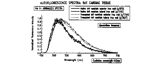
spectroscopic analyses are shown in Figures Sa through 7b. The FWHM is the
full
s width of the measured fluorescence spectrum at a level that is one-half the
maximum
height of the spectrum.
The spectroscopic analysis can alternatively, or also, comprise
comparing the ratio of the integral intensity of two or more wavelength bands
of the
spectrum that comprises the transplant fluorescent signature to the same ratio
from
1o healthy tissue. The wavelength bands for such an analysis can be selected,
for example,
by using numerical techniques to select sub-regions from the measured
fluorescence
spectrum acquired with a spectrometer or by using optical techniques, for
example
optical band pass filters, to select specific spectral bands that are measured
by
broadband optical detectors. Thus, a wavelength band is a range of wavelengths
of light
15 defined by a selected shorter wavelength limit and a selected longer
wavelength limit.
In some embodiments, the wavelength band is measured by a broad band optical
detector, which are characterized by a response to light across a broad
spectral region,
typically greater than several hundred nanometers. Examples of broad band
detectors
include silicon detectors, photomultiplier tubes (PMTs) and CCD assays.
Additionally,
2o the wavelength bands can be specific spectral bands, which can be selected
using
optical band pass filters in conjunction with the broad band detector.
As another alternative, the step of comparing can comprise comparing
the wavelength of maximum intensity of the fluorescence spectrum of the
transplanted
tissue with the wavelength of maximum intensity of the fluorescence spectrum
from the
25 healthy tissue. The wavelength of maximum intensity is the wavelength at
which the
fluorescent spectrum reaches its maximum intensity; a red-shift in the
wavelength of
maximum intensity indicates that the transplanted tissue comprises
characteristics of
rej ection.
In a preferred embodiment that applies to this and other aspects of the
3o invention, the step of collecting comprises selectively collecting a
significant portion,
CA 02283949 1999-09-10
WO 98/40007 PCT/CA98/00192
19
preferably at least about 10%, of the collected fluorescence or other induced
response
from an approximately 0.05-0.2 mm thick segment of the transplanted or other
target
tissue, the segment being located anywhere at a depth from 0-1 mm below the
surface
- of the transplanted tissue. Preferably, at least about 70% of the
fluorescence is collected
at such level, and further preferably at least about 85% of the fluorescence
is collected
at such level. Additionally, in alternative embodiments, the fluorescence is
collected
within a segment that is at a depth of about 0.05 - 0.3 mm, preferably at
about 0.05-
0.15 mm, and further preferably at about 0.1-0.2 mm. The depth of the selected
segment from the surface of the transplanted tissue is determined by measuring
perpendicularly from the center of the distal tip of the optical probe where
it contacts, or
is nearest to, the target tissue.
An example of the induction of fluorescence in different depth segments
of a target tissue is set forth in Figures 13a-13b. In Figure 13, the graphs
are contour
plots based on a Monte-Carlo calculation of the probe's sensitivity to the
tissue area
near its tip (the probe tip has a single illumination light guide centrally
disposed among
six surrounding collecting light guides, as in Figure 15). The value at a
particular r and
z is the contribution from the volume element generated by rotating the square
grid
element in the r - z plane about the r=0 axis (volume ~ 2~rOraz). In Figure
13a, the
collection fibers are hypothetically disposed immediately adjacent to the
excitation light
2o guide (and therefore extend from +100 p,m to +300 p,m and -100 ~m to -300
pm,
respectively). In Figure 13b, a hypothetical 50 p,m thick cladding/spacer
encompasses
the centrally located excitation light guide, and therefore the collection
light guides
extend from +150 p,m to +350 pm and -150 pm to -350 pm. As can be seen by the
key
to the right of the graphs, the different graph areas represent different
ranges of
sensitivity. The contours are spaced on a logarithmic scale (arbitrary units)
with each
level representing a half decade.
As can be seen from Figure 13b, the highest level of contribution to the
collected fluorescence (assuming a uniform distribution of fluorophores inside
the
tissue) occurs in the region from about 100 p,m to 300 p,m deep and at a
radial distance
of 30 p,m to 100 pm . The equivalent region in Figure 13a is much larger and
extends
CA 02283949 1999-09-10
WO 98/40007 PCT/CA98/00192
all the way to the tissue surface where the probe tip is situated. As a
result, a higher
percentage (23%) of the total signal for Figure 13a would originate in the
first 100 p,m
below the tissue surface as compared to Figure 13b (17%). Similarly, only 12%
of the
total signal in Figure 13b comes from the region from 400 ~m to 650 pm whereas
17%
5 comes from this region in Figure 13a. This percentage increases to 21% from
this
region for the case where there is a 100 p,m thick space bet<veen illumination
and
collection fibers. Thus, by varying the separation between illumination and
collection
fibers, sensitivities to different depths in the tissue can be obtained. It is
also possible to
change the depth sensitivity by changing the relative size of the collection
and emission
1o fibers or by adopting a different arrangement for the
collection/illumination array (e.g.,
a linear array as opposed to a concentric one).
Catheters or endoscopes that are useful for detection of the indicated
characteristics at different depths in the transplanted tissue (or other
tissue when
appropriate), can be made by providing at least one illumination light guide
and a
15 plurality of collection light guides, wherein the collection light guides)
and the
illumination light guides are spaced at differing distances from one another.
For
example, a single illumination light guide can be centrally disposed within
the distal tip
of the catheter or endoscope and a plurality of collection light guides can be
disposed in
a radial spiral away from the illumination light guide. Alternatively, the
illumination
20 light guide and the plurality of collection light guides can be maintained
in a line, or the
distal tip can comprise a plurality of illumination light guides and
collection light
guides in a geometric matrix, or in a random or semi-random matrix, such that
selection
of differing illumination light guides and collection light guides allows for
differing
distances between the light guides; such an arrangement can be preferable for
some
purposes because the plurality of illumination light guides provides for
implementation
of multiple light sources.
In a further aspect, the present invention provides methods of
determining the orientation of an optical probe relative to a target tissue.
The
orientation of the optical probe means the angle and/or distance of the
optical probe in
3o comparison to the target tissue. The target tissue can be transplanted
tissue as discussed
CA 02283949 1999-09-10
WO ~~ PCT/CA98/00192
21
above, or the target tissue can be any other tissue for which examination with
an optical
probe would be advantageous. The optical probe is a probe capable of
transmitting
light bi-directionally, and of emitting and gathering light, and is preferably
a probe
disposed at the distal end of a catheter or endoscope and therefore suitable
for insertion
and use within a living body. As is well known in the art, a catheter or
endoscope is a
generally tubular device for insertion into a body, typically via canals,
vessels,
passageways or body cavities for any of a variety reasons, including the
diagnostic
purposes such as those described herein as well as other purposes such as the
injection
or withdrawal of fluids or to keep a passageway open. The distal end of a
catheter or
1o endoscope is the end of the catheter or endoscope that is inserted into the
body and
directed to a target tissue; the proximal end is the end of the catheter or
endoscope that
is maintained outside the body, and typically comprises one or more handles,
knobs
and/or other control devices that allow the user to manipulate the distal end
of the
catheter and/or devices located at the distal end of the catheter or
endoscope. As used
herein, the distal end of the catheter or endoscope includes the distal tip of
the catheter
or endoscope, which is the most distal surface or opening of the catheter or
endoscope,
and the portion of the catheter or endoscope adjacent to the distal tip of the
catheter or
endoscope.
In one embodiment, the optical probe of the invention comprises at least
2o one light emitter, which means a device capable of launching light from the
optical
probe, and at least three light collectors (preferably six), which means
devices that are
capable of gathering light that strikes a receptive window of the light
collector. The at
least three light collectors are preferably equally radially distanced from
the at least one
light emitter, which typically means that the light emitters) is centrally
located and the
light collectors form a circle around the light collector. One example of such
an array is
depicted in Figure i 5, and is discussed further below.
The methods of determining the orientation of the optical probe
comprise the following steps. Light is emitted, or launched, from the at least
one light
emitter to the target tissue under conditions suitable to cause light (which
can be, for
3o example, reflectance light or fluorescent light) to emanate from the target
tissue. Such
CA 02283949 1999-09-10
WO 98/40007 PCT/CA98100192
22
return light can be termed "emanating light" and means light that is launched
from the
target tissue. Such emanating light is collected as it enters the at least
three light
collectors. Collected light is then analyzed to measure the relative intensity
of the
emanating light collected by each of the at least three radially disposed
light collectors,
which means the intensity of the emanating light is measured for each of the
collection
light guides and then assigned a value relative to the other light guides.
Equal
measurements for each of the collection light guides indicates that the
optical probe is
perpendicular to the target tissue; variance from equal gives the relative
position of the
probe. In view of the present disclosures, an artisan of ordinary skill can
also vary the
io radial distance or diameter of one or more of the light collectors from the
light
emitters) and then account for such variation when determining the orientation
of the
optical probe, and still be within the scope of this discussion. In addition,
the overall
strength of the emanating light provides information about the distance of the
optical
probe from the target tissue, and therefore the measurement of the relative
intensity of
the collected emanating light can also provide a value of the absolute
intensity of such
light and the distance from optical probe to the target tissue.
In another aspect capable of determining the orientation of an optical
probe relative to a target tissue, the present invention provides methods
wherein the
optical probe comprises at least three pairs of light emitters and light
collectors. The
light emitter in each of the at least three pairs is equally distanced from
the light
collector, which means that for each of the pairs, the light available for
collection by the
light collector is of the same relative intensity when the optical probe is
perpendicular to
the surface of the target tissue.
Generally, the methods comprise the following steps. Illumination light
(typically equal in intensity, wavelength, etc., so that the emanating light
induced by the
illumination will be equal when the probe is perpendicular to the target
tissue) is
emitted from each of the at least three light emitters to the target tissue
under conditions
suitable to cause light to emanate from the target tissue, to provide
emanating light.
The emanating light entering the at least three light collectors is collected.
And, the
3o relative intensity of the emanating light collected by each of the at least
three light
CA 02283949 1999-09-10
WO 9$/40007 PGT/CA98/00192
23
collectors is measured, therefrom providing for the determination of the
orientation of
the optical probe with respect to the target tissue.
In still another aspect, the present invention provides methods relating to
a biopsy, which comprises the removal of tissue from the body of a living
organism,
preferably a human being (human beings are the preferred subjects for each of
the
aspects and embodiments of the present invention, but the invention can be
practiced for
the benefit of other animals such as dogs, cats, horses and cows).
Generally, the methods comprise the following steps. An optical probe
comprising at least one light emitter and at least one light collector is
removably
1o attached to a target tissue in vivo, which means that the optical probe is
physically,
releasably attached to the target tissue, typically by mechanical devices that
can be
attached and released at will via the manipulations of a handle, lrnob or
other control
mechanism located at the proximal end of a catheter. Light is emitted from the
at least
one light emitter to the target tissue under conditions suitable to cause
Iight to emanate
from the target tissue, to provide emanating light. The emanating light
entering the at
least one light collector is collected and then evaluated to determine whether
the target
tissue comprises the one or more characteristics indicating that the target
tissue should
be biopsied. Such characteristics include characteristics of rejection as
discussed above,
characteristics of disease such as cancer or bacterial or viral infection, and
2o characteristics of inflammation. If the target tissue comprises one or more
characteristics indicating that the tissue may be unhealthy, additional
methods further
comprise obtaining the biopsy from the target tissue.
In a preferred embodiment, the optical probe is a part of the distal end of
a catheter or endoscope that also comprises a bioptome. In one embodiment, the
step of
removably attaching comprises clamping the jaws of the bioptome (or other
cutting
mechanism) onto the target tissue, and obtaining the biopsy comprises closing
the
bioptome about the target tissue, which means closing the bioptome
sufficiently to
separate, or remove, a piece of the target tissue from the target tissue.
Certain preferred
embodiments suitable for use for these aspects of the invention are discussed
in U.S.
3o patent application no. , filed March -, 1998 and entitled Catheters and
CA 02283949 1999-09-10
WO 98/40007 PCTlCA98/00192
24
Endoscopes Comprising Optical Probes and Bioptomes and Methods of Using the
Same.
In another preferred embodiment, the optical probe is a part of a catheter
or endosocope system, which means at least a catheter capable of conducting
light back
s and forth from its proximal end and its distal end, and a light source.
Thus, the system
comprises a catheter or endosocope comprising a light source at its proximal
end, an
optical probe at its distal end, and one or more light guides to transmit
light from and to
the proximal end and the distal end. The light emitter comprises an
illumination light
guide that transmits light from the light source to the distal end and the
light collector
to comprises a collection light guide that transmits the emanating light from
the distal end
to the proximal end of the catheter. Briefly, the illumination light guide
transmits light
from the proximal end of the catheter to the distal end, where the light is
launched onto
the target tissue. The collection light guide collects light that emanates
from the target
tissue (such as reflected or fluorescent light) and transmits it to the
proximal end of the
15 catheter, where the light is made available for analysis.
The illumination light guide and the collection light guide can be a single
light guide, which means that the same light guide can function as both the
illumination
light guide and the collection light guide. This is true for most aspects of
the present
invention. Alternatively, the illumination light guide and the collection
light guide can
2o be separate light guides.
In still yet another aspect that is similar to the aspect discussed in the
preceding paragraphs, the present invention provides methods relating to the
conduction
of a biopsy that do not require that the distal tip of the catheter be adhered
to the target
tissue. Generally, the methods comprise the following steps. An optical probe
is placed
25 adjacent to a target tissue in vivo, which means sufficiently near or in
physical contact
with the target tissue such that light can be emitted to the target tissue and
resultant
emanating light can be collected from the target tissue (the light emitted by
the target
tissue can be fluorescence, reflectance or other light that is induced by the
illumination
light and directed toward the optical probe). Light is emitted from the
optical probe to
3o the target tissue under conditions suitable to cause light to emanate from
the target
CA 02283949 1999-09-10
WO 98J40007 PCT/CA98/00192
tissue, to provide emanating light from an illumination area (which is the
area
illuminated by the light emitted by the optical probe). The emanating light
striking the
optical probe is collected and then evaluated to determine whether the target
tissue
comprises the one or more characteristics indicating that the target tissue
should be
5 biopsied, as discussed above.
If the target tissue comprises one or more characteristics indicating that
the tissue may be unhealthy {i.e., in need of biopsy), additional methods
further
comprise obtaining the biopsy from the target tissue without removing the
catheter from
the body of the patient, and preferably without moving the distal end of the
catheter
to within the patient. Keeping the distal end stationary between the optical
scan and the
biopsy facilitates obtaining the biopsy from the area of the target tissue
that was
illuminated by the optical scan, or the illumination area. Thus, these methods
permit
the same piece of tissue to be both scanned in vivo and biopsied. The methods
may be
implemented via the use of a catheter or endosocope comprising both an optical
probe
15 and a bioptome.
In yet a further aspect, the present invention provides methods for
determining whether a target tissue comprises one or more characteristics
indicating
that a target tissue should be biopsied. Such characteristics are discussed
above. The
methods comprise placing an optical probe adjacent the target tissue. The
optical probe
2o comprises at least one light emitter and at least three light collectors
that are equally
radially distanced from at the least one light emitter, and light is emitted
from the at
least one light emitter to the target tissue under conditions suitable to
cause light to
emanate from the target tissue, thereby providing emanating light. Such
emanating
light that enters the at least three light collectors is collected, and the
relative intensity
25 of the collected light is measured for each of the at least three light
collectors. This
permits determination of the orientation of the optical probe with respect to
the target
tissue, which in turn provides information about the quality of the scan taken
by the
optical probe including whether some or all of the target tissue in the
illumination area
was too far from the optical probe to provide adequately significant data
about such
area.
CA 02283949 1999-09-10
WO 98140007 PCT/CA98/00192
26
Thus, the user can next determine whether the orientation of the optical
probe relative to the target tissue is adequate to provide sufficient data
about the one or
more characteristics indicating that the target tissue be biopsied. The
orientation of the
optical probe to the target tissue is adequate to provide sufficient data
about the target
tissue when the optical probe is close enough and perpendicular enough to the
tissue
that the emanating light does not contain artifacts that interfere with the
interpretation of
the emanating light. The light emitted from the target tissue is strong enough
to be
collected by the optical probe and analyzed to provide meaningful information
for the
intended purpose, such as the determination of the presence or absence of
characteristics
of rejection. The optical probe has a suitable angle relative to the target
tissue when the
illumination light emitted by the probe strikes the target tissue generally
evenly across
the area of illumination such that the strength of the induced return light
from the target
tissue is representative of the state of the tissue across the area of
illumination and
collection. Preferably, the illumination light emitted from the optical probe
is emitted
perpendicular to the target tissue.
In view of the present specification, a person of ordinary skill in the art
will be able to routinely select a set of data points that will fit a given
situation. For
example, such a person can select only data points above a certain intensity
threshold,
select only peak data points and/or select only data points that occur within
a certain
2o wavelength or range of wavelengths. Upon determining that the orientation
is adequate,
the light collected by each of the at least three collection light guides is
then evaluated
to decide whether the target tissue comprises the one or more characteristics
indicating
that the target tissue be biopsied.
As with the aspects of the invention described above concerning the
determination of the orientation of an optical probe relative to a target
tissue, the present
invention also provides methods for determining one or more characteristics
indicating
that a target tissue should be biopsied wherein the methods comprise the use
of at least
three pairs of illumination light guides and collection light guides spaced as
described
above.
CA 02283949 1999-09-10
WO 98/40007 PCT/CA98/00192
27
In the event that the target tissue is found to comprise characteristics
indicating that a biopsy would be appropriate, then the methods optionally
further
comprise obtaining the biopsy, preferably of the same site that was optically
scanned.
In this and other methods described herein, the methods are typically
preferably performed on living animals, preferably human patients. Thus, the
illumination light is transmitted and the fluorescence, or other return light,
is collected
in vivo.
In an embodiment that is preferred for in vivo optical scanning,
particularly where the target tissue is a moving organ such as the heart, the
timing of the
to illumination and collection (i.e., the optical scanning) is controlled and
synchronized
with movement of the organism and/or the target organ to enhance the utility
of the
information that is collected and processed. Briefly, as discussed above,
measurements
of target tissue are preferably made when the target tissue receives strong
illumination
and then emits strong fluorescence or other response (preferably a signal to
noise ratio
that is greater than about 5:1, further preferably greater than about 10:1) at
a suitable
orientation to be optimally collected and evaluated.
Thus, in a preferred embodiment, the illumination and collection are
both performed during a single diastole of a single heart beat (or other
selected motion
of the target tissue). This embodiment is particularly preferred when the
target tissue is
the heart. Determination of the diastole of the heart beat can be effected by
a variety
means that will be apparent to one of ordinary skill in the art in view of the
present
specification. For example, the user can detect an electrocardiogram of the
heart beat of
the host, and then use one or more signals, such as the QRS wave or other
identifiable
event, of the electrocardiogram to initiate or trigger the steps of
transmitting and
collecting during a single diastole of the heart beat. See, e.g., Figure 17.
Alternatively,
the user can detect a pulse of the host using a blood pressure monitor, and
then use the
pulse to trigger the steps of transmitting and collecting. See, e.g., Figure
18. In a
preferred embodiment, a pulse oximeter, which measures the oxygen content of
the
blood, is used to provide the trigger that induces the scanning or date
gathering.
CA 02283949 1999-09-10
WO 98/40007 PCT/CA98/00192
28
In one preferred embodiment, the pulse is externally measured and the
blood pressure monitor is located externally to the host, which means that the
pulse is
measured non-invasively. Typically, this means that the pulse measurement
device
does not traverse the skin or any membranes of the patient, although the
device could be
inserted into a body cavity; preferably, the measurement device does not enter
the body,
including body cavities. In another preferred embodiment, measurement of the
pulse
comprises using a pulse oximeter.
In an alternative preferred embodiment, a plurality of measurements are
obtained throughout the duration of the heart beat (or other motion). An
example of this
embodiment is depicted schematically in Figure 16, which shows that the signal
intensity of the fluorescence or emanating light varies as the probe
approaches and
moves away from the target tissue (such movement can also be caused by the
target
tissue approaching and moving away from the probe). When the tissue is then
repeatedly induced to fluoresce or otherwise respond and the corresponding
fluorescence or other response is synchronously collected, the information
obtained
provides a generally repetitive series of sequentially increasing and
decreasing data
points. The increases and decreases correspond to the movement of the heart
during a
beat, and therefore provide a measure of the heart beat. The data points can
then be
selected to provide optimal information about the target tissue, for example,
by
2o selecting only data points above a certain threshold, by selecting only
peak data points
and/or by selecting data points that only occur in a certain temporal locale
within the
beat. In addition, these data point selection criteria can be combined with
physiological
triggers such as an ECG or pulse measurement.
Absent such determination of proximity, orientation, and timing, the
z5 presence of motion, a poor angle of incidence of light, or too great a
distance between
the probe and the target tissue may produce artifacts such as a "smeared"
measurement
that does not represent a discrete site.
In still another aspect, the methods of detection described above can be
used to distinguish between different grades or level of rejection, for
example by
3o correlating the status of the transplanted tissue with the ISHLT or
Billingham's levels
CA 02283949 1999-09-10
WO 98J40007 PCT/CA98/00192
29
described herein. Determination of the level of rejection can be determined,
for
instance by applying a linear discriminant function, then training the
function to
discriminate between the desired grades of rejection. For example, two groups
of
spectra are defined; a first group of spectra from a tissue known to have
ISHLT grade 0,
and a second group of spectra from the same type of tissue and known to
exhibit ISHLT
grade III rejection. Each spectrum comprises a set of features that comprise
numeric
information specific to that spectrum. In a preferred embodiment, the relative
intensities at all wavelengths are evaluated to discriminate between the
levels of
rejection (the intensity of each wavelength being a separate feature),
although the
discrimination and training can also be performed on selected wavelengths. A
computer-implemented program known as a stepwise feature selection algorithm
is used
to evaluate the wavelengths or features and then used to perform statistical
calculations
on various combinations of designated features to determine which
combinations) of
features, when used in the linear discriminant function, is (are) capable of
discriminating between the two groups of spectra. Preferably the program
selects
combinations that maximally discriminate between the two groups. This is
referred to
as "training" the discriminant function.
One example of such a linear discriminant function suitable for use as
described is the following:
DF = ao+a,I(480)+aZI(496)+a3I(504)+a4I(518)+asI(522)
ao = -5.6; a,=-64.7; a2=45.2; a3=4.8.8; a4=-115.5; as=64.1
In this function, I(480) means relative intensity at 480 nm, I(496) means
relative
intensity at 496 nm, etc. The absolute value of the coefficients (ao, a,, a2,
...) is
dependent on the methods of normalizing the spectra. Selection of wavelengths
and
normalization of spectra will be apparent to a person of ordinary skill in the
art in view
of the present disclosure.
Once the linear discriminant function has been trained it can be used to
score any spectrum, and can thereafter classify such spectrum into a desired
category,
such as ISHLT grade 0, I, II, III or IV. In addition, quadratic discriminant
functions,
CA 02283949 1999-09-10
wo 98i4ooo~ rc~ricA9aroom
neural network methods and other appropriate computer-implemented approaches
can
be used to discriminate between the grades of rejection.
Turning to apparatus provided by the present invention, catheter systems
and catheters according to the present invention generally comprise an optical
probe
5 and/or other features. The catheter is typically applied to the subject by
insertion into a
vein in the neck or leg and subsequent guidance to the target tissue. For
example, when
the target tissue is the heart, the catheter is typically inserted
percutaneously and then to
and through the superior vena cava and into the right atrium and right
ventricle of the
heart. Also typically {approximately 80% of the time), the heart biopsy is
taken from
1o the apex end of the thick septum wall dividing the left and right
ventricles in the heart.
The catheters of the present invention permit optical scanning to be performed
in areas
where biopsy was not possible and also provide information regarding the
status of the
target tissue. Materials used in the construction of the catheters of the
present
invention, when intended for human use, should meet USP Class N
biocompatibility
1 s standards.
As a general discussion of catheter systems of the present invention, such
systems comprise a catheter, as discussed above, that generally comprises a
catheter
body and a distal end designed for insertion into the patient, typically a
human patient, a
grip for physician control, and one or more light guides that can be connected
to one or
2o more light sources and one or more detectors. The light guides comprise one
or more
illumination light guides and one or more collection light guides. The
illumination light
guide can be the same light guide as the collection light guide, which means
that in
some embodiments the same light guide functions as both the illumination light
guide
and the collection light guide, and there need be only one light guide in the
catheter.
25 The illumination light guide accepts light from one or more light sources
and transmits it to its distal end, which is disposed within the distal end of
the catheter.
The light is emitted or launched, from the light guide into the tissue. The
illumination
light guide is preferably connected to the light source by an indexed
mechanical
coupling. The collection light guide collects light emitted by the target
tissue and
3o striking or entering its distal end, which is disposed within the distal
end of the catheter.
CA 02283949 1999-09-10
WO 98/40007 PCT/CA98/00192
31
The collection light guide transmits the light from the tip of the catheter to
one or more
detectors. The light emitted by the target tissue can be any kind of light
emitted by the
target tissue, such as fluorescent light, which is typically induced by the
illumination
light, or reflectance light wherein illumination light is reflected from the
target tissue.
The collection light guide is preferably connected to the detector by an
indexed
mechanical coupling. The detector is typically located at the proximal end of
the
catheter. Examples of materials suitable for use as a light guide include
optical fibers,
fiber bundles or fiber arrays. In a preferred embodiment, the illumination
light guide is
optimized for blue or near ultraviolet light transmission, particularly where
the
1o illumination light induces fluorescence in the target tissue and the
collected light is such
fluorescence. The light guide collection is optimized for visible and near IR
transmission.
At the proximal end of the catheter, the collection light guide, which
transmits light to the detector, can be directed into a fitting that positions
the fibers) of
the collection light guide concentrically with a lens or lens array such as a
gradient
refractive index (GRID lens or lens array. The GRIN lens may be cut so that
the one
end of the lens is matched to the numerical aperture (NA) of the fiber and the
opposite
end is cut to emit a collimated beam of light. If more than one fiber is used,
the GRIN
lens array can be positioned as shown in Figure 14 so that the beams can be
projected in
2o a line into a detector to facilitate wavelength separation and analysis.
The distal end of the catheter is generally designed to launch the
illumination light to the target tissue and collect the light emanating from
the target
tissue.
Turning to specific aspects of certain apparatus of the present invention,
one aspect provides an optical bioptome, which is an apparatus that combines
an optical
measurement device or probe with a tissue biopsy device or bioptome. As noted
above,
a bioptome is a device carried at the distal end of a catheter that snips off
a piece of a
target tissue for extraction from the organism and evaluation. Bioptomes often
comprise a pair of opposing jaws, but other configurations are also known. U.
S. Patent
3o No. 3,964,468; U. S. Patent No. 54,953,559; U. S. Patent No. 4,884,567; U.
S. Patent
CA 02283949 1999-09-10
WO 98/40007 PCT/CA98/00192
32
No. 5,287,857; U. S. Patent No. 5,406,959; WO 96/35374; WO 96/35382; WO
96/29936; WO 96/35374. As one example, a bioptome can comprise a fluoro-
polymer
catheter type body bonded to a metal tip that incorporates mechanically
actuated cutting
jaws. The cutting jaws can be opened and closed by actuating a control in the
hand
piece of the catheter.
The device allows optical assessment of tissue to assist the surgeon in
selecting sites and performing biopsy, and allows simple and easy biopsy with
minimal
risk and reduced harm to the patient because of shortened overall surgical
procedure
time and fewer insertions of catheters into the body.
The optical bioptome comprises a catheter or endoscope system
comprising a light source that supplies light at a proximal end of a catheter,
at least one
illumination light guide suitable for conducting light from the proximal end
to a distal
end of the catheter and for emitting the light from a distal end of the
illumination light
guide, at least one collection light guide suitable for collecting light
entering the distal
end of the collection light guide and conducting the light to the proximal end
of the
catheter, and a bioptome. In a preferred embodiment, the illumination light
guide and
the collection light guide are the same optic fiber.
The catheter comprising the optical probe and a bioptome can be
designed in a variety of configurations. For example, the catheter can be
prepared so
that when the jaws of the bioptome are opened the optical probe is extended
past the
open jaws to the target tissue, where the optical probe can then make a
measurement.
When the jaws are released (i.e., closed) the optical probe is retracted into
its lumen
within the catheter exterior. The bioptome and the optical probe can also be
disposed
side-by-side or concentrically within the catheter. Thus, the bioptome and the
optical
probe can be disposed equally extended with regard to the distal tip of the
catheter, one
can be disposed more extended than the other, or one or both can be extendible
and
retractable according to the needs of the user.
The optical probe and the bioptome can be modular or they can be
integrated in a single assembly. As with other catheters and devices described
herein,
each of the optical probe and the bioptome can be sterilized or destroyed
after use.
CA 02283949 1999-09-10
WO 98140007
PCT/CA9$/00192
33
In a further aspect, the present invention provides a catheter system
suitable for emitting and collecting light, the catheter system comprising a
light source
that supplies light at a proximal end of a catheter, at least one illumination
light guide
suitable for conducting light from the proximal end to a distal end of the
catheter and
for emitting the light from a distal end of the at least one illumination
light guide, and at
least three collection light guides, each collection light guide suitable for
collecting light
entering the distal end of the collection light guide and conducting the light
to the
proximal end of the catheter, wherein the collection light guides are equally
radially
disposed around the at least one illumination light guide. As with many other
aspects of
to this invention, the light guide ends are preferably flat cut and polished
flush with the
distal tip of the catheter.
In a functionally related aspect, the present invention provides a catheter
system suitable for emitting and collecting Light, the catheter system
comprising a light
source that supplies light at a proximal end of a catheter, at least three
pairs of light
guides, each pair comprising an illumination light guide suitable for
conducting light
from the proximal end to a distal end of the catheter and for emitting the
light from a
distal end of the illumination light guide and a collection light guide
suitable for
collecting light entering the distal end of the collection light guide and
conducting the
light to the proximal end of the catheter, and wherein the distance from the
collection
light guide to the illumination light guide is equal in the at least three
pairs.
These aspects of the invention are functionally related because they can
be used to determine if the distal end is perpendicular to the target tissue
and/or in
contact with or near to the target tissue. The spacing between the light
guides and the
diameter of the light guides, which can be routinely selected by a person of
ordinary
skill in the art in light of the present disclosure, determine the depth layer
of the target
tissue from which optical property information is collected. As discussed
above,
independent measurement of the signal intensity from each collection light
guide
permits elucidation of the orientation of the distal tip and the target
tissue, which can be
used to indicate the quality of the measurement. Thus, in sum, these aspects
are
3o preferred when the user desires to determine the orientation of the distal
end (and the
CA 02283949 1999-09-10
WO 98/40007 PCT/CA98/00192
34
optical probe carried therein) relative to the target tissue and/or to
selectively collect
light from a certain desired depth of the target tissue.
In still a further aspect, the present invention provides a catheter system
suitable for emitting and collecting light, the catheter comprising at least
one light
source that supplies light at a proximal end of the catheter and a plurality
of light
guides, wherein at least two of the light guides are suitable for conducting
light
longitudinally along the catheter, and for emitting and detecting light at
different sites
located along a distal end of the catheter, such that the catheter is capable
of emitting
and collecting light at a number of different sites along the distal end of
the catheter
to without moving the distal end. See Figures 19a-d.
Thus, the catheter comprises a plurality, preferably three or more and
further preferably six, optical ports 132 along the distal end of the
catheter. Preferably,
the ports form a spiral along the side of the distal end of the catheter,
although the ports
can also be in a line or otherwise spaced along the distal end of the
catheter. One or
more of the optical ports can also be located at the distal tip of the
catheter. The optical
ports typically provide a view or scan that is perpendicular to the axis of
the catheter,
although the optical ports can be either forward-looking or rear-looking if
desired. Each
of the optical ports provides a window for at least one illumination light
guide and at
least one collection light guide. The same light guide 130 can function as
both the
illumination light guide and the collection light guide, or the illumination
and collection
light guides can be provided in pairs (or in other combinations according to
the needs of
the user). Each of the pair of light guides is preferably positioned such that
the light
launched from the illumination light guide and the light collected by the
collection light
guide focus on the same location in the target tissue. In one embodiment, as
depicted in
Figures 19b-19d, the light guides 130 have a 45° angle 136 at their
distal ends to
provide a reflective flat surface 134 that emits and collects light in a side-
viewing
fashion.
The 45 degree surface at the distal end of the light guide is one preferred
embodiment because it facilitates perpendicular viewing from the side of the
catheter.
3o The distal ends of the light guides are then positioned to transmit
illumination light
CA 02283949 1999-09-10
PCT/CA98/00192
through the optical ports and to the target tissue, and to receive light from
the target
tissue through the window. Preferably, the distal ends of the light guides are
contacted
with the optical ports and are immobile with respect to such optical ports,
preferably via
use of an optically clear non-fluorescent epoxy. The 45 degree surface of the
light
5 guides can be coated with a reflective, non-fluorescent coating.
Each of the light guides (or pairs of light guides) can be placed in a
lumen maintained within the catheter, to give a plurality of lumens spaced
equally
around the inside of the catheter. In one embodiment, each lumen ends at a
different
distance to provide the light guides) therein access to the appropriate
optical port in the
to distal end of the catheter. In another embodiment, each of the lumens is
the same
length, and optical ports are cut into the outer side of each of the lumens at
various
distances from the distal tip of the catheter.
The resultant catheter has a plurality of optical ports that spiral up the
side of the catheter from the distal tip and can be used to make measurements
of the
15 target tissue at various distances from the apex by rotating the catheter
so that the port is
in contact with the tissue.
In still another aspect of the invention, the light guides (again preferably
flat cut and polished) are recessed from the distal tip of the catheter rather
than flush
with it. Co-luminal with the fibers is a liquid-carrying lumen that allows a
bolus of
2o non-fluorescing, non-reflecting liquid saline solution to be pumped to the
distal tip of
the catheter. The structure of the catheter tip directs the liquid around the
optical fibers
and out to the tissue so that the resultant jet of liquid pushes aside blood
or other
interfering material and acts as a liquid light path for transmission of light
to, and
collection of light from, the target tissue.
25 In still a further aspect of the invention that is somewhat similar to that
discussed in the preceding paragraph, the distal tip of the catheter is
covered with and
bonded to an elastomeric balloon comprising an optically transmissive window
that is
preferably non-fluorescing and non-reflective. Upon insertion of the catheter
into the
right ventricle, a bolus of gas or liquid is pumped into the balloon to cause
it to distend
3o and thereby contact the window with the tissue. The liquid or gas, which
can be air, in
CA 02283949 1999-09-10
WO 98/40007 PCT/CA98/00192
36
the balloon acts as an optically clear path to the tissue, while the balloon
pushes blood
and other interfering material out of the field of view.
In a preferred embodiment, the methods and catheter systems and other
apparatus described herein are operably linked to a computer containing at
least one
computer implemented program that implements at least one facet of the
methods,
catheter system and/or other apparatus. In a preferred embodiment, the program
is able
to determine the spectrum of light collected by the collection light guide,
determine an
intensity of light collected by the collection light guide, compare the
relative intensity
of light collected by a plurality of collection light guides and/or time when
light is to be
1o transmitted along the light guides in concert with a pulse or
electrocardiogram. An
example of a system is depicted in Figure 9. In Figure 9, an external
physiological
trigger 30 is received by system control and data processing computer 32,
which in turn
is linked to excitation light source 34 and transmits light via illumination
light guide 44
(contained within catheter 42) to transplanted tissue 48. The autofluorescence
emission
~5 or other response is collected and transmitted via collection light guide
46 and to
detector 36, where sensors 38 and wavelength discriminator 40 are located,
which
system then signals the system control and data processing computer 32.
Generally speaking, a computer suitable for use with the various aspects
of the present invention comprises a user interface, a system control, and
devices for
20 data acquisition, processing and management. Briefly, the user interface
typically
comprises devices such as probes, keyboards and screens for the entry and
display of
patient data and session information, system parameters and current control
parameters
and data collected. The system control typically effects system timing, light
source
pulsing and data acquisition timing. Data acquisition typically concerns
25 synchronization with physiological signals, signal conditioning and
preprocessing, and
data acquisition and storage. Data Processing typically concerns data quality
verification, data signal processing and data analysis. Data management
typically
concerns a structured data storage, data integrity check, data security, data
backup and
data reporting. Certain preferred embodiments suitable for use for these
aspects of the
3o present invention are discussed in U.S. patent application no. , filed
March -,
CA 02283949 1999-09-10
WO 98/40007 PCT/CA98100192
37
1998 and entitled Validating and Processing Fluorescence Spectral Data for
Detecting
Rejection of Transplanted Tissue.
The following discussion sets forth some of the components that are
advantageous for use with the methods and apparatus of the present invention.
Briefly,
systems suitable for implementation of optical scanning such as is described
herein
generally comprise a light source to generate appropriate excitation
wavelength(s), a
detector that selects and measures the appropriate wavelengths of the
fluorescence
emitted, and, preferably, a data processing and control system with software
that
1 o controls the timing of illumination and detection and processes the
acquired data.
Light Sources
The present invention can use any light source that provides a light that
induces fluorescence in the target tissue. For some aspects of the invention,
the light
source need not induce fluorescence, but may instead cause reflectance or
other light to
be emitted from the target tissue. Selection of an appropriate light source is
well within
the ordinary skill in the art in view of the present specification. With
regard to light
sources that induce fluorescence, the light source can be selected to provide
light from
ultraviolet (UV) through visible light. Preferably, the light comprises blue
or near-W
light. Also preferably, and particularly for in vivo aspects of the invention,
the light
does not comprise UV light because such light can induce cancer or other
problems
within the patient organism, which is preferably a human being. Further
preferably, the
light consists essentially of blue light and/or green light.
Some examples of preferred light sources to generate the required
excitation energy include a pulsed xenon flashlamp equipped with wavelength
selection
filters (Figure 10), a CW (continuous wave) mercury or xenon arc lamp equipped
with
wavelength selection filters (Figures 11 and 12), a Blue or UV CW laser, and a
Blue or
W pulsed laser. These are discussed below. The light sources in these figures
preferably have an indexed mechanical coupling adapter 82 maintained in a base
80 to
3o ensure that the illumination waveguide 84 is positioned to maximize the
light entering
CA 02283949 1999-09-10
WO 98/40007 PCTICA98/00192
38
the fiber, and are preferably controlled by system software, which controls
pulse timing
of the arc lamp power supply.
In figure 10, a pulsed xenon flashlamp 70 comprises a sealed housing arc
lamp and power supply. The arc lamp typically has an arc length of less than 2
mm and
is optionally equipped with an integral reflector to maximize energy directed
toward the
illumination light guide of the catheter or optical probe. An optical filter
or series of
filters such as a blocking filter 72 and wavelength selection filter 76 placed
in the
optical path can select the wavelength of the illumination light. The energy
emitted by
the arc lamp is collected and focused by a focusing lens 78. A collimating
lens 74 can
be placed between the filters if desired. In a preferred embodiment suitable
for use with
the present invention, the lenses are selected to direct the energy in a
converging cone
into an illumination light guide, with an apex angle that is less than or
equal to the
acceptance angle of the illumination light guide as defined by the numerical
aperture of
the illumination light guide.
is A CW mercury or xenon arc lamp light source (Figures 11 and 12)
comprises a sealed housing arc lamp 90 and power supply. The arc lamp
typically has
an arc length of less than 2 mm and is optionally equipped with an integral or
external
reflector to maximize energy directed toward the illumination waveguide of the
catheter
or optical probe. An optical filter or series of filters placed in the optical
path can select
the wavelength of the illumination light. The energy emitted by the arc lamp
is
collected and focused by a lens system. The lenses are selected to direct the
energy into
the illumination light guide in a converging cone with an apex angle that is
less than or
equal to the acceptance angle defined by the numerical aperture of the
illumination light
guide. In one embodiment, the lamp power supply operates continuously with no
pulsing. Alternatively, the lamp can be powered by a sinusoidally varying
current/voltage, which can also enhance the blue wavelength emission of the
lamp.
A blue or UV CW laser light source comprises a laser that emits light in
the blue or near ultraviolet wavelengths. Wavelength selection can be
accomplished by
using a laser such as a Helium-Cadmium (HeCd) laser or a Krypton-Argon laser
that
3o emits in the blue portion of the spectrum. Alternatively, a dye laser
pumped by a
CA 02283949 1999-09-10
WO 98J40007 PCT/CA98/00192
39
shorter wavelength laser wherein wavelength selection is a function of dye
characteristics and cavity monochrometer tuning can be used. The energy
emitted by
the laser is collected and focused by a lens system. The lenses are selected
to direct the
energy into the illumination light guide in a converging cone with an apex
angle that is
less than or equal to the acceptance angle defined by the numerical aperture
of the
illumination light guide. The laser can be equipped with a manual and/or
computer
controlled shutter.
A blue or W pulsed laser light source comprises a laser that emits light
in the blue or near ultraviolet wavelengths. The laser emits short duration
pulses,
l0 preferably under software program control. Wavelength selection can be
accomplished
by using a dye laser pumped by a shorter wavelength laser wherein wavelength
selection is a function of dye characteristics and cavity monochrometer
tuning.
Alternatively, a longer wavelength laser equipped with a frequency doubling
system
andlor an optical parametric oscillator (OPO) can be used. The energy emitted
by the
laser is collected and focused by a lens system. The lenses are selected to
direct the
energy into the illumination light guide in a converging cone with an apex
angle that is
less than or equal to the acceptance angle defined by the numerical aperture
of the
illumination light guide.
Detectors
Detectors suitable for use with the present invention separate the
fluorescence light emitted by the target tissue, and typically conducted to
the detector
from the target tissue by a collection light guide, into wavelength regions of
interest and
produces a signal proportional to the fluorescence emission of each of the
regions of
interest. The present invention can use more than one detector if desired. The
detector
is typically controlled by the system software so that start of acquisition
and integration
time can be synchronized with the shuttering and pulsing of the illumination
system and
with the physiological triggers. Exemplary detectors include a charge coupled
device
(CCD), charge injection device (CID), intensified CCD detector,
photomultiplier tube
CA 02283949 1999-09-10
WO 98/40007 PCTICA98/00192
(PMT) detector array, photo-diode array (PDA), intensified PDA and an
avalanche
photo-diode {APD) array.
The fluorescence light collected by the collection light guide can be
directed to a wavelength dispersive grating or prism and the resultant
spectrally
5 distributed light is projected onto the selected detector array(s), which
has been
calibrated for wavelength and intensity. The resulting signal then typically
undergoes
signal processing and discriminant analysis by the system software to
determine
whether the signal comprises the optical characteristics of tissue undergoing
rejection.
In a preferred embodiment, the fluorescence is collected by multiple
1o collection light guides and is projected onto the selected detector array
such that the
signal for each collection light guide can be analyzed independently. This
type of
multiple collection light guide/ 2-D detector array (which can also be
implemented with
other types of detector arrays) can be particularly helpful for analysis of
information to
elucidate probe orientation, distance and mobility relative to the target
tissue.
15 In another preferred embodiment, the fluorescence light from the target
tissue is directed into an optical beam splitter that divides the light into
two or more
spectral regions of interest. The spectrally separated components are then
each directed
to discrete detectors. In preferred embodiments, detectors can be silicon
detectors,
photomultiplier tubes or avalanche photo diodes, although other detectors can
also be
2o used advantageously with this embodiment of the invention.
The System Controller Program
The system controller program controls the timing of the emission of the
light that induces emanation of light from target tissue, typically by
controlling the
25 pulsing or shuttering of the light source. It also controls and
synchronizes the timing of
emanation light acquisition and detector integration with the operation of the
light
source, and in a preferred embodiment controls and synchronizes the timing of
such
light actions with external or internal physiological measurement triggers,
such as an
electrocardiogram {ECG) or the pulse generated by the heart beat. In one
preferred
3o embodiment, the measurement of the pulse comprises the use of a pulse
oximeter. In
CA 02283949 1999-09-10
PCT/CA98/00192
41
the ECG triggering mode, the software synchronizes illumination and collection
windows with specific signals, or waves, within the ECG, such as the QRS wave.
One
advantage of an ECG trigger is that the ECG can be obtained from an external
patient
electrocardiograph monitoring device or system. Pulse oximeter triggering mode
permits the user to trigger timing of various actions from an external pulse
oximeter
monitoring device or system.
Using the system controller program, the user can set a time interval for
desired events. For example, the user can set a time interval after the
physiological
trigger or after initiation of data acquisition when the light source will be
pulsed or
1 o shuttered, when data acquisition will begin, and/or when data will be
measured,
integrated and/or analyzed. See Figures 17 and 18. Alternatively, such
intervals) can
be set automatically by the system controller program.
In an alternative preferred embodiment, which can be considered an
internal optical triggering mode, the system continuously triggers the light
source
and/or detector at a set frequency to provide a plurality of measurements over
the
duration of a single heart beat. See Figure 16. As the optical probe comes in
proximity
with the tissue the light signal collected by the probe increases. The
software can
monitor the signal for a local temporal intensity maximum in illumination or
emission
of the target tissue, and then select a window of desired data at a selected
time at or near
2o the time of the trigger, which can before, during and/or after the trigger.
As an
alternative to, or in conjunction with, the temporal window, an intensity
threshold
window can be defined for desired data.
Physiological triggering such as ECG or pulse monitoring can be used in
conjunction with the threshold window, the temporal window and/or other data
selection methods described above.
After data acquisition, in one embodiment, the system controller
program can process the data to determine the relative responses of the
intensity of the
spectral regions of interest. This information can then be compared to the
responses
characteristic of normal and abnormal tissue and then shown by a display, such
as a
CA 02283949 1999-09-10
WO 98/40007 PCT/CA98/00192
42
numerical or graphic display, to assist the surgeon in evaluating the tissue
and/or
determining appropriate sites for biopsy.
The following Examples are offered by way of illustration, and not by
way of limitation.
EXAMPLES
Example 1. Rat Heart Transplant Model
to A. Abdominal allograft and isograft rat heart transplant models were
used to evaluate tissue rejection by detection of fluorescence. The models
allow for the
transplant recipient rat's heart as an internal control. Allografts (different
rat strains)
and isografts (same rat strains) were treated with cyclosporin or untreated.
The protocol
for this Example was depicted schematically in Figure 1.
Turning to the experiment itself, a 250-350 g F344 rat was anaesthetized
and, using a surgical microscope, a ziphopubic incision was made to free the
abdominal
aorta and inferior vena cava below the origin of the renal vessels. A donor
rat (150-
200 g Lewis rat) was then anaesthetized and the heart was isolated. Ties were
placed
around the inferior and superior venae cavae and the pulmonary artery and
thoracic
2o aorta were transected 3 to 5 mm distal to their origins. A ligature was
placed around the
pulmonary veins. The heart was removed from the thoracic cavity and placed in
saline.
A donor heart was placed in the abdominal cavity of the F344 recipient, and
microvascular anastomoses were completed between the recipient inferior vena
cava
and donor pulmonary artery and the recipient abdominal aorta and donor
thoracic aorta.
Syngeneic grafts (Lewis-to-Lewis and F344-to-F344) were performed in the same
manner.
On the day of sacrifice, the transplant and the host heart were taken from
each rat. The atria were frozen and the ventricles were cut into 3 transverse
cross
sections. The posterior walls were opened and spectral measurements were
acquired, as
3o depicted in Figure 2. Following the spectral measurements, the ventricular
sections
CA 02283949 1999-09-10
WO 98/40007 PCT/CA98/00192
43
were fixed either in 10% buffered formalin for histo-morphological studies
including
histological evaluation, tests for cell death, and assays to evaluate protein
and gene
expression, or frozen in OCT for archive.
Figure 2 was a schematic diagram of the light pathway and heart tissue
indicating the section or slice of the ventricle that was taken and a method
of achieving
the spectral measurements.
B. Fluorescence spectroscopy.
The system to measure spectroscopy, illustrated in block diagram fonm
to in Figure 3, comprised a Nitrogen Dye Laser 2, 4 with stepper motor 6 and
stepper
motor controller 8 for wavelength selection, an optical multi-channel analyzer
(OMA)
16 with gated intensified detector, and a PC computer 10 (other computers
could also be
used). A bifurcated fiber optic bundle 26 was used to conduct the excitation
or
illumination laser light and to collect and return the fluorescence light to
the detector
20. Gating electronics (pulser 12 and pulse amplifier 18) were used to achieve
a high
signal to noise ratio, allowing fluorescence measurements to be performed with
ambient
light on. Excitation wavelength changes were performed automatically and
quickly by
the stepper motor 6 which was controlled by the OMA PIA (peripheral interface
adapter) port 14 using the OMA data acquisition (DAD) program. The filter
wheel 22
2o in front of the spectrometer 24 was also controlled by the PIA port to
change the barrier
filters when different excitation wavelengths were used.
Operation of the system was illustrated in Figure 4, which was a timing
sequence for operation of the Nitrogen Dye Laser-OMA system. The DAD program
directs the OMA to send a 28 p,s pulse to the pulser. Three milliseconds
later, the pulser
triggers the pulse amplifier and the laser. Twelve microseconds after being
triggered
the laser emits a 0.5 ns wide laser pulse. The high voltage negative gating
pulse was
kept active for 15 p,s after the laser trigger pulse was fired to make sure
all the
fluorescence photons were collected. The gating pulse width is still much
shorter than
the basic OMA exposure time (=30 ms). Therefore, a high signal to noise ratio
was
3o achieved. When the spectral measurement at one excitation wavelength was
completed,
CA 02283949 1999-09-10
WO 98/40007 PCT/CA98100192
44
the stepper motor controller was triggered to change the wavelength. When the
wavelength change was complete, the stepper motor controller signals the OMA
that
spectral data acquisition can begin again. With the same laser dye, the
wavelength
change takes less than one second.
C. Results:
Ten rat abdominal cardiac allografts underwent 201 spectral
measurements. Fluorescence spectra from normal cardiac tissue and transplanted
tissue
at 442 nm excitation wavelengths were shown in Figures 5a through 7b. Normal
tissue
consistently had a narrower spectral peak with consistently dampened
fluorescence
intensity (au) as compared to transplanted tissue. The latter tissue thus had
significantly
broader and higher level fluorescence intensity, whether sampled optically in
the
endocardium (Figures Sa and Sb), in the myocardium (Figures 6a and 6b), or on
the
epicardium (Figures 7a and 7b). The waveform contour was consistently broader
in the
transplanted hearts and had a spectral peak shifted to longer wavelenths. In
Figure 8,
the difference in fluorescent qualities of normal and allograft myocardium was
represented in color images of fluorescence at different red/green image gain
ratios.
The image gain ratio was the ratio of the intensity amplification of the red
image versus
the intensity amplification of the green image where the red image was the
image in the
2o wavelength range 630 nm and above, and the green image is the image in the
wavelength range of 490 nm to 560 nm. In addition to the fact that the normal
heart
tissue was consistently green regardless of the gain ratio, certain hearts
with moderate
injury required a higher gain ratio in order for the image to be more
predominantly
orange-red, while more severely injured myocardium (images g, h, and i in
Figure 8)
becomes yellow-orange-red at a lower red-green image gain ratio. Thus, not
only the
presence of rejection may be detected, but also the severity of rejection can
be
discerned. Waveform contour analysis of other samples also showed that the
severity of
rejection can be discerned. See Figures 20-22.
CA 02283949 1999-09-10
WO 98/40007 PCT/CA98/00192
Example 2. Optical Biopsy In Yivo Of A Human Being
After determining that the patient is not excluded from the procedure,
perform a standard diagnostic cardiac catheterization procedure in order to
introduce an
optical bioptome into the heart. Such procedure can comprise a standard right
heart
5 catheterization performed percutaneously from the right internal jugular
vein as
described below. A 9 F sheath is used initially, which can accommodate a
standard 7 F
end-hold catheter or 7 F balloon-directed floating thermodilution catheter.
A cap is placed over the patient's hair and a pillow is positioned under
the shoulders and neck in order to slightly hyperextend the neck. Care is
taken to
1 o position the head in line with the long axis of the body, with the patient
facing to the
left. The right side of the neck is prepped and draped using standard sterile
technique.
Gentle pressure is then placed on the patient's mandible, and the patient is
asked to raise
his head slightly off the pillow (no more than 2 in.). This causes the
sternomastoid
muscle to contract, thus making it easier to identify landmarks and to mark
the position
15 to introduce the sheath. A point on the lateral border of the median head
of the
sternomastoid muscle at least 6 cm above the clavicle is marked. The area is
infiltrated
with 2% xylocaine using a 25-gauge needle. A 22-gauge, 1 %2-in. needle is then
attached to the syringe and xylocaine is infiltrated deeply. Care is taken not
to enter the
carotid artery. The needle is angled caudal and slightly lateral in an attempt
to locate
2o the right internal jugular vein, which lies directly under the lateral head
of the
sternocleidomastoid muscle. After the area is adequately anesthetized a small
stab
wound is made in the skin with a #11 scalpel blade and the subcutaneous tissue
is
spread apart with a small straight clamp. A 10-cc syringe containing a small
amount of
saline is attached to a 22-gauge needle. The patient is instructed to perform
the
25 Valsalva maneuver, in order to increase venous pressure. It is also helpful
to raise the
patient's legs, thus further increasing venous pressure. The needle is then
advanced
slowly, angling the tip both caudal and slightly lateral so that it penetrates
just under the
lateral head of the sternomastoid muscle. Constant suction is maintained on
the syringe
in order to identify when the needle first enters the internal jugular vein.
The needle is
3o not advanced through the back wall of the vein, thus minimizing the
possibility of
CA 02283949 1999-09-10
WO 98/40007 PCT/CA98/00192
46
bleeding into the carotid sheath. The syringe is detached and the needle left
in place in
order to act as a guide. An 18-gauge Amplatz~ or Cournand needle is attached
to the
syringe. Using constant suction, the needle is advanced slowly following the
course of
the 22-gauge needle. When the internal jugular vein is entered, a short
straight guide
wire is passed through the 18-gauge needle, the needle removed, and the false
catheter-dilator and sheath are then positioned in the internal jugular vein
using standard
technique. The side-arm of the sheath is fitted with a stopcock to prevent an
air
embolus. The side-arm and sheath are then flushed with heparanized saline.
The optical bioptome is prepared by curving it at a 45° angle
to approximately 7 cm from the tip. The bend is in alignment with the optical
bioptome
handle, thus facilitating proper manipulation of the tip in the heart. The
patient is asked
to suspend breathing and the optical bioptome is advanced quickly into the
sheath,
pointing the tip towards the lateral border of the heart (the patient's right
side).
Fluoroscopy can be used at this point in order to ensure that the optical
bioptome does
not inadvertently enter the right subclavian vein. When the optical bioptome
is in the
mid to lower third of the right atrium, the handle is rotated counterclockwise
(anterior)
to point the tip medially. The tip of the optical bioptome is then advanced
across the
tricuspid valve. Occasional difficulty in crossing the tricuspid valve will be
encountered. A slightly different bend on the optical bioptome or rotating the
optical
2o bioptome at a different level in the right atrium will facilitate crossing
the tricuspid
valve. After crossing into the right ventricle the optical bioptome handle is
rotated
further counterclockwise so that it is now pointing posteriorly; the optical
bioptome tip
should also be pointed in this direction. The posterior position of the
optical bioptome
tip can be verified using a C-arm type fluoroscope is available. The tip is
then
advanced until it meets resistance (at this point the operator should feel the
cardiac
impulse) or until ventricular premature depolarizations are induced. The tip
is now in
the area of the right ventricular apex pointing towards the ventricular
septum. On
fluoroscopy the tip should be at the level or slightly caudal to the top of
the left
diaphragm.
CA 02283949 1999-09-10
WO 98/40007 PCT/CA98/00192
47
An optical biopsy of the tissue is obtained by administering excitation
light at a wavelength of 442 nm in accordance with the procedure outlined
above in
Example 1B. The resulting fluorescence is analyzed to determine if the tissue
provides
a significantly broader and higher level fluorescence intensity, and/or is
significantly
red-shifted when compared with healthy tissue of the same type, similar to the
analysis
described above in Example 1 C.
In the event that the optical biopsy suggests that tissue biopsy is
indicated, the optical bioptome is withdrawn 1 to 2 cm and the jaws opened.
This tends
to straighten the bend in the optical bioptome, pointing the tip more towards
the apex of
to the right ventricle. Such straightening of the optical bioptome can also be
effected prior
to obtaining the optical biopsy, if desired (typically such early
straightening is effected
without opening the jaws of the bioptome). The optical bioptome is again
advanced
until resistance is felt or ventricular premature depolarizations are induced.
The jaws of
the optical bioptome are then closed. Occasionally, it is necessary to pause
approximately 2 to S sec to allow the jaws to close completely. The bioptome
is then
withdrawn rapidly and in the same motion rotated clockwise (anteriorly) back
to the
right atrium. Initially, significant resistance will be experienced, but then
there will be a
sudden release of tension and the bioptome can be Quickly withdrawn from the
right
ventricle. The patient is then asked to suspend breathing and the bioptome is
withdrawn
2o from the sheath. The jaws of the bioptome are opened and the size of the
sample is
examined. The sample is removed using fine forceps. Care is taken not to crush
the
sample. It is then placed in room temperature fixative for later study.
When manipulating the bioptome into the right ventricle, it is preferred
that the bioptome is rotated anteriorly, thus avoiding accidental entry into
the coronary
sinus. The biopsy will typically be taken from the ventricular septum in order
to avoid
the thin free wall of the right ventricle. It is also preferred to remove the
biopsy from
the apex of the right ventricle in order to avoid the conducting system.
If indicated from the optical biopsy, from three to five tissue biopsy
specimens are obtained, each measuring approximately 1 to 2 mm3. Samples are
assayed for light and electron microscopy and one specimen frozen for possible
CA 02283949 1999-09-10
WO 98/40007 PCT/CA98/00192
48
subsequent study. Specimens may also be obtained for viral culture or other
specialized
procedures.
At the conclusion of the procedure the thorax is fluoroscoped to look for
evidence of either pericardial effusion, pleural effusion, or pneumothorax. If
any
complication is suspected a standard chest film or echocardiogram may be
obtained.
The patient is then put in a sitting position to lower venous pressure, asked
to suspend
breathing, and the sheath is removed. Pressure is applied to the area above
and below
the puncture site for at least 10 min. The puncture site is then covered with
a Band-Aid
~-type bandage.
Example 3.
This evaluation employs the abdominal allograft model (Lewis to F344)
which allows for the heart from the rat receiving the transplant to serve as
an internal
control. Lewis-Lewis isografts are also planned as controls. Allografts
(different rat
strains) and isografts (same rat strains) will be treated with cyclosporin or
left untreated.
The donor and recipient rats are prepared and then sacrificed, as outlined
above in Example 1.
For each rat, sections from the native heart, the spleen and the transplant
heart are cut onto a single slide. Sections cut at 5 p.m are stained with
Hemotoxylin &
2o Eosin (H&E), Masson's trichrome and Movat's pentachrome. The slides stained
with
H&E are evaluated by a pathologist and scored using the ISHLT grading system
as well
as other pathologic indices.
Figures 23 and 24a-d depict a graph of several spectra that differ
according to the level of rejection experienced by the target tissue, along
with
photographs of corresponding tissue sections. Figures 23 and 24a-d help
illustrate
changes in the autofluorescence spectra after the onset of tissue rejection: a
shifting of
the main peak to longer wavelengths and an increase in spectral weight in the
520 nm
600 nm region. Another salient feature of the graph in Figure 23 is a distinct
sub-peak
in the control spectrum at 600 nm that largely disappears in spectra ~ from
tissue
3o exhibiting rejection.
CA 02283949 1999-09-10
WO 98/40007 PCTJCA98100192
49
Immunosuppression therapy with cyclosporin was used on a large subset
of the rats described above because of its key role in the management of heart
rejection
in human patients. It was intended to determine whether it had unforeseen
effects
regarding tissue autofluorescence that might obscure real changes associated
with
inflammation due to rejection. Figure 25a compares the average
autofluorescence
spectrum of 118 non-transplant control spectra taken from hearts that had not
been
treated with cyclosporin and 126 similar spectra taken from hearts that had
been treated
with cyclosporin. There is excellent agreement between the two average
spectra,
indicating that cyclosporin in and of itself is not contributing to any
anomalous
fluorescence that might be masked as intrinsic tissue autofluorescence. Figure
25b
depicts the same non-transplant control dataset as in Figure 25a but this time
divided
into averaged spectra corresponding to epicardium and endocardium. There is an
intrinsic difference between endocardium and epicardium: a greater ratio of
green
wavelengths (near the main peak at ~ 520 nm) to longer wavelengths is formed
in the
epicardium. This is consistent with the appearance of the epicardium, which is
brighter
green than the endocardium, when viewed under a fluorescence microscope.
To quantify the variations in the autofluorescence spectra as the tissue
rejection progresses, a linear discriminant function analysis (DFA) with a
stepwise
feature selection of single wavelength values was used. Care should be taken,
however,
that there are no spectra in the dataset used for the stepwise selection
procedures that
have clearly identifiable spectral features that are not necessarily
correlated with the
tissue rejection/inflammatory response process. A primary example is that of
absorption of light by hemoglobin: the absorption coeff cient of red blood
cells changes
by more than an order of magnitude within the visible range (480 nm - 800 nm).
Hemoglobin has a characteristic "double-bumped" absorption spectrum having
maxima
at 540 nm and 580 nm. This can be used as a signature facilitating visual
and/or linear
function discrimination. It was quite common to find a pair of minima in the
autofluorescence spectra corresponding to hemoglobin absorption. The tissue
rejection
process can involve hemorrhaging and the heterotopic transplant heart tends to
be prone
3o to thrombi. Such can have so many red blood cells present that the main
peak of the
CA 02283949 1999-09-10
WO 98/40007 PCT/CA98/00192
signal is near 640 nm with shorter wavelengths being heavily suppressed.
Before
proceeding with an analysis of the spectra in terms of their ISHLT grade,
spectra
displaying blood absorption characteristics were removed from consideration:
first, a
subset of these spectra was identified by visual inspection and then this
subset was used
5 as one group in a stepwise feature selection, the other group being all the
remaining
spectra; the linear discriminant function thus identified was then used to
score all the
spectra which isolated the majority of the blood absorption-contaminated
spectra in a
single group.
The remaining spectra were found to be free of the blood absorption
1o effects and could be analyzed with fiu-ther discriminant fimctions.
Initially, all
endocardium and epicardium spectra were group into one data set and then
separated
into an ISHLT grade 0 group and an ISHLT grade III group. DFA was performed
and a
five-feature DF generated. The wavelength values used were 480, 496, 504, 518
and
522 nm. These wavelengths were in the area of the position and shape changes
of the
15 main peak visually observed in the autofluorescence spectra. This DF was
then used to
score spectra from all grades including spectra from native hearts and control
hearts.
The results are shown in the box-plot in Figure 26a. A clear progression of
decreasing
DF scores with increasing tissue rejection severity is evident with high
statistical
differences (2-population T-test, p<0.005) between the ISHLT grade 0, grade I
and
2o grade II groups. The ISHLT grade II and III groups were statistically
different at the
p=0.05 level. The data was not divided beforehand into a training and test
set; the DF
was trained only on the ISHLT grade 1 and grade III groups. Thus the controls,
natives,
grade I and grade II groups are fair tests of the DF performance.
Figure 26b is a Receiver Operator Characteristic (ROC) plot for a test of
25 grades I, II and III versus grade 0 (which was designated the "normal"
group for this
purpose). The III vs. 0 test had the best performance although even the I vs.
0 gives a
30% False Positive/30% False Negative rate for a DF score threshold of ~ 2Ø
Other
discriminant functions ca,n be generated in view of the present disclosure
that are more
specific to grades 0/I discrimination and give better performance.
CA 02283949 1999-09-10
WO 98/40007 PCT/CA98/00192
51
Figure 27a shows the performance of a discriminant function that uses
the control group and a group consisting of grades II and III as the training
set.
Combining grades II and III into one group gives a larger training set, which
helps to
give a more robust discriminant function. Using a small training set runs the
risk of
over-fitting the data, i.e., the discriminant function performs well on the
training set but
poorly on other data sets. In this case, the grade 0 group was included along
with the
native and grade I group in the test set. The discriminant function separated
the groups.
Figure 27b is the box-plot of the DF scores of the grade II versus grade
III group where the DF was trained on these two groups. A good separation in
DF
to scores and a highly statistically significant difference between the two
groups results
although it must be noted that the training and the test sets are the same.
Figures 28a and 28b illustrate a more severe test of the discrimination
between the spectra from the different ISHLT grades. Here, the spectra were
divided
into epicardium and endocardium groups prior to any DFA. The discriminant
function
in Figure 28a was trained on endocardium spectra from grades 0 and III and the
resulting box-plot of the DF scores for the epicardium spectra is shown in the
figure -
all groups shown are independent tests of the DF performance. There is still a
monotonic decrease in mean DF scores for groups of increasing rejection
severity and
highly statistically significant differences (p<0.005) between grades 0 and
any of grades
I, II and III. However, the discrimination between intermediate stages of
rejection is not
as good as in Figure 26a. This decrease in performance is most likely due to
the smaller
amount of data used as the training set as discussed above. Another
manifestation of
this is the greater intra-group variability of the DF scores, which blurs
inter-group
differences. Figure 28b is analogous to Figure 28a except that the DF training
was done
on the epicardium spectra and the results shown are for the scored endocardium
spectra.
This box-plot is qualitatively similar to Figure 28a and the same conclusions
apply.
The training set for this DF is even smaller than for Figure 28a, and this is
reflected by
the even larger spread in DF scores within the groups. These are in spite of
the intrinsic
differences between epicardium and endocardium autofluorescence spectra
discussed
3o above.
:.
CA 02283949 1999-09-10
~ ~ _ , . ~ . ) ~7 7 7
' V1~0 98/40007 _ , " ; ;
~C3'/C~~S8~00?92~
,, ", » ..
52
Although the present invention had been described in some detail by way
of illustration and example for purposes of clarity and understanding, it will
be readily
apparent to those of ordinary skill in the art in light of the teachings of
this invention
that certain changes and modifications may be made thereto without departing
from the
s ~ii-aF scope of the appended claims.
A;rE~J~E~ S~bE