Note: Descriptions are shown in the official language in which they were submitted.
CA 02284356 1999-09-29
DEVICE FOR INTUBATING LACRIMAL DUCTS
FIELD OF THE INVENTION
The present invention relates to devices for intubating lacrimal ducts and
more particularly to intubation sets for positioning a length of tubing in the
lacrimal
ducts.
BACKGROUND OF THE INVENTION
The insertion of an elongate length of tubing member in the lacrimal ducts
is a common surgical procedure for reconstruction or other remedial purposes.
Lacrimal fluid or tears are continuously supplied from the lacrimal gland to
wash
across the sclera and other conjunctiva) components and the cornea. The excess
lacrimal fluid is drained through a network of passages commencing with the
puncta
which appear as small papilla adjacent the inner canthus or inner corner of
the eye,
the lacrimal fluid being collected in the lacrimal sac by a number of
canaliculi
connecting the puncta with the lacrimal sac. The lacrimal sac is drained
through the
nasolacrimal duct which passes into the inferior nasal meatus. This network of
passages is referred to hereinafter as the lacrimal ducts.
Closures of the lacrimal ducts can occur as a result of congenital anomalies,
accidents, inflammation, advanced aging, as well as other physiological
conditions.
The closures prevent drainage of tears so that the affected eyes are
continually
brimming over with fluid, causing much personal discomfort to the patient, and
often causing infection and/or inflammation of the mucous membranes as well as
other undesirable conditions.
Known devices for correcting blocked lacrimal ducts include an intubation set
CA 02284356 1999-09-29
disclosed in U.S. Patent No. 4,380,239 to Dr. John Crawford, et al. The
intubation
set includes a probe consisting of a light resilient wire which can be readily
deflected
through an angle of at least 90 degrees to permit the probe to pass from the
nasolacrimal duct to the inferior nasal meatus. The probe has a tip or distal
end
which is slightly enlarged and rounded to limit the possibility of damage to
tissue
when the probe is inserted, and a proximal end provided with an enlargement. A
very flexible tube of minimal rigidity has a first end which is in engagement
over the
wire at the proximal end and into contact with the enlargement. Adhesive can
be
used to improve the connection.
When in use, the probe is inserted through either an upper punctum or a
lower punctum of the lacrimal ducts and is guided downwardly through the
lacrimal
ducts to the inferior nasal meatus whereupon a tool in the form of a hook
(shown
in the Crawford patent) is used to pull the probe through the nostril leaving
a length
of tubing extending through the full length of the lacrimal ducts.
Although the Crawford intubation set provides a relatively secure connection
between the tube and the probe, in rare instances these components have been
found
to separate during the intubation procedure. The cause is believed to be
contact
with bony structure which tends to roll the tube off the wire. Although this
event
is unusual, it would be preferable to provide an intubation device which
benefits
from the principles taught in the Crawford patent and which also has a
connection
between the probe and the tube which is less likely to suffer from this
problem.
It is also possible for a closure to occur in the canaliculi and not in the
nasolacrimal duct. For instance, the canaliculi may suffer traumatic injury
while the
nasolacrimal duct remains unaffected. It is therefore also desirable to
provide an
-2-
CA 02284356 1999-09-29
intubation set adapted to position a length of tubing in the region of the
canaliculi
without probing the healthy nasolacrimal duct in order to facilitate
intubation by
a physician.
SUMMARY OF THE INVENTION
In accordance with an aspect of the invention, an intubation set is provided
having an improved connector between a tube and a probe. In a preferred
embodiment, the connector includes a deformable sleeve which at one end
receives
an end of the tube and at the other end is tapered to converge on the probe
where
it is permanently attached. The tube contains a solid insert which can be set
in a
compound such as silicone rubber and the sleeve is deformed radially inwardly
to
trap part of the tube containing the insert thereby locking the tube to the
sleeve and
hence to the probe. A method of making the intubation set is also provided.
The invention also provides an improved intubation set for use in intubating
the canaliculi without probing a healthy nasolacrimal duct. In this case the
probe
is curved in a spiral configuration and preferably lies in a plane. A tip on
the probe
can be pulled out of the plane to create a spiral for easier insertion. The
spiral can
be clockwise or anticlockwise depending on which way the tip is pulled out of
the
plane.
These and other aspects of the invention will be better understood with
reference to the following description and drawings.
BRIEF DESCRIPTION OF THE DRAWINGS
Preferred embodiments of the invention will now be described with reference
-3-
CA 02284356 2003-09-15
to the drawings in which
Fig. 1 is a diagrammatic view of an intubation set according to a first
preferred
embodiment of the invention;
Fig. 2 is a side sectional view taken generally on line 2-2 of Fig. 1 and
drawn to a
larger scale to illustrate a connector used to attach a tube to a probe of the
intubation set;
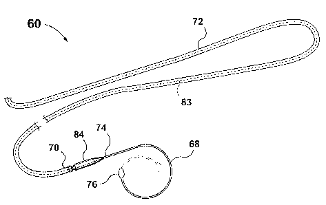
Fig. 3 is a diagrammatic view of part of an intubation set according to a
second
preferred embodiment of the invention; and
Fig. 4 is a diagrammatic view showing the intubation set of Fig. 3 in use
intubating
the canaliculi of a patient.
DETAILED DESCRIPTION OF THE PREFERRED EMBODIMENTS
Reference is first made to Fig. 1 which illustrates an intubation set
designated
generally by the numeral 20 according to a first preferred embodiment of the
invention.
The intubation set 20 can be used according to a procedure described in the
aforementioned
U.S. Patent No. 4,380,239 to Dr. Crawford. The procedure has become well
established
and uses a tool described in the Crawford patent. The same tool can be used
with the
present intubation set 20 to pull the probe from the nasal passage.
The intubation set 20 preferably includes a pair of similar thin probes 22, 24
of a light
stainless steel wire which can be resiliently deflected to pass from the
nasolacrimal duct to
the inferior nasal meatus. The probes 22, 24 have enlarged and olive-shaped
tips 28, 30,
respectively, to limit the possibility of damage to tissue when one of the
probes 22, 24 is
inserted, and proximal end portions 32, 34 respectively.
It has been found that an olive-shaped tip meets with less resistance than a
more
rounded tip, as taught by the Crawford patent and results in easier insertion
of the probe
-4-
CA 02284356 2003-09-15
through the lacrimal ducts.
The end portions 32, 34 of probes 22, 24, respectively, are securely coupled
by
connectors 33, 35 to respective first and second end portions 36, 38 of a very
flexible
resiliently deformable, medical grade silicone rubber tube 40 of minimal
rigidity, as will be
described.
A length of silk 6/O suture 41 extends through the tube 40 along its full
length and
is used to tie cut ends of the tube 40 together after the tube 40 has been
positioned in the
lacrimal ducts, as is common in the art.
Fig. 2 illustrates the connector 33 in more detail. This connector is also
typical of
connector 35 and connects the end portion 36 of the tube 40 to the proximal
end portion
32 of the probe 22.
A thin-walled stainless steel tubular sleeve designated generally by reference
numeral
42, has a tapered leading end 44 a trailing end 46 and a crimped intermediate
portion 50.
hhe leading end 44 is silver soldered to the end portion 32 of the probe 22
with a portion
of the proximal end portion 32 of the probe 22 extending within the sleeve 42.
The end portion 36 of the tube 40 extends inside the sleeve 42 and is secured
by the
combination of a cylindrical insert 48 of solid stainless steel, and the
crimped portion 50 of
the trailing end 46 of the sleeve 42 as will be described.
The insert 48 is assembled in the connector by first entering a first quantity
of
uncured silicone sealer 56 into the tube, followed by the insert 48 and
finally by a second
quantity of uncured sealer 52.
After curing, the tube, together with the sealer and the insert 48, is entered
into the
sleeve 42 and deformed by crimping to form the portion 50 which locks the tube
in place.
The cured silicone will combine with the insert 48 to create a structure which
can not be
-5-
CA 02284356 2003-09-15
withdrawn through the radially decreased crimped intermediate portion 50. The
resistance
to separation is enhanced by the rigid insert 48 because the insert is larger
than the internal
diameter of the crimped intermediate portion 50. Also, because the silicone
locks the insert
in the tube, the insert cannot escape from the tube to allow the tube to be
removed without
the insert.
The connector 33 provides a smooth tapered transition from the probe 22 to a
generally cylindrical portion 49 of the sleeve 42. Consequently, as the
intubation set 20 is
advanced in the lacrimal ducts, the smooth transition will ensure there will
be little
likelihood of snags thereby minimizing the discomfort to the patient.
The dimensions and other characteristics of the intubation set 20 will now be
discussed followed by a discussion of alternative structures.
The probes 22, 24 are of tempered stainless steel wire having a diameter of
approximately 0.4 mm and exhibit a resistance to deflection to retain their
original shape
after being subjected to small deflections as they are moved through the
lacrimal ducts.
Such fine wires would, of course, puncture tissue if the ends of the wires did
not include the
enlarged tips 28, 30.
The length of the probes 22, 24 is approximately 110 mm. The silicone rubber
tube
40 has an outer diameter of about 0.6 mm, an inner diameter of about 0.3 mm
and a length
of about 300 mm. The sleeve 42 is deformable stainless steel having a length
of about 11
mm. The generally cylindrical portion 49 of the sleeve 42 has an outer
diameter of about
0.8 mm and an inner diameter of about 0.6 mm.
One of the advantages of the present invention is that there is provided a
very secure
means of connecting the tube 40 to the probes 22, 24 such that the risk of
separation of the
tube 40 from any one of the probes 22, 24 is minimized.
-6-
CA 02284356 2003-09-15
Many variations to the intubation set 20 thus described and the method of its
manufacture are possible within the scope of the invention. For example, the
length of the
probe may range from about 50 mm to about 150 mm.
As well, the length of the sleeve may be from about 6 mm to about 16 mm.
Further,
the outer diameter of the generally cylindrical portion of the sleeve may be
from about 0.7
mm to about 1.0 mm.
Additionally, the deformation of the sleeve resulting in portion 50 may be by
any
suitable method, including rolling or swaging.
The intubation set may consist of a single probe coupled to a tube rather than
a pair
of probes coupled to respective ends of a tube.
Further, the tube need not contain a length of silk suture extending
therethrough.
Reference will now be made to Figs. 3 and 4 which illustrate an intubation set
designated generally by reference numeral 60 according to a second preferred
embodiment
of the invention. The intubation set 60 is specially adapted for intubating
only the upper
and lower canaliculi 62, 64 respectively, without probing the nasolacrimal
duct 66 (Fig. 4).
Thus, the intubation set 60 is to be preferred to the intubation set 20 when a
blockage
occurs only in the canaliculi of the patient. In such case, it would be
simpler and easier to
probe the canaliculi using the intubation set 60 than to probe the canaliculi
and nasolacrimal
duct using the intubation set 20.
The intubation set 60 is similar in every respect to the intubation set 20
except for
the dimensions of the component parts, and the configuration and material of
the probes.
Specifically, the intubation set 60 includes a pair of similar spiral probes
68, (one of which
is shown). The probe is a resilient, stiff, stainless steel wire having a
proximal end portion
74 coupled to a portion 70 of a very flexible resiliently deformable silicone
rubber tube 72
_7_
CA 02284356 2003-09-15
of minimal rigidity containing a length of 6/O silk suture 83 extending
through the full
length of the tube 72.
The probe 68 has an enlarged and olive shaped tip 76 and can be deflected to
pass
through the puncta and the canaliculi of a patient. The spiral probe 68 can be
made to
become more like a corkscrew by pulling the tip 76 transversely to a plane
containing the
remainder of the probe. This can be done in either direction to give a left or
right handed
helical spiral as preferred to facilitate engagement in the selected punctum.
The length of
the probe can be varied as indicated in ghost outline in Fig. 3.
The intubation set may be used in two ways. One way, illustrated in Fig. 4, is
to
insert the probe through an upper punctum 80, feed the probe into the upper
canaliculus
62, move the probe downwardly into and through the lower canaliculus 64, and
finally move
the probe out through the lower punctum 82. The probe 68 is then pulled away
from the
patient by the tip 76 until a portion of the tube 72 is positioned through the
canaliculi.
Afterwards, the tube 72 is cut in two locations adjacent respective upper and
lower puncta
80, 82. Portions of the tube 72 near the cut ends thereof are peeled away
revealing ends of
the silk suture 83 which are then tied together, thereby forming the portion
of the tube 72
retained in the canaliculi into a loop, in accordance with established
practice.
Another method of using the intubation set 60, which is not illustrated,
involves
inserting one of the spiral probes through one of the upper and lower puncta,
inserting the
other of the spiral probes through the other of the upper and lower puncta,
and feeding the
probes through respective canalicula until they meet at a location where the
canaliculus is
severed due to trauma to the region. The probes are then removed through an
incision or
open wound in the patient at the severed location, thereby leaving a length of
tubing in the
canaliculi. The tube is then cut in two locations adjacent the incision or
wound, to form cut
_g_
CA 02284356 2003-09-15
ends of the tube and suture. The cut ends of the tube are peeled away to
reveal cut ends of
the silk suture, and the cut ends of the silk suture are then tied together to
form the tube
into a loop.
The intubation set 60 further includes connectors 84 (one of which is seen)
coupling
the probe 68 to the tube 72 in the same manner as described with reference to
the
intubation set 20.
The wire of the probe has a diameter of about .6 mm and a length of about 50
mm.
As in the case of the intubation set 20 according to the first preferred
embodiment,
variations to the second preferred embodiment are possible without departing
from the
scope of the invention.
For example, the probe set shown in Figs. 3 and 4 has a different use from the
probe
set 20 and may be made using known connections between the tube and the probes
while
still providing a novel structure. The spiral probe may be secured to the
silicone tube in any
number of ways such as by using an adhesive or the joint taught in U.S. Patent
4,380,239
to Crawford et al.
The spiral probe may be made of annealed stainless steel which is more easily
deformable and less resilient.
The wire of the probe may have a diameter of from about 0.4 mm to about 0.8
mm,
and a length of from about 37 mm to about 75 mm.
The intubation set may consist of a single spiral probe coupled to a tube
rather than
a pair of spiral probes coupled to respective end portions of a tube.
Further, the tube need not contain a length of silk suture extending
therethrough.
It should be understood that the foregoing description of the preferred
embodiments
are by way of example only and should not be construed as limiting the scope
of the
-9-
CA 02284356 2003-09-15
invention as defined by the following claims.
-10-
