Note: Descriptions are shown in the official language in which they were submitted.
CA 02285959 1999-10-08
WO 98/46124 PCT/US98106851
1
MINIMALLY INVASIVE DETECTING DEVICE
z
TECHNICAL FIELD
4
The present invention relates to percutaneous detecting devices. More
s particularly, this invention relates to percutaneous detecting of agents,
such
as body electrolytes, glucose, alcohol, pharmaceuticals and illicit drugs
using
a transcutaneous sensors.
s BACKGROUND ART
~o
11 Interest in the percutaneous detecting of body analytes (i.e., fluid
~z electrolytes), organics (e.g., glucose), pharmaceuticals and illicit drugs
has
~s grown over the years. In recent years, a number of electrochemical sensors
have been developed for detecting each of these analytes in the blood or
interstitial fluid of a patient. For example, glucose sensors have been
~s developed for obtaining an indication of blood glucose levels in diabetic
patients. Existing electrochemical sensors require either collection of a
~s sample from the patient or some form of invasive insertion of a sensor
probe
into the patient.
zo Thin film electrochemical sensors have been developed for
z~ subcutaneous placement of sensor probes in direct contact with the
patient's
zz blood or other extracellular fluid. One such example of a thin film
zs electrochemical sensor, disclosed in U.S. Patent No. 5,391,250 issued to
za Cheney, II et al., is fabricated using thin film mask techniques. With thin
film
z5 mask techniques, three thin film conductive elements are laid down in close
zs parallel relation on a substrate and encased between flexible insulating
layers
z7 of polyimide material. The conductive elements are left exposed at the
distal
zs end of the electrochemical sensor for placement in direct contact with the
zs patient's blood. Appropriate electrode chemistries are applied to the
exposed
so conductive elements for use as a blood glucose sensor. One of the exposed
3~ conductive elements has a coating containing glucose oxidase to define a
sz working electrode. The other two exposed conductive elements are coated
ss with other suitable materials or left uncoated to define a reference
electrode
sa and a counter electrode for the electrochemical sensor. The conductive
35 elements are left exposed at the externally located proximal end for
ss connection to a suitable monitor.
, tlic~. t,~amc1 CA 02285959 1999-10-08
" ,. ,
,, _,
. , , , , " ,
:,
~, ~ ,.,.,
,, . , ;
2' , ,
1 The exposed conductive elements at the distal end of the
2 electrochemical sensor are transcutaneously placed using a sensor insertion
3 set such as disclosed in U.S. Patent No. 5,390,671 issued to Lord et al. The
4 sensor insertion set comprises a separate slotted insertion needle extending
through a mounting base that attaches onto the patient's skin. The thin film
6 sensor has a proximal end carried by the mounting base and a distal segment
7 with the exposed sensor electrodes thereon protruding from the mounting
8 base. The proximal end of the sensor is linearly offset from the distal
9 segment so that the distal segment can be fitted into the slotted insertion
needle while the proximal end is carried by the mounting base. The distal
11 segment is transcutaneously placed as the insertion needle pierces the
12 patient's skin upon the mounting base being pressed onto the patient's
skin.
13 The insertion needle is then withdrawn over the electrode from the patient
14 leaving the distal segment at the selected site and the mounting base on
the
patient's skin.
16 Insertion of the needle is comparatively invasive, painful and
17 frightening to many patients. Therefore, there is a need for a minimally
18 invasive, painless placement of electrochemical sensors in the patient's
skin.
19 Furthermore, it is desirable in some circumstances to apply the
electrochemical sensors to individual skin-piercing elements rather than in
21 close parallel relation on one sensor probe for improved manufacturability.
22 A blood glucose monitoring system is described in United States Patent
23 No. 4,953,552, by DeMarzo. This includes a patch and mounted therein a
24 ground electrode and a sensing electrode. The sensing electrode contains a
single piercing element. The piercing element is coated with appropriate
26 materials so that a nanoampere-level current is generated when the
electrode is
27 exposed to glucose. The patch is typically applied to the forearm and the
two
28 electrodes are electrically connected to a meter which reads the low level
29 current and displays an equivalent blood glucose reading on a display worn
around the wrist.
31
32 DESCRIPTION OF THE INVENTION
33
34 The present invention is a detecting device and method for placing an
electrochemical sensor in contact with a patient's interstitial fluid with
skin
36 piercing microprotrusions in a minimally invasive manner. The device of the
~L ~»~ H1 CA 02285959 1999-10-08
. n ~ n
.., v n ~
n 1 ~
~ ~ v .
2A " ,
1 present invention pierces the stratum corneum of a body surface to position
2 the electrochemical sensor just below the outermost layer of the epidermis
3 but above the patient's nerve endings and blood vessels to eliminate pain
and
4 bleeding for the patient. The present invention integrates an
electrochemical
sensor and at least one skin-piercing member into one device to achieve in
6 situ detection with a painless application.
7 In one aspect, the invention comprises a plurality of microprotrusions
8 for piercing the skin in which each microprotrusion forms an individual
9 electrode of an electrochemical sensor, instead of all of the electrodes on
one
probe, to maximize the electrode area while maintaining the small protrusion
.;_ .:_, "~[~'
n...!-..L~J Jf7[C(
CA 02285959 1999-10-08
WO 98/46124 PCT/US98/06851
3
1 size necessary for minimally invasive operation. In another aspect, the
z electrodes are coated onto each side of the microprotrusions to increase the
. s active electrode area.
4 In another aspect of the invention, the device utilizes a member having
s an opening therethrough in communication with a fluid-attracting member, a
s plurality of microprotrusions extending downward from a first side of the
member, and a thin-film electrode on the microprotrusions which form an
a electrochemical sensor. With the thin-film electrodes inserted in the
patient's
s skin, a constant flow of interstitial fluid past the electrodes can be
maintained
1o by drawing the fluid through the opening with the fluid-attracting member
11 (e.g., an osmotic salt layer).
12
1s BRIEF DESCRIPTION OF THE DRAWINGS
14
1s Figure 1 is a top plan view of a portion of a member with
1s microprotrusions having sensor electrodes thereon;
Figure 2 is a bottom perspective view of the member of Figure 1 after
1a the microprotrusions have been bent into position;
1s Figure 3 is an enlarged partial cross-sectional view of a detecting
zo device in accordance with the present invention;
z1 Figure 4 is an enlarged perspective view of the bottom side of a
22 member in accordance with another embodiment of the present invention;
zs and
24 Figure 5 is a diagrammatic cross-sectional view of an osmotic
2s detecting device in accordance with the present invention.
zs
27
28 MODES FOR CARRYING OUT THE INVENTION
zs
so Turning now to the drawings in detail, one embodiment of the skin
31 piercing member 2 of the present invention is generally shown in FIGS. 1
and
s2 2. Member 2 is used for the percutaneous detecting of an agent. The term
33 "detecting" is used broadly herein to include detection of or sensing the
sa presence or amount of an agent, as well as monitoring the presence or
ss amount of an agent. The terms "substance", "agent" and "drug" are used
ss interchangeably herein and broadly include substances such as glucose,
CA 02285959 1999-10-08
WO 98/46124 PCT/US98/06851
4
body electrolytes, alcohol, illicit drugs, pharmaceuticals, etc. that can be
sampled through the skin. The major barrier properties of the skin, such as
s resistance to agent detecting, reside with the outer most layer (i.e.,
stratum
a corneum). The inner division of the epidermis generally comprises three
layers commonly identified as stratum granulosum, stratum malpighii, and
s stratum germinativum. There is essentially little or no resistance to
movement
of an agent through the stratum granulosum, stratum malpighii, and stratum
s germinativum. The device of the present invention is used to pierce the
s stratum corneum 24 for in situ detecting of an agent with a sensor located
below the outermost layer of the patient's skin (FIG. 3).
» Member 2 comprises a plurality of microprotrusions 4 which are sized
~z and shaped for piercing the outermost stratum corneum layer of (e.g., human
~s or other animal) skin. FIG. 1 shows the microprotrusions 4 after they are
~a formed (by a photolithography process followed by a chemical etching
process described in more detail hereinafter) and after coating (e.g., by
~s printing) electrodes 14, 16, 18 and electrical traces 20 thereon. FIG. 2
shows
the microprotrusions 4 after they have been bent to extend (e.g.,
,a perpendicularly) downward from the plane of plate 6. FIG. 4 shows member
~s 2 in an inverted position to better show the microprotrusions 4. Only a
portion
Zo of the plate 6 is shown in FIGS. 1, 2 and 4. The member 2 provides for the
transcutaneous placement of a flexible sensor 22 having one or more
Zz electrodes at a selected site within the body of a patient. Particularly
member
23 2 facilitates the placement of a flexible thin film electrochemical sensor
of the
Za type used for detecting specific parameters representative of patient
25 conditions. Placing the sensor within the skin of the patient allows in
situ
2s readings to be obtained instead of relying on collecting interstitial fluid
into an
27 absorbing member. The in situ detection minimizes lag time in the readings
is compared to diagnostic methods which rely on extracting the interstitial
fluid
is before the measurement can take place. In one preferred embodiment, the
so sensor is designed to monitor glucose levels in diabetic patients.
CA 02285959 1999-10-08
WO 98/46124 PCT/US98/06851
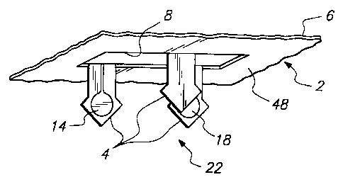
In the embodiment illustrated in FIGS. 1 and 2, the member 2
comprises a three electrode electrochemical sensor shown generally at 22
s having a sample electrode 14, common electrode 16 and reference electrode
a 18. Electrical traces 20 are routed from each electrode along the upper
s surface of the device 2 for interface with an electronic control unit or
detector
s 10 (shown schematically in FIG. 3). The three electrodes 14, 16 and 18 on
the adjacent microprotrusions 4 are moved into the orientation shown in FIG.
s 2 by placing the plate 6 of FIG. 1 on a die (not shown) and using a punch
(not
s shown) which is pushed through the opening 8. The microprotrusions 4 of the
electrochemical sensor 22 are sized appropriately so that they reach through
the stratum corneum 24 but do not contact the patient's nerve endings 26
12 (FIG. 3). For example, the tear drop shaped electrodes 14, 16 and 18 shown
~s in FIGS. 1 and 2 at the tip of each microprotrusion 4 are about 100
~a micrometers in diameter and the microprotrusions 4 have an overall length
of
about 150 micrometers. With this configuration, electrochemical sensor 22 is
responsive to changes in the presence or amount of agent in the patient's
interstitial fluid without causing a painful sensation or bleeding. Prior to
~a punching, the sensor 22 can be constructed using thin film mask techniques
is utilizing thin film conductors 20 embedded or encased between layers of
2o selected insulated material such as polyimide film. The electrodes 14, 16
and
21 18 at the distal tip of each microprotrusion are inserted into the
patient's skin
Zz in order to contact the patient's interstitial fluid when the sensor is
23 transcutaneousiy placed.
za As is known in the art and illustrated diagrammatically in FIG. 3, the
is diamond electrodes 14, 16 and 18 are in electrical communication, through
Zs conductive traces 20, with a suitable control unit 10 for detecting the
patient's
z~ condition (e.g., blood glucose concentration) in response to signals
derived
Za from the sensor electrodes. Any suitable thin film mask techniques
including
is with reference to those disclosed in U.S. Patent Numbers 5,391,250 issued
so February 21, 1995 to Cheney, II et al. and 5,108,819 issued April 28, 1992
to
s~ Heller et al. can be used in the present invention. The sensor can be used
CA 02285959 1999-10-08
WO 98/46124 PCT/US98106851
6
over a prolonged period of time for periodically or continuously detecting a
2 body electrolyte, such as glucose in a diabetic patient. Such readings are
s useful in monitoring the patient's blood glucose concentration (i.e.,
through
a appropriate software which correlates the concentration of glucose in
s interstitial fluid with the concentration of glucose in the blood) and can
further
s be used to adjust a treatment regime which typically includes administration
of insulin to the patient and/or appropriate modification of diet and/or
exercise.
s In the illustrative sensor construction shown in FIGS. 1 and 2 designed
s for use as a subcutaneous glucose sensor, each sensor 22 is shown to
~o include three parallel conductors or traces 20 corresponding with three
separate electrodes 14, 16 and 18. Appropriate electrode chemistries
defining the tear drop-shaped electrode surfaces at the distal ends of the
~s microprotrusions 4 can be applied as appropriate. In this illustrative
sensor
,a embodiment for use as a glucose sensor, electrode 14 includes glucose
~s oxidase to define a working or sample electrode. The other two electrodes,
~s counter electrode 16 and reference electrode 18 may contain other suitable
chemistries, to define a counter electrode and a reference electrode for the
electrochemical sensor 22. As is known to those skilled in the art of
electrochemical analyte (e.g., glucose) sampling, at least the working
2o electrode 14 should be coated with an excluding membrane in order to limit
electrical interference due to oxidation or reduction of extraneous species in
22 the interstitial fluid. The excluding membrane can be comprised of two
layers,
is including a first layer for keeping scar tissue or macrophages from coating
the
2a electrode and reducing the active electrode area, and a second layer for
2s excluding small molecular weight oxidizable or reducible species. In
glucose
is sensing, the second layer is typically formed of cellulose acetate and is
permeable to hydrogen peroxide but substantially less permeable to other
28 endogenous oxidizable/reducible species.
2s The reference electrode is typically formed of silver/silver chloride and
so preferably contains an electrolyte having a controlled composition as is
known
s~ to those skilled in the electrochemical sensing arts.
CA 02285959 1999-10-08
WO 98/46124 PCT/US98/06851
7
By placing each of the electrodes 14, 16 and 18 on a separate
microprotrusion 4, instead of locating aH of the electrodes 14, 16 and 18 on a
s single microprotrusion 4, the electrode area is maximized while maintaining
a
a relatively small protrusion size necessary for a minimally invasive device.
In an alternate embodiment, the electrodes are coated onto each side
s of the microprotrusions doubling the active electrode area. The separation
of
electrodes on individual microprotrusions eliminates problems that are
s associated with depositing the reference, sample and common electrodes
s close together in a small configuration. The etched space between the
electrodes guarantees safe separation of the electrode coating materials so
that there is little chance of bleeding of one coating to another electrode
during manufacturing. It is within the scope of the invention, however, to
~s utilize only a single microprotrusion 4 with all of the electrodes 14, 16
and 18
~a on that one microprotrusion. Likewise, although a glucose sensor has been
described, any detecting system can be utilized with the device 2. It is
within
the scope of the invention that the particular detecting system may have only
one or two electrodes or may have more than three electrodes. If additional
electrodes are needed for the detecting system, more microprotrusions can
be used and arranged for the best configuration. The configuration illustrated
zo in FIG. 4 utilizes multiple microprotrusions 4 around the plurality of
openings 8
2, in a redundant way such that all six microprotrusions are coated with
22 electrodes. In this way, if some of the electrodes are damaged during
is manufacturing, faulty, or do not penetrate the skin, the control unit 10
can test
Za at start up to see which electrodes are working and only utilize the
working
is electrodes for detecting the agent. Likewise, more than one set of
is microprotrusions and openings can be located on a member 2 as shown.
Also, as shown in FIG. 4, two sets of three electrode sensors are shown
za around each opening 8 for redundancy and accuracy.
is The distal ends of microprotrusions 4 can have any of a variety of
so shapes and configurations for piercing the skin or body surface, including
s~ arrow-shaped or diamond-shaped ends as shown in FIGS. 1 and 2,
CA 02285959 1999-10-08
WO 98/46124 PCT/US98/06851
8
triangular-shaped ends as shown in FIG. 4 and pins {not shown). The
z microprotrusions 4 penetrate the stratum corneum of the epidermis when
s pressure is applied to the device to facilitate the detecting of an agent
through
a a body surtace. The term "body surface" as used herein refers generally to
the outermost layer of skin, mucous membranes, and nails of an animal or
s human, and to the outer surtace of a plant.
In the illustrated embodiment, the plate 6 is formed with an opening 8
s between the microprotrusions 4. The opening 8 corresponds to the portion of
s the plate 6 occupied by each of the microprotrusions 4 prior to the
~o microprotrusions being bent into a position which is substantially
perpendicular to the plane of plate 6. The number of openings 8 per device
,2 and the number of microprotrusions 4 per device are independent. The
~s device may have only one large opening 8 with a plurality of
microprotrusions
4 around the opening. As will be described below, the opening 8 may be
covered with a fluid-attracting member for enhancing the movement of an
agent being sampled past the electrodes and into a fluid-attracting reservoir.
In another embodiment, the device does not have an opening 8 through the
is plate 6. In this latter embodiment, the microprotrusions 4 are made by
molding or casting and are then coated with the electrodes.
Zo The microprotrusions 4 are generally formed from a single piece of
material (although they need not be) and are sufficiently sharp and long for
2z puncturing at least the stratum corneum of the body surface. In one
is embodiment, the microprotrusions 4 and the plate fi are essentially
24 impermeable or are impermeable to the passage of an agent. The width of
25 each microprotrusion can be any of a range of widths. Usually, the width of
is the microprotrusion is in the range of about 25 micrometers to 500
micrometers. The length of the microprotrusions is subject to variation of the
28 body surface being penetrated and corresponds to the natural thickness of
2s the stratum corneum for one of the features of the invention is that the
sensor
so electrode detects the agent below the outermost layer of the epidermis.
s~ Usually, the microprotrusions will be about 20 micrometers to about 400
CA 02285959 1999-10-08
' ' ~ , -,' . .,
. , ~,, , ~',",. ,
' 3 ~ ,~ , ' ,,'
:,
micrometers in length. The microprotrusions 4 can have slanted (i.e., angled)
z leading edges 64 (FIG. 4) to further reduce the insertion force required to
s press the microprotrusions into the body surface. The leading edges of each
a microprotrusion can be all the same angle or can be at different angles
s suitable for piercing the body surface. Alternatively, the leading edge of
each
s microprotrusion can be arcuate (i.e., curved) in shape, having, for example,
a
convex or concave shape.
a The member 2 can also improve the attachment of the device to the
s body surface so that continuous agent detection through the body surface is
~o preserved during movement of the body surface. In the embodiment shown in
FIG. 4, projections in the form of barbs 50 on at least one of the
~z microprotrusions 4 assist in anchoring the member 2 and any corresponding
,s device or structure used in combination therewith to the body surface.
Barbs
a 50 can be on any number of the microprotrusions from one to all
~s microprotrusions. The barbs 50 are optional as other means for holding the
,s member in contact with the body surface can be used. The present invention
,~ can be used in conjunction with a wide variety of microprotrusions
,a configurations, for example, reference may be had to U.S. Provisional
~s Application No. 60/019,990 filed June 18, 1996 and subsequently published
as
zo PCT application W097148440, of which any of the disclosed configurations
z, can be used with the present invention.
zz The pattern for any of the microprotrusion array members 2 of the
z3 present invention can be produced with a photo-etching process. For
za example, reference may be had to U.S. Provisional Application No.
60/019,990
zs filed June 18, 1996 of which any of the disclosed methods can be used to
zs produce the member 2 of the present invention. A thin plate 6 of metal such
z~ as stainless steel or titanium is etched photo-lithographically with
patterns
zs containing skin piercing structures. In general, a thin laminate dry resist
or wet
zs resist is applied on the plate 6 which typically has a thickness of about 7
micrometers to about 100 micrometers, preferably about 25 micrometers to
s~ about 50 micrometers. The resist is contact exposed using a mask having the
sz desired pattern and is subsequently developed. These
AMENDED SHEEP
CA 02285959 1999-10-08
WO 98/46124 PCT/US98/06851
operations are conducted in much the same way that they are for the
z manufacture of a printed circuit board. The plate 6 is then etched using
acidic
s solutions. After the pattern has been etched through the plate, the plate 6
is
a placed on a die having a plurality of openings corresponding to the openings
s 8 in the plate. A punch having a plurality of protrusions corresponding to
the
s openings 8 in the plate 6 and openings in the die is initially located above
the
plate and the die. At the initial stage, the microprotrusions 4 are in the
same
s plane as the rest of the plate 6. The punch dies are then pressed into the
s openings 8, thus bending the microprotrusions downward to be substantially
~o perpendicular to the plane of the plate 6. The finished structure provides
microprotrusions 4 with an adjacent opening 8. In one embodiment, the
opening 8 allows the passage of interstitial fluid therethrough when the
member 2 is applied to the body surface. Rectangular openings 8 are shown
~a in the figures but the invention encompasses the use of any shape openings
including, but not limited to, square, triangular, circular and elliptical.
Generally, the microprotrusions 4 are at an angle of about 90 degrees
to the surface 48 (FIG. 3) of the plate 6 after being punched, but they can be
~a disposed at any angle forward or backward from the perpendicular position
~s that will facilitate penetration of and attachment to the body surface. In
2o addition, other anchoring elements such as barbs, openings, etc. can be
used
with the angled microprotrusions to further enhance anchoring of the device.
22 The plates 6 and microprotrusions 4 can be made from materials that
23 have sufficient strength and manufacturability to produce microprotrusions,
2a such as, glasses, ceramics, rigid polymers, metals and metal alloys.
25 Examples of metals and metal alloys include but are not limited to
stainless
2s steel, iron, steel, tin, zinc, copper, silver, platinum, aluminum,
germanium,
nickel, zirconium, titanium and titanium alloys having nickel, molybdenum or
is chromium. Each of the plate and microprotrusions can have a thin layer of
is silver, gold, platinum, iridium, titanium, rhodium plating or evaporated or
so sputtered biocompatible metals to provide for inertness, biocompatibility
and
s~ preservation of the sharpness of the edges during storage. An example of
CA 02285959 1999-10-08
WO 98/46124 PCT/US98/06851
11
glasses include a devitrified glass such as "PHOTOCERAM" available from
z Corning in Corning, NY. Examples of polymers include but are not limited to
3 polystyrene, polymethylmethacrylate, polypropylene, "BAKELITE", cellulose
a acetate, ethyl cellulose, styrene/acrylonitrile copolymers,
styrene/butadiene
copolymers, acrylonitrile/butadiene/styrene (ABS) copolymers, polyvinyl
s chloride and acrylic acid polymers including polyacrylates and
polymethacrylates.
a The number of microprotrusions 4 and electrodes of any of the
s embodiments of the member 2 is variable with respect to the redundancy
desired in the system, the agent being detected, the type of sensor being
» used, and other factors as will be evident to one of ordinary skill in the
art.
The member 2 can optionally be made to adhere to the patient's body
~s surface by various means, including an adhesive applied to the body-
contacting side of plate 6 or other anchoring elements on the member 2 of
any of the embodiments discussed herein. Further, a watch band or elastic
bandage can be used to maintain the device in contact with the skin. The
adhesive should have sufficient tack to insure that the member 2 remains in
~s place on the body surtace during normal user activity, and yet permits
reasonable removal after the predetermined (e.g., 24-hour) wear period. A
2o suitable release liner (not shown) is preferably provided for maintaining
the
integrity of the adhesive before use. In use, the release liner is stripped
from
22 the adhesive before the device is applied to the skin.
23 As mentioned, the member 2 of the present invention can also be used
Za with fluid-attracting regimes including, but not limited to, reverse
2s electrotransport (i.e., iontophoresis and/or electroosmosis), osmosis, and
2s passive diffusion. Figure 5 illustrates an osmotic device 104 in
combination
with any of the embodiments described previously for member 2. Osmotic
2a devices can be used to draw fluid from the body (i.e., interstitial fluid
or sweat)
2s which carries the agent to be detected, for example, reference may be had
to
3o U.S. Patent No. 4,756,314 of which the disclosed osmotic configurations can
31 be used with the present invention. The osmotic device 104 is attached to a
Ah~C.v5ti5 ttl CA 02285959 1999-10-08
.12 , . ,
~ body surface by means of a flexible adhesive overlay 100. Device 104 is
2 comprised of a salt layer 106 separated by semi-permeable membrane 95
s from control unit or detector 10 and member 2. The salt layer 106 draws
fluid
a from the patient's body by osmosis. The fluid drawn from the body contains
s the agent being detected. In this way, with the electrodes located at the
distal
s ends of the microprotrusions, a constant flow of interstitial fluid can be
~ maintained past the electrodes and through the openings 8. Preferably, the
s salt layer 106 is free to expand or is encapsulated in a semi-permeable
s membrane 95 so that it retains the fluid therein. With this configuration,
the
,o agent is detected in situ below the body surface as the interstitial fluid
flows
past the electrodes. Alternatively, salt layer 106 and semi-permeable
,z membrane 95 can be combined in one layer of absorbent hydrogel that stores
~s the absorbed fluid as well as the agent.
a The presently disclosed embodiments are therefore considered in all
~s respects to be illustrative and not restrictive. The scope of the invention
is
~s indicated by the appended claims rather than the foregoing description, and
all
,z changes which come within the meaning and range of equivalents thereof are
,s intended to be embraced therein.
T
A~,~~~u~~ sH~,