Note: Descriptions are shown in the official language in which they were submitted.
CA 02286897 1999-10-08
WO 98/44870 PCT/IB98/00530
-1-
ENDOVASCULAR GRAFT FOR REPAIRING ABDOMINAL AORTIC ANEURYSMS
escrW#iQa
The present invention relates to grafts for repairing aortic aneruysms.
The endovascular approach to aneurysms of the abdominal aorta is a new
technique for the therapy of aneurysms by the placement of grafts which are
transferred to the position of placement by means of the vascular lumen from
easily
anatomically approachable regions, thus avoiding the need for massive surgical
operations. For the effective therapy of .3n aneurysm by this technique, it is
necessary to have good circular application (contact) of the endovascular
graft with
the healthy, nondistended part of the blood vessef, so as to have complete
exclusion
of the distended arterial lumen by the pressure of the systemic arterial
circulation,
since the blood will now come out through the graft which substitutes for the
vascular lumen. Furthermore, the endovascular graft must have a small starting-
off
diameter, which will allow its easy introduction and advancement from the
entrance
blood vessel to the position of placement, as well as its easy technical
positioning.
In abdominal aortic aneurysms there usually is a central neck region of the
healthy
blood vessel having a normal diameter underneath the kidney arteries (at the
point
where the graft is surgically attached even by the classical operation).
However
often the aortic distention is peripherally extended up the point of the
aortic
bifurcation at the two iliac arteries. This fact excludes the possibility of
placing an
endovascular tube graft due to the absence of a healthy peripheral contact
point
(neck). This often creates the necessity for the placement of endovascular
grafts
which can be attached to healthy regions of the iliac arteries more
peripherally
positioned to the distended aortic bifurcation with the simultaneous branching
of the
aortic blood flow to two cylinders of effluence. The systems that till now
have been
presented for the placement of aortic stent grafts composed, which are
composed
of, first off, of a stent graft or of grafts compriised of two parts are often
complicated
in their placement, have a large compressed' size, and have imperfections in
their
support mechanisms and at their point of contact with the vascular wall. These
CONFIRMATION COPY
CA 02286897 1999-10-08
WO 98/44870 PCT/IB98/00530
-2-
result in the appearance of immediate or future complications or in the
inability of
placement for a sufficient number of circumstances.
Furthermore, it is a device that, when combined with the fact that it has
a limited number of parts, can serve a large number of circumstances of
different
anatomical dimensions.
According to the present invention, there is provided a graft arrangement
for repairing an aortic aneurysm, the arrangement comprising a main graft, to
be
percutaneously introduced into the aorta via an iliac artery, the main graft
being
expandable and having a proximal orifice to be located in a part of the aorta
adjacent
to the renal arteries, the main graft also having a distal orifice end which
when
expanded serves to receive the proximal erids of a pair of expandable iliac
artery
grafts, wherein each graft comprises an expandable stent and at least one
cover over
and/or in the start, and wherein the cross-sectional area of the said distal
orifice
when expanded is sufficiently less than the sum of the cross-sectional areas
of the
proximal ends of the iliac artery grafts when expanded so as to form a seal
with the
said distal orifice when the pair of grafts are expanded therein.
The main graft can be selected tc- be of a length to extend from the said
part to the bifurcation region wherein thE: aorta extends into the iliac
arteries.
Alternatively, the main graft can be of a length such that it extends only
across the
said part. The part of the main graft in the region of the distal orifice is
reinforced
in order to support the iliac artery grafts when expanded. It is preferred for
these
stents to be self expanding. The cover at the distal end of the main graft
preferably
extends beyond the distal orifice so that after the iliac grafts have been
inserted and
expanded, the extension to the cover enters the interior of the iliac grafts
and forms
an extra seal therewith. The start of the rnain graft can comprise hooks or
barbs
designed to enter the wall of the said part in order to assist in the
attachment of that
graft to the said part.
Brief Descril2tion of the Drawings
FIG. 1 is a schematic representation of the stent graft after its placement
in the aneurysm of the abdominal aorta according to the present technique.
W098/44870 CA 02286897 2007-03-28 PCT/Iii98/00530
-3-
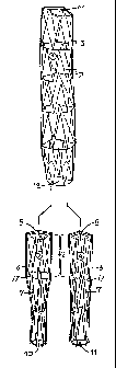
FIG. 2 is a schematic representation of the three parts of the central graft
(main cylinder) and the two limbs (pe(pheral cylinders) from which are
composed the
stent graft according to the presented technique. The two limbs (peripheral
cylinders) enter with their central region (of greater diameter) at the
peripheral or
distal region of the central cylinder and by their expansion create a
leakproof
branching of the central graft.
FIGS. 3A to 3C are a representation of the analytical magnification of the
central graft
(main cylinder) which is placed in the abdominal aorta in the region between
the out-
branching of the kidney or renal arteries and the aortic bifurcation after its
positioning
and its expansion. At the peripheral end can be discerned the refolding and
attachment of the thin-walled external covering inside the main cylinder, to
reinforce
it and to make the branching of the main cylinder more leakage proof after the
placement of the two peripheral cylinders.
FIG. 4 is a magnified representation of the cylinders in their compressed
form inside the storage tubules which are small in diameter equal to the
diameter of
the placement tubules inside the arteries as well as the propulsion device for
the
introduction and progression of the graft by means of a guide wire.
FIGS. 5A to 5E are a three-dimensional schematic representation of the
overlapping
regions of the cylinder and of the two peripheral cylinders (limbs) after the
expansion
of the limbs inside the main cylinder and their leakage-proof application, not
only
amongst the limbs, but also with the central cylinder due to their self-
expanding
character. At cross-section, one can clearly see the variety of shapes that
the central
orifices of the limbs can take when restricted at their external surface by
the internal
surface of the central cylinders at their overlapping arts.
FIGS. 6A to 6D are a schematic representation of an alternative manner of
construction of the limbs of the stent graft where the diameter of the central
orifice
at one of the limbs at the length of > 2 cm (covered by the central
cylindrical part)
is equal to the diameter of the peripheral orifice of the main cylinder while
the
diameter of the other limb is smaller at the center of the orifice and part as
well as
at the cross-section of the central orifices after the expansion of the two
limbs
inside the central tube. If they were allowed to expand naturally, the cross-
sectional
CA 02286897 1999-10-08
WO 98/44870 PCT/IB98/00530
-4-
areas of the combined limbs would be twice that of the distal, orifice or the
central
tube.
FIG. 7a is a schematic representation of the method of placement of the
central graft (main cylinder). This is found compressed inside the placement
tubule
where it is advanced since the guide wire passes through the graft and is
brought to
the point of placement with the help of the propulsion device and by the guide
wire.
The central orifice of the placement tubule has already been advanced more
central
to the aneurysm and when the graft reaches the position of placement, it is
allowed
to expand by means of maintaining the propulision device stable and by means
of the
peripheral attraction of the placement tubule toward the propulsion device in
such a
manner so that during its expansion the graft will maintain its position
unchanged.
FIG. 7b is a schematic representation after the placement of the central
graft a second guide wire is advanced from the other iliac artery and by the
peripheral
orifice of the main cylinder. On the guide wires of both sides the placement
tubules
are advanced by an X-ray shadowing ring at their central orifice, through the
iliac
arteries and through the peripheral orifice of the main cylinder inside the
peripheral
part of the main cylinder.
FIG. 7c is a schematic representation of the advancement of the two limbs
inside the guide introduction tubules using the same technique as that for the
placement of the central graft, and of their equal in height positioning by
means of
retraction of the introduction tubules when these are forwarded at an upright
position
which is at the same transverse level as the two limbs. The complete release
of the
limbs (peripheral cylinders) along with the simultaneous or the
nonsimultaneous
withdrawal of the introduction tubules of both sides results in the fact that
the
expanded parts of the limbs will come in complete contact with the peripheral
nonexpanded healthy region of the corresponding iliac blood vessei at which
the flow
of blood is directed.
FIG. 8 illustrates an alternative arrangement in which the main graft is
substantially contained within the region of the aorta containing the renal
arteries.
The distal end of the main graft is constructed in a manner similar to that in
the other
CA 02286897 1999-10-08
WO 98/44870 PCT/IB98/00530
-5-
embodiments. The two limbs extend from the iliac arteries into the distal end
of the
main graft and are sealed in a similar manner.
Detailed Descri tion
In general, this device presents a stent graft for the therapy of abdominal
aortic aneurysms without the need for the presence of healthy peripheral
aortic walls
(26), whereof the placement of one and only one graft tube would be possible.
The
need to avoid the pathological wall by airtightness in the nonexpanded part of
the
region of the iliacs is accommodated by the: branching of the central graft
(main
cylinder) at two peripheral tubes so that the blood can be driven toward both
iliac
arteries (24, 25) from the central aortic graft. Furthermore, the described
graft must
have the ability to be compressed to a small starting-off diameter (FIG. 4),
such that
the advancement and placement in its compressed form is made possible by means
of the small diameter tubes (37, 38) inside thie vascular lumen from distant
regions
(from the femoral artery in the abdominal aorta) where released it can regain
its
original large diameter. This is accomplished in such a way so that it is able
to
achieve its leakproof perimetric contact with the internal surface of the
healthy
vascular lumen central and peripheral to an aineurysm. Till today, there have
been
proposed and used various devices for the accomplishment of the above goals.
However these are often complicated and difficult in their usage with the
subsequent
appearance of complications during and after the operation. To use these
devices
it is necessary for those performing the opera1tion to acquire lengthy
experience and
to have ample abilities. Aim of the device Inrhich will be described as well
as its
technical positioning is the simplification of the placement procedure, the
minimization of the immediate and future complications and the enhancement of
the
percentage of successful clinical results witti the goal of the possibility of
a wide
usage of the method of endovascular therapy of aneurysms for those patients
where
the placement of a stent-graft is necessary.
The herein presented stent-graft is a graft which is comprised of a central
"main" cylinder or tube (1) that at the center of the orifice comes into
contact with
the external surface of the perimeter of the aorta at the level more central
to the
distended part of the aorta (13) while the periphery of the orifice sits upon
the aortic
CA 02286897 2007-03-28
WO 98/44870 PCT/IB98/00530
-6-
bifurcation (26). The diameter or cross-sectional area of the central or
proximal
orifice (14) of the main cylinder (1) is equal to or larger than the diameter
of the
healthy part of the aorta (13) on which the cylinder will be placed. The
diameter or
cross-sectional area of the peripheral or distal orifice (12) is constant and
independent of whatever aortic diameter at the level of its bifurcation (28)
where the
cylinder will be placed. The length of the main cylinder (1) is determined by
whichever length amongst the central point of contact in the aorta (13) and
its
bifurcation (26).
The main cylinder (1) comprises a stent on skeleton (cast) which is
preferably cylindrical and/or metallic with a length that is substantially
equal to the
distance between the kidney or renal arteries and the aortic bifurcation (see
FIG. 2). This
skeleton has as starting-off predetermined diameter a and a compressed
diameter
and consists of successive and connected amongst themselves cylindrical pieces
of
variable length and with a Z configuration made out of biocompatible metal
with
memory such as stainless steel wire or nitinol (a nickel titanium alloy with
thermal
memory). The connection of these parts of the skeleton can be accomplished
either
by sutures (16) which pass through the orifices of the last of the endcrests
of each
piece or by metallic joints (solderings) in such a manner so as to allow a
certain
amount of flexibility amongst the various Z parts of the skeleton whilst the
length of
the skeleton has as minimal as possible changes between its compressed and its
starting-off diameter. The present plan of the skeleton is described
extensively and
consists of the skeleton (2) of the main cylinder (1) which is described in
the figures
as well as other plans of however, the self-expanding skeletons with similar
properties and characteristics which will possibly save the basic idea of the
creation
of the bifurcation of the herein presented stent-graft. The stent can at its
proximal
end, have a plurality of barbs or hooks which can, upon expansion, rotate and
penetrate part 13 for sealing and securement purposes.
The metallic skeleton or stent of the main cylinder on its outer surface is
covered by a tube (3) to form a graft which has a central (14) orifice, and a
peripheral or distal (12) orifice. The wall is preferably thin-walled and of
PTFE,
Dacron, polyurethane or another type of biocompatible plastic. Alternatively,
the
CA 02286897 1999-10-08
PA-5166 PCT
-7-
inner and outer surfaces of the metallic skeleton (2) can be covered by
cylindrical
tubes (3), or it can be covered on its inner surface only by the cylindrical
tube. This
tube has a starting-off or proximal diameter which is equal to that of the
metallic
skeleton and it has a central and peripheral distal orifice and a main body.
The
central orifice (14) has a diameter which is preferably substantially equal to
or greater
than that of the healthy part of the aorta at the point of its contact (13)
with the
main cylinder more central to the aneurysm. The tube(s) of the graft is
refolded or
is not refolded at the central orifice (14) of the metallic skeleton (2) and
is attached
upon the central orifice of the skeleton by a series of connective sutures
(44) at the
end-crests of the central end of the (metallic) skeleton or stent. An outer
covering
can cover an outer enlargement at the outer proximal end of cylinder (1) to
firmly
engage part (13) and then a flap can extend inwardly into orifice (14) to
improve the
seal. The peripheral orifice of the graft has a diameter of 20 - 25 mm and is
refolded
(18) at a length of 0.5 -1 .0 cm at the internal side of the peripheral or
distal orifice
(12) of the metallic skeleton where it is internally attached by single
sutures at two
or at three different points of the metallic skeleton (2). Thus the flow of
blood after
the placement of the main cylinder is accomplished inside the peripheral
refolding of
the graft of the main cylinder at the peripheral end and creates two or three
pockets
(petals) (41) minimizing the surface of the peripheral orifice (12) of the
main cylinder.
Additional attachment points can be used, if clesired, to establish additional
petals.
The main cylinder (1) has a starting-off diameter, which is of the thin-walled
covering
cylinder (3) around the center (14) and periphery (12) of the orifice end
around its
body. The metallic skeleton (2) expands the cylinder to this diameter and to a
compressed diameter much smaller than that of the original diameter in such a
manner so that it can be compressed inside the storage tubule (23) which has a
small
diameter (FIG. 4) equal to that of the placemE:nt tubule (37) which is used
for the
advancement of the graft inside the blood vessels (FIG. 7a). During the
compression
of the main cylinder (1) inside the storage tubule (23) it has at its center a
catheter
(39) which is used for the insertion of the'guide wire (21) (which after this
it is
removed) through the compressed inside the storage tubule main cylinder during
the
positioning process. The progression of the main cylinder to the placement
position
AMENDED SHEET
PA-5166 PCT CA 02286897 1999-10-08
-8-
is accomplished by its propulsion by the storagie tubule (23) to the placement
tubule
(37), wh"ich as previously mentioned, has the same diameter. This is achieved
with
the help of a propulsion device (22) which moves upon the guide wire (21)
which
goes through the center of the main cylincier and in continuation through the
aneurysm.
The herein presented stent-graft is also comprised of two peripheral
cylinders (limbs) (4, 5) which preferably are self-expanding stents or
skeletons (7)
identical to that of the main cylinder (1) but of different dimensions (a
series of Z
casts). These are covered at their external surface by a thin-walled
cylindrical tube
(6), which have an identical or a different composition from that of the main
cylinder
(1), but are of different dimensions. The peripheral cylinders have central
(8, 9) and
(peripheral) distal (10, 11) orifices and a starting-off diameter and a
compressed
diameter such that it becomes possible for thern to be placed inside a storage
tubule
(23) exactly in the same way as with the main cylinder but of a smaller
diameter.
They can be advanced to the corresponding placement tubules (37, 38) inside of
the
blood vessels. The thin-walled cylindrical tube (6) covers the entire length
of the
metallic skeleton (7) and is attached to the central (8, 9) and the peripheral
(10, 11)
end of the metallic skeleton of each peripheral cylinder. The skeleton (7) of
the
peripheral cylinders (4, 5) has a diameter in i-ts expanded form equal to or
greater
than that of the thin-walled cylindrical tube (6) at each of its parts in such
a manner
so that it comes in complete contact at its external surface with the internal
surface
of the thin-walled cylindrical tube (6) which has a constant diameter at its
expanded
form and at each of its parts. The graft can be refolded to a small length <
5mm and
can also not be refolded inside the central orifice (8, 9) of the skeleton of
each limb
and is not refolded at the peripheral orifice ('10, 11) of the skeleton of
each limb
where it is attached by sutures.
The diameter or diameters of the central orifice (8, 9) of each (peripheral
cylinders) limb is preferably equal to or approximately up to about 5 mm or
more
smaller in relation to the diameter of the peripheral orifice (12) of the main
cylinder.
Alternatively, each orifice (8,9) can be equal to or greater than orifice
(12), or each
can be significantly smaller than orifice by much more than 5 mm such as 10 or
20
AMENDE[) SHEET
CA 02286897 2007-03-28
wO 98/44870 PC171898/00530
-9-
mm. Experimentation of a simple nature can determine sizes of the distal
cylinders
relative to orifice 12 in order to achieve a sealing affect between cylinders
1, 4 and
5. The diameter of the central orifice of each limb continues at a length of 2
- 2.5
cm at ttie central part (42) of the graft of each limb which is the length of
the first
of the Z casts of the preferably self-expanding stents or skeletons (7) of
each limb
and the point (41) where the first cast of the preferably metallic skeleton is
connected to (jointed to) the second cast as has previously been mentioned.
This
central part (42) of each limb is the part which enters into the distal or
peripheral end
(43) of the central cylinder for the creation of the bifurcation of the
central cylinder
as will be mentioned in continuation. The diameter of the peripheral
cylinders, more
peripfieral to t.tie previously mentioned part, has a length and a diameter
which vary
according to the length and the diameter that is necessary so that the
peripheral
orifice (10, 11) of each of those two cylinders (4, 5) can come in complete
contact
with ttie healthy part of the corresponding iliac blood vessel (24, 25). That
is to say,
ttiat the peripheral diameter and the length of each limb can differ from each
other
in relation to the dimensions of the iliac blood vessels of their healthy part
and of the
length of the damage of each, from the bifurcation of the aorta.
Alternatively, the diameter of the central orifice of the central part of each
limb can differ in size (FIGS. 6A to 6D). Specifically, the diameter of the
central orifice (30)
and of the central part (46) of one of the limbs (28) which enters from the
peripheral
orifice (12) of the main cylinder (1) is equal to the diameter of the
peripheral part and
of the orifice (12) of the main cylinder, while the diameter of the central
part (46)
and the orifice (29) of the other limb (27) can be smaller with the aim of
creating a
smaller compressed limb diameter and its progression inside of a smaller in
diameter
placement tubule, percutaneously. In this case, the expanding ability of the
metallic
skeleton (31) of the smaller in diameter limb is equal to or greater than that
of the
metallic skeleton (32) of the limb with the greater central diameter. The
length as
well as the periphery of the peripheral orifice (33, 34) of each peripheral
cylinder
(limb) (27, 28) can vary as previously mentioned according to the dimensions
of the
iliac blood vessels and their condition.
CA 02286897 1999-10-08
PA-5166 PCT
-10-
As material for the thin-walled covering cylinder (6, 35, 36) of the metallic
skeleton' (7, 31, 32) of the peripheral cylinders (4, 5, 27, 28) one can use a
cylinder
made out of thin-walled polytetrafluoroethylene, Dacron, or another type of
biocompatible plastic. This thin-walled cylinder preferably has the previously
mentioned constant dimensions of the peripheral cylinders (4, 5, 27, 28) at
its
noncompressed form as well as after its expansion by the self-expanding
metallic
skeleton (2) internally. The material of the thir-walled covering cylinder (6,
35, 36)
of the metallic skeleton of the peripheral cylinciers can cover the
cylindrical metallic
skeleton at its external surface or at its external and internal surface or at
its internal
surface.
Technical Placement - Creation of a Bifurcatioin
The herein presented stent-graft is created by the placement of three
cylinders (1,4,5) (main and two peripheral) of which it is composed and which
is
executed in the following manner:
After the percutaneous placement of the guide wire (21) from the femoral
artery and in a head on direction towards the aorta, an angiogram is performed
so as
to determine the height of the kidney arteries (20). At this point, as well as
at the
point of the aortic bifurcation, the X-ray shadowed guided position is
monitored by
X-rays. On the guide wire is advanced with the help of a diastolic device,
percutaneously, the introduction tubule (37) (sheath) inside of which the main
cylinder will be advanced during its placement. The introduction tubule has at
its
central end an X-ray shadowed ring (40) and at its peripheral end it has a
hemostatic
valve. During its endovascular advancement, the introduction tubule (37) has
inside
of it a diastolic device which makes easier its percutaneous entrance into the
artery.
The introduction tubule is advanced inside the aorta so far in as necessary so
that the
X-ray shadowed ring at its central end will be found at a more central level
than that
of the out-branching of the kidney arteries (20). In continuation, the
diastolic device
is removed from inside of the introduction tubule (37). And on the guide wire
(21)
through the hemostatic valve, the storage tubule (23) is advanced which has a
diameter equal to that of the introduction tubule which carries inside its
lumen the
compressed main cylinder (1) (of the stent graft) that has a length equal to
that of
-;.-T
PA-5166 PCT CA 02286897 2007-12-10
-11-
the distance amongst the point of the out-branching of the kidney arteries
(20) and
the aortic bifurcation (26) and a diameter of the central orifice (14) (after
its
expansion) which is equal to or greater than that of the aorta at the height
of the
central neck (13) directly underneath the kidney tubules.
The main cylinder (1) is advanced from the storage tubule (23) to the
introduction tubule (37) and through this to the placement point with the help
of a
propulsion column (propulsion device) (22), which passes through the center of
the
compressed main cylinder. When the main cylinder (1) which casts an X-ray
shadow
in its entire length (metallic skeleton) is advanced inside the introduction
tubule (37)
between the guide points that have been placed at the out-branching of the
kidney
arteries (20) and the aortic bifurcation (26), the propulsion column (22) is
maintained
in a stable condition by its central end which restrains the peripheral end
(12) of the
main cylinder at the height of the aortic bifurcation. The introduction tubule
(37) is
retracted over the column (22) in a centrifugal direction in such a manner so
that the
main cylinder (1) is progressively released frorn the introduction tubule at
its entire
length and will expand (FIG. 7a). When it comes into contact with the internal
surface (13) of the aorta directly beneath the kidney arteries (20) it will
become more
rounder whilst the peripheral orifice sits upon the aortic bifurcation (26).
In this way,
the dislocation of the main cylinder becomes impossible due to the constant
length
of the metallic skeleton (2) of the main cylinder which is supported by the
aortic
bifurcation.
In continuation and after the de novo progression of the column inside the
introduction tubule (37), it is advanced on the guide wire (21) through the
peripheral
orifice (12) inside of the main cylinder (1) so that the X-ray shadowing ring
(40) will
be found 2 - 2.5 cm more central to the peripheral orifice (12) of the main
cylinder.
In the same way, one of the two peripheral cylinders (5) is advanced inside
of the introduction tubule through the hemostatic valve. In this manner, its
central
end (orifice) (9) will reach the height of the X-ray shadowing ring (40) of
the
introduction tubule.
In continuation, percutaneous advancement of the guide wire (56) is
achieved from the femoral artery of the other side after its percutaneous
injection.
AMENDED SHEET
rA-S I bb r(%, ( CA 02286897 2007-12-10
-12-
The guide wire is centripetally directed with the help of a guide catheter
through the
iliac artery (25) and through the peripheral orifice of the main cylinder
which sits
upon the main cylinder which sits upon the aortic bifurcation inside the lumen
of the
main cylinder. Advanced on the guide wire follows the second introduction
tubule
(38) with a diastolic device inside of it and an X-ray shadowing ring (40) at
its central
orifice. The second introduction tubule (38) is centripetally advanced through
the
peripheral orifice (12) of the main cylinder and up to the point where the X-
ray
shadowing ring at its central orifice is found at the same height (2 - 2.5 cm
from the
peripheral orifice inside the main cylinder) with the X-ray shadowing ring of
the main
orifice of the introduction tubule (37) of the femur of the other side (FIG.
7b) inside
of which is already found the peripheral cylincler of the limb (5) of the
other side at
its compressed form. Inside the second introcluction tubule end with the
technique
which was previously mentioned, the second peripheral cylinder (limb) (4) is
advanced till the point where its central compressed end is at an equal height
as that
of the X-ray shadowing ring (40) of the introduction tubule (38) inside of
which it is
advanced as well as with the central end of the compressed peripheral cylinder
(5)
of the other side.
After they are X-ray monitored, the two compressed peripheral cylinders
(limbs) inside of the introduction tubules have a position of equal height,
both with
their central end, 2-2.5 cm more central and inside of the peripheral orifice
of the
main cylinder; that is to say, at the point of the union (41) (joint) of the
first with the
second Z element of their metallic skeleton which has a corresponding length,
the
introduction tubules are withdrawn simultaneously or nonsimultaneous in a
centrifugal or distal direction and the peripheral cylinders are expanded
according to
the technique which was mentioned previously for the main cylinder.
After the expansion, the two peripheral cylinders, these having a self-
expanding skeleton, preferably each with an equal strength of expansion at the
end
covered by their main cylinder part extended, are compressed due to the
greater total
diameter of both in relation to the diameter of the peripheral part of the
central
cylinder by the main cylinder. In this way, they come into leak-proof contact
with
the internal surface of the wall of the central cylinder, but also between
them but
AMENDFO SHEET
PA=5166 PCT CA 02286897 2007-12-10
-13-
however, maintaining the diameter of both central orifices (8, 9) equal to the
diameter of the peripheral part (43) of the main cylinder (1). The shape of
the central
orifice of the two peripheral cylinders can vary, without these, however,
coinciding
completely due to the equivalent expansive ability of their metallic skeletons
(FIGS. 5A to 5E).
Furthermore, the reversal of the external cover (19) of the main cylinder
at the peripheral orifice (12) creates an additional valve mechanism at this
level
which hinders the escape of blood from the niicrochasms which may occur during
the contact amongst the two peripheral cylinders, but also with the internal
surface
of the peripheral part (43) of the main cylinder.
In this manner, a blood leak-proof (without the escape of blood) bifurcation
of the main cylinder (1) is created at two peripheral cylinders (5, 4) with an
entrance
orifice (9, 8) of variable shape and area.
The peripheral part of each peripheral cylinder (limb) has a length and a
diameter of the peripheral orifice which is analogous to those of the
corresponding
iliac artery (24, 25), so that after its expansion, it will come in complete
contact with
the internal surface of the healthy part of the wall of the iliac artery.
After the placement of the "stent graft", according to the manner which
was previously mentioned, the direction of the flow of blood is achieved
inside of the
"stent" graft from the height of the kidney arteries through the orifice of
the two
limbs and more peripheral to these inside of the iliac arteries with the
simultaneous
exclusion of the systemic arterial pressure and the blood circulation of the
pathologically distended wall of the aneurysm of the aorta which also includes
the
aortic bifurcation (FIGS. 1 and 3A to 3C).
In the FIG. 8 embodiment, the graft arrangement comprises a main graft
50 which is formed in a manner similar to graft 1 of the other embodiments. It
comprises an inner stent and an outer covering 3, the latter extending upward
to
form a flap 3' which when the assembly is operational, folds into the graft
limbs to
assist the sealing process.
The main graft 50 is introduced into the part 13 of the aorta with the
proximal end 51 of the graft adjacent to the iliac arteries and with the
distal end 40
of the graft adjacent to the distal end of the good tissue 13. The graft is
expandable
AWE"IbEu SHEET
CA 02286897 2009-03-06
14
to form a force fit within part 13. If desired, the graft 50 can be provided
with
hooks, which upon expansion of graft 50, rotate and embed themselves into
part 13 in a known manner. The graft 50 has an internal cross-sectional area
at least at end 54, much less than the sum of the external cross-sectional
areas at least at end 54, much less than the sum of the external
cross-sectional areas of the limb members 6. The latter are introduced via
their respective iliac arteries in a tube such as 37 and allowed to expand and
for the seals with the main cylinder 50 and using the flap 3' of the cover
extending beyond the provisional end of the cylinder 50. The latter is quite
stable since it is supported by the firm or undiseased part 13 of the aorta
and
its position is quite fixed. The round flap can have 2 slits at appropriate
positions to faciiitate entry into the two respective tubes.
A type of stent endovascular aortic graft consists of a central branched
cylinder adjoined by two peripheral cylinders destined for the endovascular
therapy of aneurysms of the abdominal aorta by expansion at the level of the
aorta's bifurcation. The placement of the device is characterized by its
progressive positioning into the lumen of the aorta. The device is brought to
the desired position endoarterially from a distant blood vessel with the help
of
a guide-wire and a catheter both of which are positioned inside small in
diameter tubules.
First off, a main self-sustaining cylinder with a predetermined length
that is equal to the length amongst the kidney artery and the aortic
bifurcation.
It has a self-expanding central and a peripheral orifice with a diameter of
compression and one of expansion consisting of a self-expanding metallic
skeleton with memory externally covered with a thin-walled cylinder made out
of PTFE, Dacron or another type of biocompatible plastic, that has a constant
predetermined maximum diameter which defines the expanded diameter of
the main cylinder at the center of the orifice at its body end at the
periphery of
the orifice.
CA 02286897 2009-03-06
The device also has two self-expanding peripheral cylinders of variable
length each having a central and a peripheral orifice with a compressed and
an expanded diameter which are equidiameterical at their expanded diameter
at their central part and with a diameter of the central part equal to or
approximately 5 mm smaller than the diameter of the peripheral orifice and
not necessarily equidiametrical to their peripheral parts. They are composed
of self-expanding metallic skeletons with memory. Both have an equal
strength of expansion, a diameter equal to or greater than that of each
peripheral cylinder at its respective part with the same shape and material
and
of a different shape, material and dimensions from that of the main cylinder.
They are covered by a thin-walled cylinder made out of PTFE, Dacron, or
another type of biocompatible plastic with a constant predetermined maximum
diameter which defines the expanded diameter of the peripheral cylinders.
By the creation of a bifurcation of the main cylinder from the two
peripheral cylinders whose placement is by way of guide wires and tubules
which are introduced through the peripheral arteries of the bifurcation where
the graft will be positioned and through the peripheral orifice of the main
cylinder which sits upon, and is supported by the aortic bifurcation at the
same or at different times of transport and by the equal in height positioning
of
the compressed central parts of the peripheral cylinders internal to the
peripheral orifice and the peripheral expanded part of the main cylinder. In
continuation, there is the gradual or simultaneous restoration of the central
and peripheral parts of the two peripheral cylinders back to their expanded
diameter, where at the central orifice and central part of the peripheral
cylinders, is greater than the total sum of the peripheral parts and that of
the
peripheral orifice of the main graft. The graft brings in hemostatic contact,
the
outer surface of the central part and the orifices of the two peripheral
cylinders
and with the inner surface of the peripheral part and also the peripheral
orifice
of the main cylinder. It maintains open their central orifices, due to the
equal
strength of expansion of their metallic skeleton and it guides the blood flow,
branching it from the lumen of the main cylinder and into the lumen of the two
peripheral cylinders without leakage.
CA 02286897 2009-03-06
16
The thin-walled cylinder which externally covers the metallic skeleton of
the main cylinder is refolded at its inner peripheral orifice by a length less
than
half of the diameter of the peripheral orifice of the main cylinder. It is
attached
to the internal surface of the metallic skeleton of the main cylinder at two
or
three places in such a manner such that the incoming blood flow at the
refolding causes the falling forward of the parts of the refolded region of
the
thin-walled cylinder between the points of attachment and the inner peripheral
orifice of the main cylinder and will minimize the area of the peripheral
orifice
at this level, stopping leakages due to imperfect contact of the internal
surface
of the main cylinder with the external surface of the two peripheral
cylinders.
The main and the peripheral cylinders have a metallic skeleton which
may be composed of successive ring-like parts of different lengths and
diameters, consisting of a stainless steel wire with memory and each circular
part having a shape of a different number of successive Z's. The rings are
attached by a fiber to each other by means of openings at the ends of each
part or by some other type of joint or attachment amongst the ring-like parts.
The metallic skeleton of the main and of the peripheral cylinders may
consist of stainless steel self-expanding with memory or of a nickel titanium
alloy self-expanding with memory.
The metallic skeleton of the main and peripheral cylinders may differ
from each other in their shape, length, diameter and material with which they
have been constructed.
The peripheral cylinders may have orifices of different dimensions and
with the diameter of the central region inside the main cylinder being equal
to
that of the peripheral region to the peripheral orifice of the main cylinder.
The
diameter of the central orifice and the central region of the other peripheral
region will be smaller, but will have a metallic skeleton with a greater
expansion strength than that of the other cylinder, in such a manner so as to
be able to maintain the orifice of the cylinder.
CA 02286897 2009-03-06
17
In the above described embodiments, self expanding stents have been
employed. If that is not desirable for any reason, then non self expanding
stents can be employed, but that would necessitate the use of expansion
means such as balloons could be used, such as those employed in
angioplasty procedures.