Note: Descriptions are shown in the official language in which they were submitted.
CA 02287220 1999-10-21
WO 99/43253 PCT/US98/13736
IMAGE-GUIDED THORACIC THERAPY AND APPARATUS THEREFOR
TECHNICAL FIELD
The present invention relates to methods and apparatus for
performing medical procedures in the thorax of a medical or veterinary
patient.
BACKGROUND ART
Some common medical procedures require the ability to operate
on a specific location in the thorax, including locations in the respiratory
system, such as the lungs, bronchi and immediately suirounding tissues. For
example, needle aspiration biopsies have been performed heretofore using an
endoscope inserted tllrough the trachea into a bronchus. The needle is
advanced througli the eiidoscope through the bronchial wall to sample tissue
in a lymph node witllin the lung parenchyma near the exterior surface of the
bronchus. The physician can monitor placement of the endoscope and the
biopsy needle using the optical system of the endoscope. As the endoscope is
advanced toward the area to be sampled, the physician can determine where
the tip of the endoscope lies by observing features of the airway itself.
However, it is difficult to place a biopsy needle within a particular lymph
node using this approach. The physician cannot see the lymph nodes, which
lie outside of the airway. Therefore, the physician can only position the
endoscope tip and the biopsy needle at an approximate position, near the
location of the lymph node to be biopsied. For this reason, there has been a
significant need for improvement in the reliability of needle aspiration
biopsies of the lymph nodes surrounding the respiratory tract. There have
been similar needs for improvement in other biopsies procedures using a
probe advanced into the body, such as a biopsy needle or biopsy forceps to
sample tissues in the vicinity of the respiratory tract. There have been
similar
needs for improvement in other procedures where a probe is advanced into the
CA 02287220 2004-09-17
tissues of the thorax for other purposes as, for example, to perform surgical
procedures on these tissues or to administer drugs within these tissues.
Some procedures heretofore have used imaging during
advancement of the probe to provide guidance. Thus, as the probe is
advanced, the probe and the body are imaged using conventional imaging
techniques such as fluoroscopy or magnetic resonance imaging. This allows
the physician to observe the relationship between the position of the probe
and the surrounding tissues. These procedures have the disadvantage that
the imaging apparatus is occupied for the entire time required to perform the
procedure. Moreover, the use of fluoroscopic or other x-ray based imaging
modalities during the procedure exposes the physician and the patient to
radiation.
As described, for example, in U.S. Patents 5,558,091,
5,391,199; 5,443,489; and in PCT International Publication WO 96/05768,
the position, orientation or both of the distal end of a probe can be
determined by using one or more field transducers such as a Hall effect or
magnetoresistive device, coil or other antenna carried on the probe, typically
at or adjacent the distal end of the probe. One or more additional field
transducers are disposed outside the body in an external frame of reference.
The field transducers preferabiy are arranged to detect or transmit non-
ionizing fields or field components such as a magnetic field, electromagnetic
radiation or acoustical energy such as ultrasonic vibration. By transmitting
the field between the external field transducers and the field transducers on
the probe, characteristics of field transmission between these devices can be
determined. The position and/or orientation of the sensor in the external
frame of reference can then be deduced from these transmission
characteristics. Because the field transducer of the probe allows
determination of the position of the probe, such transducer is also referred
to
as a "position sensor".
-2-
CA 02287220 2004-09-17
As described, for example, in the aforementioned U.S. Patent
5,558,091, the frame of reference of the external field transducers can be
registered with the frame of reference of imaging data such as magnetic
resonance imaging data, computerized axial tomographic data, or
conventional x-ray image data. The probe position and orientation data
derived by field transmission can be displayed as a representation of the
probe superimposed on an image of the patient's body. The physician can
use this information to guide the probe to the desired location within the
patient's body, and to monitor its orientation during treatment or
measurement of the body structure. This arrangement greatly enhances
the ability of the physician to navigate the distal end of the probe through
bodily structures. Because it does not require acquisition of an optical
image of the surrounding tissues for navigation purposes, it can be used
with probes which are too small to accommodate optical elements, and
can be used for navigation of the probe within solid or semisolid tissues.
The transducer-based system also avoids the difficulties associated with
navigation of a probe by continuous imaging of the probe and patient
during the procedure. For example, it avoids exposure to ionizing radiation
inherent in fluoroscopic systems.
Some additional problems are encountered in use of systems
of this type for procedures in the thorax near the respiratory system. As
the patient breathes, the positions, sizes and shapes of the thoracic
organs change. Thus, if an image of the patient is acquired at one stage
of the respiratory cycle, the image data does not accurately represent the
patient during other stages. Therefore, if the position of the probe is
detected while the patient is in one stage of the respiratory cycle, and this
probe position data is combined with patient image data from another
stage of the respiratory cycle to provide an image with a representation of
the probe superposed thereon, the location of the probe relative to the
surrounding organs will be depicted inaccurately. As described in
International Publication WO 97/29709, problems of this nature can be
-3-
CA 02287220 1999-10-21
WO 99/43253 PCT/US98/13736
-4-
avoided by positioning a first probe, referred to as a "site probe" within the
body of the patient at a location to be treated, and providing a further
probe,
refelred to as an "instrument probe" for performing the medical procedure.
The site probe is positioned within the body at the location to be treated as,
for example, at a location to be biopsied. Using a location system such as the
magnetic location systems discussed in the aforementioned patents, the
locations of both probes are monitored during the medical procedure.
Therefore, the distance alld direction from the instrument probe to the site
probe are known during the medical procedure, despite any motion caused by
the patient's breathing. Using that directional and distance information, the
physician can navigate the instrument probe to the site probe.
PCT Publication WO 97/29682 refers to systems for
determining the "physiological motioii" such as breatliing motion or cardiac
motion of a portion of the body in which a probe is situated. Using a device
such as a belly strap to sense breathing motion, the system selects a
"correct"
image from a set of previously obtained images at each instant during the
procedure, or interpolates between iinages. Thus, the displayed image always
reflects the actual size and shape of the organs at the instant in question.
Accordingly, the representation of the probe can be accurately superposed on
the display image.
U.S. Patent 5,577,502 discloses a system in which the position
of the subject's chest is monitored by devices such as optical, ultrasound or
mechanical tracking elements. Based on that positional tracking, the image
used in a superposition system is distorted so as to provide a corrected image
which changes as the subject breathes. The position of the probe can be
superposed on the corrected image. Systems of this type require considerable
computation to distort the reference image as the patient moves through
various stages of the respiratory cycle. Moreover, additional equipment is
required for tracking the position of the patient's chest. ln an alternative
approach also discussed in the '502 patent, a series of images is acquired at
CA 02287220 1999-10-21
WO 99/43253 PCT/US98/13736
-5-
numerous stages of the respiratory cycle. As the patient moves through
different stages of the respiratory cycle, different images are employed. This
approach multiplies the task of acquiring and storing the image data.
Moreover, this approach can only be used if a set of multiple images exists.
For example, where the patient is subjected to a conventional diagnostic
imaging procedure such as an MRI or CT imaging, a single set of image data
representing the patient at only one stage of the respiratory cycle generally
is
acquired. The need for a biopsy or other procedure using a probe advanced
into the patient may only be apparent after that image has been evaluated. To
acquire a series of images, the patient must be subjected to further imaging
procedures before the interventional procedure using the probe can begin.
Thus, despite these and other efforts in the art, furtlier
improvements in interventional procedures and apparatus for perfoiming the
saine would be desirable.
DISCLOSURE OF THE INVENTION
One aspect of the present invention provides methods of
performing medical procedures on thoracic tissues, and particularly on tissues
of the respiratory system or tissues adjacent the respiratory system. A
metliod
according to this aspect of the present invention includes the steps of
providing an image of the patient in an image frame of reference representing
the patient at a selected respiratory state, such as in a selected stage of
the
normal respiratory cycle, and advancing a probe into the respiratory system of
the patient by adjacent tissues. During the advancing step, the disposition of
the probe is determined in a locating frame of reference when the patient is
at
the aforesaid selected respiratory state. The disposition of the probe
desirably
is detected by transmitting one or more non-ionizing fields to or from at
least
one probe field transducer on the probe and detecting one or more properties
of the transmitted fields. The method further includes the step of
transfonming the image, the disposition of the probe in the locating frame of
reference or both so as to place the image and the disposition of the probe
into
CA 02287220 1999-10-21
WO 99/43253 PCT/US98/13736
-6-
a comrnon frame of reference. Typically, the transforming step is performed
by transforming the disposition of the probe in the locating frame of
reference
into the image frame of reference. The method further includes the step of
displaying the image of the patient with a representation of the probe
superposed thereon, at a location corresponding to the disposition of the
probe
in the aforesaid common frame of reference.
Because the disposition of the probe used as the basis for the
superposed representation is acquired at the same stage in the respiratory
state
as the image, the motion artifact or inaccuracy caused by motion due to the
respiratory cycle is eliminated. Methods according to this aspect of the
invention thus provide a solution to the motioti artifact problem which does
not require acquisition of multiple images for massive manipulation of the
image data to distort an image. The system is compatible witli standard
ilnages acquired for diagnostic purposes, which represent only one stage in
the respiratory cycle.
The method preferably further includes the step of detecting
when the patient is in the selected stage of the respiratory cycle by
monitoring
the position of a reference point on a patient which moves in respiration.
Preferably, the system detects when the patient is in the selected respiratory
state by determining whether the position of the reference point matclies a
selected position corresponding to the selected respiratory state within a
preselected tolerance. For example, where the image was acquired at a
particular stage of respiration, the system acquires the disposition of the
probe
during each breath, at the same stage of respiration.The method may further
include the step of establishing a particular position of the reference point
which corresponds to a selected stage of the respiratory cycle by monitoring
the position of the reference point over a plurality of respiratory cycles and
finding an extreme position of the reference point which recurs in each cycle
using the data acquired by this monitoring procedure. For example, the
selected stage of the respiratory cycle may be the minimum inspiration state,
CA 02287220 1999-10-21
WO 99/43253 PCT/US98/13736
-7-
i.e., the state achieved at the end of exhalation during a normal breathing
cycle. ln this case, the system may select the position of the reference point
at
which the patient's front chest wall is closest to the patient's back. If the
patient is lying in a supine position, witli his or her back on a table, the
system
may select the position wliere a reference point on the patient's front chest
wall is closest to the table.
The step of monitoring the position of the reference point
desirably includes the step of transmitting one or more non-ionizing fields to
or from at least one reference field transducer on the reference point. Thus,
the position of the reference point can be monitored using much of the same
equipment and techniques as are used for monitoring the position of the
probe.
Desirably, the system displays a perspective image of the tissues
surrounding the probe as, for example, a perspective image of an airway and
surrounding tissues with the probe superposed thereon. The image is
displayed so that the position of the probe and the trajectory for moving the
probe to engage a target location such as a lymph node is readily visible by
viewing the displayed image. Typically, the step of advancing the probe is
perfonmed by advancing the probe through an airway as, for example, by
passing the probe tluough the wall of the airway to sample tissue at a target
location such as the lymph nodes outside of the airway. The probe may
include an endoscope and a needle. The step of advancing the probe may be
performed by advancing the endoscope until the endoscope is positioned at
the wall of the airway adjacent the target location, and then advancing the
needle through the wall of the airway.
During some portions of the probe-advancing step, the patient
may be instructed to hold his or her breath at the prescribed respiratory
state.
Thus, while the patient holds the prescribed point in the respiratory state,
the
system will continually acquire new positiotis of the probe and will
continually update the superposed representation of the probe on the image.
CA 02287220 1999-10-21
WO 99/43253 PCT/US98/13736
-8-
If the patient momentarily deviates from the prescribed stage of the
respiratory cycle, the system will stop generating new superposed positions of
the probe representation on the image and preferably will provide a warning
to the physician.
Further aspects of the present invention provide apparatus for
monitoring the respiratory cycle of a medical patient. Apparatus according to
this aspect of the iiivention desirably includes means for monitoring the
position of a reference point on a patient which moves in the respiratory
cycle
and means for finding an extreme position of a reference point which recurs in
each cycle based on the data acquired in the monitoring operation. The
apparatus desirably further includes means for determining whether the
position of the reference point matches such extreme position to within a
preselected tolerance. Apparatus according to this aspect of the present
invention can be used in the aforementioned methods. Desirably, the means
for monitoring the position of a reference point includes a reference field
transducer adapted for mounting on the exterior of a patient's thorax at the
reference point and one or more external field transducers defining a locating
frame of reference. The apparatus desirably further includes sensing means
for transmitting one or more non-ionizing fields between the external field
transducers and the reference field transducer, detecting one or more
properties of the transmitted fields and determining the position of the
reference field transducer in the locating frame of reference from the so
detected properties. The apparatus may further include a probe adapted for
insertion into the respiratory system of a patient of the surrounding tissues,
and at least one probe field transducer on the probe. The sensing means
desirably is operative to transmit one or more non-ionizing field between the
external field transducers and the probe field transducers, to detect one or
more properties of these transmitted fields and to determine the position of
the
probe field transducer in the locating frame of reference from these
properties.
As discussed above in connection with the methods, apparatus in accordance
CA 02287220 2009-01-09
-9-
with this aspect of the present invention can utilize the same position
measuring devices as employed in determining the probe position to
determine the respiratory cycle.
More particularly, the invention provides an apparatus for performing a
medical procedure on the respiratory cycle of a patient comprising:
(a) means for acquiring an image of the patient in an image frame
of reference while the patient is at a selected respiratory state, said
selected respiratory state being a stage of the patient's normal
respiratory cycle;
(b) a probe adapted to be inserted into the respiratory system of
the patient, an intrabody medical tool, and a probe field transducer;
(c) a set of external field transducers adapted to define a locating
frame of reference, said transducers being linked to a field transmitting
and receiving device and to a computer, wherein the computer, field
transmitting and receiving device and external field transducers are
arranged to cooperate with the probe field transducer to determine the
disposition of the probe in the locating frame of reference; and
(d) a reference field transducer which, when in use, is deployed on
the outside of the patient's body at a position which moves during
respiration and is connected to the field transmitting and receiving
device, wherein the computer, field transmitting and receiving device
and external field transducers are arranged to. cooperate with the
reference field transducer to determine its disposition in the locating
frame of reference;
wherein the computer is adapted to:
transform at least one of the image and disposition of the probe in said
locating frame of reference so as to place the image and the disposition
CA 02287220 2009-01-09
-9a-
of the probe in a common frame of reference;
display said image of the patient with a representation of the probe
superposed thereon at a location corresponding to the disposition of the
probe in the common frame of reference;
detect when the patient is in said selected respiratory state by
monitoring the position of the reference field transducer;
establish a position of said reference field transducer corresponding to
said selected stage of the respiratory cycle by monitoring the position of
the reference field transducer over a plurality of respiratory cycles and
finding an extreme position of the reference field transducer which
recurs in each cycle.
BRIEF DESCRIPTION OF THE DRAWINGS
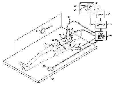
Fig. 1 is a diagrammatic perspective view depicting
elements of apparatus in accordance with one embodiment of the
invention in conjunction with a patient.
Fig. 2 is a diagrammatic sectional view depicting further
elements of the apparatus of Fig. 1.
Fig. 3 is a diagrammatic perspective view showing further
elements of the apparatus.
Fig. 4 is a graph of reference point position in operation of
the apparatus of Figs. 1-3.
MODES FOR CARRYING OUT THE INVENTION
Apparatus according to one embodiment of the present
invention includes a probe 20. The probe may incorporate essentially
any device which can be inserted or advanced into the body to perform
a medical procedure, such as treatment, measurement or observation.
CA 02287220 2009-01-09
-9b-
As used herein, the term "treatment" includes capturing samples of
tissues or materials present within the body, and thus includes
biopsies. The probe 20 desirably includes a conventional endoscope
having a tubular body 22. Body 22 has a handle portion 24 affixed to a
proximal end of the body and has a distal portion 26 remote from
handle 24. Body 22 has a bore 28 extending longitudinally from its
proximal end to its distal end and open to the outside through handle
24. Body 22 may incorporate a flexible section adjacent the distal end,
so that the distal end 26 can be bent or pivoted relative to the
remainder of the body. The endoscope may incorporate devices (not
shown) for bending the distal end of the body so as to steer the device
as it is advanced into the patient's anatomy. The endoscope may
further include a fiber optic or television
CA 02287220 1999-10-21
WO 99/43253 PCT/US98/13736
-10-
system (not shown) for visually observing the anatomical features of the
patient at the distal end of the endoscope.
The probe further includes a conventional intrabody medical
tool sucli as a biopsy needle 30 or other surgical tool operable from the
proximal end or handle of the device. Merely by way of example, instead of a
biopsy needle, the tool may be any conventional surgical tool of the type
commonly used in endoscopic, arthroscopic, a laparoscopic surgical
procedures; a biopsy forceps or other sampling device; a needle, catheter or
other drug delivery device; a measuring instrument such as a thennometer or
electrical potential measuring electrode; a device for applying therapeutic
radiation; or any other device which can be used to treat, measure or observe
structures within the body of a living subject. Needle 30 is arranged so that
it
can be advanced to an operative positioti 30' outside of the distal end of
body
22. Needle 30 is arranged so that it can be manipulated and controlled froin
the proximal end or handle 24 of the body. Thus, the needle is connected to a
manipulating handle 32 by conventional control elements or linkages. Other
expedients for manipulating and controlling a tool at the distal end of body
can be employed as, for example, electrical, electronic or optical control
linkages. Alternatively, a tool can be mounted in fixed position on body 22 or
formed integrally therewith as, for example, where body 22 is equipped with a
cutting blade.
A probe field transducer or position sensor 34 is mounted in
probe body 22 adjacent the distal end 26 thereof. Transducer 34 may be a
sensor arranged to detect magnetic or electromagnetic fields. For example,
the sensor 34 may be a multiaxis, solid-state position sensor of the type
disclosed in the aforementioned U.S. Patent 5,558,091. Such a sensor
incorporates a plurality of transducers sensitive to magnetic field components
in mutually orthogonal directions. Other suitable position sensors include
coils as disclosed in the aforementioned U.S. Patent 5,391,199 and in PCT
Application PCT/US95/01103, now published as PCT intelnational
CA 02287220 2004-09-17
Publication WO 96/05768. Such coils may be provided as a single coil or as a
plurality of orthogonal coils capable of detecting field components in
orthogonal
directions. Position sensor or field transducer 34 is connected to leads 36
which
extend through bore 28 to and beyond the proximal end 24 of body 22.
The apparatus further includes a set of external field transducers or
antennas 40 defining a locating frame of reference. For exampie, external
field
transducers 40 may be mounted to a patient-supporting bed 41. Antennas 40 are
linked to a field transmitting and receiving device 42 and a computer 44,
which
in turn is linked to a display device such as a cathode ray tube 46. The
computer
is also provided with conventional input devices 45 such as a keyboard,
trackball,
mouse and the like. Computer 44, field transmitting and receiving device 42
and
transducers 40 are arranged to cooperate with the probe field transducer 30 to
determine the dispositions of the field transducer on the probe, and hence
determine the disposition of the distal end of the probe in the locating frame
of
reference of the external field transducers or antennas 40. These elements of
the
apparatus can be as described in the aforementioned '091 or '199 patents.
Other
devices for detecting disposition of probes equipped with position sensors by
transmission of non-ionizing fields are known in the art. As is known in the
art,
electromagnetic or magnetic fields can be transmitted between an antenna or
field transducer mounted in an external frame of reference and a field
transducer
on a probe, and the disposition of the probe can be calculated from the
characteristics of the fields detected by the transducer on the probe. Thus,
the
external field transducers or antennas 40 and the position sensor or probe
field
transducer 34 on the probe cooperatively define a plurality of transmitter-
receiver
pairs. Each such pair includes one transmitter and one receiver as elements of
the pair. One element of each such pair is disposed on the probe and the other
eiement of each such pair is disposed at a known
-11-
CA 02287220 2004-09-17
disposition in the external frame of reference. Typically, at least one
element of
each transmitter-receiver pair is disposed at a different position or
orientation
than the corresponding element of the other pairs. By detecting the
characteristics of field transmission between elements of the various pairs,
the
system can deduce information concerning the disposition of the probe in the
external frame of reference. The disposition information can include the
position of the probe, the orientation of the probe or both. Although the
external
field transducers 40 are illustrated as mounted to a rigid structure such as a
patient bed, so that the external field transducers remain in fixed position
relative to one another, this is not essential. As described in commonly
assigned PCT Publication WO 97/29685, the external field transducers may be
movable relative to one another. The computer system can determine the
positions of the external field transducers by measuring the properties of
fields
transmitted between these transducers, or between the external field
transducers and calibration transducers mounted to the individual external
field
transducers.
The apparatus further includes a reference field transducer 50
mounted in a protective housing 52 effective to protect field transducer 50
from
physical damage when the field transducer is deployed at a position on the
outside of a patient's body. Thus, the housing 52 and field transducer 50 can
be mounted by any conventional expedient such as adhesive tape, bandages,
sutures or the like at a selected point on the exterior of a patient.
Optionally,
housing 52 may be provided with features such as flat pads or wings 54, suture
holes (not shown) or other physical features which further facilitate
attachment
to the exterior surface of the body. Reference field transducer 50 has
essentially the same structure as probe field transducer 34 discussed above.
Leads 56 connect the reference field transducer to the field transmitting and
receiving device 42. The field transmitter and receiver 42 and computer 44
actuate external field transducers 40 and reference field
-12-
CA 02287220 1999-10-21
WO 99/43253 -13- PCT/US98/13736
transducer 50 to transmit and receive fields in the same manner as discussed
above in connection with probe field transducer 34. Thus, the system
determines the disposition of the reference field transducer in the locating
frame of reference defined by external field transducers 40.
In a method according to one embodiment of the invention, a
patient P is imaged using any conventional imaging modality such as
computerized tomograpllic x-ray ("CAT" or "CT") imaging, magnetic
resonance imaging or any other imaging method which is capable of depicting
the internal organs of the body and, particularly, the respiratory system and
surrounding tissues. The image is acquired while the patient is at a selected
respiratory state. The selected respiratory state may be a state which is not
part of the patient's normal respiratory cycle, such as a forced inhalation or
forced exhalation. Preferably, the selected respiratory state is a stage of
the
patient's normal respiratory cycle. Preferably, the image is acquired while
the
patient is at the so-called "minimum inhalation" stage. This stage is the
stage '
during normal breathing where the patient has exhaled the normal, tidal
volume of air. The patient may be instructed to hold his or her breath at the
selected state during image acquisition. The image may be a conventional
diagnostic image acquired without regard to any special considerations for the
therapeutic procedure and indeed acquired before the need for the therapeutic
procedure is known.
The image includes at least a portion of the patient's thorax and
includes certain features of the patient's anatomy which are readily
identifiable in the image with a good degree of precision. These include
features of the skeletal system such as the scapula SC, portions of the spine
SP and the sternal notch ST. In the conventional manner, the image is
provided as computer data defining properties of structures of various
locations within the body as, for example, x-ray absorption of individual
volume elements or "voxels" in a CAT image or MRI data defining magnetic
resonance properties such as proton density, Ti or T2 for individual voxels.
CA 02287220 1999-10-21
WO 99/43253 PCT/US98/13736
-14-
After the image has been acquired, the patient is placed into
position in the locating frame of reference defined by external field
transducers 40 as, for example, by placing the patient in supine position on
the supporting table 41. Reference field transducer 50 is then engaged
successively with several of the aforementioned readily defined points on the
patient's body as, for example, with each of the scapula, with the sternal
notch
or witli readily identified points on the spine. This is done while the
patient
remains in position on the table. The table may be provided with apertures or
grooves in its surface (not shown) to allow insertion of the reference field
transducer 50 into engagement with features of the patient's back. While the
reference field transducer is in engagement with each defined point in the
patient's anatomy, the external field transducers 40, field transmitting and
receiving apparatus 42 and computer 44 are actuated to determine the location
of the reference field transducer in the locating frame of reference defined
by
the external field transducers 40. Thus, the locations of the various defined
points in the locating frame of reference are provided to the computer.
An operator can also input the locations of the same defined
points in the frame of reference of the image. For example, computer 44 can
be actuated to display depictions of the image which include the various
identifiable points in the anatomy and the operator can manually adjust a
cursor on the image as, for example, by adjusting a knob, trackball or mouse
incorporated in input devices 45. When the cursor is aligned with an
identifiable point in all dimensioiis, the operator enters a further signal
indicating to the computer that the coordinates of the cursor in the image
frame of reference correspond to the coordinates of the particular point in
the
anatomy. Once the coordinates of the identifiable points in the anatomy have
been provided to the computer in the image frame of reference and in the
locating frame of reference, the computer can derive a mathematical
transformation between the locating frame of reference and the image frame
of reference. Techniques for acquiring locations of points in the anatomy and
CA 02287220 1999-10-21
WO 99/43253 PCT/US98/13736
deriving transformations between an image frame of reference and a locating
frame of reference are well known - and are described in the aforementioned
patents and publications. In a variant of such techniques, also described in
these patents and publications, fiducial markers incorporating field
transducers are mounted on the patient before the imaging procedure, so that
the fiducial markers are visible in the image. The positions of the fiducial
markers in the locating frame of reference are acquired by actuating the field
transducers on the fiducial markers in conjunction with the external field
transducers, in the same manner as described above. In other variants, the
system acquires a succession of positions in the locating frame of reference
while a reference field transducer is moved over a well-defined contour in the
patient's anatomy. The computer system uses automatic pattern-matching
techniques to find a feature having a contour including a set of locations in
the
image frame of reference which can be mapped to the set of locations in the
locating frame of reference by a rigid-body transformation. Again, various
techniques for finding matching points in both frarnes of reference, and for
deriving a transformation between the locating and imaging frames of
reference, are well known in the art.
The reference field transducer 50 is then mounted on a point on
the outside of the patient's chest which moves during respiration. For
example, the reference field transducer can be taped or sutured in place over
one of the patient's ribs. While the patient remains in position on Table 41,
the patient breathes normally and hence reference field transducer 50 moves
cyclically in a motion corresponding to the various phases of the respiratory
cycle. Thus, the location of reference field transducer 50 varies with time.
The computer, in cooperation with the field transmitting and receiving unit
and external field transducers 40 continually monitors the position of
reference field transducer 50. The computer tracks the position of the
reference field transducer over time and generates a plot of the reference
field
transducer position in a selected direction versus time. The plot is depicted
in
CA 02287220 1999-10-21
WO 99/43253 -16 PCT/US98/13736
-
graphical fonn in Fig. 4 for ease of understanding. ln practice, the plot
consists of a series of numbers denoting the location of the reference
transducer along the selected axis at various times. The axis selected for
tracking may be a vertical axis (towards and away from the table in Fig. 1)
and hence towards and away from the patient's back; a horizontal axis
transverse to the longitudinal (head-to-toe) axis of the patient or an axis at
an
arbitrary angle between the vertical and the horizontal.
The computer selects successive extreme positions in the plot.
For example, where the location represented by the plot is location in a
vertical axis corresponding to movement towards and away from the patient's
back, the computer may be actuated to select successive minima 60 of such
location, i.e., the points wliere the reference field transducer is closest to
the
patient's back. Alternatively, wliere the location represented by the plot is
horizontal location, the computer may be actuated to select minima in the plot
corresponding to locations wliere the reference field transducer is closest
the
central axis of the patient. These minima can be found by conventional
computer-programming techniques for selecting local minimum values in a
sequence of numbers. Numerical techniques of this nature are well known in
the programming arts and are available in many standard mathematical
software packages. The minima represent the minimum inspiration point in
the patient's respiratory cycle as discussed above.
The value of the location at successive minima may not be
exactly the same. However, for a patient breathing nonnally, all of the
minima will have values close to one another. Thus, the computer calculates
a mean value 61 representing the mean location of several successive minima.
The system then applies a preselected tolerance or maximum deviation 62.
Whenever the location of reference field transducer 50 deviates from the mean
value 61 by less than a predetermined tolerance 62, the system treats the
patient as being at the minimum inspiration point of the respiratory cycle.
Thus, by monitoring the respiratory cycle, the systetn establishes a
particular
CA 02287220 1999-10-21
WO 99/43253 PCT/US98/13736
-17-
respiratory state corresponding to an extreme of the movement of the
reference transducer encountered in normal respiration.
The physician advances the distal end 26 of the probe into the
respiratory system of the patient in the conventional manner. Typically, the
distal end of the probe is advanced through an airway A as, for example,
through the larynx and trachea into the bronchi. Computer 44, field transmit
and receive unit 42 and extemal field transducers 40 cooperate with probe
field transducer 30 to determine the position of the probe field transducer
and
hence the position of the probe distal end 26 in the locating frame of
reference
defined by the external field transducers 40, and cooperate with reference
transducer 50 to determine its position. When the position of the reference
transducer is within the predetermined tolerance 62 of the mean minimum
inspiration location 61, the computer captures the location of the probe field
transducer and probe distal end in the locating frame of reference defined by
the external field transducers. Thus, the computer captures the location of
the
probe distal end when the patient is at the minimum inspiratory state.
The computer transforms the location of the probe distal end
into the frame of reference of the image and prepares a composite display
including at least a portion of the image and a representation of the probe
superposed on the image. For example, as shown in Fig. 1, the displayed
image on cathode ray tube 46 includes a depiction A' of the portion of the
airway together with a representation 26' of the probe distal end. The image
also includes a depiction T of the target tissue, in this case a lesion
outside of
the airway but adjacent thereto. Preferably, the image displayed is a
perspective view, so that the physician can readily perceive the spatial
relationships between the distal end of the probe and the target tissue. The
system may also generate a line or arrow 66 on the displayed image showing
the trajectory from the probe distal tip to the target. The physician can use
the
information shown in the displayed image to bring the probe distal end into
engagement with the target. For example, the physician can bring the distal
CA 02287220 1999-10-21
WO 99/43253 PCT/US98/13736
-18-
end of the probe body 22 into engagement with the airway adjacent the target
and can advance the biopsy needle 30 to its extended position 30' (Fig. 2) and
thus pierce the airway wall and engage the target.
Because the location of the probe distal end is captured only
when the patient is in the same respiratory state as used in image
acquisition,
the acquired position of the probe distal end, transformed into the frame of
reference of the image accurately represents the relative position of the
probe
distal end and the surrounding tissues. The patient continues to breathe while
the probe is advanced into the airway. A new probe position is acquired on
each respiratory cycle when the patient reaches the minimum inhalation state.
Each time a new probe position is acquired, the display shown on CRT screen
46 is revised to conform with the new probe position. Thus, the physician can
monitor the progress of the probe distal end towards the target tissue. The
physician can accurately align the probe with the target tissue.
The procedure discussed above can be varied in many ways.
For example, the computer can be adjusted to find the mean location for the
maximum inllalation 68 of the patient's respiratory cycle, and the image may
be acquired at a similar maximum inhalation state. In a further variant, the
computer can select an arbitrary axis for plotting motion of the reference
transducer so that the axis is aligned with the principal direction of motion
of
the reference field transducer during respiration. For example, the computer
can first track the location of the reference field transducer using a
horizontal,
vertical or other preset axis to find maxima and minima in the location on
that
axis. The computer can then compute the average time between successive
maxima or successive minima. That time corresponds to the period of the
respiratory cycle. The computer can then test various pairs of locations, each
including one point delayed in time by one full period from another point.
The computer can then calculate the distance in three dimensional space
between each pair of points. The pair of points wllich has the largest
distance
lie along ttle principal direction of movement of the reference field
transducer
CA 02287220 1999-10-21
WO 99/43253 PCT/US98/13736
-19-
during the respiratory cycle. The computer can then plot location along this
direction versus time.
The image may be acquired when the patient is at an abnormal
respiratory state such as a maximum forced exhalation obtained by
deliberately forcing exhalation with maximum voluntary effort, or a maximum
forced inhalation obtained by deliberately forcing maximum inhalation with a
maximum voluntary effort. In this case, the state used during image
acquisition will not recur during a normal respii-atory cycle. Instead, the
patient is instructed to repeat the state while the system monitors the
location
of reference field transducer 50. After the patient repeats the state in one
or
more trials, the system records the location of the reference transducer at
this
state, or the mean locatioti obtained in several trials. While the physician
is
advancing the probe into the respiratory system, the patient is instructed
periodically to repeat the same state. The system acquires the image when the
patient is holding his or her breath at the desired respiratory state. An
arbitrary respiratory state such as a state midway between the maximum and
minimum inhalation in a normal respiratory cycle can also be used. However,
the patient typically will not be able to reproduce such an arbitrary state
accurately. In methods according to a further embodiment of the invention,
the systein can provide guidance to the patient and the physician to aid in
duplicating an arbitrary respiratory state. Thus, if the reference transducer
50
is mounted on the patient before the imaging procedure, and if the reference
field transducer is visible in the image, the position of the field transducer
relative to the identifiable points on the body, such as the scapular spine or
sternum will vary witll the respiratory state of the patient. For example, if
the
reference field transducer is mounted to the ribs, the field transducer will
move outwardly, away from the central axis of the body as the patient inhales.
When the patient is placed on table 41, in proximity to the external field
transducers, the systein can track the location of the reference field
transducer
in the manner discussed above. The positiotl of the reference field transducer
*rB
CA 02287220 1999-10-21
WO 99/43253 PCT/US98/13736
-20-
can be transformed into the frame of reference of the image. If the patient is
in the same respiratory state as was used in image capture, the position of
the
reference field transducer in the image frame of reference will overlie the
depiction of the reference field transducer in the image. In a further
variant,
two or more reference transducers may be attached to the patient at locations
which move towards or away from one another during respiration. The
system can track the distance between the reference transducers as a measure
of respiratory state.
The physician can use the information as to the respiratory state
provided by the reference transducer or transducers to provide feedback to the
patient, as, for example, by instructing the patient to inhale or exhale
slightly
to better match the respiratory state used during image capture.
Alternatively,
a mechanical respirator can be controlled automatically to achieve
superposition between the position of the reference field transducer as
determined by the field transmitting and receiving apparatus and the position
depicted in the image. Thus, the respirator may be arranged to provide
substantially normal breathing followed by periods of forced breath holding
and inflation of the lungs to the extent necessary to match the position
captured in the image.
In the procedures discussed above, the probe is advanced
through the airway. However, the same advantages can be obtained in
procedures where the probe is advanced through the skin or through the
intestinal track to other organs affected by motion due to respiration.
As these and other variation and combinations of the features
discussed above can be utilized without departing from the present invention,
the foregoing description of the preferred embodiment should be taken by way
of illustration rather than by way of limitation of the invention defined by
the
claims.
CA 02287220 1999-10-21
WO 99/43253 PCT/US98/13736
-21-
INDUSTRIAL APPLICABILITY
The present invention can be applied in medical and veterinary
procedures.