Note: Descriptions are shown in the official language in which they were submitted.
CA 02287260 1999-10-22
WO 98/48718 PCT/US98/08272
THREADED FUSION CAGE ANCHORING DEVICE AND METHOD
CROSS-REFERENCE TO RELATED APPLICATIONS
This Patent Application claims the benefit of U.S.
Provisional Application No. 60/044,190 filed April 25,
1997.
1. Technical Field
The present disclosure concerns devices and methods
for stabilizing fusion inserts placed for the purpose of
fusing two adjacent joints such as vertebrae of the
spine, and more particularly, to a threaded fusion cage
anchoring device.
2. Background of Related Art
Degeneration of a joint such as a spinal segment by
a deterioration of the hard and soft tissues of the joint
complex may produce severe local or radiating pain when
that segment is in motion. Typically joint complexes
consist of two bony structures and an interposed
flexible, movable portion. In the spine the bony
structures are the vertebrae and the movable portion is
0
the intervertebral disc. The disc is composed of a
multilayered outer ligamentous belt, the annulus,
CA 02287260 1999-10-22
WO 98148718 PCT/US98/08272
constructed in concentric laminations rather like the
plies of an automobile tire. In the core of the disc
there is a small mass of flexible fibrogel, contained by
the annulus ring. The fibrogel mass, the nucleus of the
disc, is a hydrogel which on absorbing water exerts a
substantial swelling pressure to lift the vertebra and
balance the forces applied against the disc by gravity
and surrounding muscular contractions. Therefore, the
hydrogel is important for resisting potentially
disruptive forces applied to the vertebrae.
Unfortunately, as the disc degenerates, the
internally contained hydrogel begins to lose its water-
binding ability and shrinks. This shrinkage leads to a
loosening of the annulus fibers which permits an abnormal
range of motion of the segment with buckling and
delamination of the overlapping plies. Tears in as few
as several layers of the approximately 12 to 20
concentric laminations of the annulus may permit a
herniation of the pressured central nucleus material
outward through the annulus defect.
Conventional procedures for treating degenerative
vertebral discs involve fusing the discs together to stop
all motion of the bone segments. The most efficient
method of fusion places bone or a bone inducing substance
inside a supporting device surgically implanted into the
center of the disc. This supporting device construct
will obliterate the degenerated nucleus, hold the bone
material rigidly in position, protect the bone from
-2-
,,,
CA 02287260 1999-10-22
WO 98/48718 PCT/US98/08272
collapse, extrusion or invasion by residual soft tissues
of the disc and cause the opposing vertebrae to rapidly
fuse together. The preferred intervertebral fusion
device is a vertebral fusion cage. For example, U.S.
Patent No. 4,961,740 to Ray, contents of which are
incorporated herein, discloses threaded vertebral fusion
cages. The internal cavities of the cages are used to
secure the bone graft material and to permit bone growth
through and across the surgically emptied nucleus cavity
between adjacent vertebrae.
As opposed to non-threaded fusion cages which are
hammered or tapped into position, insertion of threaded
vertebral fusion cages is made more efficient because the
threaded outer surface permits easy adjustment of the
depth and penetration of the cage into the disc space.
The threaded outer surface also prevents dislodgment or
expulsion of the cage. In addition, the graft bone
packed within the threaded fusion cages presents or
effuses through these perforations and comes into
intimate contact with the bone of the adjacent vertebral
bodies. When the cage is inserted into the bored or
tapped intervertebral bed, the lateral walls of the cage
are oriented horizontally and face the disc cavity.
These lateral cage walls are blocked (i.e., contains no
apertures) and therefore are a barrier against any
potential ingrowth of residual disc tissue into the
contained graft area which could interfere with or weaken
-3-
CA 02287260 1999-10-22
WO 98/48718 PCT/US98/08272
the fusion formation of these adjacent vertebrae.
More recently, emphasis has been placed on securely
fixating the fusion cage implant within the vertebrae.
During a fusion cage implantation procedure, the surgeon
may determine that sufficient stabilization of the space
has not been achieved by implantation of the fusion cage
alone. In such situations, additional instrumentation tc
improve the stability of the vertebrae and cage is
required. Examples where additional stabilization
procedures may be used include: the vertebral bone is
weak, the cages do not fit tight enough in the vertebral
space or the central concavity of the disc space is too
deep to achieve good cage penetration along the anterior-
posterior length of the disc space. In such cases, the
surgeon ordinarily would be forced to place additional
fusion instrumentation such as pedicle screws, rods or
vertebral body plates to prevent cage dislodgment and
improve the opportunity for a good fusion. This
additional step in the surgical procedure increases the
complexity, potential hazards and cost of the procedure.
The embodiments of the present disclosure solve these and
other associated problems and provides a simple and
easily applied instrumentation to intraoperatively
achieve increased cage fixation and disc space stability.
-4-
.._.."..w,m.....T,... ... . .. ~.. .v .T...
CA 02287260 1999-10-22
WO 98/48718 PCT/C1S98/08272
S UN~IARY
The present disclosure is directed to an anchoring
device and system for stabilizing adjacent vertebral
. joints. The anchoring device includes an anchoring plate
adapted r.o be secured to at least one vertebral disc
having a central portion, extended end portions and at
least one lateral extension. The central portion
includes an anchoring nut for fastening the anchoring
plate to a vertebral implant and the end portions include
anchoring screws for fastening the anchoring plate to the
at least one vertebral disc. The vertebral implant is
preferably a threaded fusion cage, wherein the anchoring
nut is rotatably fixed to both the anchoring plate and
the threaded fusion cage.
The anchoring device preferably includes the central
portion and each extended end portion having at least one
locking tab for rotatably locking the anchoring nut and
anchoring screws. Additionally, the central portion
further includes a plurality of projecting detents along
an outer periphery thereof which mate with corresponding
slots on the threaded fusion cage.
The present disclosure is also directed to a method
of implanting an anchoring device to at least one
vertebral disc having a vertebral implant. The method
includes providing an anchoring plate having a central
portion and extended end portions. The central portion
includes an anchoring nut for fastening the anchoring
-5-
CAI02287260 1999-10-22
WO 98148718 PCT/US98/08272
plate to a vertebral implant and at least one locking tab
along an outer periphery thereof. The end portions
include anchoring screws for fastening the anchoring
plate to at least one vertebral disc and at least one
locking tab along an outer periphery thereof. The method
further includes: situating the anchoring plate in a
corresponding relationship to the at least one vertebral
disc and the vertebral implant; fastening the anchoring
nut to the vertebral implant; and fastening the anchoring
screws to the at least one vertebral disc. The locking
tabs are then positioned into engagement with the
anchoring nut and anchoring screws.
BRIEF DESCRIPTION OF THE DRAWINGS
The objects and features of the present disclosure,
which are believed to be novel, are set forth with
particularity in the appended claims. The present
disclosure, both as to its organization and manner of
operation, together with further objectives and
advantages may best be understood by reference to the
following description, taken in connection with the
accompanying drawings, in which:
FIG. 1 is a view illustrating several adjacent
spinal segments and two anchoring devices according to
the present disclosure mounted to the spinal segments;
FIG. 2 is a bottom plan view of the anchoring device
according to the present disclosure;
-6-
a t
CA 02287260 1999-10-22
WO 98/48718 PCT/US98/08272
FIG. 3 is a perspective view illustrating a
posterior aspect of a fusion cage and associated fusion
cage inserting drive shaft;
FIG. 4 is a plan view of an anchoring nut associated
with the anchoring device of the present disclosure; and
FIG. 5 is a bottom plan view illustrating an
anchoring device of an alternative embodiment according
to the present disclosure.
DETAILED DESCRIPTION OF PREFERRED EMBODIMENTS
The preferred embodiments of the apparatus and
methods disclosed herein are discussed in terms of
orthopedic spinal fusion procedures and instrumentation.
It is envisioned, however, that the disclosure is
applicable to a wide variety of procedures including,
but, not limited to joint repair, non-union fractures,
facial reconstruction, spinal stabilization and the like.
In addition, it is believed that the present method and
instrumentation finds application in both open and
minimally invasive procedures including endoscopic and
arthroscopic procedures wherein access to the surgical
site is achieved through a cannula or small incision.
The following discussion includes a description of
the threaded fusion cage anchoring device utilized in
performing a spinal fusion followed by a description of
the preferred method for implanting the threaded fusion
cage anchoring device in accordance with the present
i
CA 02287260 1999-10-22
WO 98/48718 PCT/US98108272
disclosure.
Reference will now be made in detail to the
preferred embodiments of the disclosure, which are
illustrated in the accompanying figures. Turning now to
the figures, wherein like components are designated by
like reference numerals throughout the various figures,
attention is first directed to FIGS. 1 and 2.
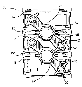
An anterior view of a fused vertebral section 10
including two implanted anchoring devices 12 across three
adjacent spinal segments 14, 16 and 18 are generally
shown at FIG. 1. The spinal segments 14, 16 and 18, for
instance, cervical spinal segments, are separated by two
interposed disc spaces 20 and 22. Threaded fusion cages
24 are implanted between spinal segments 14 and 16 and
spinal segments 16 and 18 and across the interposed disc
spaces 20 and 22, respectively.
With particular reference to FIGS. 1-3, the
anchoring device 12 of the present disclosure generally
includes an anchoring plate 26, anchoring nut 28 and
anchoring screws 30. Anchoring plate 26 is generally in
the shape of a figure-eight and includes a large central
bore 32 and extension arms 34. Each extension arm 34
includes at least one anchoring screw bore 36 for
receiving anchoring screw 30. The anchoring device 12 of
the present disclosure is preferably fabricated from a
suitable biocompatible rigid material such as titanium
and/or alloys of titanium, stainless steel, ceramic
_g_
....-.-~.-...w.~.~..... ~ , , r
CA 02287260 1999-10-22
WO 98148718 PCT/US98/08272
materials or rigid polymeric materials. Anchoring plate
26 includes a fusion cage mating side 38 (bottom, i.e.,
FIG.2) and an anchoring nut mating side 40 (top, i.e.,
FIG. 1). The fusion cage mating side 38 includes
projecting detents or pins 42 for engaging slots 44 of
fusion cage 24 (FIG. 3). The projecting detents 42 are
preferably located along the outer periphery of central
bore 32. The anchoring device 12 of the present
disclosure and associated threaded fusion cage 24 may
include any number of detents 42 and mating slots 44,
wherein the higher quantity of projecting detents 42 and
mating slots 44 provide for an optimal fixed relationship
between the fusion cage 24 and the anchoring plate 26
without a large angular change in the implanted fusion
cage 24 position. That is, the altering of the number of
slots 44 of fusion cage 24, as well as, the mating
projecting detents 42 of anchoring plate 26 will
incrementally alter the angular relationship between the
anchoring plate 26 and fusion cage 24. As such, minor
angular changes in the orientation of the anchoring plate
26 with respect to the fusion cage 24 is beneficial so
that the initial optimal depth of penetration of
implanted fusion cage 24 into the joint space need not be
markedly altered from the possible rotation attributable
to the implantation of anchoring device 12.
As best depicted in FIG. 1, the orientation or
angular displacement of the anchoring plate 26 relative
_g_
CA 02287260 1999-10-22
WO 98/48718 PCT/US98108272
to the longitudinal axis of the spinal column, may be
altered from O to 80 degrees as needed to provide
appropriate stability to the fusion cage 24 relative to
the adjacent vertebrae. This broad angular displacement
assures a safe positioning of the anchoring plate 26
relative to certain anatomical structures, such as, the
vertebral end plate, nearby traversing nerves or bony
obstructions. In this regard, when a pair of fusion
cages 24 has been implanted at the same level, the
anchoring plates 26 are preferably set substantially
parallel to each other. Hence, having a broad angular
displacement allows the angulation between each anchoring
plate 26 to be altered in order to obtain optimal
anchoring screw 30 placement in the vertebral bodies.
Similarly, when two single cages 24 or a pair of cages 24
are placed at adjacent spinal levels, the common
angulation of the anchoring plates 26 may be altered for
optimal anchoring screw 30 placement into the adjacent
vertebral bodies. Both the cages 24 and anchoring plates
26 may be placed by an anterior or posterior surgical
approach to the lumbar spine. However, in the thoracic
and cervical spinal areas an anterior method alone is
recommended.
With particular reference to FIGS. 1, 2 and 4, the
anchoring device 12 of the present disclosure is placed
over the implanted fusion cages 24 and attached to each
fusion cage 24 with a threaded anchoring nut 28 which
-10-
CA 02287260 1999-10-22
WO 98/48718 PCT/US98108272
screws into matching threads 46 inside the inner
periphery of fusion cage 24. Once installed, anchoring
nut 28 is prohibited from loosening by a plurality of
malleable nut locking tabs 48 positioned on opposing
sides of the central bore 32 of anchoring plate 26. The
bending of the nut locking tabs 48 over the anchoring nut
28 will engage at least one flat portion of the anchoring
nut collared head 50 and thereby prevent the anchoring
nut 28 from rotating and becoming loose. The extension
arms 34 of anchoring plate 26 include anchoring screw
bores 36 through which slotted head anchoring screws 30
are passed and tightened into the vertebral bodies at
convenient and safe locations. Similarly, the anchoring
screws 30 are prevented from unscrewing by a plurality of
malleable screw locking tabs 52 placed on opposing sides
of the anchoring screw bores 36 on extension arms 34.
The screw locking tabs 52 are oriented so that at least
one of them will firmly mate with at least one slot
located on the head of anchoring screws 30 when locking
tabs 52 are bent over anchoring screws 30.
The anchoring device 12 provides the additional
support needed to fully stabilize the fusion cage 24
relative to spinal segments 14, 1G and 18. By crossing
. the interposed disc space 20 and 22 and attaching the
anchoring screws 30 to the vertebral bodies at an
extended distance from fusion cage 24, a substantial
increase in mechanical fixation strength is provided.
-11-
CAI02287260 1999-10-22
WO 98/48718 PCT/US98108272
Essentially, the anchoring device 12 keeps the vertebrae
from moving apart and therefore from distracting away
from the fusion cage 24 as postoperative spinal motions
occur. Further, the anchoring plate 26 significantly
improves the initial overall rigidity of the fusion cage
system.
With reference to FIG. 3, a fusion cage 24 inserting
drive shaft 54 for seating a threaded fusion cage 24
inside a bore made between adjacent surfaces of a spinal
segment is shown. A plurality of slots 44 on the outer
edge 62 of the fusion cage 24 match projecting tabs 56 on
drive shaft 54. The fusion cage 24 attaches to a
retractable central threaded coupler 58 which rotates
freely within the drive shaft 54. The fusion cage slots
44 mate with the projecting tabs 56 of drive shaft 54.
Upon rotation of drive shaft 54, the threaded coupler 58
engages the matching fusion cage threads 46 located along
an inner periphery of fusion cage 24. The mated slots 44
and projecting tabs 56 are used to rotatably drive the
fusion cage 24 into position after which the threaded
coupler 58 is unscrewed releasing both the drive shaft 54
and threaded coupler 58 from the fusion cage 24. In the
cases where positioning of fusion cage 24 needs further
adjustment, the drive shaft 54 may be mated to the fusior_
cage 24 via projecting tabs 56 and slots 44 to torque the
fusion cage 24 into a final position without the
necessity of firmly reattaching the threaded coupler 58
-12-
,.. t
CA 02287260 1999-10-22
WO 98/48718 PCT/US98/08272
to the fusion cage 24.
With particular reference to FIG. 5, an additional
embodiment of the anchoring plate 26 is shown, wherein
r like components which correspond to those of previous
embodiments described herein are designated by like
reference numerals. Anchoring plate 26 further includes
additional lateral extensions or tangs 60 for further
stabilizing the interposed disc spaces 20 and 22 by being
forced into the spaces 20 and 22 as the anchoring nut 28
is tightened onto anchoring plate 26 and into fusion cage
24. The space between the margins of lateral tangs 60
may accommodate additional bone growth material such as
cancellous or soft bone from another human (allograft) or
from the same patient (autograft) which serves to provide
a better fusion of spinal segments 14, 16 and 18.
IMPLANTATION OF THE ANCHORING DEVICE
The implantation of the anchoring device 12 of the
present disclosure will now be described with respect to
a single anchoring device 12 although multiple anchoring
devices 12 can be implanted across one or more vertebral
discs or spinal segments 14, 16 and 18. A standard
surgical approach is used to gain access to the surface
of the vertebral bodies to be fused. This may consist of
an anterior approach in the neck and thoracic spine or an
anterior or posterior approach in the lumbar spine. One
or more bores are drilled into selected intervertebral
-13-
CA 02287260 1999-10-22
WO 98/48718 PCT/US98/08272
spaces and tissue debris is cleaned out therefrom. For
some fusion cage implants, the bore may be tapped to
match the threaded portion of the fusion cages. In other
cases, a self-tapping fusion cage may be used and no
threading will be required.
As shown generally at FIG. 3, a threaded fusion cage
24 having slots 44 along its outer edge 62 is mated to
projecting tabs 56 on the tip of the drive shaft 54,
wherein the threaded coupler 58 is threaded onto the
inner fusion cage threads 46 of fusion cage 24 to secure
the fusion cage 24 to the drive shaft 54 during insertion
thereof within the intervertebral spaces. Next, the
fusion cage 24 is screwed into its optimal position in
the prepared intervertebral bore and the threaded coupler
58 and drive shaft 54 are detached.
The placement of anchoring device 12 is dependent
upon the actual location of the implanted fusion cage 24.
If a single fusion cage 24 or alternatively a pair of
fusion cages 24 are implanted at multiple vertebral
levels, then the orientation of the anchoring plates 26
will be in parallel pairs throughout the multiple fused
vertebral segments. The projecting detents 42 located
along the mating surface 38 of anchoring plate 26 mates
with equivalently spaced slots 44 located on the outer
edge 62 of the implanted fusion cage 24. A minor angular
adjustment is made in the orientation of the anchoring
plate 26 and fusion cage 24 relative to the longitudinal
-14-
............ .. ~. , ,.
CA 02287260 1999-10-22
WO 98/48718 PCT/US98/08272
axis of the spine to maintain clearance of any anatomical
structures. This angular adjustment requires only a few
degrees of change from the initial position of the fusion
cage 24. The anchoring plate 26 may be bent slightly,
before or after being attached to the fusion cage 24, to
conform with the curving surface of the vertebral bodies
or to establish clearance from other adjacent structures.
Once an anchoring plate 26 is positioned over the
implanted fusion cage 24, the anchoring nut 28 is passed
through the central bore 32 of the anchoring plate 26 and
screwed into the corresponding mating fusion cage threads
46 portion of the implanted fusion cage 24. Since the
anchoring device 12 and fusion cage 24 can include a
plurality of projecting detents 42 and mating slots 44,
the anchoring plate 26 can be incrementally rotated to
bring the anchoring plate 26 into its most advantageous
position relative to the fusion cage 24 position. The
rotation or adjusting of the anchoring plate 26 with
respect to the fusion cage 24 is performed prior to
tightening the anchoring nut 28 to the anchoring plate 26
and fusion cage 24. Pilot holes or bores are then
drilled into the vertebrae through the anchoring screw
bores 36 of extension arms 34. The extension arms 34 are
~ next fitted with anchoring screws 30 of the appropriate
length through bores 36 which are screwed and anchored
into the vertebral bone. Both nut locking tabs 48 and
screw locking tabs 52 are bent over the edges of
_15_
CA 02287260 1999-10-22
WO 98148718 PCT/US98/08272
anchoring nut 28 and the slots of anchoring screw 30,
respectively, to prevent either from loosening or
unscrewing. Bone inducing material is then packed inside
fusion cage 24, through central bore 32 and the center of
anchoring nut 28. Alternatively, bone inducing material
may be packed into fusion cage 24 prior to insertion.
With respect to the alternative embodiment of
anchoring device 12 depicted at FIG. 5, wherein like
components and methods correspond to those of previously
described embodiments described herein and are designated
by like reference numerals. The implantation of
anchoring plate 26 further includes the addition of bone
inducing material laterally placed along fusion cage 24
prior to attaching anchoring plate 26 to fusion cage 24.
It will be understood that various modifications may
be made ~..o the embodiments disclosed herein. For
example, the anchoring device 12 of the present
disclosure may include any number of nut locking tabs 48
and screw locking tabs 52 to better secure each nut 28
and screw 30, respectively. Also, anchoring device 12
may include one or more extension arms 34 radially
displaced along an angular relationship from central bore
32 which would provide better stabilization of the
anchoring device 12, fusion cage 24 and associated bone
segments 14, 16 and 18. Therefore, the above description
should not be construed as limiting, but merely as
exemplifications of preferred embodiments. Those skilled
-16-
~.....~....~ . , , . ,
CA 02287260 1999-10-22
WO 98/48718 PCT/US98/08272
in the art will envision other modifications within the
scope and spirit of the claims appended hereto.
-17-