Note: Descriptions are shown in the official language in which they were submitted.
CA 02287947 1999-10-28
WO 98/52016 PCT/CA97/00318
-1-
MIJLTI-SPECTRAL IMAGING SYSTEM
AND METHOD FOR CYTOLOGY
. Field of the Inventioa
The present invention relates to automated biological
testing systems and more particularly to a system for generating
data for the analysis of the visual characteristics of
cytological specimens, and in particular biological specimens
obtained fox Papanicolaou (Pap) testing and prepared as a
monolayer specimen.
B_ackaround of the Iaveatioa
In the art, there are known techniques for the machine-
aided evaluation of biological or medical specimens. Many of
these embody the application of optical decomposition for image
evaluation.
Bacus, in U.S. Patent No. 5,202,931, teaches an optical
method and apparatus for protein quantification that utilizes two
band-pass optical filters centred at 500 nm and 650 nm. The
filters are optimized to produce maximal contrast between
cellular nuclei with and without diaminobenzidine precipitate
staining. While the Bacus invention is effective for application
in a quantitative immunohistochemical assay, the Bacus method is
not suitable to capture and exploit the crucial properties of a
Papanicolaou (Pap) test for automated evaluation. Specifically,
the Pap test evaluation does not reduce to a simple binary
decision, i.e. either a ~~yes~~ or a ~~no~~ for the presence of a
specific staining precipitate. The Pap test evaluation requires
the synthesis of a highly-variable and wide-ranging set of visual
and clinical circumstances in order to render a diagnostically
reliable outcome. From the perspective of machine automation,
these visual circumstances are the complete range of mathematical
~~ features ~~ which are raised as a consequence of the standardized
staining protocol. Thus, any application of the image analysis
techniques to the Pap test must be constrained to this stain and
must extract the full range of features that replicate the
appreciation gained through human visual evaluation.
CA 02287947 1999-10-28
WO 98/52016 PCT/CA97/00318
-2-
In United States Patent No. 4,191,940, Polcyn et al.
discloses a technique for the use of_a decomposed set of optical
wavelengths for a multivariate analysis of cell identification.
Though powerful in its own right, the Polcyn technique is limited
to the separation of different categories of material based on
simple absorption properties alone. As described above, the Pap
test is much more subtle and complex. The optical absorption
properties represent only the beginning of the chain of analysis
that ultimately leads to a medical diagnosis. Given the
complexity of the cervical cytology application it is usual to
apply what is known as a "classical" image analysis consisting
of segmentation, feature extraction and classification. In this
way only is it possible to arrive at a precise and accurate
classification of the myriad components that reside within a
gynaecological specimen.
The complexity of the Pap test automation task is borne
out in United States Patent No. 5,287,272 by Rutenberg et al.
Rutenberg et al. teaches a method and apparatus that draws a
clear distinction between the conventional Pap smear and the thin
layer or monolayer specimens that are the subject of the present
invention. According to Rutenberg et al., the application of
cytological image analysis is severely constrained by limitations
of the conventional Pap smear. Unlike the controlled monolayer
specimen, the conventional smear is characterized by irregular
cell groupings and distributions, thick, overlying cell clusters
and occluding debris. By avoiding the monolayer preparation,
Rutenberg et al. are restricted to a level of image analysis that
is limited in its sensitivity and specificity.
The subject invention addresses the problems and
limitations associated with the prior art. The present invention
utilizes a monolayer specimen for automated cytological analysis
and advantageously features a segmentation phase with improved
accuracy and produces a complex and extensive range of extracted
features. This allows a more refined approach to the problem of
cytological classification and improves performance and provides
cost savings. The image collection component of this invention
also features the creation of a "pseudo-coloured" image that
CA 02287947 1999-10-28
WO 98/52016 PCT/CA97/00318
-3-
retains the bulk of the visual cues required by cyto-
technologists for interactive review_purposes.
Constrained by the nature of the preparation, the fixed
' protocol of the biological staining and the necessity to bridge
the gap between machine processing and human evaluation, the
' present invention comprises a refined set of optical filters used
in conjunction with a high-speed imaging system, processing
hardware, discriminant-analysis techniques and mathematical
measures to pre-process images for cellular identification. The
images gathered generated according to the invention are also
useful for human-interactive review, a further advantage of the
system.
Brief Summary of the Invention
The present invention provides an imaging system having
the capability to simultaneously capture the same scene in
multiple spectral bands, and comprises a system having an
integrated optical system, image collection devices and a method
for pre-processing and analyzing human cervical cytology
specimens or samples. The system is particularly suited for
specimens prepared in the form of thin-layers or monolayers . The
image data produced by the system is suitable for automated
assessment of the clinically-relevant state of the specimen and
also permits the use of human-expert review for confirmation or
to establish diagnostic grade and clinical action.
The system according to the present invention comprises
three principal components (a) optical hardware (b) electronic
hardware and (c) measurement and analysis procedures and methods.
The optical hardware provides for illumination of the specimen,
magnifies the cellular components, separates the appropriate
wavelengths and directs the separated wavelengths for electronic
digitization. The electronic hardware provides for the
translation of the optical images into digital information and
for the overall control of the processing steps according to the
invention. The measurement and analysis procedures preferably
comprise processing steps embedded in hardware for pre-processing
the information for classification.
CA 02287947 1999-10-28
WO 98/52016 PCT/CA97/00318
-4-
This subject invention is intended to function with
components described in co-pending patent applications entitled
Automated Scanning of Microscope Slides International Patent
Application No. CA96/00475 filed July 18, 1996 and U.S. Patent
Application No. 60/001,220 filed July 19, 1995, Pipeline
Processor for Medical and Biological Applications U.S. Patent
Application No. 08/683,440 filed July 18, 1996 and U.S. Patent
Application No. 60/001,219 filed July 19, 1995, Multi-Spectral
Segmentation International Patent Application No. CA96/00477
filed July 18, 1996 and U.S. Patent Application No. 60/001,221
filed July 19, 1995, Neural-Network Assisted Multi-Spectral
Segmentation International Patent Application No. CA96/00619
filed September 18, 1996 and U.S. Patent Application No.
60/003,964 filed September 19, 1995, Automated Focus System
International Patent Application No. CA96/00476 filed July 18,
1996 and Window Texture Extraction International Patent
Application No. CA96/00478 filed July 18, 1996 and U.S. Patent
Application No. 60/001,216 filed July 19, 1995, all in the name
of the common owner.
In a first aspect, the present invention provides an
imaging system for capturing multi-spectral image data of a
cytological specimen, said imaging system comprising: (a) an
optical stage having a light source for illuminating the
specimen, and optical means for producing images of the
illuminated specimen in a plurality of spectral bands; (b) an
image capture camera having means for simultaneously capturing
said spectral images and generating corresponding electrical
signals corresponding to said captured spectral images; (c)
controller means for controlling the operation of said image
capture camera and said light source, said controller means
having means for converting said electrical signals corresponding
to said captured spectral images into a data form suitable for
further processing.
In another aspect, the present invention provides a
method for generating multi-spectral image data for cytological
specimen, said method comprising the steps of : (a) exposing said
cytological specimen to a short burst of broad-band light; (b)
CA 02287947 1999-10-28
WO 98/52016 PCT/CA97/00318
-5-
separating said burst of broad-band light into a plurality of
spectral bands; (c) simultaneously capturing an image for each
of said spectral bands and generating electrical signals
corresponding to each of said captured spectral images; (d)
converting the electrical signals corresponding to said captured
spectral images into a data form suitable for further processing.
Brief Description of the Drawincrs
Reference will now be made, by way of example, to the
accompanying drawings which show preferred embodiments of the
present invention, and in which:
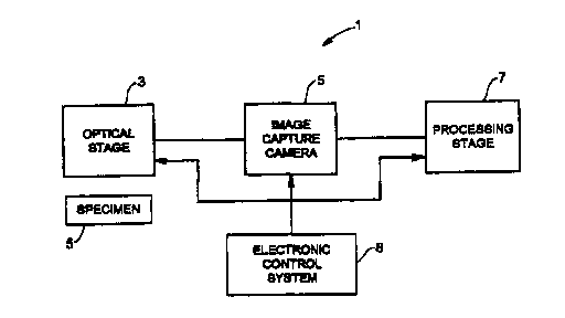
Fig. 1 shows in block diagram form a multi-spectral
imaging system according to the present invention;
Fig. 2 shows in a diagrammatic form an optical pathway
for the multi-spectral imaging system of Fig. 1;
Fig. 3 shows spectral bands for images captured;
Fig. 4 shows in block diagram forth an electronic
circuit for the multi-spectral imaging system according to the
present invention; and
Fig. 5 shows in block diagram a camera for the multi-
spectral imaging system according to the present invention.
Detailed Description of the Preferred Embodiments
Reference is first made to Fig. 1 which shows in block
diagram form a multi-spectral imaging system 1 according to the
present invention. The multi-spectral imaging system 1 comprises
an optical stage 3, an image capture camera S, and a processing
stage 7 and an electronic control system 8.
As will be described, the multi-spectral imaging system
1 provides a method and apparatus for generating data
representing the visual characteristics of a cytological specimen
denoted by reference S in Fig. 1. According to one aspect of the
invention, the data is generated in a form which facilitates
. further processing and analysis of the characteristics of the
cytological specimen S and is particularly suited for monolayer
specimens.
CA 02287947 1999-10-28
WO 98/52016 PCT/CA97/00318
-6-
Reference is made to Fig . 2 which shows the optical
stage 3 in more detail. The optical stage 3 provides the optical
path for the system 1. The optical stage 3 includes a high-
intensity electrical discharge tube 11, a condensing lens 13, a
fibre-optic bundle 15, a small aperture 17, an objection lens 19,
a telan lens 21, and a prism assembly 23. The prism assembly 23
includes an optical element 25 with filters 27, 29, 31.
The electrical discharge tube 11 is operated as a
stroboscopic lamp. Preferably, the discharge tube 11 produces
a short intense pulse of light lasting less than 6 microseconds.
The lamp 11 is selected to have a broad-band spectral output
covering a range between 400 nm and 700 nm. As will be
described, the optical filters 27, 29, 31 select the appropriate
wavelengths for image formation from this broad range. The pulse
of light must have sufficient intensity to accommodate losses
from the intervening optics. A short light pulse is preferred
because it allows the multi-spectral system 1: (a) to isolate
from the image mechanical vibrations that result in mechanical
velocities of less than 0.08 metres per second at the microscope
slide level, (b) to operate the CCD array cameras (see Fig. 4
below) without electronic or mechanical shutters thereby
increasing, the rate of image acquisition, and (c) to illuminate
the sample without the photo-bleaching or heat damage effects
associated with continuous illumination sources.
The light emitted by the strobe lamp I1 is coupled to
the fibre-optic bundle 15 by the condensing lens 13. The
condensing lens 13 comprises a known optical element which
functions to gather, concentrate, collimate and project the light
emitted by the strobe lamp 11 onto the face of a fibre-optic
bundle 15. The fibre-optic bundle 15 preferably comprises a
tightly-packed group of glass fibre-optic cables. The primary
function of the fibre-optic bundle 15 is to couple the light from
the lamp 11 to illuminate the specimen S. The use of a fibre-
optic bundle 15 as a light guide is preferred because it allows
the strobe lamp 11 to be operated at some distance from the
object plane, i.e. specimen S, of the system 1. Advantageously,
this arrangement reduces the potential occurrence of electrical
CA 02287947 1999-10-28
WO 98/52016 PCT/CA97/00318
_7_
interference from the intense electrical discharges occurring at
the lamp 11. The flexibility of the_fibre-optic bundle 15 also
permits the use of indirect optical paths from the strobe lamp
11 to the object plane and thereby eases design considerations.
As shown in Fig. 2, the small aperture 17 is centred
' on the optical axis of the objective lens 19 at the exit face of
the fibre-optic bundle 15. This arrangement is preferred because
it restricts the illumination to the region immediately
surrounding the region of interest (denoted by 16 in Fig. 2) and
advantageously reduces the contrast-reduction effects associated
with internal reflections within the optical components and
yields better-resolved images.
The light which passes through the specimen S is
collected by an objective lens 19. The objective lens 19
preferably comprises an infinite-conjugate optical system. The
objective lens 19 preferably has moderate nominal magnification
(x10 or x20) and a numerical aperture of 0.4 NA-0.75 NA. The
lens 19 is brought into the correct or optimal focus for the
nuclear material contained in the specimen S within the field of
view by means of an automatic focus module 20. The automatic
focus module 20 is preferably implemented as the apparatus and
method as substantially described in co-pending PCT Patent
Application No. CA96/0047& filed in the name of the common owner.
The automatic focus techniques which control the focus mechanism
are used in conjunction with a method of image formation by
spectral separation as will be described below in further detail .
As described in co-pending International Patent Application No.
CA96/00476 (which is hereby incorporated by reference) the
automatic focus module 20 comprises a servo-mechanical mechanism
having a magnetically-suspended voice-coil actuator 47 (Fig. 4)
which supports the objective lens 19. The voice-coil actuator
47 receives motion control instructions from the electronic
control system 8 based upon the mathematical calculations and
process control steps as described in the co-pending application
for an automated focus system.
The obj ective lens 19 preferably comprises an inf inite-
conjugate objective lens which produces a real image of the
CA 02287947 1999-10-28
WO 98/52016 PCT/CA97/00318
_g_
specimen S that is projected (theoretically) to an infinite
distance. In the optical stage 3 the light emitted from the
infinite-conjugate lens 19 is subsequently gathered by the telan
lens 21. The function of the telan lens 21 is to create and
project a real image to a finite position within the prism
assembly 23. An infinite-conjugate system is preferred for the
following reasons. First, the magnification is a function only
of the ratio of the focal length of the objective lens 19 and the
telan lens 21. This means that the magnification is not
sensitive to the relative displacement of the objective lens 19
and so the motion of the objective lens 19 during the automatic
focusing will have negligible effect upon the optical
magnification of the system 1. This is in contrast to a
conventional DIN microscope system in which the magnification is
based on a specific tube length (e.g. 160 mm with 45 mm parfocal
length). A second advantage of the present arrangement is that
the light between the objective lens 19 and the telan lens 21 is
collimated. Thus, it is possible to introduce additional optical
elements, such as beam-splitters, without suffering or incurring
spherical aberrations in the final image. Thirdly, the infinite-
conjugate objective lens 19 allows the simple alteration of the
magnification of the real image by a substitution of an objective
lens of a different focal length. Unlike conventional finite
tube length systems, the alteration of the arrangement shown in
Fig. 2 would carry no penalty with respect to the quality of the
image obtained from the specimen S.
The image re-formed by the telan lens 21 is projected
into the prism assembly 23. The prism assembly 23 comprises the
internal optical prism element 25 with the three optical filters
27, 29, 31 which are optically coupled to respective faces of the
prism assembly 23. The function of the prism assembly 25 is to
select a series of three narrow optical wavelength
representations of the image. The three optical wavelengths are
based in part on spectral decomposition principles as described
by G. Coli et al. in Olivetti Research and Technology Review Vol.
8, No. 33 (1987).
CA 02287947 1999-10-28
WO 98/52016 PCT/CA97/00318
_g_
The optical prism element 25 comprises a set of glass
wedges coated with dielectric film stacks to create the
interference band-pass optical filters 27, 29, 31. By selecting
wedge angles and dielectric film coatings the prism 23 will
simultaneously produce three images from the same scene in each
of three narrow optical regions. The width of each of these
optical regions is preferably 10 nm with a transmission
efficiency of at least 50% within the optical band. The three
centre wavelengths for these bands are selected as 530 nm (I),
577 nm (II) and 630 nm (III) as shown in Figure 3.
The arrangement according to this aspect of the
invention has specific advantages for the acquisition and
processing of images derived from Papanicolaou-stained human
epithelial cells, such as those encountered in the Pap test. The
prism assembly 23 features a compact and robust design with very
high natural vibration frequencies. Thus the prism assembly 23
is immune from the much lower frequencies that typify ambient
mechanical vibrations. Once assembled and aligned, the prism
assembly 23 is highly stable against thermal or mechanical drift
and as such reduces additional servicing over its useful
lifetime.
In another aspect, the prism simultaneously produces
three spectrally-selective images thus conferring a factor of
three reduction in the acquisition time for images needed in the
processing stages. In addition, the simultaneous capture is
advantageous because it reduces the number of strobe flashes
required of the lamp 11 by a factor of three. This, in turn,
increases the operating life of the lamp 11 and also the lifetime
of the stains that are present in the specimen S itself . The
simultaneous image acquisition feature also reduces the
possibility of image mis-alignment among the.three images due to
. vibrations.
The three spectrally-selected images produced by the
optical stage 3 are fed to the image capture camera 5 (Fig. 1).
The image capture camera 5 comprises a CCD (Charge Coupled
Device) camera which digitizes each of the three spectral images.
The image capture camera 5 is described in greater detail below
CA 02287947 1999-10-28
WO 98/5201b PCT/CA97/00318
-10-
with reference to Fig. 5. The acquisition, digitization, storage
and pre-processing of the three spectrally-selected images is
controlled by an electronic control system 8 as shown in Fig. 4.
Reference is made to Fig. 4 which shows in block
diagram the electronic control system 8 for the multi-spectral
imaging system 1. The electronic control system 8 comprises a
control processor 33, a pipeline processor 35, a camera control
subsystem 37, and a strobe unit 39. As shown in Fig. 4, the
control processor 33 provides an interface to the mechanical
subsystems 41. The mechanical subsystems 41 comprise a slide
loader 43, a scanning table 45 and the voice-coil actuator 47.
Elements of the electronic control system 8 and the mechanical
subsystems 41 are subjects of co-pending patent applications
filed in the name of the common owner and referenced by
International Patent Application No. CA96/00476 entitled
Automatic Focus System, International Patent Application No.
CA96/00475 entitled Spiral Scanner for Microscope Slides, and
U.S. Patent Application No. 08/683,440 entitled Pipeline
Processor for Medical/Biological Image Analysis.
Normal operation of the multi-spectral imaging system
1 is initiated by a call or request to the electronic control
system 8. The request is typically issued by a host/server 49
for image data and/or mathematical feature data which is derived
from a captured image.
The request from the host/server is directed to the
control processor 33 which is responsible for the overall control
of the image acquisition systems comprising the camera 37, strobe
unit 39 and mechanical subsystems 41. According to this aspect
of the invention, the control processor 33 is suitably programmed
to synchronize and integrate the operations of the mechanical
subsystems 41, camera control subsystem 37 and the pre-processing
or pipeline processor 35 so as to comply and complete the request
of the host/server.
In operation, the control processor 33 first determines
the state of the slide loader 43 and scanning table 45. (The
operation of a preferred slide loader is described in co-pending
PCT Patent Application No. CA96/00475 and U. S . Patent Application
CA 02287947 1999-10-28
WO 98/52016 PCT/CA97/00318
-11-
No. 60/001,220, and the operation of a preferred voice-coil
actuator for an automatic focusing system is described in co-
pending PCT Patent Application No. CA96/00476 and U.S. Patent
Application No. 60/001,218.) The control processor 33 determines
whether a slide carrying the specimen S is present in the
scanning table 45 or whether a slide is being loaded or unloaded.
The control processor 33 also receives signals with respect to
the precise position of the slide on the scanning table 45 in
relation to the optical axis of the system through a rotary
encoding system (not shown). The control processor 33 then
issues instructions to the voice-coil actuator 47 based on
information provided by the pipeline processor 35 with respect
to optimal focus position.
When the mechanical subsystems have been appropriately
positioned, the control processor 33 instructs the camera
subsystem 37 and the pipeline processor 35. The camera subsystem
37 initiates capture of an image, and the captured image is then
pre-processed by the pipeline processor 35 and the data generated
is sent to the host/server 49. For these functions, control
preferably devolves to the local level of the control CPU in the
pipeline processor 35 which is responsible for the image data
requests and the pre-processing timing and synchronization.
The control CPU in the pipeline processor 35 determines
the availability of memory, the timing conditions for the
pipeline processor 35 and the status of the camera subsystem 37.
If the camera 37 and mechanical subsystems 41 are ready, the
control CPU initiates a stroboscopic flash by means of a trigger
command to the strobe unit 39. Histogram processing in the
pipeline processor 35 determines if the strobe unit 39 must
adjust its intensity, and if necessary an analog signal is sent
to the strobe unit 39 for such an adjustment before the flash is
. initiated. After the light pulse from the strobe lamp 11 is
completed, the camera subsystem 37 converts the light signal into
digital information.
According to this aspect, the camera subsystem 37
simultaneously digitizes the three images produced by the optical
stage 3 (Fig. 2). After the digitization of the three
CA 02287947 1999-10-28
WO 98/52016 PCT/CA97/00318
-12-
spectrally-resolved images, all three digitized images are
simultaneously transmitted from the_camera subsystem 37 to the
input stage of the pipeline processor 35 over three separate
fibre-optic links (Fig. 5).
The pipeline processor 35, under the control of the
control processor 33, performs the pre-processing steps required
before classification procedures can be applied to the digitized
images. The pre-processing operations include one of two types
of segmentation procedures: (i) a multi-spectral segmentation
operation, or (ii) a neural-network assisted multi-spectral
segmentation operation. The multi-spectral segmentation process
is described in co-pending PCT Application No. CA96/00477 and
U.S. Patent Application No. 60/001,221, and the neural-network
assisted multi-spectral segmentation process is described in co-
pending PCT Application No. CA96/00619 and U.S. Patent
Application No. 60/003,964. The pipeline processor is described
in co-pending U.S. Patent Application No. 08/683,440 and U.S.
Patent Application No. 60/001,219. The segmentation operation
is followed by an extraction operation wherein a wide range of
features from the segmented objects within the digitized images
are extracted. The pipeline processor 35 is also responsible for
image levelling routines, focus number calculations and histogram
calculations. The histogram calculations are used for proper
light intensity control. When the segmentation and feature
extraction operations are complete, the pipeline.processor 35
sends the features to the host/server 49 along with the images
(if requested by the host/server 49) . The processed features are
then fed into a hierarchical classification system 51. The
principal function of the hierarchical classification system is
to make decisions regarding the identity of the segmented
objects, such as, identifying features or characteristics in the
nuclei of cervical cells corresponding to medical prognosis.
As described above, a feature of the present invention
is the simultaneous capture of three spectrally-resolved images
of cellular matter and the subsequent digitization and processing
of the image data. The image capture camera 5 is controlled by
the camera control subsystem 37 (Fig. 4) as described above. The
CA 02287947 1999-10-28
WO 98/52016 PCT/CA97/00318
-13
image capture camera 5 according to this aspect of the invention
is shown in more detail in Fig. 5. The primary function of the
image capture camera 5 is the digitization of the images for
processing and analysis. Referring to Fig. 5, the image capture
camera 5 comprises three image processing stages 101, 102, 103,
one for each spectral band. Each of the image processing stages
101, 102, 103 includes a Charge Coupled Device (CCD) array 105,
107, 109. The first image processing stage 101 comprises the CCD
array 105, an analog-to-digital interface module 111, and optic
communication link 113. The image processing stage 101 is
controlled by signals generated by a control module 115.
Similarly, the second and third image processing stages 102, 103
comprise respective analog-to-digital interface modules 117, 119,
fibre-optic communication links 121, 123 and control modules 125,
127. The Charge Coupled Device (CCD) arrays 105, 107, 109 are
utilized for capturing three spectrally-resolved images. Charge
Coupled Devices are preferred because they are stable, solid-
state elements which have a linear response to visible light over
a wide spectral range. The CCD arrays 105, 107, 109 provides a
high rate of image capture in a digital format that is
particularly suited to computer processing and display.
Advantageously, the CCD arrays 105, 107, 109 permit the imaging
system 1 to avoid complications associated with analogue cameras
such as baseline drift, re-sampling errors and analogue noise.
The CCD arrays 105, 107, 109 take the form of area (rather than
linear) scan arrays of 512 vertical by 768 horizontal picture
elements ("pixels"). By employing accurate timing of the scan
lines, the images drawn from the CCD arrays utilize only 512 of
the 768 pixels available in the horizontal dimension. This
allows a shift of image position by up to 50% without the need
to resort to mechanical adjustments.
According to the invention, the images of the cervical
cells are simultaneously examined by three narrow (10 nm)
interference band-pass filters 27, 29, 31 (Fig. 2). This allows
a maximization of the image contrast between the nucleus and the
cytoplasm in the specimen S and between the cytoplasm and the
background.
CA 02287947 1999-10-28
WO 98/52016 PCT/CA97/00318
-14-
The CCD arrays 105, 107, 109 used in the image capture
camera 5 preferably comprise the CCD_array manufactured by Kodak
under model number KAF-0400. The KAF-0400 model CCD array is a
full-frame image sensor, i.e. the CCD device captures and
transfers an entire video frame rather than using alternating
image "fields" composed of odd and even rows (known in the art
as the interline transfer technique). The use of a full-frame
sensor is preferred because it simplifies the electronics while
maintaining image resolution. The maximum data rate for the KAF-
0400 model CCD array device is 20 MHz which allows a theoretical
image capture limit of 40 frames/sec. The picture elements of
the CCD array are square (9 microns x 9 microns). This feature
eliminates the need for the aspect-ratio corrections as required
in television receivers for example. In addition, the CCD array
provides a 100% fill factor for the pixels. This means that a
negligible amount of light is lost to the depletion regions that
confine the photo-generated electrons to each individual pixel.
The KAF-0400 CCD array does not have an electronic "shutter"
which allows it to clear out and reset all the pixels between
capturing and transferring images. However, as the illumination
system consists of an arc-discharge strobe lamp 11 the
integration of stray light between images does not pose a
problem. In another aspect, each "line" of the CCD array 105,
107, 109 has a number of "black" reference level pixels that are
completely shielded from light. The "black" pixels are measured
to establish a baseline for the CCD array on a line-by-line
basis. This allows an immediate adjustment for drifts in
sensitivity due to temperature or electrical fluctuations in the
CCD array.
Referring to Fig. 5, each CCD array 105, 107, 109 is
coupled to the respective control module comprising a Field-
Programmable Gate-Array (FPGA) 115, 125, 127. The first FPGA 115
is also coupled to a command register 129. The command register
129 comprises a shift register which receives instructions from
an external source, in this case, the command register 129
receives control commands from the control CPU in the pipeline
processor 35. The commands issued by the pipeline processor 35
CA 02287947 1999-10-28
WO 98/52016 PCT/CA97/00318
-15-
instruct the FPGA 115 to "take a picture". The other two FPGA~s
125, 127 are coupled to the first FPGA 115 through a" daisy-
chain" and also receive the command. The FPGA~s 125, 127, 115
comprise digital logic circuits and are configured to issue
control signals in response to commands received from the control
CPU in the pipeline processor 35 for controlling the operation
of the respective image processing/capture stage 101, 102, 103.
In particular, each FPGA 115, 125, 127 is programmed to
synchronize the respective CCD array 105, 107, 109 and initiate
the timing procedures for capturing and digitizing each of the
spectrally-resolved images. In operation, each FPGA 115, 125,
127 synchronizes the respective CCD array 105, 107, 109 and
initiates the timing procedures. The first FPGA 115 then sends
a signal via the interface register 129 and pipeline processor
35 to the strobe unit 39 to initiate a flash and then the capture
of the three spectrally-resolved images. After the flash is
complete, the transfer and pre-processing of image data from the
three CCD arrays 105, 107, 109 is commenced simultaneously.
Referring to Fig. 5, the contents of each pixel in the
CCD array 105, 107, 109 are shifted out one-by-one to the
respective analog-to-digital interface module 111, 117, 119. The
analog-to-digital interface modules 111, 117, 119 are preferably
implemented using the single-channel analog-to-digital signal
interface available from Philips Semiconductors under model
number TDA-8786. The TDA-8786 analog-to-digital interface
features a Correlated Double Sampling (CDS) circuit 131,
automatic gain control (AGC) 133, a 10-bit analog-to-digital
converter 135, a reference voltage regulator 137, and is fully
programmable via a serial interface, as will be understood by one
skilled in the art.
As shown in Fig. 5, the analog-to-digital interface
modules accept and measure the electronic charge from the CCD
camera arrays 105, 107, 109 using the internal correlated double
. sampling circuitry 131. The output voltage is amplified within
the analog-to-digital interface through an internal voltage-
controlled voltage amplifier 133. The gain of this voltage
controlled voltage amplifier 133 is controlled by an on-chip
CA 02287947 1999-10-28
WO 98/52016 PCT/CA97/00318
-16-
digital-to-analog converter (not shown) that receives
instructions via a serial interface coupled to the FPGA 115, 125,
127. This arrangement allows the FPGA 115, 125, 127 to
electronically adjust the gain of the video signal produced by
the respective CCD array 105, 107, 109.
The "optical black clamp" in the analog-to-digital
interface 111, 117, 119 is timed to sense the output of the first
"black" pixels mentioned above. The voltage values extracted
from the "black" pixels are used to off-set the sample-and-hold
circuit so as to compensate for drifts in the response of the CCD
array 105, 107, 109 in a line-by-line fashion.
The output signals from the CCD arrays 105, 107, 109,
now converted to voltage values, are sent to the on-board analog-
to-digital converter 135. The analog-to-digital converter 135
is capable of 10 bits accuracy, but as will be understood by one
skilled in the art the usable output will be limited by the
bandwidth of the analog video signal received from the video
differencing amplifiers 133 contained within the analog-to-
digital signal interfaces 111, 117, 119.
The digital video signal derived from the output for
each CCD array 105, 107, 109 is transmitted via the respective
fibre-optic link 113, 121, 123 to the computational sections of
the pipeline processor 35.
As described above, a feature of the multi-spectral
imaging system 1 is the capability to simultaneously capture the
same scene in each of three narrow optical bands, 530 nm, 577 nm
and 630 nm.
The use of the spectrally-resolved images according to
the present invention as described above permits a more refined
and accurate measure of the relevant biological characteristics
of the segmented obj ects such as DNA quantification, etc . In
this aspect, the multi-spectral imaging technique both
concentrates attention on the relevant biological measures and
greatly multiplies the number of features available for the
classification stage. This is an important advantage because it
is usually not known at the outset which, if any, features will
be of value to classification. Additional applications and
CA 02287947 1999-10-28
WO 98/52016 PCT/CA97/00318
-17-
techniques for feature extraction with these spectrally-resolved
images may be found in the co-pending_PCT Patent Application No.
CA96/00478 for a Window Texture Extraction method.
Another advantage of the multi-spectral imaging system
1 is the reduction in the sensitivity to stain variations. The
- use of these three narrow optical bands reduces the sensitivity
of the classification to variations in the quality and intensity
of the Papanicolaou stain. The application of this stain
protocol is very much site-dependent, and variations are
typically only noticed when they begin to interfere with the
human interpretation of the Pap tests. If an automated analysis
system is to be commercially-viable then it must not be over-
sensitive to these stain variations . The use of the three narrow
optical bands allows the contraction of a set of stain-invariant,
or at the very least, less stain-sensitive features based on the
ratios of the three optical bands. This improves the versatility
of the classification system and advantageously its commercial
value.
The present invention may be embodied in other specific
forms without departing from the spirit or essential
characteristics thereof. Therefore, the presently discussed
embodiments are considered to be illustrative and not
restrictive, the scope of the invention being indicated by the
appended claims rather than the foregoing description, and all
changes which come within the meaning and range of equivalency
of the claims are therefore intended to be embraced therein.