Note: Descriptions are shown in the official language in which they were submitted.
CA 02288187 1999-10-25
WO 98/48b88 PCT/US98/08406
1
Surgical Instrument With Multiple Rotatably Mounted
Offset End Effectors and Method of Using the Same
BACKGROUND OF THE INVENTION
Field of the Invention:
The present invention pertains to surgical procedures conducted on bodily
or anatomical tissue and, more particularly, to an apparatus and method for
accomplishing various procedures during endoscopic and open surgery.
Discussion of the Related Art:
Various steps are accomplished in both open surgery and endoscopic
surgery. Generally the multiple steps require various operating instruments,
or
"end effectors". "Open surgery" refers to surgery wherein the surgeon gains
access to the surgical site by a relatively large incision and "endoscopic
surgery"
refers to minimally invasive surgery wherein the surgeon gains access to the
surgical site via one or more portals through which endoscopes are introduced
to
view the surgical site and through which instruments having end effectors,
such
as forceps, cutters, needle holders, cauterizers, clip applicators, and the
like, are
introduced to the surgical site.
The performance of an endoscopic procedure typically involves creation
of one or more puncture sites through a wall of an anatomical cavity using a
penetrating instrument including an obturator, such as a trocar, disposed
within
a portal sleeve. After the penetrating instrument has penetrated into the
anatomical cavity, the obturator is withdrawn leaving the sleeve in place to
form
CA 02288187 1999-10-25
WO 98/48688 PCT/US98/08406
2
a portal in the cavity wall for the introduction of instruments such as
endoscopes,
scissors, forceps, needle holders and the Like into the anatomical cavity.
The various end effectors at the distal end of the instrument are
manipulated by the surgeon using controls disposed at the proximal end of the
instrument. Of course, it is desirable to move the end effectors through
various
paths, depending on the step being performed. Traditionally, this was
accomplished by moving the entire distal end of the endoscopic instrument.
However, recently it has been proposed to provide a plurality of end effectors
on
a single endoscopic instrument to minimize the number of puncture sites and
thus
reduce the risk and healing time associated with endoscopic surgery. For
example, pending U.S. application Serial No. 081758,648 filed November 27,
1996, the disclosure of which is incorporated herein by reference discloses a
device having two needle holders for suturing.
When a plurality of end effectors are incorporated into a single endoscopic
device it is often desirable to move the end effectors individually with
respect to
one another without necessarily moving the entire distal end of the device.
Also,
it is often desirable to move the end effector through a predetermined path,
such
as an arc or the like, to manipulate tissue without repositioning the entire
endoscopic device.
Of course, it is also generally desirable to minimize the size of each
puncture site. Further, in order to permit operations on a wide range of
tissue
sizes, it is desirable to provide a wide range of relative movement between
the
end effectors. These objectives, minimal number/small size punctures and wide
range of relative movement, are seemingly contradictory. Conventional devices
have not achieved the above-noted objectives.
United States Patent 5,582,617 discloses an endoscopic instrument having
an end effector that can move from a position within the diameter of the
barrel of
the device to a position outside the diameter. However, this device must pivot
about an axis that is transverse to the axis of the barrel and an axis that is
coincident with the axis of the barrel and thus requires a complex movement
and
linkage to accomplish the disclosed functions. Accordingly, this device falls
short
CA 02288187 1999-10-25
WO 98/48688 PCT/US98108406
3
of providing an end effector that can be utilized over a large working span
for a
wide range of applications.
SUMMARY OF THE INVENTION
Accordingly, it is a primary object of the present invention to overcome the
above-mentioned disadvantages of the prior art and to improve surgical
instruments and methods of surgery including endoscopic surgery.
It is also an object of the invention to increase the working span and
minimize the insertion diameter of a surgical instrument.
Yet. another object of the present invention is to minimize the number of
puncture sites required for performing operative steps on anatomical tissue in
an
endoscopic or open surgery procedure by inserting more than one end effector
through a single puncture site or incision with an instrument that is operable
to
move the end effectors relative to one another in a cooperative manner to
operate
on anatomical tissue.
A first aspect of the present invention is generally characterized in an
instrument for operating on anatomical tissue including a barrel, at least two
shafts
extending through the barrel and at least one end effector mounted on each
shaft.
The end effectors are offset from the axis of the shaft by a connecting member
to
permit the end effector to rotate through a path that is outside of the
diametrical
dimension of the barrel. During insertion, the end effectors can be positioned
within a diametrical dimension of the device either by rotating the shaft or
by
drawing the shaft and the end effectors into the barrel in an axial manner.
In another aspect of the invention, the end effectors are manipulated
relative to one another in concert to facilitate tissue manipulation,
ligating, cutting,
clipping cauterizing or similar operations.
Other objects and advantages of the present invention will become
apparent from the following description of the preferred embodiments taken in
conjunction with the accompanying drawings, wherein like parts in each of the
several figures are identified by the same reference numerals.
CA 02288187 1999-10-25
WO 98/48688 PCT/US98/08446
4
BRIEF DESCRIPTION OF THE DRAWINGS
Fig. 1 A illustrates the suturing instrument of the first preferred
embodiment;
Fig. 1 B illustrates distal/proximal movement of the driver;
Fig. 2 is a perspective view of the barrel of the first preferred embodiment;
Fig. 3A is a perspective view of the distal end of the first preferred
embodiment in the insertion position;
Fig 3B is an end view of the distal end of the first preferred embodiment in
the insertion position;
Fig. 4 is a perspective view of the distal end of the first preferred
embodiment in an operative position;
Fig. 5A is a side view of a driver of the first preferred embodiment;
Fig. 5B illustrates an alternative driver;
Fig. 5C illustrates an alternative driver;
Fig. 5D illustrates an alternative driver;
Fig. 6 is a sectional view of the inner mechanism of the proximal controls
in an operative position taken along line 6-6 in Fig. 1;
Fig. 7 is a sectional view of the inner mechanism of the proximal controls
in an adjusting position taken along line 6-6 in Fig. 1;
Fig. 8 is a side view of the distal end of the first preferred embodiment;
Fig. 9 is a perspective view of the distal end of the second preferred
embodiment in the insertion position;
Fig. 10 is a perspective view of the distal end of the second preferred
embodiment in an operative position;
Figs. 11 A and 11 B illustrate a second preferred embodiment;
Figs. 12A and 12B illustrate a third preferred embodiment;
Figs. 7 3A and 13B illustrate a fourth preferred embodiment;
Figs. 14A and 14B illustrate a fifth preferred embodiment;
Figs. 15A and 15B illustrate a sixth preferred embodiment;
Figs. 1fiA and 16B illustrate a seventh preferred embodiment;
Figs. 17A and 17B illustrate an eight preferred embodiment;
Figs. 18A and 18B illustrate a ninth preferred embodiment;
CA 02288187 1999-10-25
WO 98/48688 PCT/US98/08406
Figs. 19A and 19B illustrate a tenth preferred embodiment;
Figs. 20A and 20B illustrate an eleventh preferred embodiment;
Fig. 21 illustrates an alternative arrangement of the proximal controls;
Fig 22 illustrates another alternative arrangement of the proximal controls
having only one set of handles;
Fig. 23 illustrates the inner switching mechanism of the proximal controls
of Fig. 22 in partial section taken along line 23-23;
Fig. 24 illustrates the inner switching mechanism of the proximal controls
of Fig. 22 in partial section taken along fine 23-23;
Fig. 25 illustrates a thirteenth embodiment of the invention having more
than two driver; and
Fig. 26 illustrates the distal end of another alternative driver.
DETAILED DESCRIPTION OF THE PREFERRED EMBODIMENTS
The instrument of the present invention can be utilized for any type of
anatomical tissue in any type of anatomical cavity. Accordingly, while the
instrument is described hereinafter for use with a portal sleeve in endoscopic
procedures, such as laparoscopy, the instrument can be used in open surgery
and with catheters and other small and large diametertubular or hollow
cylindrical
members providing access to small cavities, such as veins and arteries, as
well
as large cavities, such as the abdomen.
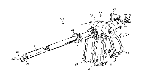
A surgical instrument according to a first preferred embodiment of the
present invention is illustrated at 30 in Fig. 1A and includes cylindrical
barrel, or
outer shaft, 32 which has an elongated passage defined therein, driver 40, and
driver 50. Driver 40 and driver 50 are substantially contained within
cylindrical
barrel 32 as is described in detail below.
As shown in Figs. 1A and 2, barrel 32 terminates distally at a distal end
which is disposed within the body cavity of a patient during use and
terminates
proximally at a proximal end which is disposed externally of the patient
during use.
As shown in Fig. 2, barrel 32 includes a plurality of operating channels 38a-d
extending longitudinally therethrough. Barrel 32 can have additional channels
for
CA 02288187 1999-10-25
WO 98/48688 PCT/US98/08406
6
receiving one or more additional instruments to be introduced in the abdominal
cavity or barrel 32 can have fewer channels as needed. Optical fibers 39
extend
through barrel 32 to transmit light from a proximal light source to the body
cavity
of a patient. Channels 38a-d can be formed by thin wall, tubular sleeves
extending longitudinally through barrel 32 or merely by void spaces defined by
optical fibers 39. Fig. 1 B illustrates that driver 40 and driver 50 can be
movable
in proximal and distal directions.
Fig 5A illustrates driver40 removed from barrel 32 for illustrative purposes.
Driver 40 includes elongated, tubular outer member 42 and elongated tubular
inner member 44 disposed within outer member 42. Outer member 42 and inner
member 24 define a shaft that is rotatable in barrel 32. Outer member 42 has a
distal end on which two diametrically enlarged flanges 46 are disposed.
Flanges
46 serve to fix collar 55, which has circumferential teeth, on outer member 42
while permitting collar 55 to rotate with respect to outer member 42. The
function
of collar 55 is described in detail below.
Fig. 5B illustrates driver 40 having pivoting jaws 50A and 50B. Fig. 5C
illustrates driver 40 having ftexible inner member 44 which can be advanced
distally to grasp a needle or other object disposed in a notch formed in outer
member 42. Fig. 5D illustrates driver 40 that is flexible and can be drawn
into
barrel 32 to be straightened. In a normal state driver 40 of Fig. 5D is
angled.
The end effectors of this embodiment are forceps and are constituted of
jaw members 50A and 50B biased to be normally disposed in an open position in
which there is a gap defined between jaw members 50A and 50B. Of course, the
inner surfaces of jaw members 50A and 50B can be shaped in any other
appropriate way to firmly grasp tissue or other objects when jaw members 50A
and 50B are in a closed position as shown in Figs. 3A, 3B, 4, and 5. The end
effectors can be of any other type including, but not limited to, cauterizing
electrodes, clip applicators, scissors, needles, biopsy devices, or the like.
Driver40 can be designed in various known ways permitting jaw members
50A and 50B to be movable between the closed position and the open position,
such as the configurations disclosed above. Jaw members 50A and 50B can be
CA 02288187 1999-10-25
WO 98148688 PCT/US98/08406
7
biased toward the open position. Arms 51 A and 51 B serve as connecting
members between jaw members 50A and 50B and inner member 44 and can be
made entirely or partly of resilient, flexible or spring materials, or
materials having
shape memory, to be resiliently biased toward the open position while being
movable to the closed position and back to the open position. Flanges 52A and
52B are respectively formed on arms 51A and 51 B. Driver 50 is similar to
driver
40 and similar elements thereof are designated with like reference numerals
having the suffix " ' ". The opening and closing movement of jaw members 50A
and 50B in this preferred embodiment is described below.
As illustrated in Fig. 1A, the proximal controls of the preferred embodiment
include two sets of scissor type handles 62 and 64 and 62' and 64', extending
from housing 89, that can be pivoted towards one another to cause movement of
the associated end effector, in this embodiment jaw members 50A and 50B and
jaw members 50A' and 50B', respectively. One set of handles is disposed on one
side of barrel 32 and the other set of handles is disposed on the other side
of
barrel 32. The operation of one set of handles 62 and 64 is discussed in
detail
below with respect to driver 40. However, the other set of handlers 62' and
64'
operate in connection with driver 50 in a similar manner.
Button 66 is provided proximate an axis of rotation of handles 62 and 64.
Depressing button 66 disengages handles 62 and 64 from driver 40 and permits
handles 62 and 64 to be rotated in concert about the axis of rotation as
indicated
by arrow A in Fig. 1 A. This allows the surgeon to orient handles 62 and 64 in
a
desired manner during surgery. Figs. 6 and 7 illustrate the internal mechanism
coupling handles 62 and 64 to driver 40 and handles 62' and 64' to driver 50.
Operating member 82 is rotatably disposed on shaft 140 and has gear portion 84
that is engaged with collar 55 on outer member 42 of driver 40. Operating
member 82 is fixed axially on shaft 80 and has radialiy extending serrated
teeth
83 formed on a side opposite gear portion 84.
Handle 62 is also rotatably mounted on shaft 80 and is slidable relative to
shaft 80. Handle 62 is fixed in axial position by projections 88 formed on an
inner
surface of housing 89. Handle 62 has serrated teeth 85 on each side thereof at
CA 02288187 1999-10-25
WO 98/48688 PCT/US98/08406
8
a top portion that is disposed around shaft 80. Shaft 80 is mounted on stem 81
and is normally biased to the right in Fig. 6 by spring 72 to press serrated
teeth
83 into engagement with serrated teeth 85 thus fixing the relative position of
operating member 82 and handle 62. Handle 64 is rotatably mounted on shaft 80
and fixed axially on shaft 80. Serrated teeth 87 are formed on each side of
handle
64 at a top portion that surrounds shaft 80 and serrated teeth 87 are normally
biased by spring 72 into engagement with teeth formed on an inner surface of
housing 89 to fix the position of handle 64 with respect to barrel 34. In this
state
handle 62 is coupled to outer member 42 of driver 40 and handle 64 is fixed in
position. Pressing handle 62 towards handle 64 will cause outer member 42 to
move over flanges 52A and 52B (see Fig. 5) to close jaws 50A and 50B.
When shaft 80 is pressed to the left in Fig.6, by depressing button 66,
serrated teeth 87 engage serrated teeth 85 to fix the relative positions of
handles
62 and 64 and serrated teeth 83 are disengaged from serrated teeth 85 to
disengage handle 62 from driver 40, as illustrated in Fig. 7. This permits the
set
of handles 62 and 64 to be rotated in concert to the desired orientation.
Button
66' is also illustrated as being depressed in Fig. 7 to illustrate the motion
of the
mechanism associated with driver 50.
As noted above, cam surfaces 52A and 52B are formed on outer surfaces
of arms 51A and 51 B respectively. When handle 62 pressed towards handle 64,
outer member 42 moves distally over cam surfaces 52A and 52B causing jaw
members 50A and 50B to move toward one another to the closed position. Cam
surfaces 52A and 52B can be formed by bent portions defined in legs 51 A and
52B or by separate elements that are attached to, or formed on, legs 51A and
51 B. Release of handles 62 and 64 causes jaw members 50A and 50B to return
to the open position due to the resilient bias of arms 51A and 51 B. Lock
protrusions 63 and 65 are disposed on handles 62 and 64 respectively (see Fig.
1A) and are serrated to interlock and allow the position of handles 62 and 64
to
be maintained in a state corresponding to a desired position of jaw members
50A
and 50B. Lock protrusions 63 and 65 can be pivoted to a position of which they
will not interlock if desired. Additionally, handles 62 and 64 can be biased
apart
CA 02288187 1999-10-25
WO 98/48688 PCTNS98/08406
9
or outer member 44 can be biased distally or proximally, depending on desired
operating characteristics.
Driver 50 is constructed similarly to driver 40 and thus further detailed
description thereof is omitted. It will be appreciated that the jaw members or
other
end effector of driver 40 and driver 50 can be of different configurations
dependent upon procedural use and other considerations such as cost. Also,
cutting elements 53 can be provided on the jaw members as needed to cut suture
material or tissue (see Fig. 4). The second set of handles 62' and 64' can be
coupled to driver 50 in a similar manner. Accordingly, control of driver 50 is
similar to that of driver 40 and further detailed description is omitted.
Also,
housing 89 and 89' can be positioned along a central transverse axis of barrel
32
and can be rotatable. In such a case an offset gear arrangement can be
provided
to couple handle 62 to collar 55.
The shafts of driver40 and driver 50 are disposed in channels 34b and 34d
respectively to extend through barrel 32 and can be rotated about their
respective
longitudinal axes relative to barrel 32 by rotating knob 48 (for driver 40) or
knob
48' (for driver 50). Push buttons 61 and 63 are respectively provided for
unlocking
knobs 48 and 48'. Also, arms 51A and 51 B of needle driver 40 can be
positioned
to extend beyond arms 51A' and 51 B' of driver 50, i.e. the transverse portion
of
the arms are in different planes, as illustrated in Fig 8, to permit the arms
to be
placed in an overlapped crossed position (illustrated in Figs. 3A and 3B). As
noted above, driver 40 and driver 50 can be movable proximally and distally.
Channel 34a and channel 34b can be used as operating channels for
suction devices, irrigation devices, or any other appropriate instrument such
as
a cautery device or the like. Also, aperture 70 is formed in a position of arm
51 B
that is proximal a distal end of inner member 44 to define an operating
channel
through driver 40 and aperture 70' is formed in arm 51 B' to define an
operating
channel through driver 50 (See Fig. 3B for example).
In use, instrument 30 is inserted into a body cavity using known techniques,
while driver 40 and driver 50 are in the position illustrated in Figs. 3A and
3B.
Note that the entire device can be inserted through a single puncture site.
Also,
CA 02288187 1999-10-25
WO 98/48688 PCT/US98/08406
in this position, jaw members 50A and 50B and 50A' and 50B', or any other
appropriate end effectors, are disposed within the diametrical dimension of
barrel
32 because the respective arms are crossed over one another. By grasping
proximal controls 60, the distal end of suturing instrument 30 is guided to
the
operative site through a portal sleeve positioned in the wall of an anatomical
cavity. The portal sleeve can be positioned in the anatomical cavity wall
using any
suitable penetrating technique, including those creating puncture sites by
means
of removable obturators, such as trocars, and can include a valve housing, if
desired, to prevent loss of pneumoperitoneum during insertion and withdrawal
of
the instrument. Further, retractable sheath 57, which is illustrated in
phantom in
Fig. 3A, (or another appropriate device) can be provided to facilitate
insertion
through a portal sleeve valve by protecting driver 50 and driver 40.
Visualization
of the endoscopic procedure can be accomplished using a conventional
endoscope incorporated into operating channel 38a, for example {known as a
single puncture procedure) or separately positioned within the anatomical
cavity
through a second portal sleeve located at another puncture site (known as a
double puncture procedure).
Prior to insertion, buttons 66 and 66' are pushed to the position illustrated
in Fig. 7 to permit the orientation of the handle sets to be adjusted as
desired.
After adjustment, buttons 66 and 66' are released and handles 62 and 64 are
set
in a desired relative position by the surgeon so that jaw members 50A and 50B
of driver 40 are in the desired position. Lock protrusions 63 and fi5 can
maintain
handles 62 and fi4 in the closed or partially closed state to permit an object
to be
securely held while freeing the surgeon's hands for other manipulation.
At any point during the operative procedure, channel 38c can be used for
irrigation or aspiration, can serve as a space for holding suture material, a
needle,
clips or the like or can be used as a portal for the introduction of other
medical
instruments such as, forceps, cutting members, ligators, orcauterydevices.
Also,
channels 38b and 38d can be used for irrigation, aspiration, insertion of an
instrument or the like by utilizing the passage through inner member 44/44' of
driver 40 and/or driver 50. Tissue can be manipulated, cut, or the like, by
CA 02288187 1999-10-25
WO 98/48688 PCT/US98/08406
11
manipulating handles 62, 64, 62', and 64' as well as knobs 48 and 48' in the
desired manner. Also, barrel 32 can be rotated to move the end effectors
From the above, it will be appreciated that the instrument according to the
present invention permits manipulation of anatomical tissue during endoscopic
procedures without the need for multiple instruments inserted through multiple
puncture sites. Driver 40 and driver 50 each are operable to move an end
effector
to manipulate or operate on anatomical tissue positioned proximate driver 40
and
driver 50, and can be moved through a large working span. While the end
effectors described above are forceps jaws, it will be understood that any end
effectors can be used. Also, any end effectors, including the forceps jaws,
can be
used as a cautery electrode by coupling an electrical power source to the end
effectorthrough electrical connector67, 6T or81 (which is illustrated in
phantom).
A surgical instrument according to a second preferred embodiment is
illustrated at 30 in Figs. 9 and 10. The second preferred embodiment includes
driver 40 and driver 50 and is similar to the first preferred embodiment
except for
the configuration of arms 51A, 51 B, 51A' and 51B' which are curved. Jaw
members 50A, 50B and 50A' and 50B' are moveably mounted on a distal end of
a respective ami to open and close in a manner similar to the jaws described
above.
As is best illustrated in Fig. 9, the arms can easily be confined within the
diametrical dimension of barrel 32 during insertion. During a procedure the
arms
can be moved, by rotating knobs 48 and 48' to cause the jaw members, or any
other appropriate end effector, to be moved through a path that is outside of
the
diametrical dimension of barrel 32. This embodiment can be used to manipulate
tissue in a mannersimiiarto the first embodiment. However, the insertion
position
of this embodiment, in which the arms and jaw members are contained within the
diametrical dimension of tubular member, does not require that the arms cross
one another. Therefore, the arms need not be disposed in different planes. The
jaw members and shafts of this embodiment can be manipulated in the same way
as the first embodiment.
CA 02288187 1999-10-25
WO 98/48688 PCT/US98/08406
12
Figs. 11 A and 11 B illustrate a third preferred embodiment. Shafts of driver
40 and driver 50 of the third preferred embodiment are offset from one another
in
both the horizontal and vertical direction as viewed in Figs. 11A and 11B.
Also,
operating channel 38a is provided in barrel 32. In other respects, the third
embodiment is similar to the first embodiment. Figs.12A and 12B illustrate a
fourth
embodiment that is similar to the third embodiment. However, the fourth
embodiment is adopted for "single puncture" procedures. Specifically,
operating
channels 38a-a are defined in barrel 36. Shafts of drivers 40 and 50 are
disposed
in operating channels 38b and 38d respectively. Operating channels 38a, c, and
a can be used for an optical endoscope for visualization and other
instruments,
such as a clip applicator or forceps, if necessary. Optical fibers 39 are
dispersed
throughout barrel 32 to direct light from a proximal light source into the
body
cavity.
A fifth embodiment is illustrated in Figs. 13A and 13B. In the fifth
embodiment, the shafts of both drivers 40 and 50 are disposed in an upper half
of barrel 32 as viewed in Figs. 13A and 13B. Operating channels 38a and 38c
are
provided for the insertion of instruments or for irrigation or aspiration. The
sixth
embodiment illustrated in Figs. 14A and 14B is similar to the fifth
embodiment.
However, in the sixth embodiment, an optical endoscope is disposed in
operating
channel 38a for viewing, operating channels 38c and 38e can accommodate
instruments, such as a clip applicator or forceps, and light transmitting
fibers 39
are provided for lighting the cavity. Alternatively fiber bundles 39',
illustrated as
dotted lines, can be provided instead of fibers 39 dispersed throughout the
cross-
sectional area of barrel 32.
In the seventh embodiment illustrated in Figs.15A and 15B the arm of
driver 50 is curved and additional operating channels 38a and c are provided.
In
the eighth embodiment illustrated in Figs. 16A and 16B, an optical endoscope
is
disposed in operating channel 38e and either distributed light fibers 39 or
fiber
bundles 39' are used for transmitting light.
Figs. 17A and 17B illustrate a ninth embodiment in which the arms of both
driver 40 and driver 50 are curved and operating channel 38a is centralized
for
CA 02288187 1999-10-25
WO 98/48688 PCT/US98/08406
13
insertion of an instrument, for suction, aspiration, or the like. The tenth
embodiment illustrated in Figs. 18A and 18B is similar to the ninth embodiment
but has optical endoscope in operating channel 38a for viewing and fiber
bundles
39' for providing fight. Of course this embodiment could have light fibers
dispersed throughout the cross-sectional area of barrel 32 instead of fiber
bundles
39'.
Figs.19A and 19B illustrate an eleventh embodiment with curved arms and
operating channels 38a and 38c that are covered by the arms when driver 40 and
driver 50 are in the insertion, or parked, position illustrated in Fig. 19A.
Centralized operating channel 38e is also provided. The twelfth embodiment
illustrated in Figs. 20A and 20B includes an optical endoscope in centralized
operating channel 38e and light fibers 39 or fiber bundles 39'.
Any appropriate proximal controls, such as those disclosed above, can be
used with the invention depending on the surgeon's preference and experience.
Fig. 21 shows a modification of the proximal controls. Handle 100 is pivotally
mounted to barrel 32 and can be locked in any desired position to facilitate
manipulation, such as in the pistol grip position illustrated in phantom. U
shaped
handle 110, having ratcheting lock device 112, extends through a slot formed
in
barrel 32 and has one leg coupled to inner member 44 and one leg coupled to
outer member 42. Compressing handle 110 thus moves outer member 42 distally
with respect to inner member 44 to lock operate the end effector of driver 40.
U-
shaped handle 120, having ratcheting device 122, is coupled to driver 50 in a
similar manner. Sliding handles 110 and 120 along respective slots 111 and 121
causes drivers 40 and 50 to move respectively in the proximal or distal
direction.
Sliding knob 330 is provided to permit movement of driver 40 and driver 50 in
concert in the proximal and the distal directions. Knobs 48 and 48' can be
rotated
to rotate driver40 and 50 respectively. Proximal apertures 90, 91, 92 and 93
are
provided for the insertion of instruments into operating channels.
Fig. 22 illustrates modified proximal controls 60 in which one set of
handles 62 and 64 are selectively coupled to driver 40 or driver 50 for
operating
end effectors. Push button 66 is used to select either driver 40 or driver 50.
CA 02288187 1999-10-25
WO 98/48688 PCT/IJS98/08406
14
Knobs 48 and 48' having locking push buttons 61, and 63 respectively are
coupled to shafts of driver40 and driver 50 to permit rotation and linear
movement
of driver 40 and apparatus 50.
Figs. 23 and 24 illustrate the internal mechanism of the proximal controls
illustrated in Fig. 22. Push button 68 is coupled to shaft 68 having gears 69
and
69' fixedly disposed thereon. In the position illustrated in Fig. 24, gear 69'
is
engaged with sleeve 55' of driver 50. Sleeve 55' is rotatably supported on
outer
member 42' of driver 55' by flanges 46'. Handles 62 and 64 are coupled to
shaft
fib to rotate shaft 68 when handles 62 and 64 are pressed together, thus
causing
relative movement between inner member 42' and outer member 44' to operate
an end effector of driver 50. Depression of push button 66 slides shaft fib
and
causes gear 69 to engage with sleeve 55 of driver 40, as illustrated in Fig.
23,
to operate an end effector of driver 40 in a similar manner. Fig. 24 also
illustrates
gears 71 and 71' that are respectively mounted on shafts of driver 40 and
driver
50. Gears 71 and 71' can slide along the shafts but cannot turn with respect
to
the shafts because of keys 73 and 73' formed on the shafts and engaged in a
keyway of the respective gears. Gears 71 and 71' are engaged respectively with
teeth formed on iriner surfaces of knobs 48 and 48'. Therefore, turning knobs
48
and 48' turns respectively the shafts of needle driver 40 and needle driver
50.
This same turning mechanism can be used for the proximal controls illustrated
in
Figs. 1 and 6. Also, the proximal control of Figs. 22-24 can have variable
orientation handles similar to those illustrated in Figs. 1 and 6. Further,
shaft 68
can be set to positions for disengaging both end effectors or for
simultaneously
operating both end effectors. Electrical connectors 67 and 81 are provided for
unipolar or bipolar cauterization.
Each of the preferred embodiments discussed above have two driver.
However, the invention can include any appropriate number of driver for
manipulating tissue or performing other procedures. Fig. 25 illustrates a
thirteenth preferred embodiment having four driver. Specifically, driver40,
driver
140, driver, driver 50, and driver 150 are each rotatably mounted in an
operating
channel defined in barrel 32. Arms of the driver lie in different planes to
permit the
CA 02288187 1999-10-25
WO 98/48688 PCT/US98/08406
driver to be placed in an insertion position similar to the embodiments
discussed
above. The operating position illustrated permits tissue to be manipulated or
other procedures to be accomplished. Of course, this embodiment can utilize
any
type of end effectors, as needed for the desired procedure. The thirteenth
embodiment is particularly suited to dual electrode cauterization, clamping
tissue
between adjacent end effectors, or separating tissue by placing adjacent end
effectors between tissue sections and moving the end effectors away from one
another. Adjacent end effectors, acting as electrodes, can be pressed against
opposing sides of tissue to cauterize the tissue. The polarity of each
electrode
can be changed to permit any two electrodes to be used in combination. An
optical endoscope can be incorporated into central channel 38f. Also,
additional
channels can be provides as needed. Of course, this embodiment can utilize
optical fibers or fiber bundles for transmitting light similar to the other
embodiments. Other aspects of this embodiment are similar to the previous
embodiments.
In each of the embodiments discussed above, two opposed jaws are
moveable toward one another. However, one of the jaw members can be fixed
and the other jaw member can be moveable. The driver can be made of flexible
or shape memory materials and can be drawn entirely into the barrel as
disclosed
in pending U.S. application Serial No. 08/847,252 entitled "Surgical
Instrument
with Rotatably Mounted Offset End Effector and Method of Using the Same", the
disclosure of which is incorporated herein by reference.
Fig. 26 illustrates an alternative driver 40 in which arm member 51A is
coupled to inner member 44 and arm member 51 B is coupled to outer member
42. Relative rotation between inner member44 and outer member42 cause jaws
50A and 50B to open/close. Rotation in concert causes the end effector 50
move.
Also, longitudinal and transverse grooves are formed in the jaws to facilitate
grasping of a needle or the like.
The two or more driver, can be of the same design or of different designs.
For example, one can have forceps jaws as an end effector and one can have a
clip applicator as an end effector. Therefore, the invention can be used for
pickup
CA 02288187 1999-10-25
WO 98/48688 PCT/US98/08406
16
and cutting, pickup and clipping, pickup and suture, or lysis of adhesion
procedures. Also, any type of end effector can be used as a cautery electrode
by
being coupled to a proximal electric power source through electrical connector
67
(see Fig. 1A). Also, a button can be provided to selectively switch the
electric
power between end effectors for unipolar cauterization. For example, button 66
can be used.
The jaw closing mechanism of the driver shown and described herein are
merely exemplary of the types of mechanisms that can be used according to the
present invention. For example, the jaw members can pivot or slide relative to
one another as disclosed in the related applications noted above and
incorporated
herein by reference. The jaw members can also carry cutting members, such as
slots with sharp edges or protruding blades, and can have opposed arcuate or
concave portions for clamping tubular objects, such as organs, without
compressing the objects. Also, only one, or more than two driver can be
provided.
The mechanisms for moving the driver relative to one another and for operating
end effectors are merely exemplary of the types of mechanisms that can be used
to perform these functions and other mechanisms can be used.
Operation of the end effectors can be automatically controlled merely by
squeezing the handles together as disclosed in the related applications
incorporated herein by reference. For example, the handle can be coupled to
one
or more end effectors to accomplish a desired procedure merely by squeezing
and releasing the handles. Squeezing the handles can perform pickup and
cutting, pickup and clipping, pickup and stapling, lysis of adhesion, or any
other
desired procedure or combination of procedures.
The components of the surgical instrument of the present invention can be
made of any suitable, medical grade materials to permit sterilization for
reuse or
disposal for single patient use. The components can be made of multiple parts
of various configurations and materials to reduce cost. The invention can have
various valves, stop-cocks and seals therein to control the flow of fluid and
medical devices through the suturing instrument.
CA 02288187 1999-10-25
WO 98/48688 PCT/US98/08406
17
In as much as the present invention is subject to many variations,
modifications and changes in detail, it is intended that all subject
matterdiscussed
above or shown in the accompanying drawings be interpreted as illustrative
only
and not be construed as limiting the scope of the invention which is defined
by the
appended claims.