Note: Descriptions are shown in the official language in which they were submitted.
CA 02288600 1999-11-02
WO 98/50431 PCT/US98/08762
A METHOD FOR MAKING MULTISPECIFIC ANTIBODIES HAVING
HETEROMULTIMERIC AND COMMON COMPONENTS
FIELD OF THE INVENTION
This invention relates to a method for making multispecificantibodies having
heteromultimericheavy
chain components and common light chain components such as bispecific
antibodies, bispecific
immunoadhesins,as well as antibody-immunoadhesinchimeras and the
heteromultimeric polypeptides made
using the method.
BACKGROUND OF THE INVENTION
BiMecific antibodies
Bispecific antibodies(BsAbs) which have binding specificities for at least two
different antigens have
significant potential in a wide range of clinical applications as targeting
agents for in vitro and in vivo
immunodiagnosis and therapy, and for diagnostic immunoassays.
In the diagnostic areas, bispecific antibodies have been very useful in
probing the functional
properties of cell surface molecules and in defining the ability of the
different Fc receptors to mediate
cytotoxicity(Fanger et al., Crit. Rev. Immunol. 12:101-124 (1992)). Nolan et
al., Biochem. Biophys. Acta.
JQ4Q:1-11 (1990) describe other diagnostic applications for BsAbs. In
particular, BsAbs can be constructed
to immobilize enzymes for use in enzyme immunoassays. To achieve this, one arm
of the BsAb can be
designed to bind to a specific epitope on the enzyme so that binding does not
cause enzyme inhibition, the
other atm of the BsAb binds to the immobilizing matrix ensuring a high enzyme
density at the desired site.
Examples of such diagnostic BsAbs include the rabbit anti-IgG/anti-ferritin
BsAb described by Hammerling
et al., J. Exp. Med. ja:1461-1473 (1968) which was used to locate surface
antigens. BsAbs having binding
specificities for horse radish peroxidase (HRP) as well as a hormone have also
been developed. Another
potential immunochemicalapplication for BsAbs involves their use in two-site
immunoassays. For example,
two BsAbs are produced binding to two separate epitopes on the analyte protein
- one BsAb binds the complex
to an insoluble matrix, the other binds an indicator enzyme (see Nolan et al.,
supra).
Bispecific antibodies can also be used for in vitro or in vivo immunodiagnosis
of various diseases
such as cancer (Songsivilai et al., Clin. Exp. Immunol. n:315 (1990)). To
facilitate this diagnostic use of the
BsAb, one arm of the BsAb can bind a tumor associated antigen and the other
arm can bind a detectable
marker such as a chelator which tightly binds a radionuclide. Using this
approach, Le Doussal et al. made a
BsAb useful for radioimmunodetectionof colorectal and thryoid carcinomas which
had one arm which bound
a carcinoembryonicantigen (CEA) and another arm which bound
diethylenetriaminepentacetic acid (DPTA).
See Le Doussal et al., Int. J. Cancer Suppl. 2:58-62 (1992) and Le Doussal et
al., J. Nucl. Med. 24:1662-1671
(1993). Stickney et al. similarly describe a strategy for detecting colorectal
cancers expressing CEA using
radioimmunodetection. These investigators describe a BsAb which binds CEA as
well as
hydroxyethylthiourea-benzyl-EDTA (EOTUBE). See Stickney et al., Cancer Res.
5,1:6650-6655 (1991).
Bispecific antibodies can also be used for humantherapy in redirected
cytotoxicity by providing one
arm which binds a target (e.g. pathogen or tumor cell) and another arm which
binds a cytotoxic trigger
-1-
CA 02288600 1999-11-02
WO 98/50431 PCT/US98/08762
molecule, such as the T-cell receptor or the Fcy receptor. Accordingly,
bispecific antibodies can be used to
direct a patient's cellular immune defense mechanisms specifically to the
tumor cell or infectious agent. Using
this strategy, it has been demonstrated that bispecific antibodies which bind
to the FcyRIll (i.e. CD16) can
mediate tumor cell killing by natural killer (NK) cell/large granular
lymphocyte (LGL) cells in vitro and are
effective in preventing tumor growth in vivo. Segal et al., Chem. Immunol.
47:179 (1989) and Segal et al.,
Biologic Therapy of Cancer 2(4) DeVita et al. eds. J.B. Lippincott,
Philadelphia (1992) p. 1. Similarly, a
bispecific antibody having one arm which binds FcyRIll and another which binds
to the HER2 receptor has
been developed for therapy of ovarian and breast tumors that overexpress the
HER2 antigen. (Hseih-Ma et
al. Cancer Research 52:6832-6839(1992) and Weineretal. Cancer Research U:94-
100 (1993)). Bispecific
antibodies can also mediate killing by T cells. Normally, the bispecific
antibodies link the CD3 complex on
T cells to a tumor-associatedantigen. A fully humanized F(ab')2 BsAb
consisting of anti-CD3 linked to anti-
p185HER2 has been used to target T cells to kill tumor cells overexpressingthe
HER2 receptor. Shalaby et al.,
J. Exp. Med. 175 :217 (1992). Bispecific antibodies have been tested in
several early phase clinical trials
with encouraging results. In one trial, 12 patients with lung, ovarian or
breast cancer were treated with
infusions of activated T-lymphocytes targeted with an anti-CD3/anti-tumor
(MOC31) bispecific antibody.
deLeij et al. Bisnecific Antibodies and Targeted Cellular Cvtotoxicitv, Romet-
Lemonne, Fanger and Segal
Eds., Lienhart (1991) p. 249. The targeted cells induced considerable local
lysis of tumor cells, a mild
inflammatory reaction, but no toxic side effects or anti-mouse antibody
responses. In a very preliminary trial
of an anti-CD3/anti-CD19 bispecific antibody in a patient with B-cell
malignancy, significant reduction in
peripheral tumor cell counts was also achieved. Clark et al. Bispecific
Antibodies and Targeted Cellular
Cytotoxicity, Romet-Lemonne,Fanger and Segal Eds., Lienhart (1991) p. 243. See
also Kroesen et al., Cancer
lmmunoI. Immunother. 37:400-407 (1993), Kroesen et al., Br. J. Cancer 70:652-
661 (1994) and Weiner et
al., J. Immunol. ,152:2385 (1994) concerning therapeutic applications for
BsAbs.
Bispecific antibodiesmay also be used as fibrinolytic agents or vaccine
adjuvants. Furthermore,these
antibodies may be used in the treatment of infectious diseases (e.g. for
targeting of effector cells to virally
infected cells such as HIV or influenza virus or protozoa such as Toxoplasma
gondii), used to deliver
immunotoxins to tumor cells, or target immune complexes to cell surface
receptors (see Fanger et al., supra).
Use of BsAbs has been effectively hindered by the difficultyof obtaining BsAbs
in sufficient quantity
and purity. Traditionally,bispecific antibodies were made using hybrid-
hybridomatechnology (Millstein and
Cuello, Nature 05:537-539 (1983)). Because of the random assortment of
immunoglobulin heavy and light
chains, these hybridomas (quadromas) produce a potential mixture of 10
different antibody molecules, of
which only one has the correct bispecific structure (see Fig. IA). The
purification of the correct molecule,
which is usually done by aff'inity chromatography steps, is rather cumbersome,
and the product yields are low.
See, for example, (Smith, W., et al. (1992) Hybridoma4:87-98; and Massimo,
Y.S., et al. (1997) J. ]mmunol.
Methods ~j0 ,:57-66). Accordingly, techniques for the production of greater
yields of BsAb have been
developed. To achieve chemical coupling of antibody fragments, Brennan et al.,
Science 229:81 (1985)
describe a procedure wherein intact antibodies are proteolyticallycleaved to
generate F(ab')2 fragments. These
fragments are reduced in the presence of the dithiol complexing agent sodium
arsenite to stabilize vicinal
-2-
CA 02288600 1999-11-02
WO 98/50431 PCT/US98/08762
dithiols and prevent intermolecular disulfide formation. The Fab' fragments
generated are then converted to
thionitrobenzoate (TNB) derivatives. One of the Fab'-TNB derivatives is then
reconverted to the Fab'-thiol
by reduction with mercaptoethylamine and is mixed with an equimolar amount of
the other Fab'-TNB
derivativeto form the BsAb. The BsAbs produced can be used as agents for the
selective immobilization of
enzymes.
Recent progress has facilitatedthe direct recovery of Fab'-SH fragments from
E. coli. which can be
chemically coupled to form bispecific antibodies. Shalaby et al., J. Exp. Med.
l 7~:217-225 (1992) describe
the production of a fully humanized BsAb F(ab')2 molecule having one arm which
binds p185HER2 and
another ann which binds CD3. Each Fab' fragment was separately secreted from
E. coli. and subjected to
directed chemical coupling in vitro to form the BsAb. The BsAb thus formed was
able to bind to cells
overexpressing the HER2 receptor and normal human T cells, as well as trigger
the lytic activity of human
cytotoxic lymphocytes against human breast tumor targets. See also Rodrigues
et al., Int. J. Cancers (Suppl.)
7:45-50 (1992).
Various techniques for making and isolating BsAb fragments directly from
recombinant cell cultures
have also been described. For example, bispecific F(ab')2 heterodimers have
been produced using leucine
zippers (Kostelny et al., J. Immunol. 148(5):1547-1553 (1992)). The leucine
zipper peptides from the Fos and
Jun proteins were linked to the Fab' portions of anti-CD3 and anti-interleukin-
2 receptor (IL-2R) antibodies
by gene fusion. The antibody homodimers were reduced at the hinge region to
form monomers and then
reoxidized to form the antibody heterodimers. The BsAbs were found to be
highly effective in recruiting
cytotoxic T cells to lyse HuT-102 cells in vitro. The advent of the "diabody"
technology described by
Hollingeret al., PNAS (USA) 2Q:6444-6448 (1993) has provided an alternative
mechanism for making BsAb
fragments. The fragments comprise a heavy chain variable domain (VH) connected
to a light chain variable
domain (VL) by a linker which is too short to allow pairing between the two
domains on the same chain.
Accordingly,the VH and VL domains of one fragment are forced to pair with the
complementary VL and VH
domains of another fragment, thereby forming two antigen-binding sites.
Another strategy for making BsAb
fragments by the use of single chain Fv (sFv) dimers has also been reported.
See Gruber et al. J. Immunol.
52: 5368 (1994). These researchers designed an antibody which comprised the VH
and VL domains of an
antibody directed against the T cell receptor joined by a 25 amino acid
residue linker to the VH and VL
domains of an anti-fluoresceinantibody. The refolded molecule bound to
fluorescein and the T cell receptor
and redirected the lysis of human tumor cells that had fluorescein covalently
linked to their surface.
It is apparent that several techniques for making bispecific antibody
fragments which can be
recovered directly from recombinant cell culture have been reported. However,
full length BsAbs may be
preferable to BsAb fragments for many clinical applicationsbecause of their
likely longer serum half-life and
= possible effector functions.
Immunoadhesins
Immunoadhesins(Ia's) are antibody-like molecules which combine the binding
domain of a protein
such as a cell-surface receptor or a ligand (an "adhesin") with the effector
functions of an immunoglobulin
constant domain. Immunoadhesins can possess many of the valuable chemical and
biological properties of
-3-
CA 02288600 1999-11-02
WO 98/50431 PCT/US98/08762
human antibodies. Since immunoadhesinscan be constructed from a human protein
sequence with a desired
specificity linked to an appropriate human immunoglobulin hinge and constant
domain (Fc) sequence, the
binding specificity of interest can be achieved using entirely human
components. Such immunoadhesins are
minimally immunogenic to the patient, and are safe for chronic or repeated
use.
Immunoadhesins reported in the literature include fusions of the T cell
receptor (Gascoigne et al.,
Proc. Natl. Acad. Sci. USA ~4:2936-2940 (1987)); CD4 (Capon et al., Nature
2L7:525-531(1989); Traunecker
et al., Nature 339:68-70 (1989); Zettmeissl et al., DNA Cell Biol. USA 9:347-
353 (1990); and Byrn et al.,
Nature 344:667-670 (1990)); L-selectin or homing receptor (Watson et al., J.
Cell. Biol. H Q:2221-2229
(1990); and Watson et al., Nature 349:164167 (1991)); CD44 (Aruffo et al.,
Cell U:1303-1313 (1990));
CD28 and B7 (Linsley et al., J. Exp. Med. ,L7,~,:721-730 (1991)); CTLA-4
(Lisley el al., J. Exp. Med. 174:561-
569 (1991)); CD22 (Stamenkovic et al., Cell 6¾:1133-1144 (1991)); TNF receptor
(Ashkenazi et al., Proc.
Natl. Acad. Sci. USA $$:10535-10539 (1991); Lesslauer et al., Eur. J. Immunol.
27:2883-2886 (1991); and
Peppel et al., J. Exp. Med. 174:1483-1489 (1991)); NP receptors (Bennett et
al., J. Biol. Chem. 266:23060-
23067 (1991)); inteferon y receptor (Kurschner et al., J. Biol. Chem. 267:9354-
9360 (1992)); 4-1BB
(Chalupny et al., PNAS (USA) $9:10360-10364 (1992)) and IgE receptor a
(Ridgway and Gonnan, J. Cell.
Biol. Vol. ,115, Abstract No. 1448 (1991)).
Examples of immunoadhesins which have been described for therapeutic use
include the CD4-lgG
immunoadhesin for blocking the binding of HIV to cell-surface CD4. Data
obtained from Phase I clinical
trials in which CD4-IgG was administered to pregnant women just before
delivery suggests that this
itnmunoadhesin may be useful in the prevention of maternal-fetal transfer of
HIV. Ashkenazi et al., Intem.
Rev. Immunol. ,]Q:219-227 (1993). An immunoadhesin which binds tumor necrosis
factor (TNF) has also
been developed. TNF is a proinflammatorycytokine which has been shown to be a
major mediator of septic
shock. Based on a mouse model of septic shock, a TNF receptor immunoadhesin
has shown promise as a
candidate for clinical use in treating septic shock (Ashkenazi et al., supra).
Immunoadhesins also have non-
therapeutic uses. For example, the L-selectin receptor immunoadhesin was used
as an reagent for
histochemical staining of peripheral lymph node high endothelial venules
(HEV). This reagent was also used
to isolate and characterize the L-selectin ligand (Ashkenazi et al., supra).
If the two arms of the
immunoadhesin structure have different specificities, the immunoadhesin is
called a "bispecific
immunoadhesin" by analogy to bispecific antibodies. Dietsch et al., J.
Immunol. Methods 162:223 (1993)
describe such a bispecific immunoadhesin combining the extracellular domains
of the adhesion molecules,
E-selectin and P-selectin. Binding studies indicated that the bispecific
immunoglobulin fusion protein so
formed had an enhanced ability to bind to a myeloid cell line compared to the
monospecific immunoadhesins
from which it was derived.
Antibody-Immunoadhesin chimeras
Antibody-immunoadhesin (Ab/la) chimeras have also been described in the
literature. These
molecules combine the binding region of an immunoadhesin with the binding
dorriain of an antibody.
Berg et al., PNAS (USA) $8:4723-4727 (1991) made a bispecific antibody-
immunoadhesin chimera
which was derived from murine CD4-IgG. These workers constructed a tetrameric
molecule having two arms.
-4-
CA 02288600 1999-11-02
WO 98/50431 PCT/US98/08762
One arm was composed of CD4 fused with an antibody heavy-chain constant domain
along with a CD4 fusion
with an antibody light-chain constant domain. The other arm was composed of a
complete heavy-chain of an
anti-CD3 antibody along with a complete light-chain of the same antibody. By
virtue of the CD4-IgG ann,
this bispecific molecule binds to CD3 on the surface of cytotoxic T cells. The
juxtaposition of the cytotoxic
cells and HIV-infected cells results in specific killing of the latter cells.
While Berg et al. supra describe a bispecific molecule that was tetrameric in
structure, it is possible
to produce a trimeric hybrid moleculethat contains only one CD4-IgG fusion.
See Chamow et al., J. Immunol.
153:4268 (1994). The first arm of this construct is formed by a humanized anti-
CD3 x light chain and a
humanized anti-CD3 y heavy chain. The second arm is a CD4-IgG immunoadhesin
which combines part of
the extracellular domain of CD4 responsible for gp120 binding with the Fc
domain of IgG. The resultant
Ab/la chimera mediated killing of HI V-infected cells using either pure
cytotoxic T cell preparations or whole
peripheral blood lymphocyte (PBL) fractions that additionally included Fc
receptor-bearing large granular
lymphocyte effector cells.
In the manufacture of the multispecific antibody heteromultimers, it is
desirableto increasethe yields
of the desired heteromultimer over the homomultimer(s). The current method of
choice for obtaining Fc-
containing BsAb remains the hybrid hybridoma, in which two antibodies are
coexpressed(Milstein and Cuello,
Nature 3 05:537-540 (1983)).
In hybrid hybridomas, heavy (H) chains typically form homodimers as well as
the desired
heterodimers. Additionally, light (L) chains frequently mispair with non-
cognate heavy chains. Hence,
coexpression of two antibodies may produce up to ten heavy and light chain
pairings (Suresh, M.R., et al.
Methods Enzymol. jn:210-228 (1986)). These unwanted chain pairings compromise
the yield of the BsAb
and inevitably impose significant, and sometimes insurmountable,purification
challenges (Smith, et al. (1992)
supra; and Massimo, et al. (1997) supra).
Antibody heavy chains have previously been engineered to drive
heterodimerization by introducing
sterically complementarymutations in multimerization domains at the CH3 domain
interface (Ridgway et al.
Protein Eng. 9:617-621(1996)) and optimization by phage display as described
herein. Chains containing the
modified CH3 domains yield up to approximately 90% heterodimer as judged by
formation of an
antibody/immunoadhesin hybrid (Ab/Ia). Heterodimerized heavy chains may still
mispair with the non-
cognate light chain, thus hampering recovery of the BsAb of interest.
SUMMARY OF THE INVENTION
This application describes a strategy which serves to enhance the formation of
a desired heteromultimeric
bispecific antibody from a mixture of monomers by engineering an interface
between a first and second
polypeptide for hetero-oligomerizationand by providing a common variable light
chain to interact with each
of the heteromeric variable heavy chain regions of the bispecific antibody.
There are three possible hetero-
and homomultimers that can form from a first and second polypeptide, each of
which is, in turn, associated
with a first and second light chain, respectively. This gives rise to a total
of ten possible chain pairings (Fig.
lA). A method of enhancing the formation of the desired heteromultimer can
greatly enhance the yield over
undesired heteromultimers and homomultimers.
-5-
CA 02288600 2007-12-24
WO 98/SO431 PCT/US98108762
The preferred interface between a first and second polypeptide of the
heteromultitneric antibody
comprises at least a part ofthe CH3 domain of an antibody constant domain. The
domain of each of the first
and second polypeptides that interacts at the interface is called the
multimerization domain. Preferably, the
multimerizationdomain promotes interaction between a specific first
polypeptide and a second polypeptide,
thereby increasing the yield of desired heteromultimer(Fig. I B). Interaction
may be promoted at the interface
by the formation of protuberance-into-cavitycomplementaryregions; the
formation of non-naturallyoccurring
disulfide bonds; leucine zipper, hydrophobic regions; and hydrophilic regions.
"Protuberances" are
constructed by replacing small amino acid side chains from the interface of
the first polypeptide with larger
side chains (e.g. tyrosine or tryptophan). Compensatory "cavities" of
identical or similar size to the
protuberancesare optionallycreated on the interface of the second polypeptide
by replacing large amino acid
side chains with smaller ones (e.g. alanine or threonine). Where a suitably
positioned and dimensioned
protuberance or cavity exists at the interface of either the first or second
polypeptide, it is only necessary to
engineer a corresponding cavity or protuberance, respectively, at the adjacent
interface. Non-naturally
occurring disulfide bonds are constructed by replacing on the first
polypeptide a naturally occurring amino
acid with a free thiol-containing residue, such as cysteine, such that the
free thiol interacts with another free
thiol-containingresidue on the second polypeptide such that a disulfide bond
is formed between the first and
second polypeptides (Fig. I B).
Single chain Fv fragments from a large non-immunizedphage display
library(Vaughan, T.). et al. (1996)
Nature BiotechnologylA:309-314 ) revealed V-gene usage in
which VH and VL sequences derived from certain germiine V-gene segments
predominated. families
predominated in the repertoire. Examples of chain promiscuity in the
repertoire were noted in which a
particular heavy or light chain is found in combination with different partner
chains (Vaughan, T.J. et al.
(1996) supro).
It is disclosed herein that the preparation of a desired heteromultimeric
multispecific antibody is
enhanced when a common light chain is provided to pair with each of the
variable heavy chains of the
multispecifi c antibody. Use of a common variable light chain reduces the
number of monomers that must
correctly pair to form the antigen binding domains by limiting the number of
light chains from two or more
light chains (in a bispecificor multispecificantibody,respectively,prior to
disclosure of the instant invention)
to one light chain (in a muhtispecific antibody of the invention, see Fig. I
C).
Accordingly,the invention relates to a method of preparing a heteromultimeric
multispecific antibody,
the antibody comprising 1) a firstpolypeptideaad a second polypeptide (and
additional polypeptides accord
to the multiplicity of the antibody) which meet at an interface, wherein the
first and additional polypeptides
(i.e., a first and second polypeptide) each include a multimerization domain
forming an interface between the
first and second (or at least one additional) polypeptides, and the
multimerization domains promote stable
interaction between first and additionalpolypeptides,and 2) a binding domain
in each of the first and at least
one additional polypeptide (i.e. a second polypeptide), each binding domain
comprising a variable heavy
chain and a variable light chain, wherein the variable light chain of the
first polypeptide and the variable light
chain of the second polypeptide have a common amino acid sequence, which
common sequence has an amino
-6-
CA 02288600 1999-11-02
WO 98/50431 PCT/US98/08762
acid sequence identity to an original light chain of each of the
polypeptidesof at least 80%, preferably at least
90%, more preferably at least 95% and most preferably 100% sequence identity.
The method comprises the
steps of
(i) culturinga host cell comprising nucleic acid encoding the first
polypeptide, the second
polypeptide, and the common light chain wherein the culturing is such that the
nucleic acid is expressed; and
(ii) recovering the multispecific antibody from the host cell culture;
In a related embodiment of the invention the nucleic acid encoding the first
polypeptide or
the nucleic acid encoding the second polypeptide, or both, has been altered
from the original nucleic acid to
encode the interface or a portion thereof.
In another embodiment of the method, the interface of the first polypeptide
comprises a free thiol-
containing residue which is positioned to interact with a free thiol-
containing residue of the interface of the
second polypeptidesuch that a disulfide bond is formed between the first and
second polypeptides. According
to the invention,the nucleic acid encoding the first polypeptidehas been
altered from the original nucleic acid
to encode the free thiol-containing residue or the nucleic acid encoding the
second polypeptide has been
altered from the original nucleic acid to encode the free thiol-containing
residue, or both.
In another embodiment of the method, the nucleic acid encoding both the first
polypeptide and at least
one additional polypeptide (i.e., a second polypeptide) are altered to encode
the protuberance and cavity,
respectively. Preferably the first and second polypeptides each comprise an
antibody constant domain such
as the CH3 domain of a human IgGI.
In another aspect, the invention provides a heteromultimer (such as a
bispecific antibody, bispecific
inununoadhesin or antibody/immunoadhesinchimera) comprising a first
polypeptide and a second polypeptide
which meet at an interface. The interface of the first polypeptide comprises a
multimerization domain which
is positioned to interact with a multimerizationdomain on the at least one
additional polypeptide(t.e., a second
polypeptide)to form an interface between the first and second polypeptide. In
preferred embodiments of the
invention, the multimerizationdomains are altered to promote interaction
between a specific first polypeptide
and a specific second polypeptide, which alterations include, but are not
limited to, the generation of a
protuberance or cavity, or both; the generation of non-naturally occurring
disulfide bonds; the generation of
complementary hydrophobic regions; and the generation of complementary
hydrophilic regions. The
heteromultimeric multispecfic antibody may be provided in the form of a
composition further comprising a
pharmaceutically acceptable carrier.
The invention also relates to a host cell comprising nucleic acid encoding the
heteromultimeric
multispecific antibody of the preceding paragraph wherein the nucleic acid
encoding the first polypeptide and
at least one additional polypeptide (i.e., a second polypeptide) is present in
a single vector or in separate
vectors. The host cell can be used in a method of making a heteromultimeric
multispecific antibody which
involves culturing the host cell so that the nucleic acid is expressed, and
recovering the heteromultimeric
antibody from the cell culture.
In yet a further aspect, the invention provides a method of preparing a
heteromultimeric multispecific
antibody comprising:
-7-
CA 02288600 2007-12-24
WO 98/50431 PCT/US98/08762
(a) selecting a first nucleic acid encoding a first polypeptide comprising an,
amino acid residue
in the interface of the first polypeptidethat is positionedto interact with an
amino acid residue of interface of
at least one additional polypeptide. In an embodiment the nucleic acid is
altered from the original to encode
the interacting amino acid residues. In another embodiment, the first nucleic
acid is altered to encode an
amino acid residue having a larger side chain volume, thereby generating a
protuberance on the first
polypeptide;
(b) altering a second nucleic acid encodinga second polypeptide so that an
amino acid residue
in the interface of the second polypeptide is replaced with an amino acid
residue having a smaller side chain
volume, thereby generating a cavity in the second polypeptide, wherein the
protuberance is positioned to
interact with the cavity;
(c) introducinginto a host cell the first and second nucleic acids and
culturing the host cell so
that expression of the first and second nucleic acid occurs; and
(d) recovering the heteromultimeric antibody formed from the cell culture.
It may also be desirable to construct a multispecific antibody (such as a
bispecific antibody) that
incorporates a previously identified antibody. Under these circumstances it is
desirable to identify a heavy
chain that when paired with the original light chain will bind specifically to
a second antigen of interest. The
methods ofFigini et al. (Figini, M. et al. (1994) J. Mol. Biol. 2Q:68-78
) may be used to identify such a heavy chain. First a phage library would be
treated with guanidine
hydrochloride to dissociate the original light chain. Next, the heavy chains
displayed on phage would be
reconstituted with the light chain of interest by removing the denaturant
(such as by dialysis). Panning against
the second antigen of interest would then be conducted to identify the desired
heavy chain. The invention
further embodies a multispecific antibody prepared by this method of selecting
a heavy chain to pair with a
chosen light chain, nucleic acid encoding the antibody, and a host cell
comprising the nucleic acid.
The invention provides a mechanism for increasingthe yields of the
heteromultimerover other unwanted
end-products such as undesired heteromultimers and/or homomultimers (see Fig.
IA-1C). Preferably, the
yields of the desired heteromultimerrecovered from recombinantcell culture are
at least greater than 80% by
weight and preferably greater than 90% by weight compared to the by-product
undesired heterodimer or
homomultimer(s).
Brief Description of the Drawines
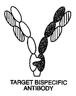
Figs. IA-iC. Fig. lA is a diagram of the formation of Fc-containing bispecific
antibodies when no
engineering is performedto enhance heteromultimerizationover
homomultimerization. Fig. I B is a diagram
showing pairing that occurs when heavy (H) chains are engineered such that
desired heteromuhimerization
is favored over undesired beteromultimerization over homomultimerization. Fig.
1C is a diagram showing
pairing that occurs when antibodiesare chosen which share the same light (L)
chain to circumventthe problem
of light chains pairing with non-cognate heavy chains.
Figs. 2A-2C. Fig. 2A diagrams a selection scheme for CH3 heterodimer using
phage display vector,
pRA2.
PhagedisplayingstableCH3heterodimersarecapturedusinganantibodydirectedtothegDfl
ag. Fig.
2B diagrams a dicistronic operon in which CH3 expressed from a synthetic gene
is co-secreted with a second
-8-
CA 02288600 1999-11-02
WO 98/50431 PCTIUS98/08762
copy of CH3 expressed from the natural gene (Ellison et al. Nucleic Acids Res.
10:4071-4079 (1982)) as a
fusion protein with M 13 gene III protein. The synthetic CH3 gene is preceded
by a sequence encoding a
peptide derived from herpes simplex virus glycoprotein D(gD flag, Lasky, L. A.
and Dowbenko, D. J. (1984)
DNA 1:23-29; Berman, P. W. et al., (1985) Science 227:1490-1492 and a cleavage
(G) site for the site-specific
protease, Genenase I (Carter, P. et al. (1989) Proteins: Structure, Function
and Genetics 6:240-248). Fig. 2C
is the nucleic acid sequence of the dicistronic operon (SEQ ID NO: 1) of Fig.
2B in which the residues in the
translated CH3 genes are numbered according to the Eu system of Kabat et al.
In Sequences of Proteins of
Immunological Interest, 5th ed. vol. 1, pp. 688-696, NIH, Bethesda, MD (1991).
Protuberance mutation
T366W is shown, as are the residues targeted for randomization in the natural
CH3 gene (366, 368, and 407).
Figs. 3A-3C. Figs. 3A and 3B are bar graphs of the results of scanning
densitometric analysis of SDS-
PAGE of protein A-purified products from cotransfection of antibody (Ab) heavy
and light chains with
immunoadhesin(la). Data presented are the mean of two independent experiments.
The x-axis indicates the
ratios of input DNA by mass (Ia:H:L) and the y-axis indicates the percentage
of each type of product multimer
with respect to total product protein. Fig. 3C is a diagram of the possible
product multimers.
Fig. 4 is a comparison of the VL sequences of eight different antibodies with
specificities for Axl, Rse,
IgER, Ob-R, and VEGF. The position of the antigen binding CDR residues
according to sequence definition
(Kabat et al. (1991) supra) or structural definition (Chothia, C. and Lesk,
A.M. J. Mol. Biol. (1987) 196:901-
917) are shown by underliningand #, respectively. Residues that differ from
the Axl.78 sequence are shown
by double underlining.
Fig. 5 is a comparison of the heavy and light chains of selected anti-Ob-R and
anti-HER3 clones. Shown
are the VH and the common VL sequences of anti-Ob-R clone 26 and anti-HER3
clone 18 used to construct
a bispecific antibody.
Fig. 6. Sandwich ELISA for detection of simultaneous binding to Mpl-IgG and
HER3-IgG. Antibodies
tested were the anti-Mpl x anti-HER3 BsIgG containing the mutations,
Y349C:T366S:L368A:Y407V/T366'W:S354'C;ogetherwith corresponding parental anti-
Mpl or anti-HER3
IgG with mutated Fc regions.
Fig. 7 is a bar graph of the results of an antibody-dependent cell-mediated
cytotoxicity (ADCC) study.
ADCC was mediated by huMAb4D5-5 (Carter, P. et al. (1992) PNAS USA $9:4285-
4289) containing either
a mutant (S354C:T366W/Y349'C:T366'S:L368'A:Y407'V) or wild-type Fc or an
isotype-matched control
antibody (E25, Presta, L.G. et al. (1993) J. Immunol. 5:2623-2632). The
antibodies (125 ng/ml) were
incubated with human peripheral blood mononuclear effector cells and SK-BR-3
target cells at the ratios
shown. Data presented are the mean of triplicate measurements and
representative of three separate
experiments.
Fig. 8 is a matrix representing the amino acid sequence identity between the
light chains of antibodies
raised to HER3 versusthe light chains of antibodiesraised to Ob-R.
Antibodieshaving light chains with 100%
sequence identity are indicated in blackened boxes. Antibodies having light
chains with 98-99% sequence
identity are indicated in white boxes. The antibody clone identity is
indicated below the matrix.
-9-
CA 02288600 2007-12-24
WO 98/50431 PCT/US98/08762
I. Definitions
In general, the following words or phrases have the indicated definitions when
used in the description,
examples, and claims:
A "heteromultimer", "heteromuhimeric polypeptide", or "heteromultimeric
multispecific antibody" is
a molecule comprising at least a first polypeptide and a second polypeptide,
wherein the second polypeptide
differs in amino acid sequence from the first polypeptide by at least one
amino acid residue. Preferably, the
heteromultimerhas bindingspecificity for at least two different ligands or
binding sites. The heteromultimer
can comprise a "heterodimer" fonmed by the first and second polypeptide or can
form higher order tertiary
structures where polypeptides in addition to the first and second polypeptide
are present. Exemplary structures
for the heteromultimer include heterodimers (e.g. the bispecific
inununoadhesin described by Dietsch et al.,
supra), heterotrimers (e.g. the Ab/la chimera described by Chamow el al.,
supra), heterotetramers (e.g. a
bispecific antibody) and further oligomeric structures.
As used herein, "multimerization domain" refers to a region of each of the
polypeptides of the
heteromultimer. The "multimerizationdomain" promotes stable interaction of the
chimeric molecules within
the heteromultimercomplex. Preferably,the multimerizationdomain promotes
interaction between a specific
first polypeptide and a specific second polypeptide, thereby enhancing the
formation of the desired
heteromultimer and substantially reducing the probability of the formation of
undesired heteromultimers or
homomultimers. The multimerizationdomains may interact via an
immunoglobulinsequence, leucine zipper,
a hydrophobic region, a hydrophilic region, or a free thiol which forms an
intermolecular disulfide bond
between the chimeric molecules of the chimeric heteromultimer. The free thiol
may be introduced into the
interface of one or more interacting polypeptides by substituting a naturally
occunring residue of the
polypeptidewith, for example, a cysteine at a position allowing for the
formation of a disulfide bond between
the polypeptides. The muhimerizationdomain may comprise an
immunoglobulinconstantregion. A possible
multimerization domain useful in the present invention is disclosed in
PCT/US90/06849
in which hybrid immunoglobulins are described. In addition a multimerization
region may be engineered such that steric interactionsnot only promote stable
interaction,but further promote
the formation of heterodimers over homodimers from a mixture of monomers. See,
for example,
PCT/US96/01598 in which a "protuberance-into-cavity"
strategy is disclosed for an interface between a first and second polypeptide
for hetero-oligomerization.
"Protuberances" are constructed by replacing small amino acid side chains from
the interface of the first
polypeptide with larger side chains (e.g. tyrosine or tryptophan).
Compensatory "cavities" of identical or
similarsize to the protuberances are optionally created on the interface of
the second polypeptideby replacing
large amino acid side chains with smaller ones (e.g. alanine or threonine).
The immunoglobulin sequence
preferably, but not necessarily, is an immunoglobulin constant domain. The
immunoglobulin moiety in the
chimeras of the present invention may be obtained from IgG I , IgG2,1gG3 or
IgG4 subtypes, IgA, IgE, IgD
or IgM, but preferably IgGI, IgG2, IgG3 or IgG4.
-10-
CA 02288600 2007-12-24
WO 98/50431 PCT/US98/08762
By "free thiol-containingcompound" is meant a compound that can be
incorporated into or reacted with
an amino acid of a polypeptide interface of the invention such that the free
thiol moiety of the compound is
positionedto interactwith a free thiol of moiety at the interface of
additional polypeptide of the invention to
form a disulfide bond. Preferably, the free thiol-containing compound is
cysteine.
The term "epitope tagged" when used herein refers to a chimeric polypeptide
comprising the entire
chimeric heteroadhesin,or a fragment thereof, fused to a "tag polypeptide".
The tag polypeptide has enough
residues to provide an epitope against which an antibody can be made, yet is
short enough such that it does
not interfere with activity of the chimeric heteroadhesin. The tag
polypeptidepreferably is fairly unique so that
the antibody thereagainst does not substantially cross-react with other
epitopes. Suitable tag polypeptides
generally have at least 6 amino acid rosidues and usually between about 8-50
amino acid residues (preferably
between about 9-30 residues). An embod'unentofthe invention encompasses a
chimericheteroadhesin linked
to an epitope tag, which tag is used to detect the adhesin in a sample or
recover the adhesin from a sample.
As used herein, "common light chain" or "common amino acid sequence of the
light chain" refers to the
amino acid sequence of the light chain in the multispecific antibody of the
invention. Panels of antibodies
were generated against at least two different antigens by panning a phage
display library such as that described
by Vaughan, et al. (1996) supra, ' with particular reference to
the method of selection of the phagemid library). The light chain sequences
were compared with respect to
the variable light chain amino acid sequences. Useful light chains from the
compared panels are those having
amino acid sequence identity of at least 80%, preferably at least 90%, more
preferably at least 95%, and most
preferably 100% identity. A common light chain sequence is a sequence designed
to be an approximation of
the two compared light chain sequences. Where the compared light chains are
100% sequence identical at the
amino acid level, the common light chain is identical to the light chains from
the selected library clones, even
though the light chain functions in a different binding domain of the
multispecific antibody. Where the
compared light chains differ as described above, the common light chain may
differ from one or the other, or
both, of the compared light chains from the library clones. ln a case in which
the common light chain differs
from one or the other, or both of the library clones, it is preferred that the
differing residues occur outside of
the antigen binding CDR residues of the antibody light chain. For example, the
position of the antigen binding
CDR residues may be determinedaccordingto a sequence definition (Kabat et al.
(1991) supra) or structural
defmition (Chothia and Lesk (1987) J. Mol. Biol. 12¾:901-917).
As used herein, "amino acid sequence identity" refers to the percentage of the
amino acids of one
sequence are the same as the amino acids of a second amino acid sequence. 100%
sequence identity between
polypeptide chains means that the chains are identical.
As used herein, "polypeptide"refers generally to peptides and proteins having
more than about ten amino
acids. Preferably, mammalian polypeptides (polypeptides that were originally
derived from a mammalian
organism) are used, more preferablythose which are directly secreted into the
medium. Examples of bacterial
polypeptides include, e.g., alkaline phosphatase and (3-lactamase. Examples of
mammalian polypeptides
include molecules such as renin, a growth hotmone, including human growth
hormone; bovine growth
hormone; growth hormone releasingfactor; parathyroidbonmone; thyroid
stimulating hormone; lipoproteins;
-11-
CA 02288600 1999-11-02
WO 98/50431 PCT/US98/08762
alpha-l-antitrypsin; insulin A-chain; insulin B-chain; proinsulin; follicle
stimulating hormone; calcitonin;
luteinizing hormone; glucagon; clotting factors such as factor VIIIC, factor
IX, tissue factor, and von
Willebrands factor; anti-clotting factors such as Protein C; atrial
natriuretic factor; lung surfactant; a
plasminogen activator, such as urokinase or human urine or tissue-type
plasminogen activator (t-PA);
bombesin; thrombin; hemopoietic growth factor; tumor necrosis factor-alpha and
-beta; enkephalinase;
RANTES (regulated on activation normally T-cell expressed and secreted); human
macrophage inflammatory
protein (MIP-1-alpha); a serum albumin such as human serum albumin; Muellerian-
inhibiting substance;
relaxin A-chain; relaxin B-chain; prorelaxin; mouse gonadotropin-
associatedpeptide; a microbial protein, such
as beta-lactamase;DNase; inhibin; activin; vascular endothelialgrowth factor
(VEGF); receptors for hormones
or growth factors; integrin; protein A or D; rheumatoid factors; a
neurotrophic factor such as bone-derived
neurotrophic factor (BDNF), neurotrophin-3, -4, -5, or -6 (NT-3, NT-4, NT-5,
or NT-6), or a nerve growth
factor such as NGF-p; platelet-derived growth factor (PDGF); fibroblast growth
factor such as aFGF and
bFGF; epidermal growth factor (EGF); transfonming growth factor (TGF) such as
TGF-alpha and TGF-beta,
including TGF-01, TGF-P2, TGF-p3, TGF-(i4, or TGF-P5; insulin-like growth
factor-I and -II (IGF-I and
IGF-II); des(1-3)-IGF-I(brain IGF-I), insulin-like growth factor binding
proteins; CD proteins such as CD-3,
CD-4, CD-8, and CD- 19; erythropoietin; osteoinductive factors; immunotoxins;
a bone morphogenetic protein
(BMP); an interferon such as interferon-alpha,-beta, and -gamma; colony
stimulating factors (CSFs), e.g., M-
CSF, GM-CSF, and G-CSF; interleukins (ILs), e.g., IL-I to IL-10; superoxide
dismutase; T-cell receptors;
surface membrane proteins; decay accelerating factor; viral antigen such as,
for example, a portion of the
AIDS envelope; transport proteins; homing receptors; addressins; regulatory
proteins; antibodies; and
fragments of any of the above-listed polypeptides.
The "first polypeptide" is any polypeptide which is to be associated with a
second polypeptide. The first
and second polypeptide meet at an "interface" (defmed below). In addition to
the interface, the first
polypeptide may comprise one or more additional domains, such as "binding
domains" (e.g. an antibody
variable domain, receptor binding domain, ligand binding domain or enzymatic
domain) or antibody constant
domains (or parts thereof) including CH2, CHI and CL domains. Normally, the
first polypeptide will comprise
at least one domain which is derived from an antibody. This domain
convenientiy is a constant domain, such
as the CH3 domain of an antibody and can form the interface of the first
polypeptide. Exemplary first
polypeptides include antibody heavy chain polypeptides, chimeras combining an
antibody constant domain
with a binding domain of a heterologouspolypeptide(i.e. an immunoadhesin, see
definition below), receptor
polypeptides (especially those which form dimers with another receptor
polypeptide, e.g., interleukin-8
receptor (IL-8R) and integrin heterodimers (e.g. LFA-1 or GPIIIb/IIIa)),
ligand polypeptides (e.g. nerve
growth factor (NGF), neurotrophin-3 (NT-3), and brain-derived neurotrophic
factor (BDNF) - see Arakawa
et al. J. Biol. Chem. 269(45): 27833-27839(1994) and Radziejewskiet al.
Biochem.32(481: 1350 (1993)) and
antibody variable domain polypeptides (e.g. diabodies). The preferred first
polypeptide is selected from an
antibody heavy chain fused to a constantdomain of an immunoglobulin,wherein
the constantdomain has been
altered at the interface to promote preferential interaction with a second
polypeptide of the invention.
-12-
CA 02288600 1999-11-02
WO 98/50431 PCT/US98/08762
The "second polypeptide" is any polypeptide which is to be associated with the
first polypeptide via
an "interface", in addition to the interface, the second polypeptide may
comprise additional domains such as
a "binding domain" (e.g. an antibody variable domain, receptor binding domain,
ligand binding domain or
enzymatic domain), or antibody constant domains (or parts thereof) including
CH2, CH1 and CL domains.
Normally, the second polypeptidewil) comprise at least one domain which is
derived from an antibody. This
domain conveniently is a constant region, such as the CH3 domain of an
antibody and can form the interface
of the second polypeptide. Exemplary second polypeptides include antibody
heavy chain polypeptides,
chimeras combining an antibody constant domain with a binding domain of a
heterologous polypeptide (i.e.
an immunoadhesin, see definition below), receptor polypeptides (especially
those which form dimers with
another receptor polypeptide, e.g., interieukin-8 receptor (IL-8R) and
integrin heterodimers (e.g. LFA- I or
GPIIIb/Illa)), ligand polypeptides(e.g. nerve growth factor (NGF),
neurotrophin-3 (NT-3), and brain-derived
neurotrophic factor (BDNF) - see Arakawa et al. J. Biol. Chem. 269(45):27833-
27839 (1994) and
Radziejewski et al. Biochem. J2L48):1350 (1993)) and antibody variable domain
polypeptides(e.g. diabodies).
The preferred second polypeptide is selected from an antibody heavy chain
fused to a constant domain of an
immunoglob ulin, wherein the constant domain has been altered at the interface
to promote preferential
interaction with a first polypeptide of the invention.
A "binding domain" comprises any region of a polypeptide which is responsible
for selectively binding
to a molecule of interest (e.g. an antigen, ligand, receptor, substrate or
inhibitor). Exemplary binding domains
include an antibody variable domain, receptor binding domain, ligand binding
domain and an enzymatic
domain. In preferred embodiments, the binding domain includes an
immunoglobulin heavy chain and light
chain. Accordingto the bispecific antibodies of the invention and the method
of making them, the light chain
for each binding domain of the bispecific antibody is a common light chain,
thereby avoiding the formation
of undesired hetenmultimers in which mispairing of heavy and light chains
occurs.
The term "antibody" as it refers to the invention shall mean a polypeptide
containing one or more
domains that bind an epitope on an antigen of interest, where such domain(s)
are derived from or have
sequence identity with the variable region of an antibody. Examples of
antibodies include full length
antibodies, antibody fragments, single chain molecules, bispecific or
bifunctional molecules, diabodies,
chimeric antibodies (e.g. humanized and PRIMATIZEDTM antibodies), and
immunoadhesins. "Antibody
fragments" include Fv, Fv', Fab, Fab', and F(ab')2 fragments.
"Humanized" forms of non-human (e.g. rodent or primate) antibodies are
specific chimeric
immunoglobulins, immunoglobulinchains or fragmentsthereofwhich contain minimal
sequence derived from
non-human immunoglobulin. For the most part, humanized antibodies are human
immunoglobulins(recipient
antibody) in which residues from a complementary determining region (CDR) of
the recipient are replaced
by residues from a CDR of a non-human species (donor antibody) such as mouse,
rat, rabbit or primate having
the desired specificity, affmity and capacity. In some instances, Fv framework
region (FR) residues of the
human immunoglobulin are replaced by corresponding non-human residues.
Furthermore, the humanized
antibody may comprise residues which are found neither in the recipient
antibody nor in the imported CDR
or framework sequences. These modifications are made to further refine and
maximize antibody performance.
-13-
CA 02288600 1999-11-02
WO 98/50431 PCT/US98/08762
In general, the humanized antibody will comprise substantially all of at least
one, and typically two, variable
domains, in which all or substantially all of the CDR regions correspond to
those of a non-human
immunoglobulinand all or substantiallyall of the FR regions are those of a
human immunoglobulin sequence.
The humanized antibody preferably also will comprise at least a portion of an
immunoglobulin constant region
(Fc), typically that of a human immunoglobulin. The humanized antibody
includes a PRIMATIZEDTM
antibody wherein the antigen-binding region of the antibody is derived from an
antibody produced by
immunizing macaque monkeys with the antigen of interest.
A "multispecific antibody" is a molecule having binding specificities for at
least two different antigens.
While such molecules normally will only bind two antigens (i.e. bispecific
antibodies, BsAbs), antibodieswith
additional specificities such as trispecific antibodies are encompassed by
this expression when used herein.
Examples of BsAbs include those with one arm directed against a tumor cell
antigen and the other arm directed
against a cytotoxic trigger molecule such as anti-FcyRI/anti-CD15, anti-
p185HER2/FcyR1II (CD16), anti-
CD3/anti-malignant B-cell (1D10), anti-CD3/anti-p185HER2, anti-CD3/anti-p97,
anti-CD3/anti-renal cell
carcinoma, anti-CD3/anti-OVCAR-3, anti-CD3/L-D1 (anti-colon carcinoma), anti-
CD3/anti-melanocyte
stimulatinghormoneanalog,anti-EGFreceptor/anti-CD3,anti-CD3/anti-CAMA1, anti-
CD3/anti-CD 19, anti-
CD3/MoV 18, anti-neural cell ahesion molecule (NCAM)/anti-CD3, anti-folate
binding protein (FBP)/anti-
CD3, anti-pan carcinoma associated antigen (AMOC-31)/anti-CD3; BsAbs with one
arm which binds
specifically to a tumor antigen and one arm which binds to a toxin such as
anti-saporin/.anti-Id-1, anti-
CD22/anti-saporin, anti-CD7/anti-saporin, anti-CD38/anti-saporin, anti-
CEA/anti-ricin A chain, anti-
interferon-a(IFN-a)/anti-hybridoma idiotype, anti-CEA/anti-vinca alkaloid;
BsAbs for converting enzyme
activated prodrugs such as anti-CD30/anti-alkaline phosphatase (which
catalyzes conversion of mitomycin
phosphate prodrug to mitomycin alcohol); BsAbs which can be used as
fibrinolytic agents such as anti-
fibrin/anti-tissue plasminogen activator (tPA), anti-fibrin/anti-urokinase-
type plasminogen activator (uPA);
BsAbs for targeting immune complexes to cell surface receptors such as anti-
low density lipoprotein
(LDL)/anti-Fcreceptor(e.,g FcyRl, FcyRII or FcyRIII); BsAbs for use in therapy
of infectious diseases such
as anti-CD3/anti-herpes simplex virus (HSV), anti-T-cell receptor:CD3
complex/anti-influenza, anti-
FcyR/anti-HIV; BsAbs for tumor detection in vitro or in vivo such as anti-
CEA/anti-EOTUBE,anti-CEA/anti-
DPTA, anti-p185HER2/anti-hapten; BsAbs as vaccine adjuvants (see Fanger et
al., supra); and BsAbs as
diagnostic tools such as anti-rabbit IgG/anti-ferritin, anti-horse radish
peroxidase (HRP)/anti-hormone, anti-
somatostatin/anti-substance P, anti-HRP/anti-FITC, anti-CEA/anti-(3-
galactosidase (see Nolan et al., supra).
Examples of trispecific antibodies include anti-CD3/anti-CD4/anti-CD37, anti-
CD3/anti-CD5/anti-CD37 and
anti-CD3/anti-CD8/anti-CD37.
As used herein, the term "immunoadhesin" designates antibody-like molecules
which combine the
"binding domain" of a heterologous protein (an "adhesin", e.g. a receptor,
ligand or enzyme) with the effector
functions of immunoglobulin constant domains. Structurally, the immunoadhesins
comprise a fusion of the
adhesin amino acid sequence with the desired binding specificity which is
other than the antigen recognition
and binding site (antigen combining site) of an antibody (i.e. is
"heterologous") and an immunoglobulin
-14-
CA 02288600 1999-11-02
WO 98/50431 PCT/US98/08762
constant domain sequence. The immunoglobulin constant domain sequence in the
immunoadhesin may be
obtained from any immunoglobulin, such as IgGI, IgG2, IgG3, or IgG4 subtypes,
IgA, IgE, IgD or IgM.
The term "ligand binding domain" as used herein refers to any native cell-
surfacereceptor or any region
or derivative thereof retaining at least a qualitative ligand binding ability,
and preferably the biological activity
of a correspondingnative receptor. In a specific embodiment, the receptor is
from a cell-surface polypeptide
having an extracellular domain which is homologous to a member of the
immunoglobulin supergenefamily.
Other typical receptors, are not members of the immunoglobulin supergenefamily
but are nonetheless
specifically covered by this definition, are receptors for cytokines, and in
particular receptors with tyrosine
kinase activity (receptor tyrosine kinases), members of the hematopoietin and
nerve growth factor receptor
superfamilies, and cell adhesion molecules, e. g. (E-, L- and P-) selectins.
The term "receptorbinding domain" is used to designate any native ligand for a
receptor, including cell
adhesion molecules, or any region or derivative of such native ligand
retaining at least a qualitative receptor
binding ability, and preferablythe biological activity of a
correspondingnative ligand. This definition, among
others, specifically includes binding sequences from ligands for the above-
mentioned receptors.
As used herein the phrase "multispecificimmunoadhesin" designates
immunoadhesins (as hereinabove
defmed) having at least two binding specificities (i.e. combining two or more
adhesin binding domains).
Multispecific immunoadhesinscan be assembled as heterodimers,heterotrirnersor
heterotetramers,essentially
as disclosed in WO 89/02922 (published 6 April 1989), in EP 314,317 (published
3 May 1989), and in U.S.
Patent No. 5,116,964 issued 2 May 1992. Preferred multispecific immunoadhesins
are bispecific. Examples
of bispecific immunoadhesins include CD41gG/TNFreceptor-IgG and CD4-IgG/L-
setectin-IgG. The last
mentioned molecule combines the lymph node binding function of the lymphocyte
homing receptor (LHR,
L-selectin), and the HIV binding function of CD4, and finds potential
application in the prevention or
treatment of HIV infection, related conditions, or as a diagnostic.
An "antibody-immunoadhesinchimera (Ab/la chimera)" comprises a molecule which
combines at least
one binding domain of an antibody (as herein defined) with at least one
immunoadhesin (as defmed in this
application). Exemplary Ab/la chimeras are the bispecific CD4-IgG chimeras
described by Berg et al., supra
and Chamow et al., supra.
The "interface" comprises those "contact" amino acid residues (or other non-
amino acid groups such as
carbohydrate groups, NADH, biotin, FAD or haem group) in the first polypeptide
which interact with one or
more "contact" amino acid residues (or other non-amino acid groups) in the
interface of the second
polypeptide. The preferred interface is a domain of an immunoglobulinsuch as a
variable domain or constant
domain (or regions thereof), however the interface between the polypeptides
forming a heteromultimeric
receptor or the interface between two or more ligands such as NGF, NT-3 and
BDNF are included within the
scope of this term. The preferred interface comprisesthe CH3 domain of an
immunoglobulinwhich preferably
is derived from an IgG antibody and most preferably a human IgG 1 antibody.
An "original" amino acid residue is one which is replaced by an "import"
residue which can have a
smaller or larger side chain volume than the original residue. The import
amino acid residue can be a naturally
occurring or non-naturallyoccurring amino acid residue, but preferably is the
former. "Naturally occurring"
-15-
CA 02288600 2007-12-24
WO 98/50431 PCT/US98/08762
amino acid residues are those residues encoded by the genetic code and listed
in Table 1 of PCT/US96/01598.
By "non-naturally occurring" amino acid residue is meant a
residue which is not encoded by the genetic code, but which is able to
covalently bind adjacent amino acid
residue(s)in the polypeptidechain. Examplesofnon-naturallyoccurring amino acid
residues are norleucine,
omithine, norvaline, homoserine and other amino acid tr.sidue analogues such
as those described in Eliman et al., Meth. Enzym. =:301-336 (1991), for
example. To generate such non-naturally occurring amino acid
residues, the proceduresof Noren et al. Science 24_4: 182 (1989) and Eliman el
al., supra can be used. Briefly,
this involves chemically activating a suppressor tRNA with a non-naturally
occurring amino acid residue
followed by in vitro transcription and translation of the RNA. The method of
the instant invention involves
replacing at least one original amino acid residue, but more than one original
residue can be replaced.
Normally, no more than the total residues in the interface of the first or
second polypeptide will comprise
original amino acid residues which are replaced. The prefen-ed original
residues for replacement are "buried".
By "buried" is meant that the residue is essentially inaccessibleto solvent.
The preferred import residue is not
cysteine to prevent possible oxidation or mispairing of disulfide bonds.
By "original nucleic acid" is meant the nucleic acid encoding a polypeptide of
interest which can be
altered to encode within the multimerization domain amino acids whose side
chains interact at the interface
between the first and second polypeptide promoting stable interaction between
the polypeptides. Such
alterations may generatewithout limitation such stable interactions as
protubetance-into-cavity, non-naturally
occurring disulfide bonds, leucinezipper, hydrophobic interactions, and
hydrophilic interations. Preferably,
the alteration is chosen which promotes specific interaction between a fnst
and second polypeptide of interest
and effectively excludes interactionsthat result in undesired heteromerpairing
or the formation of homomers.
The original or starting nucleic acid may be a naturally occurring nucleic
acid or may comprise a nucleic acid
which has been subjected to prior alteration (e.g. a humanized antibody
fragment). By "altering" the nucleic
acid is meant that the original nucleic acid is genetically engineered or
mutated by inserting, deleting or
replacing at least one codon encoding an amino acid residue of interest.
Normally, a codon encoding an
original residue is replaced by a codon encoding an import residue. Techniques
for genetically modifying a
DNA in this manner have been reviewed in Mutagenesis: a Practical Approach,
M.J. McPherson, Ed., (IRL
Press, Oxford, UK. (1991), and include site-directedmutagenesis, cassette
mutagenesis and polymerase chain
reaction (PCR) mutagenesis, for example.
The protuberance, cavity, or free thiol (such as a cysteine residue for
disulfide bond formation) can be
"introduced" into the interface of the fust or second polypeptide by synthetic
means, e.g_ by recombinant
techniques, in vitro peptide synthesis, those techniques for introducing non-
naturally occurring amino acid
residues previously described, by enzymatic or chemical coupling of peptides
or some combination of these
techniques. According, the protuberance, cavity or free thiol which is
"introduced" is "non-naturally
occurring" or "non-native", which means that it does not exist in nature or in
the original polypeptide (e.g. a
humanized monoclonal antibody).
Preferablythe import amino acid residue for forming the protuberance has a
relatively small number of
"rotamers" (e.g. about 3-6). A"rotamer" is an energetically favorable
conformation of an amino acid side
-16-
CA 02288600 1999-11-02
WO 98/50431 PCTIUS98/08762
chain. The number of rotamers of the various amino acid residues are reviewed
in Ponders and Richards, J.
Mol. Biol. 1Q3:775-791 (1987).
"Isolated" heteromultimer means heteromultimer which has been identified and
separated and/or
recovered from a component of its natural cell culture environment.
Contaminant components of its natural
environmentare materials which would interfere with diagnostic or therapeutic
uses for the heteromultimer,
and may include enzymes, hormones, and other proteinaceous or nonproteinaceous
solutes. In preferred
embodiments, the heteromultimerwill be purified (1) to greaterthan 95% by
weight of protein as determined
by the Lowry method, and most preferably more than 99% by weight, (2) to a
degree sufficient to obtain at
least 15 residues of N-tenminal or internal amino acid sequence by use of a
spinning cup sequenator, or (3)
tohomogeneitybySDS-
PAGEunderreducingornonreducingconditionsusingCoomassieblueor,preferably,
silver stain.
The heteromultimers of the present invention are generally purified to
substantial homogeneity. The
phrases "substantiallyhornogeneous", "substantially homogeneous form" and
"substantial homogeneity" are
used to indicate that the product is substantially devoid of by-products
originated from undesired polypeptide
combinations (e.g. homomultimers). Expressed in terms of purity, substantial
homogeneity means that the
amount of by-products does not exceed 10%, and preferably is below 5%, more
preferably below 1%, most
preferably below 0.5%, wherein the percentages are by weight.
The expression "control sequences" refers to DNA sequences necessary for the
expression of an operably
linked coding sequence in a particularhost organism. The control sequences
that are suitable for prokaryotes,
for example, include a promoter, optionally an operator sequence, a ribosome
binding site, and possibly, other
as yet poorly understood sequences. Eukaryoticcells are known to
utilizepromoters,polyadenylationsignals,
and enhancers.
Nucleic acid is "operably linked" when it is placed into a functional
relationship with another nucleic
acid sequence. For example, DNA for a presequence or secretory leader is
operably linked to DNA for a
polypeptide if it is expressed as a preprotein that participates in the
secretion of the polypeptide; a promoter
or enhancer is operably linked to a coding sequence if it affects the
transcription of the sequence; or a
ribosome binding site is operably linked to a coding sequence if it is
positioned so as to facilitate translation.
Generally, "operably linked" means that the DNA sequences being linked are
contiguous and, in the case of
a secretory leader, contiguous and in reading phase. However, enhancers do not
have to be contiguous.
Linking is accomplished by ligation at convenient restriction sites. If such
sites do not exist, the synthetic
oligonucleotide adaptors or linkers are used in accord with conventional
practice.
II. Preoration of the Heteromultimer
1. P=aration of the Starting Materials
As a first step, the first and second polypeptide (and any additional
polypeptides fonning the
heteromultimer)are selected. Nonnally, the nucleic acid encoding these
polypeptides needs to be isolated so
that it can be altered to encode the protuberance or cavity, or both, as
herein defmed. However, the mutations
can be introduced using synthetic means, e.g: by using a peptide synthesizer.
Also, in the case where the
import residue is a non-naturally occurring residue, the method of Noren et
al., supra is available for making
-17-
CA 02288600 1999-11-02
WO 98/50431 PCT/US98/08762
polypeptid es having such substitutions. Additionally, part of the
heteromultimer is suitably made
recombinantly in cell culture and other part(s) of the molecule are made by
those techniques mentioned above.
Techniques for isolating antibodies and preparing immunoadhesins follow.
However, it will be
appreciated that the heteromultimercan be formed from, or incorporate, other
polypeptides using techniques
which are known in the art. For example, nucleic acid encoding a polypeptide
of interest (e.g. a ligand,
receptor or enzyme) can be isolated from a cDNA library prepared from tissue
believed to possess the
polypeptide mRNA and to express it at a detectable level. Libraries are
screened with probes (such as
antibodies or oligonucleotides of about 20-80 bases) designed to identify the
gene of interest or the protein
encoded by it. Screening the cDNA or genomic library with the selected probe
may be conducted using
standard procedures as described in chapters 10-12 of Sambrook et al.,
Molecular Cloning: A Laboratory
Manual (New York: Cold Spring Harbor Laboratory Press, 1989).
(I) Antibody prgparation
Several techniques for the production of antibodies have been described which
include the traditional
hybridoma method for making monocional antibodies, recombinant techniques for
making antibodies
(including chimeric antibodies, e.g. humanized antibodies), antibody
production in transgenic animals and the
recently described phage display technology for preparing "fully human"
antibodies. These techniques shall
be described briefly below.
Polyclonal antibodies to the antigen of interest generally can be raised in
animals by multiple
subcutaneous (sc) or intraperitoneal (ip) injections of the antigen and an
adjuvant. It may be useful to
conjugate the antigen (or a fragment containing the target amino acid
sequence) to a protein that is
immunogenic in the species to be immunized, e.g., keyhole limpet hemocyanin,
serum albumin, bovine
thyroglobulin, or soybean trypsin inhibitor using a bifunctional or
derivatizing agent, for example
maleimidobenzoyl sulfosuccinimide ester (conjugation through cysteine
residues), N-hydroxysuccinimide
(through lysine residues), glutaraldehyde, succinic anhydride, SOC12, or
R'N=C=NR, where R and Ri are
different alkyl groups. Animals are immunized against the immunogenic
conjugates or derivatives by
combining 1 mg of 1 g of conjugate (for rabbits or mice, respectively) with 3
volumes of Freud's complete
adjuvant and injecting the solution intradermally at multiple sites. One month
later the animals are boosted
with 1/5 to 1/10 the original amount of conjugate in Freud's complete adjuvant
by subcutaneous injection at
multiple sites. 7 to 14 days later the animals are bled and the serum is
assayed for antibody titer. Animals are
boosted until the titer plateaus. Preferably, the animal is boosted with the
conjugate of the same antigen, but
conjugated to a differentprotein and/or through a differentcross-
iinkingreagent. Conjugates also can be made
in recombinantcell culture as protein fusions. Also, aggregating agents such
as alum are used to enhance the
immune response.
Monoclonal antibodies are obtained from a population of substantially
homogeneous antibodies using
the hybridoma method first described by Kohler and Milstein, Nature 2,.,5¾:495
(1975) or may be made by
recombinant DNA methods (Cabilly et al., U.S. Patent No. 4,816,567). In the
hybridoma method, a mouse
or other appropriate host animal, such as hamster, is immunized as hereinabove
describedto elicit lymphocytes
that produce, or are capable of producing, antibodies that will specifically
bind to the protein used for
-18-
CA 02288600 2007-12-24
WO 98/50431 PCT/US98/08762
immunization. Alternatively, lymphocytes may be immunized in vitro.
Lymphocytes then are fused with
myeloma cells using a suitable fusing agent, such as polyethylene glycol, to
form a hybridoma cell (Goding,
Monoclonal Antibodies: Principles and Practice, pp.59-103 (Academic Press,
1986)). The hybridoma cells
thus prepared are seeded and grown in a suitable culture medium that
preferably contains one or more
substances that inhibit the growth or survival of the unfused, parental
myeloma cells. For example, if the
parental myeloma celis lack the enzyme hypoxanthineguanine
phosphoribosyltransferase (HGPRT or HPRT),
the culture medium for the hybridomas typically will include hypoxanth ine,
aminopterin, and thymidine (HAT
medium), which substancespreventthe growth of HGPRT-deficient cells. Preferred
myeloma cells are those
that fuse efficiently, support stable high level expression of antibody by the
selected antibody-producing cells,
and are sensitiveto a medium such as HAT medium. Among these, preferred
myeloma cell lines are murine
myeloma lines, such as those derived from MOPC-21 and MPC-11 mouse tumors
available from the Salk
Institute Cell Distribution Center, San Diego, California USA, and SP-2 cells
available from the American
Type Culture Collection, Rockville, Maryland USA. Humanmyelomaandmouse-
humanheteromyelomacell
lines also have been described for the production of human monoclonal
antibodies (Kozbor, J. Immunol.,
LU:3001 (1984);and Brodcur et al., Monoclonal Antibody Production Techniques
and Applications, pp.51-
63, Marcel Dekker, Inc., New York, 1987). See, also, Boerner et al., J.
Immunol., 147(l):86-95 (1991) and
WO 91/17769,publishedNov 28, 1991, for techniques for the production of human
monoclonal antibodies.
Culture medium in which hybridoma cells are growing is assayed for production
of monoclonal antibodies
directed against the antigen of interest. Preferably,the binding specificity
of monoclonal antibodies produced
by hybridoma cells is determined by immunoprecipitation or by an in vitro
binding assay, such as
radioimmunoassay (RIA) or enryme-linked immunoabsorbent assay (ELISA). The
binding affinity of the
monoclonal antibody can, for example, be detetmined by the Scatchard analysis
of Munson and Pollard, Anal.
Biochem. M:220 (1980). After hybridoma cells are identified that produce
antibodies of the desired
specificity,affinity, and/or activity, the clones may be subcloned by limiting
dilution procedures and grown
by standard methods. Goding, MonoclonalAntibodies: Principles and Practice,
pp.59-104 (Academic Press,
1986). Suitable culture media for this purpose include, for example,
Dulbecco's Modified Eagle's Medium
or RPMI-1640 medium. In addition, the hybridoma cells may be grown in vivo as
ascites tumors in an animal.
The monoclonalantibodiesseoretedby the subclonesare suitably separated from
the culture medium, ascites
fluid, or serum by conventional immunoglobulin purification procedures such
as, for example, protein A-
*
Sepharose, hydroxylapatite chromatography, gel electrophoresis, dialysis, or
affmity chromatography.
Alternatively, it is now possible to produce transgenic animals (e.g. mice)
that are capable, upon
immunization, of producing a full repertoire of human antibodies in the
absence of endogenous
immunoglobulin production. For example, it has been describedthat the
homozygous deletion of the andbody
heavy chain joining region (JH) gene in chimeric and germ-line mutant mice
results in complete inhibition of
endogenous antibody production. Transferof the human germ-line
immunoglobulingene array in such germ-
line mutant mice will result in the production of human antibodies upon
antigen challenge. See, e.g.,
Jakobovits et al., Proc. Natl. Acad. Sci. USA 2Q:2551-255 (1993); Jakobovits
et al., Nature M:255-258
*-trademark _19_
CA 02288600 1999-11-02
WO 98/50431 PCT/US98/08762
(1993); Fishwild, D.M., et al. (1996) Nat. Biotech j4:845-851; and Mendez, M.
J., et al. (1997) Nat. Genetics
1,.5:146-156).
In a further embodiment, antibodies or antibody fragments can be isolated from
antibody phage libraries
generated using the techniques described in McCafferty et al., Nature, 348:552-
554 (1990), using the antigen
of interest to select for a suitable antibody or antibody fragment. Clackson
et al., Nature, 352:624-628 (1991)
and Marks et al., J. Mol. Biol., M:581-597 (1991) describe the isolation of
murine and human antibodies,
respectively, using phage libraries. Subsequent publications describe the
production of high affinity (nM
range) human antibodies by chain shuffling (Mark et a1., Bio/Technol. 1 Q:779-
783 (1992)), as well as
combinatorial infection and in vivo recombination as a strategy for
constructing very large phage libraries
(Waterhouse et al., Nuc. Acids Res., 1:2265-2266 (1993); Griffiths, A. D., et
al. (1994) EMBO J. U:3245-
3260; and Vaughan, et al. (1996) supra). Thus, these techniques are viable
alternatives to traditional
monoclonal antibody hybridoma techniques for isolation of "monoclonal"
antibodies (especially human
antibodies) which are encompassed by the present invention.
DNA encoding the antibodies of the invention is readily isolated and sequenced
using conventional
procedures (e.g., by using oligonucleotide probes that are capable of binding
specifically to genes encoding
the heavy and light chains of murine antibodies). The hybridoma cells of the
invention serve as a preferred
source of such DNA. Once isolated, the DNA may be placed into expression
vectors, which are then
transfected into host cells such as simian COS cells, Chinese hamster ovary
(CHO) cells, or myeloma cells that
do not otherwise produce immunoglobulin protein, to obtain the synthesis of
monoclonal antibodies in the
recombinant host cells. The DNA also may be modified, for example, by
substituting the coding sequence
for human heavy and light chain constant domains in place of the homologous
murine sequences, Morrison
et al., Proc. Nat. Acad. Sci. $1:6851(1984). In that manner, "chimeric"
antibodies are prepared that have the
binding specificity of an anti-antigen monoclonal antibody herein.
Methods for humanizing non-human antibodies are well known in the art.
Generally, a humanized
antibody has one or more amino acid residues introduced into it from a source
which is non-human.
Humanization can be perfonmed essentially following the method of Winter and
co-workers (Jones et al.,
Nature M:522-525 (1986); Riechmann et al., Nature 332:323-327 (1988);
Verhoeyen et al., Science
~9:1534-1536 (1988)), by substituting rodent CDRs or CDR sequences for the
corresponding sequences of
a human antibody. Accordingly, such "humanized" antibodies are chimeric
antibodies (Cabilly, supra),
wherein substantially less than an intact human variable domain has been
substituted by the corresponding
sequence from a non-human species. In practice, humanized antibodies are
typically human antibodies in
which some CDR residues, and possibly some FR residues, are substituted by
residues from analogous sites
in rodent antibodies. It is importantthat antibodiesbe humanizedwith retention
of high affinity for the antigen
and other favorable biological properties. To achieve this goal, according to
a preferred method, humanized
antibodies are prepared by a process of analysis of the parental sequences and
various conceptual humanized
products using three dimensional models of the parental and humanized
sequences. Three dimensional
immunoglobulin models are familiar to those skilled in the art. Computer
programs are available which
illustrate and display probable three-dimensional confonmational structures of
selected candidate
-20-
CA 02288600 1999-11-02
WO 98/50431 PCTIUS98/08762
immunoglobulinsequences. Inspection of these displays permits analysis of the
likely role of the residues in
the functioning of the candidate immunoglobutin sequence, i.e., the analysis
of residues that influence the
ability of the candidate immunoglobulin to bind its antigen. In this way, FR
residues can be selected and
combined from the consensus and import sequence so that the desired antibody
characteristic, such as
increased affinity for the target antigen(s), is achieved. For further details
see WO 92/22653, published Dec
23, 1992.
( i i) Immunoadhesiapreparation
Immunoglobulins (Ig) and certain variants thereof are known and many have been
prepared in
recombinant cell culture. For example, see U.S. Patent No. 4,745,055; EP
256,654; Faulkner et al., Nature
29$:286 (1982); EP 120,694; EP 125,023; Morrison, J. Immun. ,123:793 (1979);
K6hler et al., Proc. Natl.
Acad. Sci. USA 77:2197 (1980); Raso et al., Cancer Res. 41:2073 (1981);
Monrison et al., Ann. Rev.
Immunol. 2:239 (1984); Morrison, Science M:1202 (1985); Morrison et al., Proc.
Natl. Acad. Sci. USA
$1:6851 (1984); EP 255,694; EP 266,663; and WO 88/03559. Reassorted
immunoglobulin chains also are
known. See, for example, U.S. Patent No. 4,444,878; WO 88/03565; and EP 68,763
and references cited
therein.
Chimeras constructed from an adhesin binding domain sequence linked to an
appropriate
immunoglobulin constant domain sequence (immunoadhesins)are known in the art.
Immunoadhesinsreported
in the literature include fusions of the T cell receptor (Gascoigne et al.,
Proc. Natl. Acad. Sci. USA ~4:2936-
2940 (1987)); CD4 (Capon et al., Nature 337:525-531 (1989); Traunecker et al.,
Nature 339:68-70 (1989);
Zettmeissl et al., DNA Cell Biol. USA 9_:347-353 (1990); and Byrn et al.,
Nature ~4,:667-670 (1990)); L-
selectin (homing receptor) (Watson et al., J. Cell. Biol. 1,1-0:2221-2229
(1990); and Watson et al., Nature
34Q:164-167 (1991)); CD44 (Aruffo et al., Cell ¾1.:1303-1313 (1990)); CD28 and
B7 (Linsley et al., J. Exp.
Med. ,J73:721-730 (1991)); CTLA-4 (Lisley et al., J. Exp. Med. 174:561-569
(1991)); CD22 (Stamenkovic
et al., Cell ¾¾:1133-1144 (1991)); TNF receptor (Ashkenazi et al., Proc. Natl.
Acad. Sci. USA $$:10535-
10539 (1991); Lesslauer et al., Eur. J. Immunol. 22:2883-2886 (1991); and
Peppel et a1., J. Exp. Med.
174:1483-1489(1991)); and IgE receptor a (Ridgway and Gorman, J. Cell. Biol.
Vol. 1,15, Abstract No. 1448
(1991)).
The simplest and most straightforward immunoadhesin design combines the
binding domain(s) of the
adhesin (e.g. the extracellular domain (ECD) of a receptor) with the hinge and
Fc regions of an
immunoglobu iin heavy chain. Ordinarily, when preparing the immunoadhesins of
the present invention,
nucleic acid encodingthe binding domain of the adhesin will be fused C-
terminally to nucleic acid encoding
the N-terminus of an immunoglobulin constant domain sequence, however N-
terminal fusions are also
possible.
Typically, in such fusions the encoded chimeric polypeptide will retain at
least functionally active hinge,
CH2 and CH3 domains of the constant region of an immunoglobulin heavy chain.
Fusions are also made to
the C-terminus of the Fc portion of a constant domain, or immediately N-
terminal to the CHI of the heavy
chain or the correspondingregion of the light chain. The precise site at which
the fusion is made is not critical;
-21-
CA 02288600 1999-11-02
WO 98/50431 PCT/US98/08762
pariicular sites are well known and may be selected in order to optimize the
biological activity, secretion, or
binding characteristics of the Ia.
In a preferred embodiment, the adhesin sequence is fused to the N-terminus of
the Fc domain of
immunoglobulin GI (IgG i). It is possible to fuse the entire heavy chain
constant region to the adhesin
sequence. However, more preferably, a sequence beginning in the hinge region
just upstream of the papain
cleavage site which defines IgG Fc chemically(i.e. residue 216, taking the
first residue of heavy chain constant
region to be 114), or analogous sites of other immunoglobulinsis used in the
fusion. In a particularlyprefen:ed
embodiment, the adhesin amino acid sequence is fused to (a) the hinge region
and CH2 and CH3 or (b) the
CH 1, hinge, CH2 and CH3 domains, of an IgG 1, IgG ~ or IgG 3 heavy chain. The
precise site at which the
fusion is made is not critical, and the optimal site can be detenmined by
routine experimentation.
For bispecific immunoadhesins, the immunoadhesins are assembled as multimers,
and particularly as
heterodimers or heterotetramers. Generally, these assembled immunoglobulins
will have known unit
structures. A basic four chain structural unit is the form in which IgG, IgD,
and IgE exist. A four chain unit
is repeated in the higher molecular weight immunoglobulins;IgM generally
exists as a pentamer of four basic
units held together by disulfide bonds. IgA globulin, and occasionally IgG
globulin, may also exist in
multimeric fonm in serum. In the case of multimer, each of the four units may
be the same or different.
Various exemplary assembled immunoadhesins within the scope herein are
schematically diagrammed
below:
(a) ACL-ACL;
(b) ACH-[ACH, ACL-ACH, ACL-VHCH, or VLCL-ACH];
(c) ACL-ACH-[ACL-ACH, ACL-VHCH, VLCL-ACH, or VLCL-VHCH];
(d) ACL-VHCH-[ACH, or ACL-VHCH, or VLCL-ACH];
(e) VLCL-ACH-[ACL-VHCH, or VLCL-ACH]; and
(f) [A-Y]n [VLCL-VHCH]2,
wherein each A represents identical or different adhesin amino acid sequences;
VL is an immunoglobulin light chain variable domain;
VH is an immunoglobulin heavy chain variable domain;
CL is an immunoglobulin light chain constant domain;
CH is an immunoglobulin heavy chain constant domain;
n is an integer greater than 1;
Y designates the residue of a covalent cross-linking agent.
In the interests of brevity, the foregoing structures only show key features;
they do not indicate joining
(J) or other domains of the immunoglobulins,nor are disulfide bonds shown.
However, where such domains
are required for binding activity, they shall be constructed to be present in
the ordinary locations which they
occupy in the immunoglobulin molecules.
Alternatively, the adhesin sequences can be inserted between immunoglobulin
heavy chain and light
chain sequences, such that an immunoglobulin comprising a chimeric heavy chain
is obtained. In this
embodiment, the adhesin sequences are fused to the 3' end of an immunoglobulin
heavy chain in each arm of
-22-
CA 02288600 1999-11-02
WO 98/50431 PCT/US98/08762
an immunoglobulin, either between the hinge and the CH2 domain, or between the
CH2 and CH3 domains.
Similar constructs have been reported by Hoogenboom, et al., Mol. Immunol.
28:1027-1037 (1991).
An immunoglobulin light chain might be present either covalently associated to
an adhesin-
immunoglobulin heavy chain fusion polypeptide, or directly fused to the
adhesin. In the former case, DNA
encoding an immunoglobulin light chain is typically coexpressed with the DNA
encoding the adhesin-
immunoglobulinheavy chain fusion protein. Upon secretion, the hybrid heavy
chain and the light chain will
be covalently associated to provide an immunoglobulin-like structure
comprising two disulfide-linked
immunoglobulin heavy chain-light chain pairs. Methods suitable for the
preparation of such structures are,
for example, disclosed in U.S. Patent No. 4,816,567, issued 28 March 1989.
In a preferred embodiment, the immunoglobulin sequences used in the
construction of the
immunoadhesins of the present invention are from an IgG immunoglobulinheavy
chain constant domain. For
human immunoadhesins,the use of human IgG I and IgG3 immunoglobulin sequences
is preferred. A major
advantage of using IgG I is that IgG I immunoadhesins can be purified
efficiently on immobilized protein A.
In contrast, purification of IgG3 requires protein G, a significantly less
versatile medium. However, other
structural and functional properties of immunoglobulins should be considered
when choosing the Ig fusion
partner for a particular immunoadhesinconstruction. For example, the IgG3
hinge is longer and more flexible,
so it can accommodate larger "adhesin" domains that may not fold or function
properly when fused to 1gG j.
Another considerationmay be valency; IgG immunoadhesins are bivalent
homodimers, whereas lg subtypes
like IgA and IgM may give rise to dimeric or pentameric structures,
respectively, of the basic lg homodimer
unit. For inununoadhesins designed for in vivo application, the
pharmacokinetic properties and the effector
functions specified by the Fc region are important as well. Although IgG 1,
IgG2 and IgG4 all have in vivo
half-lives of 21 days, their relative potencies at activating the complement
system are different. IgG4 does not
activate complement, and IgG2 is significantlyweaker at complement activation
than IgG 1. Moreover, unlike
IgG 1, IgG2 does not bind to Fc receptors on mononuclear cells or neutrophils.
While IgG3 is optimal for
complement activation, its in vivo half-life is approximately one third of the
other IgG isotypes. Another
important consideration for immunoadhesins designed to be used as human
therapeutics is the number of
allotypic variants of the particular isotype. In general, IgG isotypes with
fewer serologically-definedallotypes
are preferred. For example, IgG I has only four serologically-defined
allotypic sites, two of which (G 1 m and
2) are located in the Fc region; and one of these sites, G1m1, is non-
immunogenic. In contrast, there are 12
serologically-definedallotypes in IgG3, all of which are in the Fc region;
only three of these sites (G3m5, 11
and 21) have one allotype which is nonimmunogenic. Thus, the potential
immunogenicity of a y3
immunoadhesin is greater than that of a y I immunoadhesin.
Immunoadhesinsare most convenientlyconstructedby fusing the cDNA sequence
encoding the adhesin
portion in-frame to an Ig cDNA sequence. However, fusion to genomic Ig
fragments can also be used (see,
e.g. Gascoigneet al., supra; Aruffo et al., Cell 61:1303-1313 (1990); and
Stamenkovic et al., Cell C¾:1133-
1144 (1991)). The latter type of fusion requires the presence of lg regulatory
sequences for expression.
cDNAs encoding IgG heavy-chain constant regions can be isolated based on
published sequences from cDNA
libraries derived from spleen or peripheral blood lymphocytes, by
hybridization or by polymerase chain
-23-
CA 02288600 2007-12-24
WO 98/50431 PCT/US98/08762
reaction (PCR) techniques. The cDNAs encoding the "adhesin" and the Ig parts
of the immunoadhesin are
inserted in tandem into a plasmid vector that directs efficient expression in
the chosen host cells.
2. GgOerating a Protuberance and/or Cavitv
As a first step to selecting original residues for fotming the protuberance
and/or cavity, the three-
dimensional structure of the heteromultimeris obtained using techniques which
are well known in the art such
as X-ray crystallography or NMR. Based on the three-dimensional structure,
those skilled in the art will be
able to identify the interface residues.
The preferred interface is the CH3 domain of an immunoglobulin constant
domain. The interface
residues of the CH3 domains of IgG, IgA, IgD, IgE and IgM have been identified
(see, for example,
PCT/US96/01598 ), including those which are optimal for
replacingwith import residues; as were the interface residues of various IgG
subtypes and "buried" residues.
The basis for engineering the CH3 interface is that X-ray crystallography has
demonstrated that the
intetmolecular association between human IgG, heavy chains in the Fc region
includes extensive
protein/protein interaction between CH3 domains whereas the glycosylated CH2
domains interact via their
carbohydrate (Deisenhofer, Biochem. 2Q:2361-2370 (1981)). In addition there
are two inter-heavy chain
disulfide bonds which are efficiently formed during antibody expression in
mammalian cells unless the heavy
chain is truncated to remove CH2 and Cjj3 domains (King et al., Biochem. J.
ZU:317 (1992)). Thus, heavy
chain assembly appears to promote disulfide bond formation rather than vice
versa. Taken together these
structural and functional data led to the hypothesisthat antibody heavy chain
association is directed by the CH3
domains. It was further speculated that the interface between CH3 domains
might be engineered to promote
fotmation of heterornultimersof differentheavy chains and hinder assembly of
correspondinghomomultimets.
The experiments described herein demonstrated that it was possible to promote
the formation of
heteromultimers over homomultimersusing this approach.Thus, it is possible to
generate a polypeptidefusion
comprising a polypeptide of interest and the CH3 domain of an antibody to form
a first or second polypeptide.
The preferred CH3 domain is derived from an IgG antibody, such as an human
IgGt.
Those interface residues which can potentially constitute candidates for
forming the protuberance or
cavity are identified. It is preferableto select "buried" residues to be
replaced. To detenninewhethera residue
is buried, the surface accessibility program of Lee et al. J. Mol. Biol. 5,-,-
i:379-400 (1971) can be used to
calculatethe solvent accessibility (SA) of residues in the interface. Then,
the SA for the residues of each of
the first and second polypeptide can be separately calculated after removal of
the other polypeptide. The
difference in SA of each residue between the monomer and dimer fon:ns of the
interface can then be calculated
using the equation: SA (dimer) - SA (monomer). This provides a list of
residues which lose SA on formation
of the dimer. The SA of each residue in the dimer is compared to the
theoretical SA of the same amino acid
in the tripeptideGly-X-Gly, where X = the amino acid of interest (Rose et al.
Science =2:834-838 (1985)).
Residues which (a) lost SA in the dimer compared to the monomer and (b) had an
SA less than 26% of that
in their corresponding tripeptide are considered as interface residues. Two
categories may be delineated:
those which have an SA < 10% compared to their convsponding tripeptide (i.e.
"buried") and those which
-24-
CA 02288600 1999-11-02
WO 98/50431 PCTiUS98/08762
have 25% > SA > 10% compared to their corresponding tripeptide (i.e.
"partially buried") (see Table 1,
below).
TABLE 1
SA Lost Monomer -- Dimer % Tripeptide
Residue Polypeptide A Polypeptide B Polypeptide A Polypeptide B
No.f
Q347 22.1 31.0 25.0 26.5
Y349 79.8 83.9 5.2 5.7
L351 67.4 77.7 3.9 2.0
S354 53.4 52.8 11.3 11.7
E357 43.7 45.3 0.4 1.3
S364 21.5 15.1 0.5 1.4
T366 29.3 25.8 0.0 0.1
L368 25.5 29.7 1.0 1.1
K370 55.8 62.3 11.5 11.0
T394 64.0 58.5 0.6 1.4
V397 50.3 49.5 13.2 11.0
D399 39.7 33.7 5.7 5.7
F405 53.7 52.1 0.0 0.0
Y407 89.1 90.3 0.0 0.0
K409 86.8 92.3 0.7 0.6
T411 4.3 7.5 12.7 9.8
t residue numbering as in IgG crystal structure (Deisenhofer, Biochemistry
2Q:2361-2370 (1981)).
The effect of replacing residues on the polypeptide chain structure can be
studied using a molecular
graphics modeling program such as the InsightTM program (Biosym Technologies).
Using the program, those
buried residues in the interface of the first polypeptidewhich have a small
side chain volume can be changed
to residues having a iarger side chain volume (i.e. a protuberance), for
example. Then, the residues in the
interface of the second polypeptide which are in proximity to the protuberance
are examined to fmd a suitable
residue for forming the cavity. Normally, this residue will have a large side
chain volume and is replaced with
a residue having a smaller side chain volume. In certain embodiments,
examination of the three-dimensional
structure of the interface will reveal a suitably positioned and dimensioned
protuberance on the interface of
-25-
CA 02288600 1999-11-02
WO 98/50431 PCT/US98/08762
the first polypeptide or a cavity on the interface of the second.polypeptide.
In these instances, it is only
necessary to model a single mutant, i.e., with a synthetically introduced
protuberance or cavity.
With respect to selecting potential original residues for replacement where
the first and second
polypeptide each comprise a CH3 domain, the CH3/CH3 interface of human IgG 1
involves sixteen residues on
each domain located on four anti-parallel p-strands which buries 1090 A2 from
each surface (Deisenhofer,
supra) and Miller, J. Mol. Biol. 216:965 (1990)). Mutations are
preferablytargeted to residues located on the
two central anti-parallel p-strands. The aim is to minimize the risk that the
protuberances which are created
can be accommodated by protruding into surrounding solvent rather than by
compensatory cavities in the
partner CH3 domain.
Once the preferred originaUimport residues are identified by molecular
modeling, the amino acid
repiacementsare introduced into the polypeptide using techniques which are
well known in the art. Normally
the DNA encoding the polypeptide is genetically engineered using the
techniques described in Mutagenesis:
a Practical ARproach, supra.
Oligonucleotide-mediated mutagenesis is a preferred method for preparing
substitution variants of
the DNA encoding the first or second polypeptide. This technique is well known
in the art as described by
Adelman et al., DNA, 2:183 (1983). Briefly, first or second polypeptide DNA is
altered by hybridizing an
oligonucleotideencoding the desired mutation to a DNA template, where the
template is the single-stranded
form of a plasmidor bacteriophagecontainingthe unaltered or native DNA
sequence of heteromultimer. After
hybridization,a DNA polymerase is used to synthesize an entire second
complementary strand of the template
that will thus incorporate the oligonucleotide primer, and will code for the
selected alteration in the
heteromultimer DNA.
Cassette mutagenesis can be performed as described Wells et al. Gene 34:315
(1985) by replacing
a region of the DNA of interest with a synthetic mutant fragment generated by
annealing complimentary
oligonucleotides. PCR mutagenesis is also suitable for making variants of the
first or second polypeptide
DNA. While the following discussion refers to DNA, it is understood that the
technique also fmds application
with RNA. The PCR technique generally refers to the following procedure (see
Erlich, Science, 2,_25 :1643-
1650 (1991), the chapter by R. Higuchi, p. 61-70).
This invention also encompasses, in addition to the protuberance or cavity
mutations, amino acid
sequence variants of the heteromultimerwhich can be prepared by introducing
appropriate nucleotide changes
into the heteromultimer DNA, or by synthesis of the desired heteromultimer
polypeptide. Such variants
include, for example, deletions from, or insertionsor substitutionsof,
residues within the amino acid sequences
of the first and second polypeptides forming the heteromultimer. Any
combination of deletion, insertion, and
substitution is made to arrive at the final construct, provided that the final
construct possesses the desired
antigen-binding characteristics. The amino acid changes also may alter post-
translational processes of the
heteromultimer, such as changing the number or position of glycosylation
sites.
A useful method for identification of certain residues or regions of the
heteromultimer polypeptides
that are preferred locations for mutagenesis is called "alanine scanning
mutagenesis," as described by
Cunningham and Wells, Science, 2~L4:1081-1085 (1989). Here, a residue or group
of target residues are
-26-
CA 02288600 1999-11-02
WO 98/50431 PCT/US98/08762
identified (e.g. charged residues such as arg, asp, his, lys, and glu) and
replaced by a neutral or negatively
charged amino acid (most preferably alanine or polyalanine) to affect the
interaction of the amino acids with
the surrounding aqueous environment in or outside the cell. Those domains
demonstrating functional
sensitivity to the substitutions then are refined by introducing further or
other variants at or for the sites of
substitution. Thus, while the site for introducingan amino acid sequence
variation is predetermined,the nature
of the mutation per se need not be predetermined.
Normally the mutations will involve conservative amino acid replacements in
non-functional regions
of the heteromultimer. Exemplary mutations are shown in Table 2.
Table 2
Original Residue Exemplary Substitutions Preferred Substitutions
Ala (A) Val; Leu; lie Val
Arg (R) Lys; Gin; Asn Lys
Asn (N) Gln; His; Lys; Arg Gln
Asp (D) Glu Glu
Cys (C) Ser Ser
Gin (Q) Asn Asn
Glu (E) Asp Asp
Gly (G) Pro; Ala Ala
His (H) Asn; Gin; Lys; Arg Arg
Ile (I) Leu; Val; Met; Ala; Phe; Leu
Norleucine
Leu (L) Norleucine; Ile; Val; Met; Ile
Ala; Phe
Lys (K) Arg; Gin; Asn Arg
Met (M) Leu; Phe; Ile Leu
Phe (F) Leu; Val; Ile; Ala; Tyr Leu
Pro (P) Ala Ala
Ser (S) Thr Thr
Thr(T) Ser Ser
Trp (W) Tyr; Phe Tyr
Tyr (Y) Trp; Phe; Thr; Ser Phe
Val (V) Ile; Leu; Met; Phe; Ala; Leu
Norleucine
-27-
CA 02288600 1999-11-02
WO 98/50431 PCT/US98/08762
- Covalent modifications of the heteromultimer polypeptides are included
within the scope of this
invention. Covalent modifications of the heteromultimer can be introduced into
the molecule by reacting
targeted amino acid residues of the heteromultimer or fragments thereof with
an organic derivatizing agent
that is capable of reacting with selected side chains or the N- or C-terminal
residues. Anothertype of covalent
modification of the heteromultimerpolypeptide included within the scope of
this invention comprises altering
the native glycosylation pattern of the polypeptide. By altering is meant
deleting one or more carbohydrate
moieties found in the original heteromultimer, and/or adding one or more
glycosylation sites that are not
present in the original heteromultimer. Addition of glycosylation sites to the
heteromultimer poiypeptide is
conveniently accomplished by altering the amino acid sequence such that it
contains one or more N-linked
glycosylation sites. The alteration may also be made by the addition of, or
substitution by, one or more serine
or threonine residues to the original heteromultimersequence (for 0-linked
glycosylation sites). For ease, the
heteromultimeramino acid sequence is preferably altered through changes at the
DNA level, particularly by
mutating the DNA encoding the heteromultimer polypeptide at preselected bases
such that codons are
generated that will translate into the desired amino acids. Another means of
increasing the number of
carbohydrate moieties on the heteromultimerpolypeptide is by chemical or
enzymatic coupling of glycosides
to the polypeptide. These methods are described in WO 87/05330 published 11
September 1987, and in Aplin
and Wriston, CRC Crit. Rev. Biochem., pp. 259-306 (1981). Removal of
carbohydrate moieties present on the
heteromultimer may be accomplished chemically or enzymatically.
Another type of covalent modification of heteromultimer comprises linking the
heteromultimer
polypeptide to one of a variety of nonproteinaceouspolymers, e.g.,
polyethylene glycol, polypropyleneglycol,
or polyoxyalkylenes,in the mannerset forth in U.S. Patent Nos. 4,640,835;
4,496,689; 4,301,144; 4,670,417;
4,791,192 or 4,179,337.
Since it is often difficult to predict in advance the characteristics of a
variant heteromultimer, it will
be appreciated that some screening of the recovered variant will be needed to
select the optimal variant.
3. Fxpression of Heteromultimer havine common light chains
Following mutation of the DNA and selection of the common light chain as
disclosed herein, the
DNA encoding the molecules is expressed using recombinanttechniqueswhich are
widely available in the art.
Often, the expression system of choice will involve a mammalian cell
expression vector and host so that the
heteromultimer is appropriatelyglycosylated(e.g. in the case of
heteromultimerscomprisingantibody domains
which are glycosylated). However, the molecules can also be produced in the
prokaryotic expression systems
elaborated below. Normally, the host cell will be transformed with DNA
encoding both the first polypeptide,
the second polypeptide, the common light chain polypeptide, and other
polypeptide(s) required to form the
heteromultimer, on a single vector or independent vectors. However, it is
possible to express the first
polypeptide, second polypeptide, and common light chain poiypeptide (the
heteromultimer components) in
independent expression systems and couple the expressed polypeptides in vitro.
The nucleic acid(s) (e.g., cDNA or genomic DNA) encoding the heteromultimer
and common light
chain is inserted into a replicable vector for further cloning (amplification
of the DNA) or for expression.
Many vectors are available. The vector components generally include, but are
not limited to, one or more of
-28-
CA 02288600 1999-11-02
WO 98/50431 PCT/US98/08762
the following: a signal sequence, an origin of replication, one or more marker
genes, an enhancer element,
a promoter, and a transcription termination sequence.
The polypeptides of the heteromultimer components may be produced as fusion
polypeptides with
a signal sequence or other polypeptide having a specific cleavage site at the
N-terminus of the mature protein
or polypeptide. In general, the signal sequence may be a component of the
vector, or it may be a part of the
DNA that is inserted into the vector. The heterologous signal sequence
selected preferably is one that is
recognized and processed (i.e., cleaved by a signal peptidase) by the host
cell. For prokaryotic host cells, the
signal sequence may be substituted by a prokaryotic signal sequence selected,
for example, from the group
of the alkaline phosphatase, penicillinase, lpp, or heat-stable enterotoxin II
leaders. For yeast secretion the
native signal sequence may be substituted by, e.g., the yeast invertase
leader, alpha factor leader (including
Saccharomycesand Kltryveromyces a-factor leaders, the latter described in U.S.
Pat. No. 5,010,182 issued 23
April 1991), or acid phosphatase leader, the C. albicans glucoamylase leader
(EP 362,179 published 4 April
1990), or the signal described in WO 90/13646 published 15 November 1990. In
mammalian cell expression
the native signal sequence (e.g., the antibody or adhesin presequence that
normally directs secretion of these
molecules from human cells in vivo) is satisfactory, although other mammalian
signal sequences may be
suitable as well as viral secretory leaders, for example, the herpes simplex
gD signal. The DNA for such
precursorregion is ligated in reading frame to DNA encoding the polypeptides
forming the heteromultimer.
Both expression and cloning vectors contain a nucleic acid sequence that
enables the vector to
replicate in one or more selected host cells. Generally, in cloning vectors
this sequence is one that enables the
vector to replicate independently of the host chromosomal DNA, and includes
origins of replication or
autonomously replicating sequences. Such sequences are well known for a
variety of bacteria, yeast, and
viruses. The origin of replication from the plasmid pBR322 is suitable for
most Gram-negative bacteria, the
211 plasmid origin is suitable for yeast, and various viral origins (SV40,
polyoma, adenovirus, VSV or BPV)
are useful for cloning vectors in mammalian cells. Generally, the origin of
replication component is not
needed for mammaiian expression vectors (the SV40 origin may typically be used
only because it contains
the early promoter).
Expression and cloning vectors should contain a selection gene, also termed a
selectable marker.
Typical selection genes encode proteins that (a) confer resistance to
antibioticsor other toxins, e.g., ampicillin,
neomycin, methotrexate, or tetracycline, (b) complement auxotrophic
deficiencies, or (c) supply critical
nutrients not available from complex media, e.g., the gene encoding D-alanine
racemase for Bacilli. One
example of a selection scheme utilizes a drug to arrest growth of a host cell.
Those cells that are successfully
transformed with a heterologous gene produce a protein conferring drug
resistance and thus survive the
selection regimen. Examples of such dominant selection use the drugs neomycin
(Southern et al., J. Molec.
Appl. Genet. j:327 (1982)), mycophenolic acid (Mulligan el al., Science
M2:1422 (1980)) or hygromycin
(Sugden et al., Mol. Cell. Biol. 5:410-413 (1985)). The three examples given
above employ bacterial genes
under eukaryotic control to convey resistance to the appropriate drug G418 or
neomycin (geneticin), xgpt
(mycophenolic acid), or hygromycin, respectively.
-29-
CA 02288600 1999-11-02
WO 98/50431 PCT/US98/08762
Another example of suitable selectable markers for mammalian cells are those
that enable the
identification of cells competent to take up the heteromultimer nucleic acid,
such as DHFR or thymidine
kinase. The mammalian cell transfotmantsare placed under selection pressure
that only the transformants are
uniquely adapted to survive by virtue of having taken up the marker. Selection
pressure is imposed by
culturing the transformants under conditions in which the concentration of
selection agent in the medium is
successively changed, thereby leading to amplification of both the selection
gene and the DNA that encodes
heteromultimer. Increased quantities of heteromultimer are synthesized from
the amplified DNA. Other
examples of amplifiable genes include metallothionein-I and -II, preferably
primate metallothionein genes,
adenosine deaminase, ornithine decarboxylase, etc.
For example, cells transformed with the DHFR selection gene are first
identified by culturing all of
the transfotmantsin a culture medium that contains methotrexate (Mtx), a
competitive antagonist of DHFR.
An appropriate host cell when wild-type DHFR is employed is the Chinese
hamster ovary (CHO) cell line
deficient in DHFR activity, prepared and propagated as described by Urlaub and
Chasin, Proc. Natl. Acad. Sci.
USA 77:4216 (1980). The transfotmed cells are then exposed to increased levels
of methotrexate. This leads
to the synthesis of multiple copies of the DHFR gene, and, concomitantly,
multiple copies of other DNA
comprising the expression vectors, such as the DNA encoding the components of
the heteromultimer. This
amplification technique can be used with any otherwise suitable host, e.g.,
ATCC No. CCL61 CHO-K1,
notwithstanding the presence of endogenous DHFR if, for example, a mutant DHFR
gene that is highly
resistant to Mtx is employed (EP 117,060).
Altetnatively, host cells (particularly wild-type hosts that contain
endogenous DHFR) transformed
or co-transformed with DNA sequences encoding heteromultimer, wild-type DHFR
protein, and another
selectable marker such as aminoglycoside 3'-phosphotransferase (APH) can be
selected by cell growth in
medium containing a selection agent for the selectable marker such as an
aminoglycosidic antibiotic, e.g.,
kanamycin, neomycin, or G418. See U.S. Patent No. 4,965,199.
A suitable selection gene for use in yeast is the trpl gene present in the
yeast plasmid YRp7
(Stinchcomb et al., Nature 282:39 (1979); Kingsman et al., Gene 7:141 (1979);
or Tschemper et al., Gene
jQ:157 (1980)). The trpl gene provides a selection marker for a mutant strain
of yeast lacking the ability to
grow in tryptophan, for example, ATCC No. 44076 or PEP4-1(Jones, Genetics $
5:12 (1977)). The presence
of the trpl lesion in the yeast host cell genome then provides an effective
environment for detecting
transformationby growth in the absence of tryptophan. Similarly,Leu2-
deficientyeast strains (ATCC 20,622
or 38,626) are complemented by known plasmids bearing the Leu2 gene.
In addition, vectors derived from the 1.6 m circular plasmid pKDI can be used
for transfonnation
of Kluyveromyces yeasts. Bianchi et al., Curr. Genet. 12:185 (1987). More
recently, an expression system
for large-scale production of recombinant calf chymosin was reported for K.
lactis. Van den Berg,
Bio/Technology $:135 (1990). Stable multi-copy expression vectors for
secretion of mature recombinant
human serum albumin by industrial strains of Kluyveromyces have also been
disclosed (Fleer et al.,
Bio/Technology 2:968-975 (1991)).
-30-
CA 02288600 1999-11-02
WO 98/50431 PCTIUS98/08762
Expression and cloning vectors usually contain a promoter that is recognized
by the host organism
and is operably linked to the heteromultimer nucleic acid. A large number of
promoters recognized by a
variety of potential host cells are well known. These promoters are operably
linked to heteromultimer-
encoding DNA by removing the promoter from the source DNA by restriction
enzyme digestion and inserting
the isolated promoter sequence into the vector.
Promoters suitable for use with prokaryotic hosts include the P-lactamase and
lactose promoter
systems (Chang et al., Nature M:615 (1978); and Goeddel et al., Nature 2$1:544
(1979)), alkaline
phosphatase, a tryptophan (trp) promoter system (Goeddel, Nucleic Acids Res.,
$:4057 (1980) and EP 36,776)
and hybrid promoters such as the tac promoter (deBoer et al., Proc. Natl.
Acad. Sci. USA $Q:21-25 (1983)).
However, other known bacterial promoters are suitable. Their nucleotide
sequences have been published,
thereby enabling a skilled worker operably to ligate them to DNA encoding the
heteromultimer (Siebenlist
et al., Cell 2Q:269 (1980)) using linkers or adaptors to supply any required
restriction sites. Promoters for use
in bacterial systems also will contain a Shine-Dalgarno(S.D.) sequence
operably linked to the DNA encoding
the heteromultimer.
Promoter sequences are known for eukaryotes. Virtually all eukaryotic genes
have an AT-rich region
located approximately25 to 30 bases upstream from the site where transcription
is initiated. Another sequence
found 70 to 80 bases upstream from the start of transcription of many genes is
a CXCAAT region where X
may be any nucleotide. At the 3' end of most eukaryotic genes is an AATAAA
sequence that may be the
signal for addition of the poly A tail to the 3' end of the coding sequence.
All of these sequences are suitably
inserted into eukaryotic expression vectors.
Examples of suitable promoting sequences for use with yeast hosts include the
promoters for 3-
phosphoglyceratekinase (Hitzeman et a1., J. Biol. Chem. =:2073 (1980)) or
other glycolytic enzymes (Hess
et al., J. Adv. Enzyme Reg. 1:149 (1968); and Holland, Biochemistry 1,7:4900
(1978)), such as enolase,
glyceraldehyde-3-phosphate dehydrogenase, hexokinase, pyruvate decarboxylase,
phosphofructokinase,
glucose-6-phosphate isomerase, 3-phosphoglycerate mutase, pyruvate kinase,
triosephosphate isomerase,
phosphoglucose isomerase, and glucokinase.
Other yeast promoters, which are induciblepromoters having the
additionaladvantageof transcription
controlled by growth conditions, are the promoterregions for alcohol
dehydrogenase2, isocytochromeC, acid
phosphatase, degradative enzymes associated with nitrogen metabolism,
metallothionein, glyceraldehyde-3-
phosphate dehydrogenase, and enzymes responsible for maltose and galactose
utilization. Suitable vectors
and promoters for use in yeast expression are further described in Hitzeman et
al., EP 73,657A. Yeast
enhancers also are advantageously used with yeast promoters.
Heteromult imer transcription from vectors in mammalian host cells is
controlled, for example, by
promoters obtained from the genomes of viruses such as polyoma virus, fowlpox
virus (UK 2,211,504
published 5 July 1989), adenovirus (such as Adenovirus 2), bovine papilloma
virus, avian sarcoma virus,
cytomegalovirus, a retrovirus, hepatitis-B virus and most preferably Simian
Virus 40 (SV40), from
heterologous mammalian promoters, e.g., the actin promoter or an
immunoglobulin promoter or from heat-
shock promoters.
-31-
CA 02288600 1999-11-02
WO 98/50431 PCT/US98/08762
The early and late promoters of the SV40 virus are conveniently obtained as an
SV40 restriction
fragmentthat also contains the SV40 viral origin of replication. Fiers et al.,
Nature 273:113 (1978); Mulligan
and Berg, Science (~9:1422-1427 (1980); Pavlakis et al., Proc. Natl. Acad.
Sci. USA 7$:7398-7402 (1981).
The immediate early promoter of the human cytomegalovirus is conveniently
obtained as a HindIlI E
restriction fragment. Greenaway et al., Gene J,$:355-360 (1982). A system for
expressing DNA in
mammalian hosts using the bovine papilloma virus as a vector is disclosed in
U.S. Patent No. 4,419,446. A
modificationofthis system is described in U.S. Patent No. 4,601,978. See also
Gray et al., Nature 295:503-
508 (1982) on expressingcDNA encoding immune interferon in monkey cells; Reyes
et al., Nature 227:598-
601 (1982) on expression of human P-interferon cDNA in mouse cells under the
control of a thymidine kinase
promoter from herpes simplex virus; Canaani and Berg, Proc. Natl. Acad. Sci.
USA 2_9:5166-5170 (1982) on
expression of the human interferon (31 gene in cultured mouse and rabbit
cells; and Gorman et al., Proc. Natl.
Acad. Sci. USA ZQ:6777-6781(1982) on expression of bacterial CAT sequences in
CV-1 monkey kidney cells,
chicken embryo fibroblasts, Chinese hamster ovary cells, HeLa cells, and mouse
NIH-3T3 cells using the Rous
sarcoma virus long terminal repeat as a promoter.
Transcription of DNA encoding the heteromultimer components by higher
eukaryotes is often
increased by inserting an enhancer sequence into the vector. Enhancers are
relatively orientation and position
independent, having been found 5' (Laimins et al., Proc. Natl. Acad. Sci. USA
11:993 (1981)) and 3' (Lusky
et al., Mol. Cell Bio. 3:1108 (1983)) to the transcription unit, within an
intron (Banerji et al., Cell 31:729
(1983)), as well as within the coding sequence itself (Osborne et al., Mol.
Cell Bio. 4:1293 (1984)). Many
enhancer sequences are now known from mammalian genes (globin, elastase,
albumin, a-fetoprotein, and
insulin). Typically, however, one will use an enhancer from a eukaryotic cell
virus. Examples include the
SV40 enhancer on the late side of the replication origin (bp 100-270), the
cytomegalovirus early promoter
enhancer, the polyoma enhancer on the late side of the replication origin, and
adenovirus enhancers. See also
Yaniv, Nature 7:17-18 (1982) on enhancing elements for activation of
eukaryotic promoters. The enhancer
may be spliced into the vector at a position 5' or 3' to the heteromultimer-
encodingsequence, but is preferably
located at a site 5' from the promoter.
Expression vectors used in eukaryotic host cells (yeast, fungi, insect, plant,
animal, human, or
nucleated cells from other multicellularorganisms) will also contain sequences
necessary for the termination
of transcription and for stabilizing the mRNA. Such sequences are commonly
available from the 5' and,
occasionally 3', untranslatedregions of eukaryotic or viral DNAs or cDNAs.
These regions contain nucleotide
segments transcribed as polyadenylated fragments in the untranslated portion
of the mRNA encoding the
heteromultimer.
Construction of suitable vectors containing one or more of the above listed
components employs
standard ligation techniques. Isolated plasmids or DNA fragments are cleaved,
tailored, and religated in the
form desired to generate the plasmids required.
For analysis to confirm correct sequences in plasmids constructed, the
ligation mixtures are used to
transform E. coli K12 strain 294 (ATCC 31,446) and successful transformants
selected by ampicillin or
tetracycline resistance where appropriate. Plasmids from the transformants are
prepared, analyzed by
-32-
CA 02288600 1999-11-02
WO 98/50431 PCT/US98/08762
restriction endonuclease digestion, and/or sequenced by the method of Messing
et al., Nucleic Acids Res.
2:309 (1981) or by the method of Maxam et al., Methods in Enzymology ¾,5:499
(1980).
Particularly useful in the practice of this invention are expression vectors
that provide for the transient
expression in mammalian cells of DNA encoding heteromultimer. In general,
transient expression involves
the use of an expression vector that is able to replicate efficiently in a
host cell, such that the host cell
accumulates many copies of the expression vector and, in turn, synthesizeshigh
levels of a desired polypeptide
encoded by the expression vector. Sambrook et al., supra, pp. 16.17 - 16.22.
Transient expression systems,
comprising a suitable expression vector and a host cell, allow for the
convenient positive identification of
polypeptides encoded by cloned DNAs, as well as for the rapid screening of
heteromultimers having desired
binding specificities/affinities or the desired gel migration characteristics
relative to heteromultimers or
homomultimers lacking the non-natural disulfide bonds generated according to
the instant invention.
Other methods, vectors, and host cells suitable for adaptation to the
synthesis of the heteromultimer
in recombinantvertebrate cell culture are described in Gething et al., Nature
2U:620-625 (1981); Mantei et
al., Nature 2$j:40-46 (1979); EP 117,060; and EP 117,058. A particularly
useful plasmid for mammalian cell
culture expression of the heteromultimer is pRK5 (EP 307,247) or pSVI6B (PCT
pub. no. WO 91/08291
published 13 June 1991).
The choice of host cell line for the expression of heteromultimer depends
mainly on the expression
vector. Another consideration is the amount of protein that is required.
Milligram quantities often can be
produced by transient transfections. For example, the adenovirus EIA-
transfonmed 293 human embryonic
kidney cell line can be transfected transientiy with pRK5-based vectors by a
modification of the calcium
phosphate method to allow efficient heteromultimerexpression. CDM8-based
vectors can be used to transfect
COS cells by the DEAE-dextran method (Aruffo et al., Cell kl:1303-1313 (1990);
and Zettmeissl et al., DNA
Cell Biol. (US) 9:347-353 (1990)). If larger amounts of protein are desired,
the immunoadhesin can be
expressed after stable transfection of a host cell line. For example, a pRK5-
based vector can be introduced
into Chinese hamster ovary (CHO) cells in the presence of an additional
plasmid encoding dihydrofolate
reductase (DHFR) and conferring resistance to G418. Clones resistant to G418
can be selected in culture.
These clones are grown in the presence of increasing levels of DHFR inhibitor
methotrexate and clones are
selected in which the number of gene copies encoding the DHFR and
heteromultimer sequences is co-
amplified. If the immunoadhesin contains a hydrophobic leader sequence at its
N-terminus, it is likely to be
processed and secreted by the transfected cells. The expression of
inununoadhesins with more complex
structures may require uniquely suited host cells. For example, components
such as light chain or J chain may
be provided by certain myeloma or hybridoma host cells (Gascoigne et al.,
supra=, and Martin et al., J. Virol.
61:3561-3568 (1993)).
Other suitable host cells for cloning or expressing the vectors herein are
prokaryote, yeast, or other
higher eukaryote cells described above. Suitable prokaryotes for this purpose
include eubacteria, such as
Gram-negative or Gram-positiveorganisms, for example, Enterobacteriaceaesuch
as Escherichia, e.g., E. coli,
Enterobacter, Erwinia, Klebsiella, Proteus, Salmonella, e.g., Salmonella
typhimurium, Serratia, e.g., Serratia
marcescans, andShigella, as well as Bacilli such.as B. subtilis and B.
licheniformis (e.g., B. lichenrformis 41P
-33-
CA 02288600 1999-11-02
WO 98/50431 PCT/US98/08762
disclosed in DD 266,710 published 12 April 1989), Pseudomonas such as P.
aeruginosa, and Streptomyces.
One preferredE. coli cloning host is E. coli 294 (ATCC 31,446), although other
strains such as E. coli B, E.
coli X1776 (ATCC 31,537), and E. coli W3110 (ATCC 27,325) are suitable. These
examples are illustrative
rather than limiting. Strain W3110 is a particularly preferred host or parent
host because it is a common host
strain for recombinant DNA product fermentations. Preferably,the host cell
should secrete minimal amounts
of proteolytic enzymes. For example, strain W3110 may be modified to effect a
genetic mutation in the genes
encoding proteins, with examples of such hosts including E. coli W3110 strain
27C7. The complete genotype
of 27C7 is tonAd ptr3 phoAdE15 d(argF-lac)169 ompTd degP4lkan'. Strain 27C7
was deposited on 30
October 1991 in the American Type Culture Coliection as ATCC No. 55,244.
Alternatively, the strain of E.
coli having mutant periplasmicprotease disclosed in U.S. PatentNo. 4,946,783
issued 7 August 1990 may be
employed. Alternatively, methods of cloning, e.g., PCR or other nucleic acid
polymerase reactions, are
suitable.
In addition to prokaryotes, eukaryotic m icrobes such as filamentous fungi or
yeast are suitable cloning
or expression hosts for heteromultimer-encoding vectors. Saccharomyces
cerevisiae, or common baker's
yeast, is the most commonly used among lower eukaryotic host microorganisms.
However, a number of other
genera, species, and strains are commonly available and useful herein, such as
Schizosaccharomyces pombe
(Beach and Nurse, Nature M:140 (1981); EP 139,383 published May 2, 1985);
Kluyveromyces hosts (U.S.
Patent No. 4,943,529; Fleer et al., supra) such as, e.g., K lactis (MW98-8C,
CBS683, CBS4574; Louvencourt
et al., J. Bacteriol., DI (1983)), K. fragilis (ATCC 12,424), K. bulgaricus
(ATCC 16,045), K wickeramii
(ATCC 24,178), K. waltii (ATCC 56,500), K. drosophilarum (ATCC 36,906; Van den
Berg et aL, supra), K
. thermotolerans, and K. marxianus;yarrowia (EP 402,226); Pichia pastoris (EP
183,070; Sreekrishna et al.,
J. Basic Microbiol. 21265-278 (1988)); Candida; Trichodermareesia (EP
244,234); Neurosporacrassa (Case
et al., Proc. Natl. Acad. Sci. USA 7¾:5259-5263 (1979)); Schwanniomyces such
as Schwanniomyces
occidentalis (EP 394,538 published 31 October 1990); and filamentous fungi
such as, e.g., Neurospora,
Penicillium, Tolypociadium (WO 91/00357 published 10 January 1991), and
Aspergillus hosts such as A.
nidulans (Ballance et al., Biochem. Biophys. Res. Commun. 112:284-289 (1983);
Tilburn et al., Gene 26:205-
221 (1983); Yelton et al., Proc. Natl. Acad. Sci. USA 11:1470-1474 (1984)) and
A. niger (Kelly and Hynes,
EMBO J. 4:475-479 (1985)).
Suitable host cells for the expression of glycosylated heteromultimer are
derived from multicellular
organisms. Such host cells are capable of complex processing and glycosylation
activities. In principle, any
higher eukaryotic cell culture is workable, whether from vertebrate or
invertebrate culture. Examples of
invertebrate cells include plant and insect cells. Numerous baculoviral
strains and variants and corresponding
permissive insecrhost cells from hosts such as Spodopterafrugiperda
(caterpillar), Aedes aegypti (mosquito),
Aedes albopictus (mosquito), Drosophila melanogaster (fruitfly), and Bombyx
mori have been identified. See,
e.g., Luckow et al., Bio/Technology¾:47-55 (1988); Miller et al., in Genetic
Engineering, Setlow et al., eds.,
Vol. 8 (Plenum Publishing, 1986), pp. 277-279; and Maeda et al., Nature
315:592-594 (1985). A variety of
viral strains for transfection are publicly available, e.g., the L-1 variant
of Autographa californica NPV and
-34-
CA 02288600 1999-11-02
WO 98/50431 PCT/US98/08762
the Bm-5 strain of Bombyx mori NPV, and such viruses may be used as the virus
herein according to the
present invention, particularly for transfection of Spodopterafrugiperda
cells.
Plant cell cultures of cotton, corn, potato, soybean, petunia, tomato, and
tobacco can be utilized as
hosts. Typically,plant cells are transfected by incubation with certain
strains of the bacterium Agrobacterium
tumefaciens, which has been previously manipulated to contain the
heteromultimer DNA. During incubation
of the plant cell culture with A. tumefaciens, the DNA encoding the
heteromultimer is transferred to the plant
cell host such that it is transfected, and will, under appropriate conditions,
express the heteromultimer DNA.
In addition, regulatory and signal sequences compatible with plant cells are
available, such as the nopaline
synthase promoter and polyadenylation signal sequences. Depicker et a1., J.
Mol. Appl. Gen. _L:561 (1982).
In addition, DNA segments isolated from the upstream region of the T-DNA 780
gene are capable of
activating or increasingtranscriptionlevels of plant-expressible genes in
recombinant DNA-containing plant
tissue. EP 321,196 published 21 June 1989.
The prefenred hosts are vertebrate cells, and propagation of vertebrate cells
in culture (tissue culture)
has become a routine procedure in recent years (Tissue Culture, Academic
Press, Kruse and Patterson, editors
(1973)). Examples of useful mammalian host cell lines are monkey kidney CV 1
line transformed by SV40
(COS-7, ATCC CRL 1651); human embryonic kidney line (293 or 293 cells
subcloned for growth in
suspension culture, Graham et al., J. Gen Virol. 1¾:59 (1977)); baby hamster
kidney ceils (BHK, ATCC CCL
10); Chinese hamster ovary cells/-DHFR (CHO, Urlaub and Chasin, Proc. Natl.
Acad. Sci. USA 77:4216
(1980)); mouse sertoli cells (TM4, Mather, Biol. Reprod. 2~:243-251 (1980));
monkey kidney cells (CVI
ATCC CCL 70); African green monkey kidney cells (VERO-76, ATCC CRL-1587);
human cervical
carcinoma cells (HELA, ATCC CCL 2); canine kidney cells (MDCK, ATCC CCL 34);
buffalo rat liver cells
(BRL 3A, ATCC CRL 1442); human lung cells (W138, ATCC CCL 75); human liver
cells (Hep G2, HB
8065); mouse mammary tumor (MMT 060562, ATCC CCL51); TRI cells (Mather et al.,
Annals N.Y. Acad.
Sci. JU:44-68 (1982)); MRC 5 cells; FS4 cells; and a human hepatoma line (Hep
G2).
Host cells are transfected with the above-described expression or cloning
vectors of this invention
and cultured in conventional nutrient media modified as appropriate for
inducing promoters, selecting
transformant s, or amplifying the genes encoding the desired sequences.
Depending on the host cell used,
transfection is done using standard techniques appropriate to such cells. The
calcium treatment employing
calcium chloride, as described in section 1.82 of Sambrook et al., supra, or
electroporation is generally used
for prokaryotes or other cells that contain substantial cell-wall barriers.
Infection with Agrobacterium
tumefaciens is used for transformationof certain plant cells, as described by
Shaw et al., Gene 23:315 (1983)
and WO 89/05859 published 29 June 1989. In addition, plants may be transfected
using ultrasound treatment
as described in WO 91/00358 published 10 January 1991.
For mammalian cells without such cell walls, the calcium phosphate
precipitation method of Graham
and van der Eb, Virology 51:456-457 (1978) is preferred. General aspects of
mammalian cell host system
transformations have been described by Axel in U.S. Patent No. 4,399,216
issued 16 August 1983.
Transformations into yeast are typically carried out according to the method
of Van Solingen et al., J. Bact.
jM:946 (1977) and Hsiao et al., Proc. Natl. Acad. Sci. (USA) 71:3829 (1979).
However, other methods for
-35-
CA 02288600 2007-12-24
WO 98/50431 PCT/US98/08762
introducing DNA into cells, such as by nuclear
microinjection,electroporation,bacterialprotoplastfusion with
intact cells, or polycations,e.g., polybrene, polyornithine, etc., may also be
used. For various techniques for
transfotmingmammaliancells, see Keown et al., Methods in Enzymology (1989),
Keown et al., Methods in
Enzymoiogy ]U:527-537 (1990), and Mansour et a1., Nature M:348-352 (1988).
Prokaryotic cells used to produce the heteromultimer polypeptide of this
invention are cultured in
suitable media as described generally in Sambrook et al., supra.
The mammalian host cells used to produce the heteromultimer of this invention
may be cultured in
a variety of media. Commercially available media such as I-lam's F10 (Sigma),
Minimal Essential Medium
((MEM), Sigma), RPMI-1640 (Sigma), and Dulbecco's Modified Eagle's Medium
((DMEM), Sigma) are
suitable for culturing the host cells. In addition, any of the media described
in Ham and Wallace, Meth. Enz.
51:44 (1979), Bames and Sato, Anal. Biochem. ,1~0 :255 (1980), U.S. Patent
Nos. 4,767,704; 4,657,866;
4,927,762; or 4,560,655; WO 90/03430; WO 87/00195; U.S. Patent Re. 30,985; or
U.S. PatentNo. 5,122,469,
,may be used as culture media for the host
cells. Any of these media may be supplemented as necessary with hormones
and/or other growth factors (such
as insulin,transfen-in,or epidermal growth factor), salts (such as sodium
chloride, calcium, magnesium, and
phosphate), buffers (such as HEPES), nucleosides (such as adenosine and
thymidine), antibiotics (such as
GentamycinTM drug), trace elements (defined as inorganic compounds usually
present at fmal concentrations
in the micromolarrange), and glucose or an equivalentenergy source. Any other
necessary supplements may
also be included at appropriate concentrations that would be known to those
skilled in the art. The culture
conditions, such as temperature, pH, and the like, are those previously used
with the host cell selected for
expression, and will be apparent to the ordinarily skilled artisan.
In general, principles, protocols, and practical techniques for maximizing the
productivity of
mammalian cell cultures can be found in Mammalian Cell Biotechnology: a
Practical Approach, M. Butler,
ed., IRL Press, 1991.
The host cells referred to in this disclosure encompass cells in culture as
well as cells that are within
a host animal.
4. Reco= of the Heteromultimer
The heteromuhimer preferably is generally recovered from the culture medium as
a secreted
polypeptide, although it also may be recovered from host cell lysate when
directly produced without a
secretory signal. If the beteromultimer is membrane-bound, it can be released
from the membrane using a
suitable detergent solution (e.g. Triton X 100).
When the heteromultimer is produced in a recombinant cell other than one of
human origin, it is
completely free of proteins or polypeptides of human origin. However, it is
necessary to purify the
heteromultimer from recombinant cell proteins or polypeptides to obtain
preparations that are substantially
homogeneous as to heteromultimer. As a fitst step, the culture medium or
lysate is nonaally centrifuged to
remove particulate cell debris.
Heterod imers having antibody constant domains can be conveniently purified by
hydroxylapatite
chromatography, gel electrophoresis,dialysis, or affinitychromatography,with
aff"utitychromatographybeing
-36-
*-trademark
CA 02288600 1999-11-02
WO 98/50431 PCT/US98/08762
the preferred purification technique. Where the heteromultimer comprises a CH3
domain, the Bakerbond
ABXTM resin (J. T. Baker, Phillipsburg, NJ) is useful for purification. Other
techniques for protein
purification such as fractionation on an ion-exchange column, ethanol
precipitation, reverse phase HPLC,
chromatography on silica, chromatography on heparin Sepharose, chromatography
on an anion or cation
exchange resin (such as a polyaspartic acid column), chromatofocusing, SDS-
PAGE, and ammonium sulfate
precipitation are also available depending on the polypeptide to be recovered.
The suitability of protein A as
an affinity ligand depends on the species and isotype of the immunoglobulin Fc
domain that is used in the
chimera. Protein A can be used to purify immunoadhesinsthat are based on human
y 1, y2, or y4 heavy chains
(LindmarketaL, J. Immunol. Meth. ¾?:1-13 (1983)). Protein G is recommended for
all mouse isotypes and
for human y3 (Guss et al., EMBO J. 1:15671575 (1986)). The matrix to which the
affinity ligand is attached
is most often agarose, but other matrices are available. Mechanically stable
matrices such as controlled pore
glass or poly(styrenedivinyl)benzene allow for faster flow rates and shorter
processing times than can be
achieved with agarose. The conditions for binding an immunoadhesin to the
protein A or G affinity column
are dictated entirely by the characteristics of the Fc domain; that is, its
species and isotype. Generally, when
the proper ligand is chosen, efficient binding occurs directly from
unconditioned culture fluid. One
distinguishing feature of immunoadhesins is that, for human y 1 molecules, the
binding capacity for protein
A is somewhat diminished relative to an antibody of the same Fc type. Bound
immunoadhesin can be
efficiently eluted either at acidic pH (at or above 3.0), or in a neutral pH
buffer containing a mildly chaotropic
salt. This affinity chromatography step can result in a heterodimer
preparation that is >95% pure.
5. Uses for a Heteromultimeric Multiseecific Antibody Having Common Lieht
Chains
Many therapeutic applications for the heteromultimer are contemplated. For
example, the
heteromultim er can be used for redirected cytotoxicity (e.g. to kill tumor
cells), as a vaccine adjuvant, for
delivering ihrombolyticagents to clots, for convertingenzyme activated
prodrugs at a target site (e.g. a tumor),
for treating infectious diseases, targeting immune complexes to cell surface
receptors, or for delivering
immunotoxinsto tumor cells. For example, tumor vasculature targeting has been
accomplished by targeting
a model endothelial antigen, class II major histocompatibility complex, with
an antibody-ricin immunotoxin
(Burrows, F.J. and Thorpe, P.E. (1993) Proc Natl Acad Sci USA 20:8996-9000).
Significantly greater
efficacy was achieved by combining the anti-endothelial immunotoxin with a
second immunotoxin directed
against the tumor cells themselves (Burrows, F.J. and Thorpe, P.E. (1993)
supra). Recently, tissue factor was
successfully targeted to tumor vasculatureusing a bispecific antibody,
triggering local thrombosis that resulted
in significant anti-tumor efficacy (Huang, X. et al. (1997) Science 2U:547-
550). In addition, bispecific
diabodies have been used successfully to direct cytotoxic T-cells to kill
target breast tumor cells and B-cell
lymphoma cells in vitro (Zhu, Z. et al. (1996) Bio/Technology 14:192-196; and
Holliger, P. et al. (1996)
Protein Engin. 9:299-305).
Therapeutic fonnulations of the heteromultimer are prepared for storage by
mixing the
heteromultimer having the desired degree of purity with optional
physiologically acceptable carriers,
excipients, or stabilizers (Remington's Pharmaceutical Sciences, 16th edition,
Osol, A., Ed., (1980)), in the
form of lyophilized cake or aqueous solutions. Acceptable carriers, excipients
or stabilizers are nontoxic to
-37-
CA 02288600 2007-12-24
WO 98/50431 PCT/US98/08762
recipients at the dosages and concentrations employed, and include buffers
such as phosphate, citrate, and
other organic acids; antioxidants including ascorbic acid; low molecular
weight (less than about 10 residues)
polypeptides; proteins, such as serum albumin, gelatin, or immunoglobulins;
hydrophilic polymers such as
polyvinylpyrrolidone; amino acids such as glycine, glutamine, asparagine,
arginine or lysine;
monosaccharides,disaccharides,and other carbohydrates including glucose,
mannose, or dextrins; chelating
agents such as EDTA; sugar alcohols such as mannitol or sorbitol; salt-forming
counterions such as sodium;
and/or nonionic surfactants such as Tween, Pluronics or polyethylene glycol
(PEG).
The heteromultimeralso may be entrapped in microcapsulesprepared, for example,
by coacervation
techniques or by interfacial polymerization (for example,
hydroxymethylcellulose or gelatin-microcapsules
and poly-[methyhnethacylate]microcapsules, respectively), in colloidal drug
delivery systems (for example,
liposomes,albuminmicrospheres,microemulsions,nano-particles and nanocapsules),
or in macroemulsions.
Such techniques are disclosed in Remington's Pharmaceutical Sciences, supra.
The heteromultimerto be used for in vivo administrationmust be sterile. This
is readily accomplished
by filtrationthrough sterile filtration membranes, prior to or following
lyophilization and reconstitution. The
heteromultimer ordinarily will be stored in lyophilized form or in solution.
Therapeutic heteromultimercompositionsgenerallyare placed into a
containerhaving a sterile access
port, for example, an intravenous solution bag or vial having a stopper
pierceable by a hypodermic injection
needle.
The route of heteromultimer administration is in accord with known methods,
eg., injection or
infusion by intravenous, intraperitoneal, intracerebral, intramuscular,
intraocular, intraarterial, or intralesional
routes, or by sustained release systems as noted below. The heteromultimer is
administered continuously by
infusion or by bolus injection.
Suitable examples of sustained-release preparations include semipenmeable
matrices of solid
hydrophobic polymers containing the protein, which matrices are in the form of
shaped articles, e.g., films,
or microcapsules. Examples of sustained-release matrices include polyesters,
hydrogels (e.g., poly(2-
hydroxyethyl-methacrylate) as described by Langer et al., J. Biomed. Mater.
Res. 11:167-277 (1981) and
Langer, Chem. Tech. J2:98-105 (1982) or poly(vinylalcohol)), polylactides
(U.S. Patent No. 3,773,919, EP
58,481), copolymersof L-glutamic acid and gamma ethyl-L-glutamate (Sidman et
al., Biopolymers 22:547-
556 (1983)), non-degnsdableethylene-vinylacetate (Langeret al., supra),
degradabie lactic acid-glycolic acid
copolymers such as the Lupron DepotTM (injectable microspheres composed of
lactic acid-glycolic acid
copolymer and leuprolide acetate), and poly-D-(-}3-hydroxybutyric acid (EP
133,988).
While polymers such as ethylene-vinyl acetate and lactic acid-glycolic acid
enable release of
molecules for over 100 days, certain hydrogels release proteins for shorter
time periods. When encapsulated
proteins remain in the body for a long time, they may denature or aggregate as
a result of exposure to moisture
at 37 C, resulting in a loss of biological activity and possible changes in
immunogenicity. Rational strategies
can be devised for protein stabilization depending on the mechanism involved.
For example, if the
aggregation mechanism is discovered to be intermoiecular S-S bond formation
through thio-disulfide
interchange, stabilization may be achieved by modifj+ing sulfhydryl residues,
lyophilizing from acidic
*-trade,mark -38-
CA 02288600 1999-11-02
WO 98/50431 PCT/US98/08762
solutions, controllingmoisture content, using appropriate additives, and
developing specific polymer matrix
compositions.
Sustained-releaseheteromultimercompositionsalso include liposomally entrapped
heteromultimer.
Liposomes containing heteromultimerare prepared by methods known perse: DE
3,218,121; Epstein et al.,
Proc. Natl. Acad. Sci. USA _U:3688-3692 (1985); Hwang et al., Proc. Natl.
Acad. Sci. USA 22:4030-4034
(1980); EP 52,322; EP 36,676; EP 88,046; EP 143,949; EP 142,641; Japanese
patent application 83-118008;
U.S. Patent Nos. 4,485,045 and 4,544,545; and EP 102,324. Ordinarily the
liposomes are of the small (about
200-800 Angstroms) unilamellar type in which the lipid content is greater than
about 30 mol. % cholesterol,
the selected proportion being adjusted for the optimal heteromultimer therapy.
An effective amount of heteromultimer to be employed therapeutically will
depend, for example,
upon the therapeuticobjectives,the route of administration, and the condition
of the patient. Accordingly, it
will be necessary for the therapist to titer the dosage and modify the route
of administration as required to
obtain the optimal therapeutic effect. A typical daily dosage might range from
about 1 g/kg to up to 10
mg/kg or more, depending on the factors mentioned above. Typically, the
clinician will administer
heteromultimer until a dosage is reached that achieves the desired effect. The
progress of this therapy is easily
monitored by conventional assays.
The heteromultimersdescxibed herein can also be used in enzyme immunoassays.
To achieve this,
one arm of the heteromultimer can be designed to bind to a specific epitope on
the enzyme so that binding
does not cause enzyme inhibition, the other arm of the heteromultimer can be
designed to bind to the
immobilizing matrix ensuring a high enzyme density at the desired site.
Examples of such diagnostic
heteromultimers include those having specificity for IgG as well as ferritin,
and those having binding
specificities for horse radish peroxidase (HRP) as well as a hormone, for
example.
The heteromultimerscan be designed for use in two-site immunoassays. For
example, two bispecific
heteromultimers are produced binding to two separate epitopes on the analyte
protein - one heteromultimer
binds the complex to an insoluble matrix, the other binds an indicator enzyme.
Heteromultimers can also be used for in vitro or in vivo immunodiagnosis of
various diseases such
as cancer. To facilitate this diagnostic use, one arm of the heteromultimer
can be designed to bind a tumor
associated antigen and the other arm can bind a detectable marker (e.g. a
chelator which binds a radionuclide).
For example, a heteromultimerhaving specificities for the tumor associated
antigen CEA as well as a bivalent
hapten can be used for imaging of colorectal and thryroid carcinomas. Other
non-therapeutic, diagnostic uses
for the heteromultimer will be apparent to the skilled practitioner.
For diagnostic applications,at least one arm of the heteromultimer typically
will be labeled directly
or indirectlywith a detectable moiety. The detectable moiety can be any one
which is capable of producing,
either directly or indirectly, a detectable signal. For example, the
detectable moiety may be a radioisotope,
such as 3H, 14C, 32p, 35S, or 1251; a fluorescent or chemiluminescent
compound, such as fluorescein
isothiocyanate, rhodamine, or luciferin; or an enzyme, such as alkaline
phosphatase, beta-galactosidase or
horseradish peroxidase (HRP).
-39-
CA 02288600 2007-12-24
WO 98/50431 PGT/US98/08762
Any method known in the art for separately conjugatingthe heteromultimer to
the detectable moiety
may be employed, including those methods described by Hunter et al., Nature
4J~:945 (1962); David et al.,
Biochemistryj,'},:1014 (1974); Pain et al., J. Immunol. Meth. 44:219 (1981);
and Nygren, J. Histochem. and
Cytochem. 20:407 (1982).
The heteromultimers of the present invention may be employed in any known
assay method, such
as competitive binding assays, direct and indirect sandwich assays, and
immunoprecipitation assays. Zola,
Monoclonal Antibodies: A Manual of Techniques, pp.147-158 (CRC Press, Inc.,
1987).
Competitive binding assays rely on the ability of a labeled standard to
compete with the test sample
analyte for binding with a limited amount of heteromultimer. The amount of
analyte in the test sample is
inversely proportional to the amount of standard that becomes bound to the
heteromuhimer. To facilitate
determining the amount of standard that becomes bound, the heteromultimers
generally are insolubilized
before or after the competition, so that the standard and analyte that are
bound to the heteromultimers may
conveniently be separated from the standard and analyte which remain unbound.
The heteromultimers are particularly useful for sandwich assays which involve
the use of two
molecules, each capable of binding to a different immunogenic portion, or
epitope, of the sample to be
detected. In a sandwich assay, the test sample analyte is bound by a first arm
of the heteromultimer which is
immobilized on a solid support, and thereafter a second atm of the
heteromultimer binds to the analyte, thus
forming an insoluble three part complex. See, e.g., US Pat No. 4,376,110. The
second arm of the
heteromultimermay itself be labeled with a detectable moiety (direct sandwich
assays) or may be measured
using an anti-immunoglobulinantibodythat is labeled with a detectable moiety
(indirectsandwich assay). For
example, one type of sandwich assay is an ELISA assay, in which case the
detectable moiety is an enzyme.
Below are examples of specific embodiments for carrying out the present
invention. The examples
are offered for illustrative purposes only, and are not intended to limit the
scope of the present invention in
any way.
EXAMPLES
A strategy is presented for preparing Fc-containing BsAb (Fig. 1C). In this
strategy, we have
engineeredthe CH3 domain of antibody heavy chains so that they heterodimerize
but do not homodimerize.
This was accomplished by installing inter-chain disulfide bonds in the CH3
domain in conjunction with
sterically complimentary mutations obtained by rational design (Ridgway et
al., supra (1996)) and phage
display selection as described herein. Use of a single light chain for both
antigen binding specifcities
circumvents the problem of light chain mispairing (Fig lA-1C). Antibodies with
the same light chain were
readily isolated by panning a very large human scFv library (Vaughan, T. J.,
et al., (1996) supra).
FxamRJg 1: Generation of ctrotuberance-into-cavi heteromuhimer immunoadhesins
The CH3 interface between the humanized anti-CD3/CD4-IgG chimera previously
described by
Chamow et al. J. Immunol. jU:4268 (1994) was engineered to maximize the
percentage of heteromultimers
-40-
CA 02288600 1999-11-02
WO 98/50431 PCT/US98/08762
which could be recovered. Protuberance-into-cavityand wild-type CH3 variants
were compared in their ability
to direct the formation of a humanized antibody-immunoadhesin chimera (Ab/Ia)
anti-CD3/CD4-IgG.
Thus, mutations were constructed in the CH3 domain of the humanized anti-CD3
antibody heavy
chain and in CD4-lgG by site-directed mutagenesis using mismatched
oligonucleotides (Kunkel et al.,
Methods Enzymol. 154:367 (1987) and P. Carter, in Mutagenesis: a Practical
Approach, M. J. McPherson,
Ed., IRL Press, Oxford, UK, pp. 1-25 (1991)) and verified by dideoxynucleotide
sequencing (Sanger et al.,
Proc. Nati. Acad. Sci. USA 14:5463 (1977)). See Table 3 below.
TABLE 3
Most Preferred Mutants
CH3 of anti-CD3 C 3 of CD4-IgG
T366Y Y407T
T366W Y407A
F405A T394W
Y407T T366Y
T366Y:F405A T394W:Y407T
T366W:F405W T394S:Y407A
F405W:Y407A T366W:T394S
Preferred Mutants
F405W T394S
Residue T366 is within hydrogen-bonding distance of residue Y407 on the
partner CH3 domain.
Indeed the principal intermolecular contact to residue T366 is to residue Y407
and vice versa. One
protuberance-into-cavitypair was created by inverting these residues with the
reciprocal mutations of T366Y
in one CH3 domain and Y407T in the partner domain thus maintaining the volume
of side chains at the
interface. Mutations are denoted by the wild-type residue followed by the
position using the Kabat numbering
system (Kabat et al. (1991)supra) and then the replacement residue in single-
letter code. Multiple mutations
are denoted by listing component single mutations separated by a colon.
Phagemids encoding anti-CD3 light (L) and heavy (H) chain variants (Shalaby et
al., J. Exp. Med.
175:217 (1992) and Rodrigues et al., Int. J. Cancer (Suppl.) 2:45 (1992)) were
co-transfected into human
embryonic kidney cells, 293S, together with a CD4-IgG variant encoding
phagemid (Byrn et al., Nature
344:667 (1990)) as previously described (Chamow et al., J. Immunol. 5 :4268
(1994)). The total amount of
transfectedphagemidDNAs was fixed whereas the ratio of different DNAs was
varied to maximize the yield
of Ab/la chimera. The ratio (by mass) of la : heavy chain : light chain input
DNAs (15 g total) was varied
as follows: 8:1:3; 7:1:3; 6:1:3; 5:1:3; 4:1:3; 3:1:3; 1:0:0; 0:1:3.
-41-
CA 02288600 1999-11-02
WO 98/50431 PCTIUS98/08762
The products were affinity purified using Staphylococcal protein A (ProSep A,
BioProcessing Ltd,
UK) prior to analysis by SDS-PAGE followed by scanning LASER densitometry.
Excess light over heavy
chain DNA was used to avoid the light chain from being limiting. The identity
of products was verified by
electroblotting on to PVDF membrane (Matsudaira, J. Biol. Chem. 62~:10035
(1987)) followed by amino
terminal sequencing.
Co-transfection of phagemidsfor light chain togetherwith those for heavy chain
and Ia incorporating
wild-type CH3 resulted in a mixture of Ab/la chimera, IgG and la homodimer
products as expected (Chamow
et al., J. Immunol. 153:4268 (1994)). The larger the fraction of input DNA
encoding antibody heavy plus light
chains or la the higher the fraction of correspondinghomodimers recovered. An
input DNA ratio of 6:1:3 of
la:H:L yielded 54.5 % Ab/la chimera with similar fractions of Ia homodimer
(22.5 %) and IgG (23.0 %).
These ratios are in good agreement with those expected from equimolar
expression of each chain followed by
random assortment of heavy chains with no bias being introduced by the method
of analysis: 50 % Ab/la
chimera, 25 % Ia homodimer and 25 % IgG.
In contrast to chains containing wild-type CH3, Ab/la chimera was recovered in
yields of up to 92
% from cotransfectionsin which the anti-CD3 heavy chain and CD4-IgG Ia
contained the Y407T cavity and
T366Y protuberance mutations, respectively. Similar yields of
antibody/immunoadhesin chimera were
obtained if these reciprocal mutations were installed with the protuberance on
the heavy chain and the cavity
in the Ia. In both cases monomer was observed for the chain containing the
protuberance but not the cavity.
Without being limited to any one theory, it is believed that the T366Y
protuberance is more disruptive to
homodimerformation than the Y407T cavity. The fraction of Ab/la hybrid was not
significantly changed by
increasing the size of both protuberance and cavity (Ab T366W, Ia Y407A). A
second protuberance and
cavity pair (Ab F405A, Ia T394W) yielded up to 71 % Ab/Ia chimera using a
small fraction of Ia input DNA
to offset the unanticipated proclivity of the Ia T394W protuberance variant to
homodimerize. Combining the
two independent protuberance-into-cavity mutant pairs (Ab T366Y:F405A, Ia
T394W:Y407T) did not
improve the yield of Ab/la hybrid over the Ab T366Y, Ia Y407T pair.
The fraction of Ab/la chimera obtained with T366Y and Y407T mutant pair was
virtually
independent of the ratio of input DNAs over the range tested. Furthermore the
contaminating species were
readily removed from the Ab/la chimera by ion exchange chromatography (0-300
mM NaCl in 20 mM Tris-
HCI , pH8.0) on a mono S HR 5/5 column (Pharmacia, Piscataway,NJ). This augurs
well for the preparation
of larger quantities Ab/Ia chimeras using stable cell lines where the relative
expression levels of Ab and Ia are
less readily manipulated than in the transient expression system.
The protuberance-into-cavitymutations identified are anticipated to increase
the potential applications
of Fc-containing BsAb by reducing the complexity of the mixture of products
obtained from a possible ten
major species (Suresh et al., Methods Enzymol. M:210 (1990)) down to four or
less (Figs. lA-1B). It is
expected that the T366Y and Y407T mutant pair will be useful for generating
heteromultimersof other human
IgG isotypes (such as IgG2, IgG3 or IgG4)since T366 and Y407 are fully
conserved and other residues at the
CH3 domain interface of IgG 1 are highly conserved.
Example 2: Generation of non-naturally occurring disulfide linkages in
heteromultimeric immunoadhesins
-42-
CA 02288600 1999-11-02
WO 98/50431 PCT/US98/08762
A. DesiQn of CH3 inter-chain disulfide bonds.
Three criteria were used to identify pairs of residues for engineering a
disulfide bond between partner
CH3 domains: i) The Ca separation preferably is similar to those found in
natural disulf ide bonds (5.0 to 6.8
A) (Srinivasan, N., et al., Int. J. Peptides Protein Res. L6:147-155 (1990)).
Distances of up to 7.6 A were
permitted to allow for main chain movement and to take into account the
uncertainty of atomic positions in
the low resolution crystal structure (Deisenhofer, Biochemistry M:2361-2370
(1981)). ii) The Ca atoms
should be on different residues on the two CH3 domains. iii) The residues are
positioned to permit disulfide
bonding (Srinivasan, N., et al., (1990) supra).
B. Modeling of disulfide bonds. Disulfide bonds were modeled into the human
1gG, Fc
(Deisenhofer, supra) as described for humAb4D5-Fv (Rodrigues et al., Cancer
Res. 51:63-70 (1995)) using
Insight II release 95.0 (Biosym/MSI).
C. Construction of CH3 variants. Mutations were introduced into the C 3Hdomain
of a
humanized anti-CD3 heavy chain or CD4-IgG by site-directedmutagenesis(Kunkel,
et al., Methods Enzymol.
].~4:367-382 (1987)) using the following synthetic oligonucleotides:
Y349C, 5' CTCTTCCCGAGATGGGGGCAGGGTGCACACCTGTGG 3' (SEQ. ID NO: 1)
S354C, 5' CTCTTCCCGACATGGGGGCAG 3' (SEQ. ID NO: 2)
E356C, 5' GGTCATCTCACACCGGGATGG 3' (SEQ. ID NO: 3)
E357C, 5' CTTGGTCATACATTCACGGGATGG 3' (SEQ. ID NO: 4)
L351C, 5' CTCTTCCCGAGATGGGGGACAGGTGTACAC 3' (SEQ. ID NO: 5)
D399C, 5' GCCGTCGGAACACAGCACGGG 3' (SEQ. ID NO: 6)
K392C, 5' CTGGGAGTCTAGAACGGGAGGCGTGGTACAGTAGTTGTT 3' (SEQ. ID NO: 7)
T394C, 5' GTCGGAGTCTAGAACGGGAGGACAGGTCTTGTA 3' (SEQ. ID NO: 8)
V397C, 5' GTCGGAGTCTAGACAGGGAGG 3' (SEQ. ID NO: 9)
D399S, 5' GCCGTCGGAGCTCAGCACGGG 3' (SEQ. ID NO: 10)
K392S, 5' GGGAGGCGTGGTGCTGTAGTTGTT 3' (SEQ. ID NO: 11)
C231S:C234S 5'GTTCAGGTGCTGGGCTCGGTGGGCTTGTGTGAGTTTTTG 3' (SEQ. ID NO: 12)
Mutations are denoted by the amino acid residue and number (Eu numbering
scheme of Kabat et al.,
supra (1991), followed by the replacement amino acid. Multiple mutations are
represented by the single
mutation separated by a colon. Mutants were verified by dideoxynucleotide
sequencing (Sanger et al., supra
(1977)) using Sequenase version 2.0 (United States Biochemicals, Cleveland,
OH).
D. An inter-chain disulfide enhances heterodimer formation. Six pairs of
molecules containing
inter-chain disulfide bonds in the CH3 domain ("disulfide-CH3" variants; v 1-
v6, Table 4) were compared with
parent molecules in their ability to direct the formation of an Ab/la hybrid,
anti-CD3/CD4-IgG (Chamow et
a1., supra (1994)). Plasmids encoding CD4-IgG and anti-CD3 heavy chain
variants were co-transfected into
293S cells, along with an excess of plasmid encodingthe anti-CD3 light chain.
The yield of heterodimer was
optimized by transfecting with a range of Ia:H chain:L chain DNA ratios. The
Ab/la heterodimer, IgG and Ia
homodimer products were affmity-purifiedusing Staphylococcalprotein A and
quantified by SDS-PAGE and
scanning laser densitometry (Ridgway el al., supra (1996)).
-43-
CA 02288600 1999-11-02
WO 98/50431 PCTIUS98/08762
Each disulfide-CH3 pair gave rise to three major species, similar to the
parent molecules. However,
Ab/la heterodimer from disulfide-CH3 variants was shifted in electrophoretic
mobility, consistent with
formation of an inter-chain disulfide in the CH3 domain. Further evidence of
disulfide bond formation was
provided by the inter-chain disulfides in the hinge. Covalently bonded Ab/Ia
hybrids were observed by SDS-
PAGE for disulfide-CH3 variants but not for molecules with wildtype CH3
domains in which hinge cysteines
were mutated to serine. Disulfide-CH3 variants were prepared and designated
Y349C/S354'C, Y349C/E356'C,
Y349C/E357'C, L351C/E354'C, T394C/E397'C, and D399C/K392C. Only one variant
(D399C/K392'C)
substantially increased the yield of Ab/la hybrid over wildtype (76% vs. 52%,
respectively) as determined by
SDS-PAGE analysis of the variants. Mutations are denoted by the amino acid
residue and number (Eu
numbering scheme of Kabat et al. (1991) supra), followed by the replacement
amino acid. Mutations in the
first and second copies of CH3 come before and after the slash, respectively.
Residues in the second copy of
CH3 are designated with a prime ('). This
improvementapparentlyreflectsdisulfidebond formationratherthan
replacement of residues K392 and D399, since the mutations K392S/D399'S gave
both a similar Ab/la yield
and Ab/la electrophoretic mobility relative to wildtype. Homodimers migrated
similarly to those with
wildtype Fc domains, demonstratingpreferential engineered inter-chain
disulfide bond formation in the CH3
domain of heterodimers. All disulfide-CH3 variants were expressed at
approximately the same level.as the
parent molecules in 293S cells.
E. Disulfides combined with nrotuberance-into-cavitv engineering increases the
yield of
heterodimer to 95%. The best disulfide pair increased the percent of
heterodimer to 76%. and the
protuberance-into-cavitystrategy increased the percent of heterodimer to 87%
(Table 4; see also Ridgway et
al., (1996) supra). These two strategies rely on different principles to
increase the probability of generating
heterodim er. Therefore, we combined the two strategies, anticipating further
improvement in the yield of
heterodimer. Two of the modeled disulfides, containing L351 C or T394C, could
potentially form disulfide-
bonded homodimers as well as disulfide-bonded heterodimers (L351C/S354'C and
T394CN397'C), thus
decreasing their utility. The remaining four disulfide pairs were installed
into the phage-selected heterodimer
(variants v9-v 16) and assayed for the yield of heterodimer (Table 4). Yields
of approximately 95%
heterodimer were obtained. Again, the heterodimer showed an electrophoretic
mobility shift compared to
wildtype and v8 variants.
-44-
CA 02288600 1999-11-02
WO 98/50431 PCT/US98/08762
Table 4
Yields of Heterodimers from CH3 Variants
Mutations
Yield of
Variant Subunit A Subunit B heterodimer (%)
wildtype - - 51 I
v 1 Y349C S354C 54 f 4
v2 Y349C E356C 55 t 6
v3 Y349C E357C 57 t 4
v4 L351 C E354C 56 f 3
v5 T394C E397C 57 t 2
v6 D399C K392C 73 3
0 D399S L392S 55 1
v8 T366W T366S:L368A:Y407V 86.7 2.3
v9 1366W:D399C T366S:L368A:K392C:Y407V 86.5 0.5
vl 1 S354C:T366W Y349C:T366S:L368A:Y407V 95 2
v12 E356C:T366W Y349C:T366S:L368A:Y407V 94 f 2
v13 E357C:T366W Y349C:T366S:L368A:Y407V 93 2
04 T366W:K392C T366S:D399C:L368A:Y407V 92 1
v15 Y349C:T366W S354C:T366S:L368A:Y407V 90 1
v16 Y349C:T366W E356C:T366S:L368A:Y407V 95.5 0.5
v17 Y349C:T366W E357C:T366S:L368A:Y407V 91.0 t 1.0
Example 3: Structure-guided p]laEe dis,2la.v selection for comnlementarv
mutations that enhance rotein-
protein intefaction in heteromultimers
The following strategy is useful in the selection of complementary mutations
in polypeptides that
interact at an interface via a multimerization domain. The strategy is
illustrated below as it applies to the
selection of complementary protuberance-into-cavity mutations. However, the
example is not meant to be
limiting and the strategy may be similarly applied to the selection of
mutations appropriate for the formation
of non-naturally occurring disulfide bonds, leucine zipper motifs, hydrophobic
interactions, hydrophilic
interactions, and the like.
A. Phgge display selection. A phage display strategy was developed for the
selection of stable
CH3 heterodimers and is diagramed in Fig.2. The selection uses a protuberance
mutant, T366W (Ridgway
er al., supra (1996)), fused to a peptide flag (gD peptide flag, for example,
Lasky, L. A. and Dowbenko, D.
-45-
CA 02288600 1999-11-02
WO 98/50431 PCT/US98/08762
J. (1984) DNA 3:23-29; and Berman, P. W., et al. (1985) Science ~27:1490-1492)
that is coexpressed with
a second copy of CH3 fused to M 13 gene III protein. A library of cavity
mutants was created in this second
copy of CH3 by randomization of the closest neighboring residues to the
protuberance on the first CH3
domain. Phage displaying stable CH3 heterodimers were then captured using an
anti-flag Ab.
A CH3 phage display library of 1.1 x 10⁵ independent clones was constructed by
replacement of a
segment of the natural CH3 gene with a PCR fragment. The fragment was obtained
by PCR amplification
using degenerate primers to randomize positions 366, 368 and 407 using
standard techniques.
After 2 to 5 rounds of selection, the fraction of full length clones was 90%,
60%, 50% and 10%,
respectively, as judged by agarose gel electrophoresis of single-stranded DNA.
Phagemids containing full
length clones were gel-purified after 5 rounds of selection. Two thousand
transformants were obtained after
retransforming XLl-BLUETM cells (Stratagene).
A mean of > 106 copies of each clone was used per round of panning. Thus,
numerous copies of each
clone in the library were likely available for selection, even though some
deletion mutants arose during
panning.
After 7 rounds of panning, the CH3 mutants obtained approached a consensus
amino acid sequence
at the randomized residues. Virtually all clones had serine or threonine at
residue 366 indicating a very strong
preference for a(3-hydroxyl at this position. A strong preference for
hydrophobic residues was observed for
residues 368 and 407, with valine and alanine predominating. Six different
amino acid combinations were
recovered at least twice, including the triple mutant, T366S:L368A:Y407V,
which was recovered 1 I times.
None of these phage selectants has an identical sequence to a previously
designed heterodimer,
T366W/Y407'A (Ridgway, J. B. B., et al., (1996), supra. The phage selectants
may be less tightly packed
than the wild-type CH3 homodimer as judged by a 40-80 A3 reduction in total
side chain volume of the
domain interface residues.
CH3 variants encoded on the expression plasmid pAK 19 (Carter et al. 1992)
were introduced into
E.coli strain 33B6, expressed, and secreted from E. coli grown to high cell
density in a fermentor. The
T366S:L368A:Y407Vmutant purified by DEAE-Sepharose FF, ABx and Resource S
chromatography gave
a single major band following SDS-PAGE. Other CH3 variants were recovered with
similar purity. The
molecular masses of wild-type CH3 and T366S:L368A:Y407V, T366W and Y407A
variants determined by
high resolution electrospray mass spectrometry were as expected.
B. Phagg-selectedheterodimerstabilitv. The stability of CH3 heterodimers was
first assessed
by titrating corresponding phage with guanidine hydrochloride, followed by
dilution and quantification of
residual heterodimer by enzyme-linked immunosorbent assay (ELISA). The
guanidine hydrochloride
denaturation assay with CH3-phage provides a means to screen selectants
rapidly.
Phage were prepared from individual clones following 7 rounds of selection and
also from the control
vector, pRAI. Briefly, phagemids in XLI-BLUETM were used to inoculate 25 ml LB
broth containing 50
gg/ml carbenicillinand 10 pg/ml tetracycline in the presence of 109 pfu/ml M
13K07 and incubated overnight
at 37 C. The cells were pelleted by centrifugation (6000 g, 10 min, 4 C).
Phage were recovered from the
supernatantby precipitation with 5 m120 % (w/v) PEG, 2.5 M NaCI followed by
centrifugation (12000 g, 10
-46-
CA 02288600 1999-11-02
WO 98/50431 PCT/US98/08762
min; 4 C) and then resuspended in I ml PBS. 180 l 0-6 M guanidine
hydrochloride in PBS was added to
20 l phage preparationsand incubated for 5.0 min at approximately-25 C.
Aliquots (20 l) of each phage
sample were then diluted 10-fold with water. The presence of CH3
heterodimerwas assayed by ELISA using
5B6-coated plates and detecting the phage with an anti-M 13 polyclonal Ab
conjugated to horseradish
peroxidase, using o-phenylenediamine as the substrate. The reaction was
quenched by the addition of 50 l
2.5 M H2SO4 and the absorbance measured at 492 nm. The absorbance data were
plotted against the guanidine
hydrochloride concentration during the melt and fitted to a 4 parameter model
by a non-linear least squares
method using Kaleidagraph 3Ø5 (Synergy Software).
The most frequentlyrecoveredheterodimer,T366W/T366'S:L368'A:Y407'V, is similar
in stability
to other phage-selected heterodimers. This phage-selected heterodimer is
significantly more stable than the
designed heterodimer, T366W/Y407'A but less stable than the wild-type CH3. All
CH3 variants, both
individually and in combination, were found to be dimers by size exclusion
chromatography under the
conditionsthat these same molecules were studied by calorimetry (1.75 mg/mI,
in phosphate-buffered saline
(PBS)). The only exception was the T366S:L368A:Y407Vmutant alone which had a
slightly shorter retention
time than CH3 dimers.
A 1:1 mixture of T366W, protuberance, and T366S:L368A:Y407V, cavity, mutants
melts with a
single transition at 69.4 C, consistent with subunit exchange and formation
of a stable heterodimer. In
contrast, the T366W protuberance homodimer is much less stable than the
T366W/T366'S:L368'A:Y407'V
protuberance-into-cavity heterodimer(ATm = -15.0 C). The
T366S:L368A:Y407Vcavity mutant on its own
is prone to aggregate upon heating and does not undergo a smooth melting
transition.
The designed cavity mutant, Y407A, melts at 58.8 C and 65.4 C in the absence
and presence of the
T366W protuberance mutant, respectively. This is consistent with subunit
exchange and formation of a
T366W/Y407'A heterodimerthat has greater stability than either T366W (ATm =
11.0 C) or Y407A (ATm
= 6.6 C) homodimers. The phage-
selectedheterodimer,T366W/T366'S:L368'A:Y407'V, is more stablethan
the designed heterodimer, T366W/Y407'A, (ATm = 4.0 C), but is less stable
than the wild-type CH3
homodimer (ATm = -11.0 C).
C. Multimerization of a nhage-selected antibody immunoadhesin (Ab/la) in vivo.
Phage-
selected and designed CH3 mutants were compared in their ability to direct the
formation of an Ab/la hybrid,
anti-CD3/CD4-IgG in vivo (Chamow et al., (1994), supra. This was accomplished
by coexpression of
humanized anti-CD3 light (L) and heavy chains together with CD4-IgG. Formation
of heterodimers and
homodimers was assessed by protein A purification followed by SDS-PAGE and
scanning laser densitometry
(Ridgway, et al., (1996), supra). Comparable yields of Ab/la hybrid were
recovered from cotransfections in
which the anti-CD3 heavy chain contained the designed protuberancemutation,
T366W, and the Ia contained
either the phage-selected mutations, T366S:L368A:Y407V, or designed cavity
mutation, Y407A (Fig. 3).
Phage-selected and designed CH3 mutants were next evaluated in their
propensity to form
homodimers. The protuberance mutation, T366W, is apparently very disruptive to
homodimerization since
cotransfection of corresponding antibody heavy and light chains leads to an
excess of HL monomers (may
include non disulfide-bondedIgG) over IgG. In contrast, IgG but no HL monomers
are observed for the same
-47-
CA 02288600 1999-11-02
WO 98/50431 PCTIUS98/08762
aniibody containing wild-type CH3 domains. The cavity mutations,
T366S:L368A:Y407V, are somewhat
disruptive to homodimerization since transfection of the corresponding
phagemid leads to a mixture of
predominantly Ia dimers with some Ia monomers. The cavity mutation, Y407A, is
minimally disruptive to
homodimerization as judged by the presence of la dimers but no Ia monomers
following transfection of the
corresponding phagemid.
The phage display selection strategy described herein allows the selection in
favor of CH3 mutants
that form stable heterodimers and selection against mutants that form stable
homodimers. The counter
selection against homodimers occurs because "free" CH3 mutants will compete
with the flagged CH3 knob
mutant for binding to available CH3 mutant-gene III fusion protein. The free
CH3 mutants arise as a result
of the amber mutation between the natural CH3 gene and M 13 gene 111. In an
amber suppressor host such as
XL1-Blue, both CH3-gene III fusion protein and corresponding free CH3 will be
secreted.
Guanidine hydrochloride denaturation proved to be a useful tool for the
preliminary screening of the
stability of CH3 heterodimers on phage. Phage maintain infectivity for E. coli
even after exposure to 5 M
guanidine hydrochloride (Figini et al., J. Mol. Biol. 3:68-78 (1994)). Thus,
guanidine may also be useful
to increase the stringency of mutant selection.
Rational design and screening of phage display l ibraries are
complementaryapproachesto remodeling
a domain interface of a homodimer to promote heterodimerization. In the case
of CH3 domains, designed
mutants identified domain interface residues that could be recruited to
promote heterodimerization. Phage
display was then used here to search permutations of 3 residues neighboring a
fixed protuberance for
combinations that most efficientlyform heterodimers. Phage selectants are
useful to facilitate further rational
redesign of the domain interface, while the phage selection strategy described
herein demonstrates its
usefulness for remodeling protein-protein interfaces.
Examnle 4: Generation and assembly of heteromultimeric antibodies or
antibodL/immunoadhesins having
common Ijght chains
The following example demonstrates preparation of a heteromultimeric
bispecific antibody sharing
the same light chain according to the invention and the ability of that
antibody to bind its target antigens.
A. IdentificationQf antibodiesthat share the same light chain: Comparison of
antibody libraries raised
to eleven antigens.
A large human single chain Fv (scFv) antibody library (Vaughan et al. (1996),
supra) was panned
for antibodies specific for eleven antigens including Axl(human receptor
tyrosine kinase ECD), GCSF-R
(human granulocyte colony stimulating factor receptor ECD), IgE (murine IgE),
IgE-R (human IgE receptor
a-chain), MPL (human thrombopoietinreceptortyrosine kinase ECD), MusK (human
muscle specific receptor
tyrosine kinase ECD), NpoR (human orphan receptorNpoR ECD), Rse (human
receptortyrosine kinase, Rse,
ECD), HER3 (human receptor tyrosine kinase HER3/c-erbB3 ECD), Ob-R (human
leptin receptor ECD), and
VEGF (human vascular endothelial growth factor) where ECD refers to the
extracellular domain. The
nucleotide sequence data for scFv fragments from populations of antibodies
raised to each antigen was
translated to derive corresponding protein sequences. The VL sequences were
then compared using the
program "align" with the algorithm of Feng and Doolittle (1985, 1987, 1990) to
calculate the percentage
-48-
CA 02288600 2007-12-24
WO 98/50431 PCT/US98/08762
identity between all pairwise combinations of chains (Feng, D.F. and
Doolittle, R.F. (1985) J. Mot. Evol.
2].:112-123; Feng, D.F. and Doolittle, R.F. (1987) J. Mol. Evol. 21:351-360;
and Feng, D.F. and Doolittle,
R.F. (1990) Methods Enzymol. M:375-387). The percent sequence identity results
of each pairwise light
chain amino acid sequence comparison were an-anged in matrix format
For most pairwise comparisons, at least one common light chain sequence was
found. Table 5 is a
comparison of the VL chains showing the frequencies of scFv sharing identical
light chains (100% identity)
determined by alignment of 117 VL amino acid sequences. For example, the entry
4/9 (HER3 x Ob-R,
highlighted in a black box), denotes that 4 ciones that bind HER3 were found
to share their VL sequence with
one or more anti-Ob-R clones, whereas 9 clones binding the Ob-R share their VL
sequence with one or more
anti-HER3 clones. The entries on the diagonal represent the number of antibody
clones within a population
that share a VL sequence with one or more clones in the population. For
example, examination of the MPL
clones revealed 5 clones that shared their VL sequence with one or more other
MPL clones. In the cases where
no common light chain sequence was observed, such as for (IgE x Axl) or (NpoR
x IgE-R), the number of
fragments compared for at Ieast one specificitywas very small (5 or less).
Given the number of common light
chains found, it is likely that common light chains can be found for any VL
comparison if a sufficient number
of clones are compared.
The amino acid sequences of light chains were examined for the positions of
amino acid residue
differenceswhen the sequence identity relative to a chosen common light chain
was 98% and 99''/0. Fig. 4 is
a comparison of VL sequences of eight different antibodies with specificities
for Axl (clone Axl.78), Rse
(clones Rse.23, Rse.04, Rse.20, and Rse.15), IgER (clone IgER.MAT2C 1 G I
1),Ob-R (clone obr.4), and VEGF
(clone vegf.5). The position of the antigen binding CDR residues according to
a sequence definition (Kabat,
G. A., et al. (1991) supra) or structural definition (Chothia and Lesk, (1987)
J. Mol. Biol. 12¾:901-917) are
shown by underlining and #, respectively. Light chain residues that differ
from the Axl.78 sequence are shown
by double underlining. Of the 9 light chains compared, 6 are identical. The
light chains of Rse.04 and obr.4
(approximately99%sequence identity) differby one residue outside of the
antigen binding CDRs. The light
chain of Rse.20 (approximately98% sequence identity) differs by two residues
outside of the antigen binding
CDRs. The amino acid residue changes may have little or no affect on antigen
binding. Thus, the sequence
similarity of these light chains makes them candidates for the common light
chain of the invention.
Alternatively, according to the invention, such light chains having 98-99%
sequence identity with the light
chain of a prospective paired scFv (Ax1.78, for example) may be substituted
with the paired light chain and
retain binding specificity.
B. Identificationof aa!tibodiesthat share the same lieht chain and
cons!ructionof a bisr_xcifica_nt'bodv
sharing that lieht chain: Anti-Ob-R/Anti-HER3.
ScFv fragmentsthat bound human leptin receptor (Ob-R) or the
extracellulardomain of the HER3/c-
erbB3 gene product (HER3) were obtained by three rounds of panning using a
large human scFv phage library
(Vaughan et a!. (1996), supra). Leptin receptor-IgG and HER3-1gG (10 g in I
ml PBS were used to coat
separate Immunotubes(Nunc; Maxisorp) ovemight at 40C. Panning and phage rescue
were then performed
as described by Vaughan et a1. (1996), supra, with the following
modifications. A humanized antibody,
-49-
CA 02288600 2007-12-24
WO 98/50431 PCT/US98/08762
huMAb4DS-8 (Carter, P. et a!. (1992) PNAS USA $2:4285-4289) or humanized anti-
IgE (Presta, L. el al.
(1993) J. Immuno1.1,15 :2623-2632)at a concentrationof 1 mg/ml was included in
each panning step to absorb
Fc-bindingphage. In addition, panning in solution (Hawkins, R. E., et al.
(1992) J. Mol. Biol. V¾:889-896)
was also used to identify scFv binding leptin receptor. The leptin receptor
was separated from the Fc by site-
specific proteolysis of leptin receptor-1gG with the engineered protease,
Genenase (Carter, P., et al. (1989)
Proteins: Structure, Function and Genetics ¾:240-248) followed by protein A
Sepharose chromatography. The
leptin receptorwas biotinylatedand used at a concentration of 100 nM, 25 nM
and SnM for the fust, second,
and third rounds of panning, respectively. Phage binding biotinylated antigen
were captured using
streptavidin-coated paramagnetic beads (Dynabeads, Dynal, Oslo, Norway).
Clones from rounds 2 and 3 of each panning were screened by phage and scFv
ELISA using the
corresponding antigen and also a control immunoadhesin or antibody. The
diversity of antigen-positiveclones
was analyzed by PCR-amplificationof the scFv insert using the primers,
fdtetseq and PUC reverse (Vaughan
et al. (1996), supra) and by digestion with BstNl (Marks et al. (1991) supra).
One to five clones per BstNl
fingerprint were then cycle-sequencedusing fluorescentdideoxy chain
terminators (Applied Biosystems) using
PCR heavy link and myc seq 10 primers (Vaughan et al. (1996), supra). Samples
were analyzed using an
Applied Biosystems Automated DNA Sequencer and sequences analyzed using SeqEd.
lt is also noted that
the quanidine hydrochloride antibody denaturation and in vitro chain shuffling
method of Figini combined
with phage display selection is useful as a method of selecting antibodies
having the same light chain (Figini,
M. et al. (1994), supra,
)=
Using the method described above, eleven different anti-HER3 clones and 18
anti-Ob-R clones (11
form panning using coated antigen and 7 from panning with biotinylated
antigen) were obtained. The clones
were sequenced by standard techniques to determine the sequences of the light
chains associated with each
binding domain (Fig. 5). The sequences are the VH and common VL sequences of
the anti-Ob-R clone 26 and
anti-HER3 clone 18 used to construct a bispecific antibody (see below). The
residues are numbered according
to (Kabat, E. A., et al. (1991) supra). The position of the antigen binding
CDR residues according to a
sequence definition (Kabat et al. (1991) supra) or structural defmition
(Chothia and Lesk, (1987) J. Mol. Biol.
(1987) j2¾:901-917)are shown by underliningand overlining, respectively.
Identity between residues in the
VH sequences is indicated by *.
The sequences of the light chainswere compared for multiple anti-HER3 clones
relative to multiple
anti-Ob-R clones (Fig. 8 and Table 5). It was observed that four out of eleven
anti-HER3 clones share
identical VL with one or more anti-Ob-Rreceptorclones. Conversely, nine out of
eighteen anti-Ob-R clones
share the same VL as one of the anti-HER3 clones (See Table 5, blackened box).
-50-
CA 02288600 1999-11-02 ,
P1099R2
M O ^ N N N \ V` N
> N O N N ~
N
.`r,.
~ ~ r~ 00
O N M v~l ^ N vl i,."V'"
M
W N N ~ ^ (~V N V M
~ N N (~ N N [~
Q
I
04
OLO O O
Y z O N M
~ r~ -~- - ~
O O
U cd r]
cn M
^ ^ '^
td
> ~ N N
[r.,
U
T a
fl w ~ N ~ p
OUA ~ =--= N '-=
tG
> ~ ~ ^ O
0
(d
C/] c) N o
0 Ci.
Q N
3t U > N M 00
h
4.) w OG
=C y (/J ~ .] H p
N
¾~ Q c~ 00 11,0 ~~ z x x o>
-51-
SUBSTITtJTE SHEET
CA 02288600 1999-11-02
WO 98/50431 PCT/US98/08762
Construction of anti-Ob-R/anti-HER3, a bispecific antibody having a common
light chain was
performed as follows. Altered CH3 first and second polypeptides having the
complementary protuberances
and cavities as well as the non-naturallyoccurring disulfide bonds between the
first and second polypeptides
were used in the construction of a Fc-containing bispecific antibody. The VL
from anti-Ob-R clone #26 and
anti-HER3 clone # 18, which clones share the same light chain, as well as the
heavy chains from each antibody
were used to prepare the bispecific antibody according to the procedures
disclosed herein.
This antibody had an electrophoretic mobility shift in apparent molecular
weight relative to a
bispecific antibody that differed only by a lack of alterations for generating
non-natural disulfide bonds. An
8% SDS-PAGE gel of heterodimeric antibody variants with and without non-
naturally occurring disulfide
bonds showed a mobility shift from approximately 230 apparent MW for wild type
heterodimer to
approximately 200 apparent MW for a heterodimerhaving one non-natural
disulfide bond. The MW shift was
sufficient to allow determination of the percent of each variant that
successfully formed the non-natural
disulfide bond.
The binding specificityfor both Ob-R and for HER3 of the bispecific antibody
is tested by standard
ELISA procedures such as the following method. Ob-R binding is demonstrated in
an ELISA assay with Ob-R
present as an Ob-R-Ig fusion protein. The Ob-R-Ig fusion protein is coated
onto the well of a 96-well
microtitre plate and the bispecific antibody is added.
The well is washed several times to eliminate non-specific binding to Ob-R-Ig.
As a second
component in the same assay, a biotinylated HER3-Ig fusion protein is added
and detected by means of
streptavidin-horseradishperoxidase complex binding to the biotinylated HER3-Ig
fusion protein. Binding is
detected by generation of a color change upon addition of hydrogen peroxide
and TMB peroxidase substrate
(Kirkegaard and Perry Laboratories, Gaithersburg, MD).
Under the conditions just described, the binding of a bispecific antibody to
both Ob-R-Ig and to
HER3-Ig would be observed as detectable label immobilized on the surface of
the microtitre well due to the
formation of a complex comprising immobilized Ob-R-Ig/bispecific antibody/HER3-
Ig biotin/detectably
labeled streptavidin. Antibodies that bind Ob-R-1g, but not HER3-Ig, do not
form the above complex,
providing a negative result. Similarly, antibodies that bind to HER3-Ig, but
not Ob-R-Ig, do not form the
above complex and provide a negative result. In contrast, the bispecific
antibody expected to bind both Ob-R-
Ig and HER3-Ig, forms the complex yielding a positive result in the assay,
demonstrating that the bispecific
antibody, having a common light chain, binds both HER3 and Ob-R.
Expression and purification of the anti-(Ob-R/HER3)bispecific antibody was
performed as follows.
Human embryonic kidney 293S cells were transfectedwith three plasmid DNAs each
separately encoding anti-
Ob-R heavy chain, anti-HER3 heavy chain, or the light chain from clone 26 or
18 that was common to each
of the antibodies, as describedsupra. For each transfection, the ratio of
heavy chain-encoding DNA to light
chain-encodingDNA was 1:3 so that light chain would not be limiting for
assembly of anti-Ob-R/anti-HER3
bispecific antibody. Both heavy chains were transfected in a 1:1 ratio with
respect to each other. 12 jig of
total plasmid DNA was then co-transfected into 293S cells by means of calcium
phosphate precipitation
(Gorman, C., DNA Cloning, Vol. II, D.M. Glover, ed., IRL Press, Oxford, p. 143
(1985)). The cells were
-52-
CA 02288600 1999-11-02
WO 98/50431 PCT/US98/08762
washed with PBS prior to adding growth media intended to enhance protein
expression. Fc-containing
proteins were purified from cell supernatants using immobilized protein A
(ProSep A, BioProcessing Ltd.,
UK) and buffer-exchangedinto PBS. Iodoacetamidewas added to protein
preparationsto a final concentration
of 50 mM to prevent reshuffling of disulfide bonds.
As an additional example, expression and purification of an anti-(CD3/CD4)
antibody/immunoadhesin was perfotmed as follows. Human embryonic kidney 293S
cells were transfected
with three plasmid DNAs, each plasmid separately encoding anti-CD3 light
chain, anti-CD3 IgG 1 heavy chain,
or anti-CD4 IgG, immunoadhesin. For each transfection, the ratio of light
chain-encoding DNA to heavy
chain-encoding DNA was 3:1 so that light-chain would not be limiting for
assembly of anti-CD3 IgG.
Additionally, because the immunoadhesin is poorly expressed, the ratio of
immunoadhesin encoding plasmid
was added in excess to heavy chain encoding plasmid. The ratios tested ranged
from 3:1:3 through 8:1:3 for
immunoadhesin:heavychain:light chain phagemids. 10 ug total plasmid DNA were
then co-transfected into
293S cells by means of calcium phosphate precipitation (Gorman, C.(1985),
supra), washing cells with PBS
prior to transfection. Fc-containing proteins were purified from cell
supernatants using immobilized protein
A (ProSep A, BioProcessingLtd., UK) and buffer-exchanged into PBS.
lodoacetamide was added to protein
preparations to a final concentration of 50 mM to prevent reshuffling of
disulfide bonds.
In each of the above preparations,protein samples were electrophoresed on 8%
polyacrylamide gels
(Novex) and visualized by staining with Serva blue. Gels were de-stained
leaving a faint background in an
effort to allow visualization and quantitation of minor contaminants. Dried
gels were scanned with the
scanning densitometer (GS-670, BioRad) and protein products were quantitated
with Molecular Analyst
software.
Non-natural(engineered)disulfide bonds introduced into the CH3 domain has been
disclosed herein
to enhance heterodimerformation. One pair of
polypeptides,K392C/D399'C,enhanced heterodimerforma6on
by generating up to 76% heterodimer (Table 4, variant v6). Moreover, when the
presence of an inter-chain
disulfide bond was combined with the protuberance-into-cavity technology,
approximately 95% heterodimer
was obtained (Table 4 variants v11, v12, and v16). Thus, the method of the
invention of increasing specific
protein/protein interaction between the first and second polypeptides of a
bispecific antibody increases the
yield of desired heteromultimerand minimizesthe formation of undesired
heteromultimersor homomultimers.
ln addition, the method of characterizing the product heteromultimers by
electrophoretic mobility
analysis allows for the determination of the relative amount of desired
heteromultimers relative to undesired
products.
Selection of a common light chain as described herein further increases yield
of the desired
heteromultimer by eliminating the possibility of mispairing between variable
heavy chains and light chains
of a multispecific antibody.
C. Identificationof antibodiesthat share the same light chain and construction
of a bisoecific antibody
sharinQ that lie t chain: Anti-Mnl/Anti-HER3.
-53-
SUBSTITUTE SHEET (RULE 26)
CA 02288600 1999-11-02
WO 98/50431 PCT/US98/08762
Identification, construction and expression of another bispecific antibody of
the invention is
demonstrated herein. The methods described in Parts A and B of this example
were utilized for the preparation
of the anti-Mpl/anti-HER3 bispecific antibody.
Using the methods described in Section A of this example (Comparison of
antibody libraries raised
to eleven antigens), supra, the VH and VL amino acid sequences of the anti-
HER3 scFv were compared with
23 scFv that bind to the human thrombopoietin receptor, c-Mpl. Five of the
eleven anti-HER3 clones share
an identical VL amino acid sequence with one or more Mpl-binding clones.
Conversely, seven out of twenty-
three anti-Mpl scFv shared the same VL as one of the anti-HER3 clones (see
Table 5, supra, open box). In
contrast, the VH amino acid sequences were much more diverse, with an identity
level of 40 to 90% between
any anti-Mpl and anti-HER3 clone.
The anti-Mpl scFv, 12B5 (Genbank accession number AF048775; SEQ ID NO:27) and
anti HER3
scFv clone H6 (Genbank accession number AF048774; SEQ ID NO:28) utilize
identical VL sequences and
substantiallydifferent VH sequences. These scFv fragments were used to
construct the anti-Mpl/anti-HER3
bispecific IgG antibody capable of efficient heterodimerizationdue to the
shared light chain as well as through
the use of knobs-into-holes mutations (described herein) and an engineered
disulfide bond between the CH3
domains. Antibodiesthat share the same L chain were chosen to circumvent the
problem of L chains pairing
with non-cognate H chains. Two naturally occurring hinge region disulfide
bonds were also present. The
common L chain was cotransfected with the two H chains containing the CH3
mutations from variant vl I.
The IgG products were purified by protein A affinity chromatography and
analyzed by SDS-PAGE using
standard techniques.
The bispecific IgG antibody (BsIgG) preparation gave rise to a single major
band showing greater
mobility than IgG containingwild-typeCH3 domains. This increase in
electrophoreticmobility was consistent
with the formation of the engineered disulfide bond in the BsIgG forming a
more compact protein species.
The ability of the engineeredanti-Mpl/anti-HER3Bs1gG antibody to bind both Mpl
and HER3 ECD
antigens was assessed using an ELISA as follows. Using PBS buffer in all
steps, individual wells of a 96 well
plate (Maxisorp, Nunc) were coated overnight with HER3-IgG or Mpl-IgG at 5
g/ml, washed and then
blocked for 1 hour with 0.5% (w/v) BSA. The primary antibodies were the anti-
Mpl x anti-HER3 BsIgG
containing the mutations, Y349C:T366S:L368A:Y407V/T366' W:S354'Cpd
correspondingparental anti-Mpl
or anti-HER3 IgG with mutated Fc regions. The primary antibodies (1 g/mL)
were individually incubated
at 2 h at 23 C with biotinylated HER3-IgG and a 1:5000 dilution of
streptavidin-horse radish peroxidase
conjugate (Boehringer Mannheim) and then added to the wells and incubated for
an additional I h at 23 C.
Peroxidase activity was detected with TMB reagents as directed by the vendor
(Kirkegaard and Perry
Laboratories, Inc., Gaithersburg, MD).
As anticipated,the anti-MpUanti-HER3BsIgG bound efficiently and
simultaneouslyto each Mpl and
HER3 ECD antigens individually as well as to both antigens simultaneously. By
contrast, the parental anti-
Mpl and parental anti-HER3 IgG bound only to their corresponding cognate
antigen (Fig. 6).
D. Antibodies containin agn engineered Fc reeian are capable of efficient
antibody-dependent cell-
mediated cvtotoxicb.
-54-
CA 02288600 2007-12-24
WO 98/50431 PCTIUS98/08762
To demonstrate that the engineered F. region (CH3 mutations, supra) utilized
in generating the
exemplified bispecific antibodies of the invention is capable of efficient
antibody-dependent cell-mediated
cytotoxicity (ADCC), the following experiment was performed.
The CH3 mutations maintain the ability to support efficient antibody-dependent
cell-mediated
cytotoxicity (ADCC) as demonstratedusing the method of Lewis, G.D. et al.
(Lewis, G.D, et al. (1993) Cancer
Immunol. lmmunother. 17:255-263, ). Briefly, cytotoxicity
assays were perfotnled with 5 ICr-labeledSK-BR-3 and HBL-100 target cells
(ATCC accession numbers HTB-
30 and 45509, respectively)and human peripheral blood lymphocytesas effector
cells. However, unlike Lewis
et al., the lymphocytes were not activated with IL-2.
The CH3 mutations S354:T366W and Y349:T366S:L368A:Y407V were introduced
separately into
the H chain of the humanized anti-HER2 antibody, huMAb4D5-5 prepared by Carter
et al. (Carter, P. et al.
(1992) PNAS USA $Q:4285-0289). Antibodies containing remodeled and wild-type
Fc regions had similar
potency in ADCC with the HER2-overexpressingbreast cancer cell line, SK-BR-3
(Fig. 7). Both remodeled
and wild-type antibodies showed comparable, low activity against the notmal
breast epitheliat cell line. The
effects in the H-chain are independent of the binding domains, predicting that
these BsIgG's will function in
antibody-dependent cell-mediated cytotoxicity.
The instant invention is shown and described herein in what is considered to
be the most practical,
and the preferredembodiments. It is recognized,however,that departures may be
made therefrom which are
within the scope of the invention, and that obvious modifications will occur
to one skilled in the art upon
reading this disclosure.
-55-
CA 02288600 1999-11-02
SEQUENCE LISTING
(1) GENERAL INFORMATION:
(i) APPLICANT: GENENTECH, INC.
(ii) TITLE OF INVENTION: A METHOD FOR MAKING MULTISPECIFIC ANTIBODIES
HAVING HETEROMULTIMERIC AND COMMON COMPONENTS
(iii) NUMBER OF SEQUENCES: 28
(iv) CORRESPONDENCE ADDRESS:
(Aj- ADDRESSEE: Genentech, Inc.
(B) STREET: 1 DNA Way
(C) CITY: South San Francisco
(D) STATE: California
(E) COUNTRY: USA
(F) ZIP: 94080
(v) COMPUTER READABLE FORM:
(A) MEDIUM TYPE: 3.5 inch, 1.44 Mb floppy disk
(B) COMPUTER: IBM PC compatible
(C) OPERATING SYSTEM: PC-DOS/MS-DOS
(D) SOFTWARE: WinPatin (Genentech)
(vi) CURRENT APPLICATION DATA:
(A) APPLICATION NUMBER: PCT/US98/08762
(B) FILING DATE: 30-Apr-1998
(C) CLASSIFICATION:
(vii) PRIOR APPLICATION DATA:
(A) APPLICATION NUMBER: 08/850058
(B) FILING DATE: 02-MAY-1997
(viii) ATTORNEY/AGENT INFORMATION:
(A) NAME: Conley, Deirdre L.
(B) REGISTRATION NUMBER: 36,487
(C) REFERENCE/DOCKET NUMBER: P1099R2PCT
(ix) TELECOMMUNICATION INFORMATION:
(A) TELEPHONE: 650/225-2066
(B) TELEFAX: 650/952-9881
(2) INFORMATION FOR SEQ ID NO:1:
(i) SEQUENCE CHARACTERISTICS:
(A) LENGTH: 36 base pairs
(B) TYPE: Nucleic Acid
(C) STRANDEDNESS: Single
(D) TOPOLOGY: Linear
(xi) SEQUENCE DESCRIPTION: SEQ ID NO:1:
CTCTTCCCGA GATGGGGGCA GGGTGCACAC CTGTGG 36
(2) INFORMATION FOR SEQ ID NO:2:
(i) SEQUENCE CHARACTERISTICS:
(A) LENGTH: 21 base pairs
(B) TYPE: Nucleic Acid
-55/1-
CA 02288600 1999-11-02
(C) STRAri.sDNESS: Single
(D) TOPOLOGY: Linear
(xi) SEQUENCE DESCRIPTION: SEQ ID NO:2:
CTCTTCCCGA CATGGGGGCA G 21
(2) INFORMATION FOR SEQ ID NO:3:
(i) SEQUENCE CHARACTERISTICS:
(A) LENGTH: 21 base pairs
(S)- TYPE: Nucleic Acid '
(C) STRANDEDNESS: Single
(D) TOPOLOGY: Linear
(xi) SEQUENCE DESCRIPTION: SEQ ID NO:3:
GGTCATCTCA CACCGGGATG G 21
(2) INFORMATION FOR SEQ ID NO:4:
(i) SEQUENCE CHARACTERISTICS:
(A) LENGTH: 24 base pairs
(B) TYPE: Nucleic Acid
(C) STRANDEDNESS: Single
(D) TOPOLOGY: Linear
(xi) SEQUENCE DESCRIPTION: SEQ ID NO:4:
CTTGGTCATA CATTCACGGG ATGG 24
(2) INFORMATION FOR SEQ ID NO:5:
(i) SEQUENCE CHARACTERISTICS:
(A) LENGTH: 30 base pairs
(B) TYPE: Nucleic Acid
(C) STRANDEDNESS: Single
(D) TOPOLOGY: Linear
(xi) SEQUENCE DESCRIPTION: SEQ ID NO:5:
CTCTTCCCGA GATGGGGGAC AGGTGTACAC 30
(2) INFORMATION FOR SEQ ID NO:6:
(i) SEQUENCE CHARACTERISTICS:
(A) LENGTH: 21 base pairs
(B) TYPE: Nucleic Acid
(C) STRANDEDNESS: Single
(D) TOPOLOGY: Linear
(xi) SEQUENCE DESCRIPTION: SEQ ID NO:6:
GCCGTCGGAA CACAGCACGG G 21
-55/2-
CA 02288600 1999-11-02
(2) INFORMATION r.)R SEQ ID NO:7:
(i) SEQUENCE CHARACTERISTICS:
(A) LENGTH: 39 base pairs
(B) TYPE: Nucleic Acid
(C) STRANDEDNESS: Single
(D) TOPOLOGY: Linear
(xi) SEQUENCE DESCRIPTION: SEQ ID NO:7:
CTGGGAGTCT AGAACGGGAG GCGTGGTACA GTAGTTGTT 39
(2) INFORMATION FOR SEQ ID NO:B:
(i) SEQUENCE CHARACTERISTICS:
(A) LENGTH: 33 base pairs
(B) TYPE: Nucleic Acid
(C) STRANDEDNESS: Single
(D) TOPOLOGY: Linear
(xi) SEQUENCE DESCRIPTION: SEQ ID NO:8:
GTCGGAGTCT AGAACGGGAG GACAGGTCTT GTA 33
(2) INFORMATION FOR SEQ ID NO:9:
(i) SEQUENCE CHARACTERISTICS:
(A) LENGTH: 21 base pairs
(B) TYPE: Nucleic Acid
(C) STRANDEDNESS: Single
(D) TOPOLOGY: Linear
(xi) SEQUENCE DESCRIPTION: SEQ ID NO:9:
GTCGGAGTCT AGACAGGGAG G 21
(2) INFORMATION FOR SEQ ID NO:10:
(i) SEQUENCE CHARACTERISTICS:
(A) LENGTH: 21 base pairs
(B) TYPE: Nucleic Acid
(C) STRANDEDNESS: Single
(D) TOPOLOGY: Linear
(xi) SEQUENCE DESCRIPTION: SEQ ID NO:10:
GCCGTCGGAG CTCAGCACGG G 21
(2) INFORMATION FOR SEQ ID NO:11:
(i) SEQUENCE CHARACTERISTICS:
(A) LENGTH: 24 base pairs
(B) TYPE: Nucleic Acid
(C) STRANDEDNESS: Single
(D) TOPOLOGY: Linear
-55/3-
CA 02288600 1999-11-02
(xi) SEQUENCE uESCRIPTION: SEQ ID NO:11:
GGGAGGCGTG GTGCTGTAGT TGTT 24
(2) INFORMATION FOR SEQ ID NO:12:
(i) SEQUENCE CHARACTERISTICS:
(A) LENGTH: 38 base pairs
(B) TYPE: Nucleic Acid
(C) STRANDEDNESS: Single
(D) TOPOLOGY: Linear
(xi) SEQUENCE DESCRIPTION: SEQ ID NO:12:
GTTCAGGTGC TGGGCTCGGT GGGCTTGTGT GAGTTTTG 38
(2) INFORMATION FOR SEQ ID NO:13:
(i) SEQUENCE CHARACTERISTICS:
(A) LENGTH: 821 base pairs
(B) TYPE: Nucleic Acid
(C) STRANDEDNESS: Single
(D) TOPOLOGY: Linear
(xi) SEQUENCE DESCRIPTION: SEQ ID NO:13:
AACGCGTACG CTCTGAAAAT GGCGGACCCG AACCGTTTTC GTGGTAAAGA 50
TCTGGCTGCA CACTACGGCC AGCCGCGGGA ACCTCAGGTG TATACCCTGC 100
CACCGTCTCG AGAAGAAATG ACTAAAAACC AGGTCTCTCT GTGGTGCCTG 150
GTCAAAGGTT TCTATCCGAG CGATATCGCC GTGGAATGGG AAAGCAACGG 200
TCAACCGGAA AACAACTACA AAACCACTCC ACCGGTGCTG GATTCTGATG 250
GCTCCTTCTT TCTGTATTCG AAGCTGACCG TTGACAAAAG CCGTTGGCAG 300
CAAGGCAACG TTTTCAGCTG TTCTGTTATG CACGAGGCCT TGCACAACCA 350
CTACACCCAG AAAAGCCTGT CCCTGTCTCC CGGGAAATAA GCTGAGGCTC 400
CTCTAGAGGT TGAGGTGATT TTATGAAAAA GAATATCGCA TTTCTTCTTG 450
CATCTATGTT CGTTTTTTCT ATTGCTACAA ACGCGTACGC TGGGCAGCCC 500
CGAGAACCAC AGGTGTACAC CCTGCCCCCA TCCCGGGAAG AGATGACCAA 550
GAACCAGGTA AGCTTGTACT GCCTGGTCAA AGGCTTCTAT CCCAGCGACA 600
TCGCCGTGGA GTGGGAGAGC AATGGGCAGC CGGAGAACAA CTACAAGACC 650
ACGCCTCCCG TGCTGGACTC CGACGGCTCC TTCTTCCTCT ACAGCTTTCT 700
CACCGTCGAC AAGAGCAGGT GGCAGCAGGG GAACGTCTTC TCATGCTCCG 750
TGATGCATGA GGCTCTGCAC AACCACTACA CGCAGAAGAG CCTCTCCCTG 800
-55/4-
CA 02288600 1999-11-02
TCTCCGGGTA AATAcwGGCC C 821
(2) INFORMATION FOR SEQ ID NO:14:
(i) SEQUENCE CHARACTERISTICS:
(A) LENGTH: 50 amino acids
(B) TYPE: Amino Acid
(D) TOPOLOGY: Linear
(xi) SEQUENCE DESCRIPTION: SEQ ID NO:14:
Ser Asn Arg Phe Ser Gly Ser Lys Ser Gly Asn Thr Ala Ser Leu
1 5 10 15
Thr Ile Ser Gly Leu Gln Ala Glu Asp Glu Ala Asp Tyr Tyr Cys
25 30
Ser Ser Tyr Thr Thr Arg Ser Thr Arg Val Phe Gly Gly Gly Thr
35 40 45
Lys Leu Thr Val Leu
15 50
(2) INFORMATION FOR SEQ ID NO:15:
(i) SEQUENCE CHARACTERISTICS:
(A) LENGTH: 50 amino acids
(B) TYPE: Amino Acid
20 (D) TOPOLOGY: Linear
(xi) SEQUENCE DESCRIPTION: SEQ ID NO:15:
Ser Asn Arg Phe Ser Gly Ser Lys Ser Gly Asn Thr Ala Ser Leu
1 5 10 15
Thr Ile Ser Gly Leu Gln Ala Glu Asp Glu Ala Asp Tyr Tyr Cys
20 25 30
Ser Ser Tyr Thr Thr Arg Ser Thr Arg Val Phe Gly Gly Gly Thr
40 45
Lys Leu Thr Val Leu
30 (2) INFORMATION FOR SEQ ID NO:16:
(i) SEQUENCE CHARACTERISTICS:
(A) LENGTH: 50 amino acids
(B) TYPE: Amino Acid
(D) TOPOLOGY: Linear
35 (xi) SEQUENCE DESCRIPTION: SEQ ID NO:16:
Ser Asn Arg Phe Ser Gly Ser Lys Ser Gly Asn Thr Ala Ser Leu
1 5 10 15
Thr Ile Ser Gly Leu Gln Ala Glu Asp Glu Ala Asp Tyr Tyr Cys
20 25 30
40 Ser Ser Tyr Thr Thr Arg Ser Thr Arg Val Phe Gly Gly Gly Thr
-55/5-
CA 02288600 1999-11-02
35 40 45
Lys Leu Thr Val Leu
(2) INFORMATION FOR SEQ ID NO:17:
5 (i) SEQUENCE CHARACTERISTICS:
(A) LENGTH: 50 amino acids
(B) TYPE: Amino Acid
(D) TOPOLOGY: Linear
(xi) SEQUENCE DESCRIPTION: SEQ ID NO:17:
10 Ser Asn Arg Phe Ser Gly Ser Lys Ser Gly Asn Thr Ala Ser Leu
1 5 10 15
Thr Ile Ser Gly Leu Gln Ala Glu Asp Glu Ala Asp Tyr Tyr Cys
20 25 30
Ser Ser Tyr Thr Thr Arg Ser Thr Arg Val Phe Gly Gly Gly Thr
15 35 40 45
Lys Leu Thr Val Leu
(2) INFORMATION FOR SEQ ID NO:18:
(i) SEQUENCE CHARACTERISTICS:
20 (A) LENGTH: 50 amino acids
(B) TYPE: Amino Acid
(D) TOPOLOGY: Linear
(xi) SEQUENCE DESCRIPTION: SEQ ID NO:18:
Ser Asn Arg Phe Ser Gly Ser Lys Ser Gly Asn Thr Ala Ser Leu
25 1 5 10 15
Thr Ile Ser Gly Leu Gln Ala Glu Asp Glu Ala Asp Tyr Tyr Cys
20 25 30
Ser Ser Tyr Thr Thr Arg Ser Thr Arg Val Phe Gly Gly Gly Thr
35 40 45
30 Lys Leu Thr Val Leu
(2) INFORMATION FOR SEQ ID NO:19:
(i) SEQUENCE CHARACTERISTICS:
(A) LENGTH: 50 amino acids
35 (B) TYPE: Amino Acid
(D) TOPOLOGY: Linear
(xi) SEQUENCE DESCRIPTION: SEQ ID NO:19:
Ser Asn Arg Phe Ser Gly Ser Lys Ser Gly Ser Thr Ala Ser Leu
1 5 10 15
40 Thr Ile Ser Gly Leu Gln Ala Glu Asp Glu Ala Asp Tyr Tyr Cys
-55/6-
CA 02288600 1999-11-02
20 25 30
Ser Ser Tyr Thr Thr Arg Ser Thr Arg Val Phe Gly Gly Gly Thr
35 40 45
Lys Leu Thr Val Leu
50
(2) INFORMATION FOR SEQ ID NO:20:
(i) SEQUENCE CHARACTERISTICS:
(A) LENGTH: 50 amino acids
(B)" TYPE: Amino Acid
(D) TOPOLOGY: Linear
(xi) SEQUENCE DESCRIPTION: SEQ ID NO:20:
Ser Asn Arg Phe Ser Gly Ser Lys Xaa Gly Asn Thr Ala Ser Leu
1 5 10 15
Thr Ile Ser Gly Leu Gln Ala Glu Asp Glu Ala Asp Tyr Tyr Cys
20 25 30
Ser Ser Tyr Thr Thr Arg Ser Thr Arg Val Phe Gly Gly Gly Thr
35 40 45
Lys Leu Thr Val Leu
20 (2) INFORMATION FOR SEQ ID NO:21:
(i) SEQUENCE CHARACTERISTICS:
(A) LENGTH: 50 amino acids
(B) TYPE: Amino Acid
(D) TOPOLOGY: Linear
25 (xi) SEQUENCE DESCRIPTION: SEQ ID NO:21:
Ser Asn Arg Phe Ser Gly Ser Lys Ser Gly Asn Thr Ala Ser Leu
1 5 10 15
Thr Ile Ser Gly Leu Gln Ala Glu Asp Glu Ala Asp Tyr Tyr Cys
20 25 30
30 Ser Ser Tyr Thr Thr Arg Ser Thr Arg Val Phe Gly Gly Gly Thr
35 40 45
Lys Leu Thr Val Leu
(2) INFORMATION FOR SEQ ID NO:22:
35 (i) SEQUENCE CHARACTERISTICS:
(A) LENGTH: 50 amino acids
(B) TYPE: Amino Acid
(D) TOPOLOGY: Linear
(xi) SEQUENCE DESCRIPTION: SEQ ID NO:22:
40 Ser Asn Arg Phe Ser Gly Ser Lys Ser Gly Asn Thr Ala Ser Leu
-55/7-
CA 02288600 1999-11-02
1 5 10 15
Thr Ile Ser Gly Leu Gln Ala Glu Asp Glu Ala Asp Tyr Tyr Cys
20 25 30
Ser Ser Tyr Thr Thr Arg Ser Thr Arg Val Phe Gly Gly Gly Thr
35 40 45
Lys Leu Thr Val Leu
(2) INFORMATION FOR SEQ ID NO:23:
(i) SEQUENCE CHARACTERISTICS:
10 (A) LENGTH: 122 amino acids
(B) TYPE: Amino Acid
(D) TOPOLOGY: Linear
(xi) SEQUENCE DESCRIPTION: SEQ ID NO:23:
Gln Val Gln Leu Val Gln Ser Gly Gly Gly Leu Val Gln Pro Gly
15 1 5 10 15
Gly Ser Leu Arg Leu Ser Cys Ala Ala Ser Gly Phe Thr Phe Ser
20 25 30
Ser Tyr Glu Met Asn Trp Val Arg Gln Ala Pro Gly Lys Gly Leu
35 40 45
20 Glu Trp Val Ser Gly Ile Ser Gly Ser Gly Gly Ser Thr Tyr Tyr
50 55 60
Ala Asp Ser Val Lys Gly Arg Phe Thr Ile Ser Arg Asp Asn Ser
65 70 75
Lys Asn Thr Leu Tyr Leu Gln Met Asn Arg Leu Arg Ala Glu Asp
25 80 85 90
Thr Ala Val Tyr Tyr Cys Ala Arg Asp Asn Gly Trp Glu Leu Thr
95 100 105
Asp Trp Tyr Phe Asp Leu Trp Gly Arg Gly Thr Met Val Thr Val
110 115 120
30 Ser Ser
122
(2) INFORMATION FOR SEQ ID NO:24:
(i) SEQUENCE CHARACTERISTICS:
(A) LENGTH: 123 amino acids
35 (B) TYPE: Amino Acid
(D) TOPOLOGY: Linear
(xi) SEQUENCE DESCRIPTION: SEQ ID NO:24:
Glu Val Gln Leu Val Glu Ser Gly Pro Gly Leu Val Lys Pro Ser
1 5 10 15
40 Gln Thr Leu Ser Leu Thr Cys Thr Val Ser Gly Gly Ser Ile Ser
-55/8-
CA 02288600 1999-11-02
20 25 30
Ser Gly Gly Tyr Tyr Trp Ser Trp Ile Arg Gln His Pro Gly Lys
35 40 45
Gly Leu Glu Trp Ile Gly Tyr Ile Tyr Tyr Ser Gly Ser Thr Tyr
50 55 60
Tyr Asn Pro Ser Leu Lys Ser Arg Val Thr Ile Ser Val Asp Thr
65 70 75
Ser Lys Asn Gln Phe Ser Leu Lys Leu Ser Ser Val Thr Ala Ala
80 85 90
Asp Thr Ala Val Tyr Tyr Cys Ala Arg Val Asp Leu Glu Asp Tyr
95 100 105
Gly Ser Gly Ala Ser Asp Tyr Trp Gly Gln Gly Thr Leu Val Thr
110 115 120
Val Ser Ser
123
(2) INFORMATION FOR SEQ ID NO:25:
(i) SEQUENCE CHARACTERISTICS:
(A) LENGTH: 107 amino acids
(B) TYPE: Amino Acid
(D) TOPOLOGY: Linear
(xi) SEQUENCE DESCRIPTION: SEQ ID NO:25:
Asp Ile Gln Met Thr Gln Ser Pro Ser Thr Leu Ser Ala Ser Ile
1 5 10 15
Gly Asp Arg Val Thr Ile Thr Cys Arg Ala Ser Glu Gly Ile Tyr
20 25 30
His Trp Leu Ala Trp Tyr Gln Gln Lys Pro Gly Lys Ala Pro Lys
40 45
Leu Leu Ile Tyr Lys Ala Ser Ser Leu Ala Ser Gly Ala Pro Ser
50 55 60
30 Arg Phe Ser Gly Ser Gly Ser Gly Thr Asp Phe Thr Leu Thr Ile
65 70 75
Ser Ser Leu Gln Pro Asp Asp Phe Ala Thr Tyr Tyr Cys Gln Gln
80 85 90
Tyr Ser Asn Tyr Pro Leu Thr Phe Gly Gly Gly Thr Lys Leu Glu
35 95 100 105
Ile Lys
107
(2) INFORMATION FOR SEQ ID NO:26:
(i) SEQUENCE CHARACTERISTICS:
(A) LENGTH: 261 amino acids
-55/9-
CA 02288600 1999-11-02
(B) TYPE: Amino Acid
(D) TOPOLOGY: Linear
(xi) SEQUENCE DESCRIPTION: SEQ ID NO:26:
Asn Ala Tyr Ala Leu Lys Met Ala Asp Pro Asn Arg Phe Arg Gly
1 5 10 15
Lys Asp Leu Ala Ala His Tyr Gly Gln Pro Arg Glu Pro Gln Val
20 25 30
Tyr Thr Leu Pro Pro Ser Arg Glu Glu Met Thr Lys Asn Gln Val
35 40 45
Ser Leu Trp Cys Leu Val Lys Gly Phe Tyr Pro Ser Asp Ile Ala
50 55 60
Val Glu Trp Glu Ser Asn Gly Gln Pro Glu Asn Asn Tyr Lys Thr
65 70 75
Thr Pro Pro Val Leu Asp Ser Asp Gly Ser Phe Phe Leu Tyr Ser
80 85 90
Lys Leu Thr Val Asp Lys Ser Arg Trp Gln Gln Gly Asn Val Phe
95 100 105
Ser Cys Ser Val Met His Giu Ala Leu His Asn His Tyr Thr Gln
110 115 120
Lys Ser Leu Ser Leu Ser Pro Gly Lys Xaa Met Lys Lys Asn Ile
125 130 135
Ala Phe Leu Leu Ala Ser Met Phe Val Phe Ser Iie Ala Thr Asn
140 145 150
Ala Tyr Ala Gly Gln Pro Arg Glu Pro Gln Val Tyr Thr Leu Pro
155 160 165
Pro Ser Arg Glu Glu Met Thr Lys Asn Gln Val Ser Leu Tyr Cys
170 175 180
Leu Val Lys Gly Phe Tyr Pro Ser Asp Ile Ala Val Glu Trp Glu
185 190 195
Ser Asn Gly Gin Pro Glu Asn Asn Tyr Lys Thr Thr Pro Pro Val
200 205 210
Leu Asp Ser Asp Gly Ser Phe Phe Leu Tyr Ser Phe Leu Thr Val
215 220 225
Asp Lys Ser Arg Trp Gin Gin Gly Asn Val Phe Ser Cys Ser Val
230 235 240
Met His Glu Ala Leu His Asn His Tyr Thr Gln Lys Ser Leu Ser
245 250 255
Leu Ser Pro Gly Lys Xaa
260 261
(2) INFORMATION FOR SEQ ID NO:27:
55/10-
CA 02288600 1999-11-02
, .
(i) SEQUENCE CtsARACTERISTICS:
(A) LENGTH: 717 base pairs
(B) TYPE: Nucleic Acid
(C) STRANDEDNESS: Single
(D) TOPOLOGY: Linear
(xi) SEQUENCE DESCRIPTION: SEQ ID NO:27:
CAGGTGCAGC TGGTGCAGTC TGGGGGAGGC TTGGTCCGGC CCGGGGGGTC 50
CCTGAGTCTC TCCTGTGCAG TCTCTGGAAT CACCCTCAGG ACCTACGGCA 100
TGCACTGGGT CCGCCAGGCT CCAGGCAAGG GGCTGGAGTG GGTGGCAGGT 150
ATATCCTTTG ACGGAAGAAG TGAATACTAT GCAGACTCCG TGCAGGGCCG 200
ATTCACCATC TCCAGAGACA GTTCCAAGAA CACCCTGTAT CTGCAAATGA 250
ACAGCCTGAG AGCCGAGGAC ACGGCTGTGT ATTACTGTGC GAGAGGAGCA 300
CATTATGGTT TCGATATCTG GGGCCAAGGG ACAATGGTCA CCGTCTCGAG 350
TGGTGGAGGC GGTACAGGCG GAGGTGGCAG CGGCGGTGGC GGATCGGACA 400
TCCAGATGAC CCAGTCTCCT TCCACCCTGT CTGCATCTAT TGGAGACAGA 450
GTCACCATCA CCTGCCGGGC CAGCGAGGGT ATTTATCACT GGTTGGCCTG 500
GTATCAGCAG AAGCCAGGGA AAGCCCCTAA ACTCCTGATC TATAAGGCCT 550
CTAGTTTAGC CAGTGGGGCC CCATCAAGGT TCAGCGGCAG TGGATCTGGG 600
ACAGATTTCA CTCTCACCAT CAGCAGCCTG CAGCCTGATG ATTTTGCAAC 650
TTATTACTGC CAACAATATA GTAATTATCC GCTCACTTTC GGCGGAGGGA 700
CCAAGCTGGA GATCAAA 717
(2) INFORMATION FOR SEQ ID NO:28:
(i) SEQUENCE CHARACTERISTICS:
(A) LENGTH: 732 base pairs
(B) TYPE: Nucleic Acid
(C) STRANDEDNESS: Single
(D) TOPOLOGY: Linear
(xi) SEQUENCE DESCRIPTION: SEQ ID NO:28:
CAGGTGCAGC TGGTGCAATC TGGGGGAGGC TTGGTACAGC ATGGAGGGTC 50
CCTGAGACTC TCCTGTGCAG CCTCTGGATT CACCTTCAGT AGTTATGAAA 100
TGAACTGGGT CCGCCAGGCT CCAGGGAAGG GGCTGGAGTG GGTCTCAGGT 150
ATTAGTGGTA GTGGTGGTAG CACATACTAC GCAGACTCCG TGAAGGGCCG 200
GTTCACCATC TCCAGAGACA ATTCCAAGAA CACGCTGTAT CTGCAAATGA 250
-55/11-
CA 02288600 1999-11-02
. . r = t
ACAGACTGAG AGCTGAGGAC ACGGCTGTGT ATTACTGTGC GAGAGATAAT 300
GGGTGGGAAC TAACGGACTG GTACTTCGAT CTCTGGGGCC GGGGGACAAT 350
GGTCACCGTC TCCTCAGGTG GAGGCGGTTC AGGCGGAGGT GGCAGCGGCG 400
GTGGCGGATC GGACATCCAG ATGACCCAGT CTCCTTCCAC CCTGTCTGCA 450
TCTATTGGAG ACAGAGTCAC CATCACCTGC CGGGCCAGTG AGGGTATTTA 500
TCACTGGTTG GCCTGGTATC AGCAGAAGCC AGGGAAAGCC CCTAAACTCC 550
TGATCTATAA GGCCTCTAGT TTAGCCAGTG GGGCCCCATC AAGGTTCAGC 600
GGCAGTGGAT CTGGGACAGA TTTCACTCTC ACCATCAGCA GCCTGCAGCC 650
TGATGATTTT GCAACTTATT ACTGCCAACA ATATAGTAAT TATCCGCTCA 700
CTTTCGGCGG AGGGACCAAG CTGGAGk'T'CA AA 732
-55/12-