Note: Descriptions are shown in the official language in which they were submitted.
CA 02289637 1999-11-12
PCT/AU98/00356
Received 06 August 1999
- 1 -
THOI ALM A 12ATYTS R MONTTORIN . AF.MODVNAMTC'
FUNCT Y CN
The present invention relates to a method and
apparatus for monitoring haemodynamic function in humans
and animals and, particularly, but not exclusively, to a
method and apparatus for monitoring haemodynamic function
in humans and animals during anaesthesia and surgery, and
its relationship to anaesthetic depth.
During anaesthesia and surgery on a human or animal
subject, the aubjects haemodynamic respiratory,
neuromuscular and neurological functions are monitored as
indicators of the condition of the health of the subject
as anaesthesia and surgery progxess. In general, as
anaesthetic (depth) increases, haemodynamic, respiratory
and neurological function are depressed or decrease (ie.
there is a dose-dependent relationship). During any
operation, it is important that adequate perfusion is
maintained (ie. oxygenated blood reaches all vital organs
including the brain, heart and kidneys). Tissue oxygen
delivery is dependent on the level of perfusion or blood
flow (cardiac output [CO]) and the amount of oxygen in the
arterial blood (Arterial Oxygen Content, Ca02).
Haemodynamic function (causing blood flow to vital organs)
is therefore carefully monitored and any changes which
indicate that haemodynamic function may not be optimum
will alert the anaesthetist who may adjust the anaesthetic
dose to compensate ie., to vary the depth of anaesthesia
by adjusting anaesthetic depth.
Traditional monitoring of haemodynamic function in
anaesthetised patients undergoing surgery, in particular
humans, is based on cardiac auscultation, an ECG (electro
.cardi.ogram) and blood pressure measurement. Cardiac
auscultation will detect the rate of heart beats. The ECG
directly monitors cardiac rhythm (electrical rhythm of the
heart) and indirectly monitors the pulse rate (assuming
the electrical rhythm causes an organised heart muscle
contraction). Blood pressure monitoring devices measure
AMENDED SHEET (Article 34) (IPEA/AU)
CA 02289637 1999-11-12
PCT/AU98/00356
Received 06 August 1999
- 2 -
blood pressure, usually measure pulse rate and the
information obtained is used by clinicians/anaesthetists
to indirectly make inference about (estimate) haemodynamic
function, i.e., cardiac output (total blood flow) and
organ perfusion. The pulse rate, cardiac rhythm, blood
pressure, and inference about haemodynamic functions
provide the information necessary to give the anaesthetist
an overall picture of haemodynamic function during
anaesthesia and surgery.
Thia type of traditional monitoring of haemodynamic
function, in particular the use of blood preesure
monitors, is subject to a number of problems.
Indirect blood pressure monitors (systems using a
pneumatic cuff and a method to detect the arterial pulse)
are inaccurate in small animals, horses and human inf ants
and automated devices can be expensive. Direct blood
pressure monitors (systems using a catheter placed in an
artery, connected to a pressure measuring device) are
accurate but invasive, complex and expensive.
Catheterisation of an artery is also NOT done without some
risk of complication to the patient.
Further, the general perception in anaesthesia has
been that good blood pressure equals good haemodynamic
function. That is, if the blood pressure is good, it is
taken as an indication that there is adequate blood flow
to ensure perfusion of all the vital organs. During
anaesthesia and surgery good blood pressure together with
good results for the other indicators (cardiac rhythm,
pulse rate, etc) has generally been taken to mean that
everything is going well for the patient.
The majority of anaesthetic agents depress cardiac
output in a dose dependent fashion. Generally, therefore,
low blood pressure has been taken to indicate that
anaeethetic dose should be lightened and high blood
pressure that anaesthetic dose should be increased
(although the other indicators also have a bearing on
anaesthetic dose and the anaesthetist will take all
AMENDED SHEET (Article 34) (IPEA/AU)
CA 02289637 1999-11-12 - - - "
PCT/AU98/00356
Received 06 August 1999
- 3 --
indicators into account before deciding on the appropriate
action).
The present applicants have realised that blood
pressure is not in fact as good an estimator of cardiac
output or perfusion during anaesthesia and surgery as has
traditionally been considered. Firstly, indirect
measurement of blood pressure is inaccurate and secondly
it is, in fact, frequently negatively related to total
blood flow (cardiac output) and tissue oxygen delivery.
There is a recognised relationship between blood
pressure, cardiac output and vascular resistance, as
follows:
Cardiac Output = Blood Pressure (MAP-Right Atrial Press) =
Vascular Resistance.
One major problem with the usual assumption that
blood pressure gives an indication of cardiac output is
that none of the usual clinical measurements
(auscultation, electrocardiogram, blood pressure) provide
any information about vascular resistance.
During surgical procedures at usual anaesthetic
levels, it is believed that the subjects body may still
experience and respond to painful stimulation, although
the subject is not consciously aware of the pain. The
body, however, produces its standard sympathetic nervous
system response to the painful stimuli, including
catecholamine release, resulting in vasoconstriction. The
applicants believe that such responses lead to increases
in blood pressure during surgery being accompanied by a
r3Pr_rPage in cardiac output. This is exactly opposite to
the relationship between blood pressure and cardiac output
which clinical anaesthetists have traditionally assumed.
During painful surgery, therefore, rather than a direct
positive relationship between blood pressure and blood
flow there is believed to be a variable relationship which
may even be in a negative d3.rection.
Given the above obeervation, and also the fact that
non-invasive blood pressure monitors are inherently
AMENDED SHEET (Article 34) (IPEA/AU)
CA 02289637 2006-08-28
-4-
inaccurate, it is clear that, in anaesthetised patients undergoing surgery,
blood pressure
cannot be relied on as an accurate estimator of haemodyanmic function.
The present invention provides a method of monitoring haemodyanmic
function in a human or animal subject, comprising the steps of non-invasively
monitoring changes in blood flow in a peripheral blood vessel or tissue bed,
and
correlating the monitored changes in blood flow to provide an indication of
changes in
cardiac output.
The method preferably fmds most application during anaesthesia and surgery.
It is believed by the applicant that the monitoring of changes in peripheral
blood flow will provide a more accurate indication of changes in cardiac
output than
that inferred from monitoring blood pressure. It is thought that an increase
in blood
flow in a part of the body is more likely to indicate an increase in cardiac
output, as
compared to an increase in blood pressure, considering the limitations
discussed above
relating to using blood pressure as a cardiac output indicator during
anaesthesia in
surgery.
In anaesthesia and surgery, it is all important that haemodyanmic function be
maintained such that sufficient oxygenated blood reaches the vital organs,
e.g. brain,
liver, etc. Good cardiac output is a good indicator of whether there is
sufficient blood
flow to perfuse the vital organs, particularly during anaesthesia where
patients usually
breathe high inspired concentrations of oxygen.
Blood flow in an anaesthetised subject may be monitored in a number of ways.
Cardiac output can be monitored directly, using indicator dilution techniques
such as by
the insertion of a pulmonary artery, thermo-dilution, cardiac catheter, for
example. This
method is intermittent, invasive, requiring cardiac catheterisation, which is
not risk free
and is not preferred, although insertion of such catheters provides an
accurate
measurement of total blood flow (cardiac
CA 02289637 1999-11-12
PCT/AU98/00356
Received 06 August 1999
_ 5 _
output). Indirect cardiac output or aortic blood flow
measurement may also be made using 2 or 3-dimensional
pulsed Doppler cardiac ultrasound, but with computer
generated colour flow enhancement display this is very
expensive, not accurate, technically difficult and is very
sensitive to probe position, movement of the subject or
the measuring probe such as occurs during surgical
manipulation. In addition it requires a person to
continuously hold the transducer on the body in a constant
position.
There are a number of devices on the market which the
applicant has found could be adapted for monitoring blood
flow in blood vessels or tisaue beds, non-invasively,
relatively inexpensively and generally being relatively
non-movement sensitive. Such devices are particularly
suitable for monitoring changes in blood flow in
peripheral blood vessels, which the applicants believe
will still provide a relatively good indication of changes
in cardiac output. The method of the present invention is
preferably applied by continuously monitoring changes in
blood flow, preferably in a peripheral blood vessel, to
provide an indication of, changes in cardiac output. For
practical clinical application, it is preferred to monitor
blood flow in parts of the body where access is easier
and, in particular, blood flow in peripheral blood
vessels. It may be difficult to measure the actual blood
flow in a peripheral blood vessel as, unless an invasive
technique is used, the diameter of the peripheral
vessel(s) can only be estimated. Changee in blood flow in
peripheral vessel(s) can be monitored reliably, however.
These changes can be used to estimate changes in cardiac
output (total blood flow) we believe, quite reliably.
Changes in blood flow in the peripheral vessel during
anaesthesia and surgery can, therefore, be utilised by the
anaesthetist to adjust dose, eg. if blood flow in the
peripheral vessel should fall, then the anaesthetist can
imply corresponding falling cardiac output and can reduce
AMENDED SHEET (Article 34) (IPEA/AU)
CA 02289637 1999-11-12
PCT/AU98/00356
Received 06 August 1999
- 6 -
anaesthetic dose to compensate (also taking into account
other monitored factors, as discussed above). Changes in
blood flow in the peripheral vessel, therefore, give a
relative indication of changes in total blood flow
(cardiac output).
Blood flow devices are known which detect blood flow
in peripheral blood vessels of subjects, by employing an
ultrasound eensor which uses the Doppler effect to detect
either red blood cell motion or blood vessel wall motion.
A signal is produced to simply indicate that motion is
occurring (ie. the signal is either on or off/present or
absent). An example of such a device is produced by Parks
Electronics of Aloha, Oregon, IISA. Presently, such a
peripheral blood flow monitor is used together with a
occlusive cuff and aneroid manometer to indirectly measure
blood presaure. The occlusive cuff is tightened to the
point that the monitor registers that there is no blood
flow in a peripheral artery and the pressure is then read
from the manometer. This method only allows the operator
to obtain systolic arterial blood pressure. The Doppler
monitor is therefore only used in this application to
determine whether there is blood flow or whether there is
not any blood flow, ie ."on" or "off".
A more advanced continuous wave Doppler device can
print a pulsatile wave form based on the frequency and
volume of the reflected Doppler, and calculate the peak
and mean velocity of the blood flow. Such a device is
manufactured by Hiashi Denki Company Limited in Japan (the
ES-1000 SPM and ES-1000 SP).
AS far as the applicants are aware, no such Doppler
monitor has been used for the purpose of monitoring
haemodynamic function during anaesthesia. Indeed, none of
the prior art devices are suitably adapted to be useful
for use in such an application.
The present applicants have utilised a Doppler
ultrasound device as a blood flow monitor, to provide a
signal whose characteristics preferably varies depending
AMENDED SHEET (Article 34) (IPEA/AUP
CA 02289637 1999-11-12
PCT/AU98/00356
Received 06 August 1999
- 7 -
on the amount of blood flowing in a particular peripheral
artery, in order to provide at least a relative indication
of changes in total blood flow (cardiac output). This
device is used in one preferred embodiment of the method
of the present invention.
Pulse oximeters measure the absorption of infra-red
radiation by red blood cells in a peripheral vascular bed
in order to determine the oxygen saturation of the blood.
Since the amount of infra-red radiation absorption depends
on the amount of blood, such a device may be adapted, in
accordance with an embodiment of the present invention, to
provide an i.ndication of relative changes in blood flow in
the peripheral vascular bed. This measurement of changes
in blood flow may be used as an indication of changes in
total blood flow.
In yet a further embodiment, a colour chart may be
utilised to estimate changes in blood flow in a tissue bed
that has a high density of superficial blood vessels by
reference to the colour of the mucous membrane in that
tissue bed, eg. gums, tongue, lips, etc. Again, this
provides a relative estimate of changes in total blood
flow. Colour charts are designed by clinical observation
of control subjects under various conditions and relating
the observed colour to measurements of blood flow. In the
limit, a colour chart is not even necessary to carry out
the method of the invention, mere practiced observation of
an appropriate tissue bed by a skilled anaesthetist could
be used to estimate changes in mucous membrane colour and
therefore in blood flow in that area and therefore provide
a relative estimate of total blood flow.
The information obtained from monitoring blood flow
will be used together with information from an electro
cardiogram and measurement of blood pressure to provide a
total picture of the haemodynamic condition of a subject
during anaesthesia and surgery. This will give sufficient
information for the anaesthetist to properly evaluate the
haemodynamic condition of the subject and vary anaesthetio
AMENDED SHEET (Article 34) (IPEA/AU)
CA 02289637 2006-08-28
-8-
dose accordingly.
Preferably, where a blood flow monitor is used, the method of the present
invention includes the further step of applying a regression analysis to the
signal produced
by the blood flow monitor. Preferably, the regression analysis applied
involves the steps of
monitoring in an animal or human subject either cardiac output, tissue 02
delivery (in a
subject under anaesthesia breathing a high inspired amount of 02, arterial
oxygen content is
generally constant as changes in tissue oxygen delivery reflect changes in
cardiac output)
against the signal from the blood flow monitor. The data can be used to
produce a plot
which can be described by regression analysis. The regression equation can be
used to
calibrate the actual output of the blood flow monitor to provide a more
accurate relative
indication of CO or tissue oxygen delivery.
Preferably, the method also includes the further step of making a further
adjustment to the signal output by the blood flow monitor by applying changes
in heart rate
as a co-variant factor. This has been found to further improve the estimate of
CO of tissue
oxygen delivery.
The present invention further provides a device for monitoring haemodynamic
function in a human or animal subject, comprising a blood flow monitor
arranged to non-
invasively monitor changes in blood flow in a peripheral vessel or tissue bed
and to provide
an indication of changes in cardiac output based on the changes in blood flow.
Changes in blood flow in a peripheral vessel can preferably be used to provide
an
indication of changes in cardiac output. By "changes in blood flow" is meant
changes of
degree, not merely presence or absence of flow.
Preferred blood flow monitors are able to non-invasively monitor blood flow in
peripheral blood vessels and provide an output signal who's characteristics
vary depending
upon actual blood flow in the peripheral vessel(s) being monitored. As
discussed above in
relation
CA 02289637 1999-11-12
PCT/AU98/00356
Received 06 August 1999
- 9 -
to the previous aspect of the present invention, changes
in blood flow in a peripheral vessel provides a relative
indication of changes in total blood flow (cardiac
output). Preferably, the device comprises a display or
indication means, and means for receiving the signal from
the blood flow monitor and procesaing it to drive a
display or other indication means to provide an indication
of blood flow, preferably changes in blood flow, which can
be monitored by the clinician, such ae an anaesthetist.
In a preferred embodiment, the device may be
pre-calibrated for a particular subject by, firstly,
taking the strength of the blood flow signal from the
blood flow monitor when the patient is at rest prior to
induction of anaesthesia and surgery and, then using an
occlusive cuff to shut off blood flow to the peripheral
vessel, obtaining a zero signal. The display on the
device can then preferably be set between the upper rest
resting blood flow rate and the zero blood flow rate. The
device preferably includes an alarm warning indication
means to provide an indication of an alarm situation, if
the blood flow in the peripheral vessel drops below a
certain pre-determined amount.
The device is preferably adapted to give an output
which is particularly designed to be useful for an
anaesthetist monitoring a subject under surgery. The
display preferably provides indications of changes in
blood flow in the patient and, preferably, an alarm is
provided to sound or provide an indication of an alarm
condition when a blood flow change occurs which indicates
that a person is either anaesthetised too deeply or not
deeply enough. The display may be graded with markings
indicating the changes in blood flow in relation to
anaesthetic conditions, i.e., too much anaesthetic, too
little anaesthetic, etc.
The device is also preferably arranged to apply an
adjustment factor to the blood flow monitor signal, the
adjustment factor being based on a regression analysis of
AMENDED SHEET (Article 34) (IPEA/AU)
CA 02289637 1999-11-12
PCT/AU98/00356
= , Received 06 August 1999
- 10 -
actua7, subjects. The device is also preferably arranged
to provide a further adjustment to the signal by taking a
co-variant as an input to adjust the signal, and,
preferably, the co-variant is heart rate. The adjustment
preferably results in an improved output signal.
The blood flow signal may be derived from a pulse
oximeter, Doppler monitor, as discussed above.
In an alternative embodiment, the blood flow monitor
may comprise a colour chart including coloured patches to
be compared with an area of the body of the subject, eg.
the lips or tongue. The colour chart would be
pre-determined for an "average" subject of the particular
animal type (or human being) to give an indication of
blood flow depending 'upon the colour of the body part at
the time.
A blood flow monitor and method in accordance with
the present invention may have applications other than
during anaesthesia. For example, a device which is
arranged to monitor changes in blood flow in peripheral
vessels or peripheral tissue beds may have application in
cardiac stress testing, and other applications.
Features and advantages of the preeent invention will
become apparent from the following description of
embodiments thereof, by way of example only, with
reference to the accompanying drawings, in which:
Figure 1 is a schematic block diagram of a device in
accordance with one embodiment of the present invention;
Figure 2 is a echematic perepective view of a
external appearance of a device in accordance with the
embodiment of figure 1;
Figure 3 is a view of an example operating display of
the device of figure 1, for a human subject during
anaesthesia in surgery;
Figures 4 through 7 shQw vaxiaus displays of the
programming (set up) and alarm setting functions,
displayed as for animal operation;
Figure 8 is a view of a "colour chart" in accordance
AMENDED SHEET (Article 34) (IPEA/AU)
CA 02289637 1999-11-12
PCT/AU98/00356
Received 06 August 1999
- 11 -
with an embodiment of the present invention; and
Figure 9 is an example plot of cardiac output or
tissue 0z delivery against "perfusion index" to
demonstrate how regression analysis is to be applied to
the output signal of a blood flow monitor in accordance
with an embodiment of the present invention.
A device in accordance with an embodiment of the
present invention, for use with a method in accordance
with the present invention, is illustrated in figures 1
through 7. The device can be used as discussed in the
preamble, to monitor changes in blood flow in a peripheral
blood vessel of a human or animal subject during
anaesthesia and surgery. This gives an indication of
relative changes in total blood flow (cardiac output) as
one of the indicators for enabling the anaesthetist to
monitor the subjects haemodynamic condition and suitably
adjust anaesthetic dose. Monitoring peripheral blood flow
to provide an indication of changes in cardiac output, as
opposed to using blood pressure, runs contrary to
anaesthesia practice over the past one hundred years where
blood pressure is u$ed in surgery to indicate changes in
haemodynamic function or cardiac output. As discussed
above, the present applicants believe that, because of
responses to painful stimuli during surgery, blood
pressure ia neither a reliable or positive indicator of
changes in cardiac output. They believe that either
monitoring of total, blood flow or, as in the preferred
embodiment of the invention, monitoring of changes in
blood flow in a peripheral artery during anaesthesia in
surgery, will provide a much better positive indication of
relative changes in total cardiac output.
The method of monitoring haemodynamic function during
anaesthesia and surgery in accordance with the preferred
embodiment vf the present invention, also preferably
includes the steps of monitoring blood pressure, using
standard equipment, monitoring ECG, using standard
equipment and monitoring respiration using an airway
AMENDED SHEET (Article 34) (IPEA/AU)
CA 02289637 1999-11-12
PCT/AU98/00356
= Received 06 August 1999
-- 12 -
thermistor. The heart rate may be monitored using the ECO
device. The pulse rate may be monitored using the device
in accordance with the present invention, being determined
from the peripheral blood flow. These parameters,
together with blood flow, provide the total "picture"
required by the anaesthetist to enable monitoring and
adjustment of anaesthetic dose to ensure the haemodynamic
health of the subject.
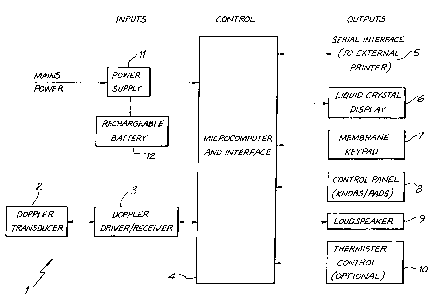
Figure 1 is a functional block diagram showing
components of an ultrasound based device for monitoring
blood flow, in accordance with an embodiment of the
present invention. The device, generally indicated by
reference numeral. 1, comprises a Doppler transducer 2 for
monitoring blood flow in a peripheral blood vessel of a
human or animal subject. In operation, the transducer
will be affixed tp the appropriate body part of the
subject eg. placed distally on the wrist or ankle of a
human being, or where an animal is the subject, on the
tail. Note that as an alternative to a Doppler transducer
2, a pulse oximeter adapted to monitor blood flow could be
used as the blood flow detector (transducer). In fact,
any device which is capable of detecting blood flow, in
the preferred embodiment in a peripheral vessel, could be
used.
Note that a further alternative, in accordance with
an alternative embodiment of the present invention, is to
use a device such as a pulse oximeter in addition to using
the Doppler transducer 2 to manitQr the changes in blood
flow. The pulse oximeter is, in accordance with this
embodiment, adapted to monitor blood volume in a
peripheral tissue bed (rather than oxygen saturation which
is usually constant during anaesthesia where patients
inspire high concentrations of oxygen) and this may be
used to improve the estimate of changes in blood flow or
to enable estimation of changes in vascular resistance.
In this alternative embodiment, the device of figure 1
would also include a sensor and a pulse oximeter device
AMENDED SHEET (Article 34) (IPEA/AU)
CA 02289637 1999-11-12
= PCT/AU98/00356
Received 06 August 1999
- 13 -
providing an input about changes in tissue blood volume to
the micro computer 4 for processing together with the
perfusion input from the Doppler device. The following
description, however, relates to an embodiment which
employs a Doppler monitor only.
In this embodiment, a continuous wave Doppler
driver/receiver 3 is connected to the Doppler transducer
for transmitting and receiving ultrasound signals
therefrom. A microcomputer and interface 4 is arranged to
process the signal from the receiver 3, and drive the LCD
display 6 to produce an output indicative of changes in
cardiac output (substantially equivalent to tissue oxygen
delivery under high inspired concentrations of 02). It
also controls and/or responds to the other peripherals, as
follows:
a serial interface 5 to an external printer;
a liquid crystal visual display 6;
a membrane keypad 7;
a control pane7. 8;
a loud speaker 9; and
a thermistor controller 10 for controlling a airway
thermistor (not shown).
Power is provided from the mains via a power supply
regulator 11, which is also provided with a back-up
rechargeable battery 12, in case of failure of the mains.
In operation, the microcomputer controller 4 operates
to process the signal from the Doppler transducer 2 to
determine changes in the blood flow rate in the peripheral
veseel and to control the liquid crystal display 6 to
provide an indication, preferably graphical indication, of
the instantaneous relative cardiac output at any time
during anaesthesia and surgery. It is preferred to give
an output of rela .iv_ cardiac output, rather than
attempting to produce an output indicative of actual
cardiac output. Attempting to obtain a measurement giving
actual cardiac output is very difficult because a) vessel
diameter is required or b) it assumes that changes in
AMBNDED SHEET (Article 34) (IPEA/AU)
CA 02289637 1999-11-12
PCT/AU98/00356
Received 06 August 1999
- 14 --
blood flow or vessel diameter in one vessel similarly
reflect changes in the whole animal. Monitoring changes
in blood flow to provide an output relative to a
reference, such as the signal output by the blood flow
monitor when the patient is at rest prior to anaesthesia
and surgery, is much more convenient, and provides
sufficient indication to the anaesthetist to guide him to
control anaesthetic depth. The loudspeaker 9 is
controlled by the controller 4 to provide an audible pulse
signal and alarms should the blood flow fall below or rise
above pre-set levels. Preferably, the display 6 also
provides a visual alarm indication. The control panel 8
can be used to pre-set the blood flow display and alarm
limits, depending upon, for example, the size of the
subject and the species of the aubject. It is envisaged
that a device would be provided suitable for operation on
a human subject and a separate device suitable for
operation on animal subjects, the animal subject device
preferably being adapted for use with a number of animal
species, control limits being pre-set for species and
animal size by the control panel S. The microcomputer and
interface 4 is arranged to process the Doppler signal
output to give an indication of blood flow changes based
on the strength of the signal.
Figure 2 shows the external appearance of an example
device 1. Equivalent items to figure i are given the same
reference numerals. The entire device 1is housed within
a robust housing 13. Brackets 14 are provided to hold a
reference manual giving operating instructions on the
device 1. The device is mounted on rubber feet 15 and has
a carrying handle 16. A plug 17 is provided for
connection to a mains power supply.
In operation, before a subject is anaesthetised, the
Doppler transducer (sensor) 2 is positioned on the skin
surface, overlying a peripheral artery such as located in
the human forearm at the level of the wrist (radial or
ulna artery), on the plantar surface of the foot of a dog
WNDED SHEET (Article 34) (IPEA/AU)
CA 02289637 1999-11-12
PCT/AU98/00356
' = . Received 06 August 1999
- 15 -
or cat (pedal artery) or on the ventral surface of the
tail (coccygeal artery). The device is attached to the
subject at rest while conscious and a flow rate
determined. The control pad 8 is then used to set a "base
line flow" rate and a base bar (reference number 20,
figure 3) will appear on the operating display. The base
bar will be used as a reference by the anaesthetist as the
"normal" flow rate of the conscious resting subject (ie.
prior to induction of anaesthesia). As an alternative,
the device may also be arranged to store a series of
"standard" base bars, being default settings for a
particular animal species/size of animal. This would be
necessary for animals which may not tolerate attachment of
the transducer while conscious. For a human subject,
however, it is preferable to pre-set the levels and the
display by monitoring of the individual subject.
Figure 3 shows an example operating display for a
human subject during anaeethesia and surgery. The left
hand side of the display, indicated by reference numeral
21, is taken up by a bar graph which graphically
continuously indicates peripheral blood flow rate based on
the signal obtained from the peripheral vessel, processed
by the controller 4 to provide the display. The base bar
20 is permanently in place on the graphical display and is
pre-set by monitoring the flow rate of the conscious
subject at rest, prior to the induction of anaesthesia.
All flow rates and flow alarms are determined relative to
this base bar 20. A high limit bar 22 and low limit bar
23 are also displayed. These can either be pre-set by the
anaesthetist or pre-stored in memory to automatically be
displayed depending upon the set base bar level and other
subject factore, eg. weight, age, etc. For example,
appropriate limits could be determined by clinical trials
and then stored in the memory of the device.
A moving flow marker 24 is also displayed. This
shows the actual real-time flow rate (relative to the base
bar). it is this marker 24 that the anaesthetist will
AMENDED SHEET (Article 34) (IPEA/AU)
CA 02289637 1999-11-12
PCT/AU98/00356
Received 06 August 1999
- 16 -
watch carefully to obtain an indication of changes in
haemodynamic function. Preferably, the flow marker is
arranged to flash. Should the rate fall to the lower
limit bar 23 or rise to the high limit bar 22 an audible
alarm will sound and the numeric flow display 26 will
flash. The anaesthetiste attention will thus be drawn to
the alarming level of perfusion or blood flow and
appropriate action can be taken (eg. altering anaesthetic
dose, administration of IV fluid, inotropic drugs etc.).
Note that it is unlikely during appropriate levels of
anaesthesia during surgery in normal, healthy patients
that blood flow will ever rise much above the base bar.
This is because standard anaesthetics tend to depress
(rather than stimulate) cardiac output in a dose dependent
fashi.on.. Such a monitoring device can also be used for
monitoring haemodynamic function during critical care such
as post cardiac surgery. On this point, a novel device
such as this is likely to provide precise clinical data on
the effect of anaesthetics and surgical manipulation on
peripheral blood flow in humans and animals. However,
there are applications of this device, such as cardiac
stress testing (treadmill testing) of conscious humans or
race horses, where blood flow could increase above the
base line measurement.
Referring again to figure 3,.the controller 4 also
determines the pulse rate of the subject from the Doppler
flow signal. This is displayed in the top right hand
portion 25 of the display 6. The anaesthetist can also
therefore view pulse rate, at a glance. The bottom right
hand corner of the display 26 displays the actual
(instantaneous) peripheral blood flow rate in
alphanumeric.
Should the probe signal change caused by transducer
or skin movement relative to the artery or loss of
acoustic coupling or otherwise malfunction, a"probe
error" display will flash 27.
Switching the device on and taking no further action
AMENDED SHEET (Article 34) (IPEA/AU)
CA 02289637 1999-11-12
PCT/AU98/00356
Received 06 August 1999
17 -
defaults the screen to the monitoring display (figure 2).
All input and control of the device is set by rotating
knob 80 (figure 2) to select function or value and
pressing enter to accept function or value.
Upper and lower limit thresholds may also be set for
pulse rate, such that if the thresholds are reached
audible alarms/visual alarms are provided. A breath to
breath audible output and a numeric display of respiratory
rate, may also be provided it an airway thermistor is
employed.
Figuree 4 through 7 show examples of screen displays
which may appear during initial set up of the apparatus
prior to operation on a human or animal subject. The
example screens are based on the device as designed for
animal use. This is generally the same as what would
appear in the device as designed for human subjects,
except that it is envisaged that there would be no screen
for default species settings (figure 5) although default
settings based on body size dould be introduced.
Alternatively, all the settings for the alarm function
could be entered manually (figure 4). After selecting
either the default settings (figure 5) or entering the
alarm settings manually (figure 4), the device will then
display the result and settinga as selected (figure 6)
before reverting to the running" display Associated with
the continuous monitoring function (a running display is
shown in figure 3 for a human being, but a similar display
would be shown for animal).
The boxed items of display (figure 4) ("Run", "Pause"
etc) are what can be selected by turning the knob 80. A
selected function displays as inverse display (ie. white
letters on black background). Depressing the knob will
then cause the numerical value to increase in magnitude to
a maximum number. Subsequently turning the knob by 10
will move the selection to the next boxed item in a left
to right, top to bottom flow with wrap-around at bottom.
Turning the knob counter clockwise will reverse the
AMENDED SHEET (Article 34) (IPEA/AU)
CA 02289637 1999-11-12
PCT/AU98/00356
Received 06 August 1999
- 18 -
selection highlighting.
Figure 4 shows a typical data entry display for
manual entry of the alarm settings, which enables entry of
pulse rate high/low limits and flow rate high/low limits
ie. minimum, base and maximum levels for each item. These
values can be set manually based on the
preference/clinical experience of the anaesthetist.
Alternatively, selection of alarm limits may be based on
default settings as shown for animals.
Figure 5 shows a display for default settings which
can be selected, which will be based on clinical trials
for the particular species/weight of animal (Note that
manually five entered default settings may be stored by
the user in memory.) Figure 6 illustrates the screen with
the default settings which were either entered manually
(figure 4) or selected (figure 5). Devices may obviou$ly
be designed with different default settings for different
species and animal eizee, depending upon application.
Figure 6 is a diagram of the entered/selected alarm
setting display, also showing the rest of the control
panel from figure 2, incorporating screen selection knob
80, mode button 31, enter button 32 and on/off switch 33.
For this example (10-20kg dog) using figure 6 "enter"
can be pressed while the selection knob is set on "Animal
Class" to display the Animal Class display from figure 5.
A 10-20kg dog will be class "3", the knob is turned 10
clockwise to highlight the numerical animal class function
number 3 which results in the various high/low default
limits shown in figure 6. Enter button is then pressed
which now selects the default settings (for class number
3) and changes the display screen to figure 6. Turning
the knob 5 will increment by one value resulting in the
display value being 1. Thus turning the knob to
approximately 55 clockwise will set the value to 11 (a
15kg dog). The knob can be rotated counter-clockwise to
decrement the values. Again the "enter" button is pressed
which records and accepts the value. At this point all
AMENDED SHEET (Article 34) (IPEA/AU)
CA 02289637 1999-11-12
PCT/AU98/00356
= Received 06 August 1999
- 19 -
the values on the Data Enter display will change to the
default values for a 15kg dog. The highlighted box will
move to the RUN box assuming the "enter" button will be
pressed to accept all the default values and change the
display to Figure 7 - If a Run Display of particular value
is to be changed, eg. warning tone to OFF, the knob is
turned either clockwise or counter-clockwise to the
desired box. Pressing enter will toggle the value (to
on/off etc) and move the selection to next value (left to
right, top to bottom). When all values on the Data Enter
display are set and RUN is entered, the display changes to
the RUN display.
In the PAUSE mode (Figure 7), the display will be
inverse. All Data Enter values will be displayed on the
RUN display format.
The Doppler sensor is secured with the animal
sedated.
RUN is selected by turning the knob counter-clockwise
approximately 10 and "enter" button pressed. The monitor
will now start to function, updating the display
approximately every 15 seconds, showing heart rate, flow,
and moving the flow marker above or below the base value.
At any time during operation the knob can be turned to
highlight any value on the run display.
During the procedure, the base value may need to be
adjusted. Such as with re-positioning the patient for
surgery. Turn the knob to highlight the base value eg 2.0
Figure 3, press enter, turn the knob (clockwise'or
counter-clockwise) to display the desired base flow, then
press enter. The monitor will accept the new base flow
number and readjust the High/Low limit bars.
With regard to the embodiments discussed above, the
output signal from the Doppler transducer is a signal the
amplitude and/or frequency of which varies depending upon
the rate of blood flow in the peripheral vessel being
monitored. As discussed above, the signal can therefore
be processed by the micro computer 4 to control a display
AMENDED SHEET (Article 34) (IPEA/AU)
CA 02289637 1999-11-12
= PCT/AU98/00356
, Received 06 August 1999
-- 20 ~
to give an output indicative of changes in total blood
flow as the changes in blood flow in the peripheral ve$sel
correlate with changes in total cardiac output (CO). In a
clinical situation, such as during anaesthesia in surgery,
the accuracy of this correlation is important, i.e., it is
important that the displayed changes correlate well with
the actual changes in cardiac output or tissue oxygen
delivery. If the display gives an inaccurate reading,
particularly in the critical range (i.e., in the region of
the alarm levels) then information given to the
anaesthetist can be misleading and ultimately lead to a
dangerous situation.
The present applicants have found that the accuracy
of the correlation between the changes in the output
signal from the Doppler transducer and changes in cardiac
output can be much improved by further processing of the
signal to adjust the signal by a factor which is based on
regression analysis of actual experimental subjects. They
have also found that the correlation can be even further
improved by adjusting the processed signal by employing a
co-variant factor, in the preferred embodiment being heart
rate. Adjustment of the signal using these factors
preferably leads to a more accurate output and the
microprocessor is preferably arranged to process the
signal from the Doppler transducer by including
adjustments based on these factors.
Figure 9 is a schematic plot of "Perfusion Index" in
relation to cardiac output (CO) or tissue oxygen delivery,
for a notional experimental subject, to illustrate how
regression analysis may be applied in accordance with this
embodiment of the invention. Perfusion zndex is a term
the applicants have chosen to represent the processed
output of the Doppler device (or where another device is
being used to monitor blood flow, the output from that
device). The processed signal from the Doppler device,
which is a voltage output proportional to doppler
frequency change, whether it be amplitude or frequency,
AMENDED SHEET (Article 34) (IPEA/AU)
CA 02289637 1999-11-12
PCT/AU98/00356
= Received 06 August 1999
- 21 -
provides an output known as the Perfusion Index. Ideally,
this output will be directly proportional to cardiac
output or tissue oxygen delivery (curve A of figure 9).
During anaesthesia, high inspired amounts of oxygen are
applied so that the arterial oxygen content is relatively
constant. Changes in cardiac output can be taken to be
substantially the same as changes in tissue oxygen
delivery, therefore, in these circumstances.
The ideal, unfortunately, is not the case. From
experiments with subjects, however, it is possible to plot
Perfusion Index against CO or tissue oxygen delivery, by
monitoring cardiac output with another device arranged to
directly monitor cardiac output, and by applying a device
such as a Doppler monitor to monitor "Perfusion Index", on
an experimental subject, to give a realistic plot, plot A
in figure 9. The equation for the curve is:
y=ax+b
where y is in this case cardiac output or tissue oxygen
delivery, x is Perfusion Index, a is the slope and b is
the intercept (see figure 9).
By adjusting the output of the Doppler device by
modifying it by a factor corresponding to a and b, i.e.,
modifying it by using a regression analysis employing a
experimental subject, a more accurate correlation of
Perfusion Index (i.e., the new adjusted Perfusion Index)
with cardiac output or tissue oxyqpn dnlivary can be
obtained. In the preferred embodiment, therefore, the
micro computer 4 is arranged to modify the output of the
Doppler receiver 3 by a factor relating to the regression
analysis. This has been found to provide a much improved
output, i.e., a more accurate indication of the cardiac
output.
In application, therefore, regression analysis is
carried out by a monitoring perfusion index against
cardiac output or tissue oxygen delivery for a plurality
of subjects. The results of the regression analysis are
then used to calculate a weighting factor to be applied to
AMENDED SHEET (Article 34) (IPEA/AU)
CA 02289637 1999-11-12
PCT/AU98/00356
Received 06 August 1999
- 22 -
the output from the Doppler morxitor, by the device in
accordance with the embodiment of the present invention,
in order to adjust that output to create a more accurate
output indicative of cardiac output or tissue oxygen
delivery. In the example given in figure 9, a and b are
calculated and y with the new adjusted output, is produced
in accordance with the formula y = ax + b.
Note that tissue oxygen delivery = tissue blood flow
(cardiac output) x arterial oxygen content.
A further improvement to the correlation of Perfusion
Index to cardiac output can be made by further modifying
the output signal from the Doppler transducer by making an
adjustment for a co-variate factor.
Cardiac output = heart rate x stroke volume.
Cardiac output also = mean arterial pressure/vascular
resistance.
There are therefore a number of variants which
influence cardiac output and which may also determine the
accuracy of an output signal from the Doppler monitor.
The applicants have found that, in patients anaesthetised
for surgery, including a co-variate factor based on heart
rate also results in an increase in the accuracy of the
final output of the device. A co-variate factor relating
to mean arterial pressure does not improve the output and
in fact degrades it.
Preferably, therefore, in accordance with the
preferred embodiment of the invention, the output of the
Doppler monitor is also adjusted by applying a co-variate
factor, based on the heart rate of the patient. Again, a
number of experimental subjects are monitored to see what
variation of the output of the Doppler monitor (perfusion
index) occurs with pulse rate. A weighting factor is then
applied to the output from the signal in accordance with
detected heart rate foz a patient, to further improve the
response of the device.
A further modification which may be made to the
device is to process the output to provide an indication
AIvfENDED SHEET (Article 34) (IPEA/AU)
CA 02289637 1999-11-12
= j PCT/AU98/00356
Received 06 August 1999
- 23
of the "trend" of the output and also provide a display of
the trend. All measurements are stored periodically, for
example every one to five seconds, and a display which
gives the directxon that the output is taking, i.e.,
either up or down, is provided for the anaesthetist. This
"trend" display can be useful in anaesthesia, and will
generally provide more direction to an anaesthetist as far
as anaesthetic dose required is concerned, than a straight
forward "number" display not indicating any trend.
As discussed above, the preferred Doppler device to
be used with the present f:nvention is a continuous wave
Doppler. These are preferably cheap, easy to build and
portable. In operation, the ultra sound beam is
transmitted from one crystal and the reflected wave
received by another. The change in frequency of the
reflected signal is in part due to the velocity of the red
blood cell flow. The change in the amplitude of the
signal depends on the vessel, distance and tissue density
differences.
Vessel wall motion alters the high amplitude, of the
signals which influences the shape of the amplitude/time
epectrum of the reflected wave. This problem can be
minimised by using Doppler crystals with higher sound
frequencies (8 to 10 MHz). In addition uee of front end
clutter filters dc3t;i gnRd to optimise the illumination of
reflected sound from skin, subcutaneous tissue and fat can
be employed, and this is preferred. Since the amplitude
and time lay of the reflected noise depends on the depth
and size of the blood vessel being analysed, the filters
are preferably specific for either body size (e.g., adult
human, child or neonate) or species (e.g., cat, dog,
horse). A toggle switch preferably enables the operator
to select the desired clutter filter (not.shown in the
f igure s ) .
The change in time difference between the reflected
signal from the proximal and distal wall of the blood
vessel can be analysed and will indicate changes in blood
AMENDED SHEET (Article 34) (IPEA/AUl
CA 02289637 1999-11-12
PCT/AU98/00356
= , Received 06 August 1999
-24-
vessel diameter. An eatimate of blood vessel diameter
combined with the estimate of velocity of blood flow, can
be used to give index of blood flow, which can be modified
in accordance with the factors discussed above to give the
desired output (perfused index) which accurately
correlates with Cardiac Output. As discussed in the
preamble of the specification, other devices which are
capable of monitoring blood flow could be used instead of
continuous wave Dopplers.
As discussed above, a pulse oximeter may also be used
to provide a monitoring device in accordance with the
present invention.
Pulse oximeters are currently designed to measure the
transmission of red and infra-red light from haemoglobin
of the arterial blood and estimate the arterial oxygen
saturation. However, changes in the reflective wavelength
of the light from the tissue bed depend on:
A. changes in the oxy-haemoglobin level.
B. Changes in the total mass of t,issue including
red blood cells.
Once a pulse oximeter is functioning on a patient, it
assumes that the background tissue and blood massn is
constant (fixed), it focuses on the pulsatile part of
perfusions or blood flow wave form and therefore assumes
that changes in the wavelength of the light are due to
changes in oxygenation.
Typically during anaesthesia, patients breath high
inspired concentrations of oxygen. Therefore, changes in
light absorption are far more commonly due to changes in
the mass of red blood cells (i.e., the assumed to be
constant light absorption) than to changes in arterial
oxygenation.
To modify a pulse oximeter, we need to work form the
principle that using two light wavelengths (one in the
visible red spectrum and one in the infra-red spectrum):
at the isobestic wavelength, the absorbing power of
oxyhaemQglobin in the reduced haemoglobi.n is the same.
AMENDED SHEET (Article 34) (IPEA/AU)
CA 02289637 1999-11-12
PCT/AU98/00356
Received 06 August 1999
-25-
Therefore total absorbency depends only on the sum of the
two and not the state of oxygenation. Therefore the total
absorbency depends only on the total amount of blood
present. As tissue blood flow increases or decreases, the
total absorbency at the isobestic point will change and
this can be used to give a measure of the relative change
in blood (mass) flow in the tissue bed. Such a device can
therefore be used to monitor changes in blood flow in
peripheral tissue beds.
Electromagnetic flow meters have been designed to be
surgically implanted around large blood vessels such as
the aorta and renal artery. It is possible that such a
device may be adapted to be placed around a peripheral
tissue bed, such as a finger or tail, to provide an
indication of relative changes in blood flow. This may
not be accurate, however.
There is no reason that an electromagnetic flow meter
could not be used in the present invention, by
implantation of a cuff type flow meter around a blood
vessel. This is, however an invasive technique, and
although it falls within the scope of the present
invention it is not preferred.
Othex' available devices which could be adapted in
accordance with the present invention are non-invasive
optical flow meters. These devices measure the absorption
characteristics of light scattered by blood flowing
through tissues such as skin surface, detecting this
reflected light, analysing the frequency of the wave forms
to obtain the mean peak light frequencies in estimating
blood flow. Problems with this approach are that the
device only measures very superficial (i.e., skin surface)
blood flow, which during anaesthesia is altered by vaso
constriction such as caused by changes in body
temperature. The device is also subject to movement
artefacts/vibrations such as caused by patient
positioning, movement by surgical manipulations,
restorations, vibrations from re-circulating water beds,
AMENDED SHEET (Article 34) (IPEA/AU)
CA 02289637 1999-11-12
PCT/AU98/00356
Received 06 August 1999
-26-
etc. It is therefore difficult to get a continuous
measure from a wave (pre-anaesthesia) through to
anaesthesia when positioned for surgery.
Further, the signal requires considerable damping to
get a stable measurement, which sacrifices the accuracy of
the "real time" measurement. It also relies on estimating
the Doppler signal change in the scattered light to obtain
the peak frequency and fails to measure perfusion of
deeper tissues. Nevertheless, although not preferred, it
is quite possible that such a device could be used in the
present invention.
The above description is of a relatively
sophisticated device which can be used with the method in
accordance with the present invention. As discussed in
the preamble, a primitive device, in the form of a "colour
chart" can also be used. Colours indicating various flow
rates would be established by clinical trials for various
species in order to produce the colour chart. An
anaesthetist will then have reference to the colour chart
and compare with the colour of the part of the body
concerned such as the oral mucosa, in order to monitor
flow rate in the subject. An example colour chart is
schematically illustrated in figure B.
It will be appreciated by persons skilled in the art
that numerous variations and/or modifications may be made
to the invention as shown in the specific embodimento
without departing from the spirit or scope of the
invention as broadly described. The present embodiments
are, therefore, to be considered in all respects as
illustrative and not restrictive.
AMENDED SHEET (Article 34) (IPEA/AU)