Note: Descriptions are shown in the official language in which they were submitted.
CA 02295005 1999-12-20
WO 98/58590 PCTNS98/10146
STRETCH RESISTANT VASO-OCCLUSIVE COILS (II)
RELATED APPLICATIONS
This is a continuation-in-part of U.S. 081607,593, filed February 27, 1996,
and
08/717,285, filed September 20, 1996 and 08/717,285, filed September 20, 1996
is a
continuation-in-part of 08/497,331, filed June 30, 1995 (issued as U.S. Patent
No.
5,582,619 on December 10, l 996), the entirety of which are incorporated by
reference.
FIELD OF THE INVENTION
This invention is an implantable vaso-occlusive device. It is typically a vaso-
occlusive coil comprising a primary helically wound coil which may then be
wound into a
secondary shape. Central to the invention is the use of a stretch-resisting
member
extending through the lumen formed, which stretch-resisting member is fixedly
attached,
directly or indirectly, to the coil in at least two locations. The stretch-
resisting member in
this variation desirably is heat-treated in situ when the coil is in the
secondary shape. This
heat treatment allows the stretch-resisting member to conform to the shape of
the coil in its
secondary configuration. Desirably, the member does not appreciably affect the
inherent
secondary shape of the coil. The stretch-resisting member is preferably
somewhat loose
within the interior of the lumen so to prevent the coil from collapsing,
binding, and
therefore stiffening during passage of turns through the human body. The coil
should bend
easily. In some variations of the invention, the stretch-resisting member may
be formed
into coil tips at the ends of the coil using simple equipment such as
soldering irons or the
like. The tips are typically of the same diameter as is the coil body itself.
This stretch-
resisting member is for the primary purpose of preventing stretching of the
coil during
movement of that coil, e.g., by retrieval or repositioning after deployment.
The device
may have a self forming secondary shape made from a pre-formed primary linear
helically
wound coil, although it need not have the secondary form. Desirably, the coil
is extremely
flexible and is controllaby released using a severable or mechanical joint
such as an
electrolytically detachable joint. External fibers may be attached to the
device and affixed
to the pre-formed linear member to increase thrombogenicity. The extremely
flexible
CA 02295005 1999-12-20
WO 98/58590 PCT/US98/10146
variation of the invention may be hydraulically delivered through the lumen of
a catheter
and is so flexible that it may be retrievably delivered therethrough a flow-
directed catheter.
The vaso-occlusive member may be also be covered with a fibrous braid. The
device is
typically introduced into the body through a catheter. The device is passed
axially through
the catheter sheath and assumes its secondary form upon exiting the catheter.
BACKGROUND OF THE INVENTION
Vaso-occlusion devices are surgical implements or implants that are placed
within
the vasculature of the human body, typically via a catheter, either to block
the flow of
blood through a vessel making up that portion of the vasculature via the
formation of an
embolus or to form such an embolus within an aneurysm stemming from the
vessel. One
widely used vaso-occlusive device is a helical wire coil having windings which
may be
dimensioned to engage the walls of the vessels. Other less stiff, helically
coiled devices
have been described, as well as those involving woven braids. Virtually all
such vaso-
occlusive implants are delivered by wire-guided catheters which devices are
pushed
through the catheter. Because of the need for a pusher and concerns for
recovery of such
vaso-occlusive devices should they be malplaced in the body, it is unlikely
that prior to this
invention has there been a vaso-occlusive device of a form similar to this
delivered through
a flow directed catheter.
As an instance of an early vaso-occlusive device, US Patent No. 4,994,069, to
Ritchart et al., describes a vaso-occlusive coil that assumes a linear helical
configuration
when stretched and a folded, convoluted configuration when relaxed. The
stretched
condition is used in placing the coil at the desired site (by its passage
through the catheter)
and the coil assumes a relaxed configuration -- which is better suited to
occlude the vessel -
- once the device is so placed. Ritchart et al. describes a variety of shapes.
The secondary
shapes of the disclosed coils include "flower" shapes and double vortices. A
random
secondary shape is described, as well.
Vaso-occlusive coils having attached fibrous elements in a variety of
secondary
shapes are shown in US Patent No. 5,304,194, to Chee et al. Chee et al.
describes a
helically wound device having a secondary shape in which the fibrous elements
extend in a
sinusoidal fashion down the length of the coil. These coils, as with Ritchart
et al., are
2
CA 02295005 1999-12-20
WO 98/58590 PCT/US98/10146
produced in such a way that they will pass through the lumen of a catheter in
a generally
straight configuration and, when released from the catheter, form a relaxed or
folded shape
in the lumen or cavity chosen within the human body. The fibrous elements
shown in
Chee et al. enhance the ability of the coil to fill space within the
vasculature and to
facilitate formation of embolus and subsequent allied tissue.
There are a variety of ways of discharging shaped coils and linear coils into
the
human vasculature. In addition to those patents which apparently describe only
the
physical pushing of a coil out into the vasculature (e.g., Ritchart et al.),
there are a number
of other ways to release the coil at a specifically chosen. time and site. US
Patent No.
5,354,295 and its parent, 5,122,136, both to Guglielmi et al., describe an
electrolytically
detachable embolic device.
A variety of mechanically detachable devices are also known. For instance, US
Patent No. 5,234,437, to Sepetka, shows a method of unscrewing a helically
wound coil
from a pusher having interlocking surfaces. US Patent No. 5,250,071, to
Palermo, shows
an embolic coil assembly using interlocking clasps mounted both on the pusher
and on the
embolic coil. US Patent No. 5,261,916, to Engelson, shows a detachable pusher-
vaso-
occlusive coil assembly having an interlocking ball and keyway-type coupling.
US Patent
No. 5,304,195, to Twyford et al., shows a pusher-vaso-occlusive coil assembly
having an
affixed, proximally extending wire carrying a ball on its proximal end and a
pusher having
a similar end. The two ends are interlocked and disengage when expelled from
the distal
tip of the catheter. US Patent No. 5,312;415, to Palermo, also shows a method
for
discharging numerous coils from a single pusher by use of a guidewire which
has a section
capable of interconnecting with the interior of the helically wound coil. US
Patent No.
5,350,397, to Palermo et al., shows a pusher having a throat at its distal end
and a pusher
through its axis. The pusher sheath will hold onto the end of an embolic coil
and will then
be released upon pushing the axially placed pusher wire against the member
found on the
proximal end of the vaso-occlusive coil.
Vaso-occlusive coils having little or no inherent secondary shape have also
been
described. For instance, in US Patent Application 07/978,320, filed November
18, 1992,
entitled "Ultrasoft Embolization Coils with Fluid-Like Properties" by
Berenstein et al., is
found a coil having little or no shape after introduction into the vascular
space.
3
CA 02295005 1999-12-20
WO 98/58590 PCT/US98/1O146
None of these devices are helical coils which contain a stretch-resisting
member
contained therein.
SUMMARY OF THE INVENTION
This invention is a vaso-occlusive device comprising a helically wound coil
which
is formed by winding a wire into a first or primary helix to form an outer
helical member
having first and second ends. A stretch resistant member extending through the
lumen
thus-formed is fixedly attached, directly or indirectly, to the coil in at
least two locations.
The stretch-resisting member is preferably loose within the coil to prevent
binding of the
coil during passage of the coil through turns in the vasculature.
The primary helix or "primary form" may be wound into a secondary form and
heat-treated to preserve that form, desirably prior to the step of including
the stretch-
resisting member into the coil. The coil, with its included stretch-resisting
member, will
be again heat-treated to shape that the stretch-resisting member into the
coil's secondary
form. The secondary form may be one which, when ejected from a delivery
catheter,
forms a specific shape. Such a shape might, e.g., fill a vascular cavity such
as an
aneurysm, or perhaps, a fistula or AVM. The stiffness of the various parts of
the coil may
be tailored to enhance the utility of the device for specific applications.
Extremely flexible
coils are highly desirable. Fibrous materials may be woven into the member or
tied or
wrapped onto it to enhance the thrombogenicity.
Once the secondary form of the coil has been achieved, the stretch-resisting
member is then inserted into the lumen, and secured to the coil. The assembly
is then
gently heat-treated to allow the stretch-resisting member to assume the
secondary form of
the coil.
The device is used simply by temporarily straightening the device, as
necessary,
and introducing it into a suitable catheter, the catheter already having been
situated so that
its distal opening is at the selected site in the body. The device is then
pushed through the
catheter and, upon its ejection from the distal end of the catheter into the
vascular cavity,
assumes its relaxed or secondary shape.
4
CA 02295005 1999-12-20
WO 98/58590 PCT/US98/10146
The device is typically used in the human vasculature to form emboli but may
be
used at any site in the human body where an occlusion such as one produced by
the
inventive device is needed.
Also forming an important aspect of this invention is the combination of this
inventive vaso-occlusive device with a flow-directed catheter.
5
CA 02295005 1999-12-20
WO 98/58590 PCT/US98/10146
BRIEF DESCRIPTION OF THE DRAWINGS
Figure lA shows a side view, partial cutaway of a vaso-occlusive coil made
according to the invention having a generally linear fibrous stretch-resisting
member.
Figure 1B shows a side view, partial cutaway of a vaso-occlusive coil made
according to the invention having a generally linear wire stretch-resisting
member.
Figure 1 C shows a side view, partial cutaway of a vaso-occlusive coil made
according to the invention having a generally helical stretch-resisting
member.
Figures 2A, 2B, and 2C show side view, partial cutaways of typical ends of the
inventive vaso-occlusive coils.
Figures 3A, 3B, and 3C show side view cutaways of electrolytically severable
joints in combination with a vaso-occlusive coil made according to the
invention.
Figures 4A and 4B show a side view, partial cutaway of a typical mechanically
detachable joint in combination with a vaso-occlusive roil made according to
the
invention.
Figure 5 shows a "C" shaped secondary configuration for the inventive vaso-
occlusive device.
Figure 6 shows a clover-leaf secondary shape for the inventive vaso-occlusive
device.
Figure 7 shows a double-looped secondary shape for the inventive vaso-
occlusive
device.
Figure 8 shows attachment of external fibrous material to the inventive vaso-
occlusive device.
Figure 9 shows attachment of external braided fibrous material to the
inventive
vaso-occlusive device.
Figure 10 shows the vaso-occlusive device of the invention in which a polymer
is
introduced into the lumen of a coil after it has been shaped to return to its
secondary form.
Figure 11 shows the combination of the vaso-occlusive device of this invention
in
assembly with a flow-directed catheter.
Figures 12A-12D show a procedure for introducing a vaso-occlusive coil such as
found in the other Figures into an aneurysm.
6
CA 02295005 1999-12-20
WO 98/58590 PCT/US98/10146
DESCRIPTION OF THE INVENTION
Figures lA, 1B, and 1 C show side-view partial cross-sections (or cutaways) of
highly desirable variations of the inventive coil (100, 200, 210).
The variations shown in Figures lA and 1B are made up of a helically wound
outer
coil (102, 202) having a first end (104, 204) and a second end (106, 206). We
refer to this
form as the as the "primary" winding or shape or form. These variations
include a stretch-
resisting member (108, 208, 214) which is shown to be fixedly attached both to
the first
end (104, 204) and to the second end (106, 206). In certain circumstances, it
may be
desirable to attach the stretch-resisting member ( 108, 208) only to one of
the two ends, to
at least one site between the to ends, or to neither of the two ends. Clearly,
for attaining
stretch resistance, the stretch-resisting member must be attached to at least
two points on
the coil.
The stretch-resisting member (108) of the variation shown in Figure lA is
fibrous
and desirably polymeric. The stretch-resisting member (108) may be
thermoplastic or
thermosetting and comprise a bundle of threads or a single filament melted
onto, glued, or
otherwise fixedly attached to the vaso-occlusive coil ( 100).
In this variation of the invention, the stretch-resisting member is preferably
a
polymer (natural or synthetic) which may be heat-set in the secondary form in
situ. The
use of heat-treated or heat-formed polymeric filaments (monofilaments or
threads) should
not affect the secondary shape of the coil and provides stretch resistance
while allowing the
selected form of the device to perform its occlusive function without
interference from the
safety component. In some instances, it may also be desirable to include one
or more
metallic strands in the stretch-resisting member (108) to provide stiffness or
electrical
conductance for specific applications.
The stretch-resisting member (208) of the variation shown in Figure 1B is a
simple
wire or "ribbon" which is soldered, brazed, glued, or otherwise fixedly
attached to the first
end (204), second end (206), or to the coil at one or more locations
intermediate to those
the ends.
The variation shown in Figure 1 C includes a stretch-resisting member (214)
which
is comprised of a heiically wound coil which is soldered, brazed, glued, or
otherwise
fixedly attached to the first end (204) or second end {206) or in one or more
intermediate
7
CA 02295005 1999-12-20
WO 98!58590 PCT/US98/10146
locations. The stretch-resisting member (214) in this configuration provides a
greater
measure of lateral flexibility than the wire variation (208 in Figure 1B). It
may be wound
in either the same direction as is the outer coil (202) or in the alternate
direction. A modest
drawback to this variation is that it will stretch more than the Figure 1B
variation when
axially stressed.
The materials used in constructing the vaso-occlusive coil ( 102, 202) and the
stretch-resisting member (108, 208, 214) may be any of a wide variety of
materials;
preferably, a radio-opaque material such as a metal or a polymer is used.
Suitable metals
and alloys for the wire making up the primary coil (102, 202) and the stretch-
resisting
member (108, 208, 214) include the Platinum Group metals, especially platinum,
rhodium,
palladium, rhenium, as well as tungsten, gold, silver, tantalum, and alloys of
these metals.
These metals have significant radio-opacity and in their alloys may be
tailored to
accomplish an appropriate blend of flexibility and stiffness. They are also
largely
biologically inert. Highly preferred is a platinum/tungsten alloy, e.g., 8%
tungsten and the
remainder platinum.
In some variations of the invention, the ribbon or coil stretch-resisting
members
(208, 214) may be of any of a wide variety of stainless steels if some
sacrifice of radio-
opacity and flexibility may be tolerated. Very desirable materials of
construction, from a
mechanical point of view, are materials which maintain their shape despite
being subjected
to high stress. Certain "super-elastic alloys" include various nickel/titanium
alloys (48-58
atomic % nickel and optionally containing modest amounts of iron); copper/zinc
alloys
(38-42 weight % zinc); copper/zinc alloys containing 1-10 weight % of
beryllium, silicon,
tin, aluminum, or gallium; or nickel/aluminum alloys (36-38 atomic %
aluminum).
Particularly preferred are the alloys described in US Patent Nos. 3,174,851;
3,351,463; and
3,753,700. Especially preferred is the titanium/nickel alloy known as
"nitinol". These are
very sturdy alloys which will tolerate significant flexing without deformation
even when
used as very small diameter wire.
If a super-elastic alloy such as nitinol is used in the device, the diameter
of the coil
wire may be significantly smaller than that used when the relatively more
ductile platinum
or platinum/tungsten alloy is used as the material of construction.
8
CA 02295005 1999-12-20
WO 98/58590 PCT/US98/10146
The coils may be made of radiolucent fibers or polymers (or metallic threads
coated
with radiolucent or radio-opaque fibers) such as Dacron (polyester),
polyglycolic acid,
polylactic acid, fluoropolymers (polytetrafluoro-ethylene), Nylon (polyamide),
or even
cotton or silk. Should a polymer be used as the major component of the vaso-
occlusive
coil member, it is desirably filled with some amount of a radio-opaque
material such as
powdered tantalum, powdered tungsten, bismuth oxide, barium sulfate, and the
like.
The coil material is first wound into a primary coil (102, 202). The primary
coil is
typically linear after it has been wound. Generally speaking, when the coil (
102, 202) is a
metallic coil and that coil is a platinum alloy or a super-elastic alloy such
as nitinol, the
diameter of the wire used in the production of the coil ( 102, 202) will be in
the range of
0.00025 and 0.006 inches. The wire is wound into a primary coil (102, 202)
having a
primary diameter of between 0.003 and 0.025 inches. For most neurovascular
indications,
the preferable primary coil (102, 202) diameter is 0.008 to 0.018 inches. We
have
generally found that the coil wire may be of sufficient diameter to provide a
hoop strength
to the resulting device sufficient to hold the device in place within the
chosen body site,
lumen or cavity, without substantially distending the wall of the site and
without moving
from the site as a result of the repetitive fluid pulsing found in the
vascular system.
However, this inventive concept allows the user to utilize extremely flexible
coil
assemblies having very high packing efficiencies. For instance, coil wires
having wire
diameters of 0.00015" and less are suitable for such highly flexible devices.
Typically the
coil diameter will be 0.01 S" and less. They will "droop" more than about
20°, preferably
35° to 90° when about 1 centimeter of the primary form of the
coil having a free end is
held horizontally.
The axial length of the primary coil will usually fall in the range of 0.5 to
100 cm,
more usually 2.0 to 40 cm. Depending upon usage, the coil may well have 10-75
turns per
centimeter, preferably 10-40 turns per centimeter. All of the dimensions here
are provided
only as guidelines and are not critical to the invention. However, only
dimensions suitable
for use in occluding sites within the human body are included in the scope of
this
invention.
Once the primary coil (102, 202) is wound, the stretch-resisting member (108,
208)
is inserted into the lumen of the primary coil (102, 202) and secured to the
coil as desired.
9
CA 02295005 1999-12-20
WO 98/58590 PCT/US98/10146
Ends (104, 204, 106, 206) are preferably of the same diameter as is the
primary coil (102,
202).
Alternatively, the primary coil is shaped into its secondary form, and heat
treated so
that the coil will return to the secondary form when relaxed. The stretch-
resistant member
is then inserted into the lumen of the coil and secured as desired. The
stretch-resisting
member does not substantially affect the shape of the coil when the coil
returns to the
secondary form. Preferably, the stretch-resistant member is attached to a hook
inside the
lumen and heat treatment used to fuse at least parts of the polymer to the
coil. The coil is
then allowed to relax to form its secondary form and any stretch-resistant
filaments
extending from the coil are heat sealed to the coil. It is required that there
be some amount
of slack in the polymer to allow the coil to pass through the catheter as
described herein
and to allow the coil to return to its secondary form. The secondary coil may
be heated
treated. Preferably, heat treatment occurs at a temperature from at least
about the Tb of the
polymer to a temperature below the melting point of polymer.
Suitable polymeric materials for the polymeric stretch-resisting member (108)
can
be either thermosetting or thermoplastic. For this variation of the invention,
however, the
polymer should be one for which a filament may be heat-treated to accept a
form
corresponding to the secondary form. Thermoplastics are preferred because they
allow
simplification of the procedure for constructing the device ( 100) since they
may be melted
and formed into the end or ends (104, 106). Simple devices such as soldering
irons may be
used to form the ends. Thermosetting plastics would typically be held in place
by an
adhesive. Suitable polymers include most biocompatible materials which may be
made
into fibers but include thermoplastics, e.g., polyesters such as
polyethyleneterephthalate
(PET) especially Dacron; polyamides including the Nylons; polyolefins such as
polyethylene, polypropylene, polybutylene, their mixtures, alloys, block and
random
copolymers; polyglycolic acid; polylactic acid; fluoropolymers
(polytetrafluoro-ethylene),
or even silk or collagen. The stretch-resistant polymer may be made from
materials used
as dissolvable sutures, for instance polylactic acid or polyglycolic acid, to
encourage cell
growth in the aneurysm after their introduction. Preferred because of the long
history of
safe and effective usage in the human body are fibrous PET (sold as Dacron)
and
polypropylene. Highly preferred is polypropylene, for instance, in the form of
10-0 and 9-
CA 02295005 1999-12-20
WO 98/58590 PCT/US98/10146
0 polypropylene suture material. We have found that the diameter of the
polymer is
typically between about 0.0001 inches and about 0.01 inches.
Figure 2A shows a side-view partial cross-section of one end of inventive coil
( 100). Figure 2A also shows the helically wound outer coil ( 102) having an
end ( 106)
which is formed from a formerly molten fiber which also makes up the stretch-
resisting
member (114). An end of this type may be considered to have modestly higher
vaso-
occluding properties than a metallic end. Other functional equivalents to this
structure
include ends ( 106) formed of glues such as epoxies and their equivalents, and
which are
mechanical in nature.
Figure 2B shows an external knot (112) which fixes the length of the coil
member
(102) and keeps it from stretching; Figure 2C shows a reformed mass of
formerly molten
polymer or of glue which is of a diameter larger than the inner diameter of
coil (102) and
prevents the coil from stretching. The knot ( 112) and block ( 114) are not
shown to be
attached to the coil (102) but may be.
The variations shown in Figures 1 A, 1 B, 1 C and 2A, 2B, and 2C are designed
to be
deployed by use of a pusher and a catheter in the manner discussed in Ritchart
et al,
discussed above. Other methods (and concomitant fixtures or joints to
accomplish those
methods) may also be used.
For instance, the end of the device may be adapted to accept an
electrolytically
severable joint of the type discussed in US Patent No. x,354,295 and its
parent, 5,122,136,
both patents to Guglielmi and Sepetka, described above. Figures 3A and 3B
depict, in
partial cross section, such variations. The vaso-occlusive coil (130, 230) is
attached to a
fill member or bushing (132, 232). The fill member or bushing (132, 232)
preferably
comprises a thermoplastic formed into place or an epoxy or the like and
adheres, in turn,
both to the stretch resistant member (134, 234) and the core wire (136, 236).
The stretch-
resisting member (134, 234) is thusly indirectly attached to the vaso-
occlusive coil (130,
230) via the fill member or bushing (132, 232). The core wire (136, 236) in
this variation
has an enlarged member which is embedded in the fill member (132, 232). The
core wire
(136, 236) is insulated, typically with a combination of
polytetrafluoroethylene and
PARYLENE (polyparaxyxylene), except for a small sacrificial joint (138, 238)
which is
intended to be the site of the electrolysis as the joint (138, 238) is eroded
or severed and
11
CA 02295005 1999-12-20
WO 98/58590 PCT/US98/10146
the coil deployed into the body site. The details of this variation (sans
stretch-resistant
member (136, 236)) are discussed in Gia et al, US Patent application Serial
No.
08/367,061, filed December 30, 1994, the entirety of which is incorporated by
reference.
Figure 3C shows an especially preferred variation of the inventive device. The
S assembly (131) employs a stretch-resisting member (I33) which is connected
indirectly to
the coil (135). Specifically the stretch-resisting member (133) is a
thermoplastic fiber or
fibers which are melted to form a coil tip (137) at one end of the coil (135)
and is looped
about a hook (139) at (or in the vicinity of) the other end of the coil (135).
An anchor coil
(141) is coaxially situated between the vaso-occlusive coil {135) and the
pusher wire (136).
The hook (139) forms the final turn or half turn ofthe anchor coil (14i). The
stretch-
resisting member (133) is thusly indirectly attached to the vaso-occlusive
coil (135) via the
anchor coil (141). The anchor coil (141) and the vaso-occlusive coil (135) are
preferably
welded together.
Figure 3C also shows the vaso-occlusive coil (135) in its maximum stretched
1 S condition. The stretch-resisting member (I33) is shown resisting further
axial stretching of
the assembly. When the vaso-occlusive coil (135) is not stretched, the stretch-
resisting
member (133) would obviously be loose, i.e., normally longer than the lumen,
in the
lumen of the assembly (131 ). If the stretch-resisting member (133) is not
allowed to have
such a loose axial fit, the adjacent turns of the coil (135) would "bottom"
against each
other during passage through turns in the vasculature and cause the assembly
(I31) to
become stiff.
Figure 4A shows still another variation of a joint for releasing the inventive
coil
into a site within the human body. In this instance, the joint is mechanically
deployed.
The primary coil ( 140) incorporates interlocking clasps, one ( 142) located
on an end of the
coil (140) and one (144) located on the end of a pusher (146). The stretch-
resisting
member (148) is attached to the interlocking clasp (142) via a filler block
(154). Again,
the filler block (154) comprises a material (e.g., a thermoplastic or adhesive
material)
which may be placed in the coil and will adhere to the stretch-resistant
member (148). The
coil assembly (150), made up of the primary coil (140), interlocking clasp
(142), and
stretch-resisting member ( 148) is deployed by retracting catheter body (or
sheath) ( 152).
12
CA 02295005 1999-12-20
WO 98/58590 PCT/US98/10146
Figure 4B shows a variation of the device depicted in Figure 4A which does not
employ
special filler block material (154) for adhering to the stretch-resistant
member.
Other mechanically deployable joints suitable for use with the inventive coil
are
described in:
S - US Patent No. 5,234,437, to Sepetka, (shows a method of unscrewing a
helically wound coil from a pusher having interlocking surfaces).
- US Patent No. 5,250,071, to Palermo, (shows an embolic coil assembly
using interlocking clasps mounted both on the pusher and on the embolic
coil)
- US Patent No. 5,261,916, to Engelson, (shows a detachable pusher/vaso-
occlusive coil assembly having an interlocking ball and keyway-type
coupling)
- US Patent No. 5,304,195, to Twyford et al. (shows a pusher-vaso-occlusive
coil assembly having an affixed, proximally extending wire carrying a ball
on its proximal end and a pusher having a similar end, which two ends are
interlocked and disengage when expelled from the distal tip of the catheter)
- US Patent No. 5,312,41 S, to Palermo (also shows a method for discharging
numerous coils from a single pusher by use of a guidewire which has a
section capable of interconnecting with the interior of the helically wound
coil).
- US Patent No. 5,350,397, to Palermo et al. (shows a pusher having a throat
at its distal end and a pusher through its axis. The pusher sheath will hold
onto the end of an embolic coil and will then be released upon pushing the
axially placed pusher wire against the member found on the proximal end of
the vaso-occlusive coil).
The entirety of which are incorporated by reference.
As was noted above, the devices of this invention may have the simple linear
shape
shown in Figures 1 and 2 or may have shapes which are not so simple. Figures
5, 6, and 7
. show what are termed "secondary" shapes in that they are formed from the
primary coil by
the simple act of winding the primary coil on a form of a desired shape and
then heat
treating the so-formed shape. Figure 5 shows a "C" shaped coil assembly {160)
having a
13
CA 02295005 1999-12-20
WO 98/58590 PCT/US98/10146
stretch-resistant member (162). Figure 6 shows a clover-leaf shaped coil
assembly (164)
also having a stretch-resistant member ( 162). Figure 7 shows a double-loop
coil assembly
(166). These are indicative of the various secondary shapes suitable for this
invention.
Additionally, these inventive devices may also be used in conjunction with
various
external fiber adjuncts. Figure 8 shows a partial side-view of a linear
variation of the
inventive device (170) having filamentary material (172) looping through the
coil (174).
This method of attachment is described in greater detail in US Pat. Nos.
5,226,911 and
5,304,194, to Chee et al, the entirety of which are incorporated by reference.
A further
description of a desirable fiber attachment is shown in US Pat. application
No. 08/265,188,
to Mirigian et al, filed June 24, 1994.
Figure 9 shows a partial cutaway of a device (180) having a braided covering
(182)
of a filamentary material and a stretch-resisting member (184). This method of
enveloping
a coil is described in greater detail in US Pat. Nos. 5,382,259, to Phelps et
al, the entirety
of which is incorporated by reference.
1 S The fibrous woven or braided tubular materials may be made from a
biocompatible
materials such as Dacron (polyester), polyglycolic acid, polylactic acid,
fluoropolymers
(polytetrafluoroethylene), Nylon (polyamide), or silk. The strands forming the
braid
should be reasonably heavy, e.g., having tensile strength of greater than
about 0.15 pounds.
The materials mentioned, to the extent that they are thermoplastics, may be
melted or fused
to the coils. Alternatively, they may be glued or otherwise fastened to the
coils. Preferred
materials include Dacron.
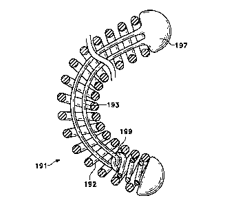
Figure 10 shows a variation in which the stretch-resistant member is a heat-
set
polymer introduced into the interior lumen after the coil has been shaped to
return to its
secondary shape. The coil (191) is wound to a primary shape and is then shaped
into a
secondary form. The coil is treated, for instance by heat-treatment, so that
it will maintain
that secondary form. One end of the coil has an interior lumen (192) and a
hook (199)
within the lumen (192). The coil is then positioned so that the stretch-
resistant thread
(193) is introduced through the lumen (192) of the coil (191) and extended to
catch the
hook portion (199) in the lumen (192) of the coil (191). The end of the coil
with the hook
is then heated so that several turns of the exterior coil contact and are
melted to the stretch-
resistant polymer (193). The coil (191) is then allowed to form its secondary
shape. Any
14
CA 02295005 1999-12-20
WO 98/5859(1 PCT/US98/10146
filaments of stretch-resistant polymer which extend from the coil (191) are
heat-sealed
{ 197). Some amount of slack in the filament is required. The stretch-
resistant polymer
through the lumen must be flexible enough so that they do not change the
secondary shape
of the coil. The entire coil ( 191 ) is then heat-treated at a temperature
below the melting
point of the polymer. Preferably, the temperature is above the polymer's Tb
range.
Figure 11 shows a highly preferred assembly incorporating a number of
desirable
aspects of the invention. Specifically, the very flexible variation of the
inventive vaso-
occlusive device noted above, e.g., wherein the vaso-occlusive device is
capable of
"drooping" 20° or more and having a polymeric stretch-resisting member
included therein,
is especially suitable for inclusion in a flow-directed catheter and
particularly when used
with an electrolytically severable joint. Figure 10 shows the flow-directed
catheter (200)
containing a very flexible vaso-occlusive coil (202) as described above and
utilizing a
similarly flexible strain-resistant member {204). The flow directed catheter
(200) may
have a distal radio-opaque marker (206) if so desired.
Proximally of the vaso-occlusive coil (202) is a connective wire (208) which
is
insulated at all points proximal of the electrolytic joint (210).
The flow directed catheter (200) may be of any known design such as is found,
e.g.,
in U.S. Pat. No. 5,336,205, to Zenzen et al, the entirety of which is
incorporated by
reference. "Flow-directed catheters" axe directed to the treatment site in the
human body
through the vasculature by the motive power of natural blood flow. The more
distal
segments of flow-directed catheters are often of materials having significant
elastomeric
properties but with high burst strengths, e.g., polyurethane,
polyvinylchloride, silicones,
etc. They are often quite "rubbery" in feel. Consequently, flow directed
catheters are not
usually especially suitable for use with guidewires and the like.
In use of this variation of the vaso-occlusive device, however, since the vaso-
occlusive device is so compliant and able to be delivered using hydraulic
pressure alone (as
with saline), they may be used with flow-directed catheters. Further, since
the vaso-
occlusive device (202) contains a stretch-resisting member (204), the vaso-
occlusive
device (202) may be withdrawn into the catheter (200) using the connective
wire (208).
The connective wire (208) used therein should be very flexible so not to
interfere
with the movement of the catheter (200). It is conductive and insulated
proximally of the
CA 02295005 1999-12-20
WO 98/58590 PCT/US98/10146
electrolytic joint (210). Introduction of an electric current into the
connective wire (208)
will cause the electrolytic joint (210) to erode and the vaso-occlusive device
(202) to
become detached. Complete description of the operation of such a device is
found in U.S.
Patent Nos. 5,122,136 and 5,354,295, both to Guglielmi and Sepetka.
Figures 12A-12D depict a common deployment method for introduction of the
inventive vaso-occlusive devices described here. It may be observed that these
procedures
are not significantly different than those described in the Ritchart et al.
patent mentioned
above. Specifically, Figure 12A shows the distal tip of a delivery catheter
(310) which is
within the opening (312) of an aneurysm (314) found in an artery (316). The
distal or end
section of the vaso-occlusive device (318) is shown within the catheter (310).
In Figure
12B, the distal end portion of the vaso-occlusive device (318) has exited the
distal end of
the catheter (310) and has wound into a secondary shape within the aneurysm
(314).
Figure 12C shows the completion of the formation of the secondary shape within
the
aneurysm (314). Figure 12D shows the separation of the vaso-occlusive device
(318) from
the pusher, placement within the aneurysm (314), and the withdrawal of the
catheter from
the mouth of the aneurysm.
Once the inventive coil is in place in an aneurysm or other site, there may be
an
occasion during which the coil must be moved or even withdrawn. For instance,
in Figure
12D, the coil might extend through the mouth (312) of the aneurysm into the
artery.
Occlusion would not be desirable in the artery. A device such as the
endovascular snare
shown in US Pat. No. 5,387,219, to Rappe, may then be used to grasp the
exposed coil and
move it or retrieve it from the body. The stretch-resisting member of this
invention
prevents the coil from stretching into a single strand of wire and multiplying
in length.
Modification of the above-described variations of carrying out the invention
that
would be apparent to those of skill in the fields of medical device design
generally, and
vaso-occlusive devices specifically, are intended to be within the scope of
the following
claims.
16