Note: Descriptions are shown in the official language in which they were submitted.
CA 02298088 2003-11-03
FRAME FOR PRONE SURGICAL POSITIONING
BACKGROUND OF THE INVENTION
1. FIELD OF THE INVENTION
The present invention relates to surgical appliances, and
S particularly to a frame placed on a conventional surgical operating
table which positions the patient in a prone position for spinal
surgery, and is especially suited for positioning the patient for a
lumbar laminectomy with spinal fusion.
2. DESCRIPTION OF THE RELATED ART
' Surgery on the spine is usually performed in either the lateral
recumbent or the prone position. The lateral recumbent position is
usually used for procedures where both an anterior and posterior
approach are used. However, the position does not permit a wide view
of the intervertebral disks and it is difficult to control bleeding.
Therefore, a prone position is normally used for a posterior approach.
Originally the prone position simply involved having the patient
in a recumbent position with his abdomen on the surface of the
operating treatment. However, in this position there was copious
1
CA 02298088 2003-11-03
bleeding due to pressure on the inferior vena cava . It was found that
there was less bleeding if the patient were elevated so that the
abdomen was distended and might hang freely. The simplest method for
accomplishing this is to position chest rolls or bolsters on the table
under the axillae and along the sides of the chest from clavicles to
iliac crests. However, this has not been found to be completely
satisfactory, and a number of devices for positioning the patient in a
prone position with the abdomen distended have been developed. A
number of devices may be distinguished by the degree to which the hips
and knees are flexed.
German Patent No. 882,476, published October 23, 1952, shows an
adaptor for a surgical table having a T-connector for supporting the
hips which attaches to leg support brackets of a conventional surgical
table. A system of bars describing a U-shape is attached to the bottom
of the T-connector. The other upright of the U-shape has supports
under the axillae, a support for the upper chest, a head support and
arm supports. While the abdomen is distended, the T-connector may
produce enough pressure across the pelvis to impair venous return, and
the use of shoulder supports directly under the axillae is questionable
due to the possibility of impaired blood flow and damage to the
brachial plexus. The device is not currently used.
The Relton-Hall frame is described in J. Bone Joint Surg. [BrJ ,
49 (2) , 327 (1967) . An example of positioning the patient on a Relton-
2
CA 02298088 2003-11-03
Hall frame is shown in "Positioning Techniques in Spinal Surgery" , R.A.
Callahan and M.D. Brown, Clinical Orthopaedics and Related Research,
Jan. -Feb. 1981, No. 154, pp. 22-26. The Relton-Hall frame is a frame
which is placed on top of a conventional operating table, the frame
' having a generally rectangular base frame, four vertical posts clamped
onto the frame and adjustable longitudinally and laterally, and pads
having a 45° inward tilt at the top of the vertical posts. The pads
are positioned under the antero-lateral aspects of the pelvic girdle
and under the lateral aspects of the upper thoracic cage as close to
the midline as possible. The hips may be flexed up to 60°. One
problem with the Relton-Hall frame is that intraoperative x-rays are
rendered difficult by the metal frame.
A modification to the Relton-Hall frame to overcome this problem
is shown in "A Radiolucent Spine Frame: A Modification of the Relton-
Hall Spine Frame" , Kumar, et al . , Journal of Pediatric Orthopaedics,
14:383 (1994). The modification describes a base composed of two
sheet layers having a space between the two layers for containing an x-
ray cassette . The base measures 35" x 18" , the bottom layer comprising
high density polyethylene glued to soft Aliplast, the top layer
comprising Plexiglass covered by Velcro°. Four vertical support posts .
are attached to the base by Velcro° strips, the top of the posts being
.
tilted at a 45° angle and capped with pads of vinyl-covered temper
foam.
3
CA 02298088 2003-11-03
A Hastings frame is described in "A Simple Frame for Operations
on the Lumbar Spine", D.E. Hastings, The Canadian Journal of Surgery,
12:251 (1969) . The frame includes a pair of parallel horizontal beams,
a pair of parallel vertical posts mounted at right angles to the beams,
a pair of diagonal struts between the beams and posts, a seat mounted
between the vertical posts, an adjustable cross beam placed between the
struts about the patients feet, and a pair of metal straps on the
vertical posts for mounting the frame to the operating table. The
patient is placed on the table in the knee-chest position with the
buttocks against the seat, the feet against the cross beam, the chest
supported on a box between four and eighteen inches high, depending on
whether a spinal fusion is being performed, and the table is tilted in
a reverse Trendlenberg to position the spine horizontally in a prone
position. The hips are hyperflexed somewhat more than 90°, flexing the
lumbar spine to spread the vertebrae and provide open access to the
disks, while also reducing hemorrhage.
An improved kneeling attachment for an Andrews frame is described
in U.S. Patent No. 4,662,619, issued May 5, 1987 to Ray, et al. The
Andrews frame includes a rigid thigh support pivotally attached to an
operating table, the thigh supports having a rail on either side, rigid
lower leg supports slidingly and lockably engaging the rails, and a
rack and pinion drive for sliding the lower leg platform up and down on
the rails, the Ray patent describing improvements in the kneeling
4
CA 02298088 2003-11-03
attachment . The Andrews frame has since been improved to a table, as
described in U.S. Patent No. 5,444,882, issued August 29, 1995 to
Andrews, et al. The table includes a plurality of hydraulic cylinders
for adjusting segments of the operating table and rotating the table.
The patient lies flat on the table with the hips extended, the lower
leg support is rotated to flex the knees at 90° vertically, the thigh
supports are rotated to 60° to place the patient in a prone kneeling
position, in which x-rays may be taken through a "radiolucent opening" ,
and the thigh supports are rotated to the operative position, in which
both the hips and knees are flexed at 90°
The Wilson frame is shown as prior art in Figs . 1 to 4 in U. S .
Patent No. 5, 584, 302, issued December 17, 1996 to Sillaway, et al . , and
photographically in Ale.Yander~s Care of the Patient in Surgery,
published by Mosby in 1995 at p. 107. The Wilson frame includes a pair
f5 of spaced apart panels on a base frame, the panels being flexible and
the base being adjustable by a hand crank mechanism which arches the
panels. The patient is supported by pads on the panels extending from
about the axillae to the hips. With the patient lying prone on the
flat frame, the surgeon may raise the panels using the crank to obtain
the desired flexion of the spine.
The Jackson table is shown in U.S. Patent Nos. 5, 088, 706, issued
February 18, 1992, and 5,131, 106, issued July 21, 1992, to R.P.Jackson.
The Jackson table includes a U-shaped base in a horizontal plane with
5
CA 02298088 2003-11-03
vertical end supports and a pair of hydraulic lifts. A pair of
vertical posts rising from the end members is equipped with a rotating
mechanism. An open, rectangular patient support frame having a fabric
stretched across its lower end for support of the patients legs is
removably mounted in the rotating mechanism. The table has two pairs
of pads mounted on the sides of the rectangular patient support frame
for support of the antero-lateral aspects of the pelvis and a pair of
pads for support of the lateral aspects of the thoracic area. The
frame is adjustable longitudinally, but only in conjunction with
changing the angle of the bed, and the patient support frame is
apparently not adjustable laterally, since the ends of the rectangular
frame comprise rigid, U-shaped structures. The '106 patent added a
strap about the hips to hold the patient prone and altered the pads,
providing a pair of pads to support the chest, hips, and thighs,
1'5 respectively, the chest pads being larger than the hip and thigh pads
and being angled towards the patient's head, all of the pads being
trapezoidal in shape and angled downwards towards the centerline. The
Jackson table may support the patient with the hips flexed about 30'
U.S. Patent No. 5,009,407, issued April 23, 1991 to R.S. Watanabe,
shows a surgical table for microscopic lumbar laminectomy surgery,
having a horizontal base, vertical columns at each end of the base, one
of the columns supporting a knee rest and the other supporting a
cantilevered table top with shoulder rests and hip xests, the height of
6
CA 02298088 2003-11-03
the columns being adjustable and the table top also being adjustable
angularly around a pivot transverse through the vertical column. The
table positions the patient with the hips and knees flexed 90°.
Other devices considered less relevant include : U. S . Patent No .
516,587, issued March 13, 1894 to A.H. Campbell (combination sofa,
chair, and surgeon's table) ; U.S. Patent No. 4, 579, 111, issued April 1,
1986 to J.C. Ledesma (pad to prevent lumbar laminectomy patient from
rol l ing during surgery) ; and U. S . Patent No . 5 , 014 , 3 75 , issued May
14 ,
1991 to Coonrad, et al. (resilient foam surgical pad with hole in the
center to support the torso).
Each of the above frames and tables have their advantages and
disadvantages, the choice of the device often being dictated by the
particular surgical procedure. Frames which support the patient with
the hips and knees flexed at least 90°, such as the Andrews table and
15' Hastings frame, offer wide exposure of the lumbar disks and reduced
bleeding. However, recent studies have indicated that when spinal
fusion with instrumentation or surgical procedures involving internal
fixation are concerned, it is important to maintain an intraoperative
curvature of the spine close to the normal lordotic curve of the spine
in the standing position, for which the Jackson table, four poster
frames like the Relton-Hall, and other frames which support the patient
with 60° or less flexion of the hips are better suited, although some
studies show that the four poster frames are less effective in doing
7
CA 02298088 2003-11-03
so than chest rolls. See Guanciale, et al . , Spine, 21 (8) , 964 (1996) ,
Peterson, et al . , Spine, 20 (12) , 1419 (1995) , Stephens, et al . , Spine,
21(15), 1802 (1996), Tan, et al., Spine, 19(3), 314 (1994). In
addition, for such procedures it is important to have the capacity for
performing C-arm fluoroscopy or x-rays intraoperatively to ensure
proper alignment. A third consideration is cost, surgical tables with
hydraulic equipment designed particularly for prone position surgery
being more expensive and less compact and portable than frames used in
conjunction with standard operating tables.
None of the above inventions and patents, taken either singularly
or in combination, is seen to describe the instant invention as
claimed. Thus a prone surgical positioning frame solving the
aforementioned problems is desired.
SUMMARY OF THE INVENTION
The present invention is a frame for prone surgical positioning
adapted for use in positioning a patient in a prone position for
surgery. The frame includes a first lateral beam, a second lateral
beam, and a pair of opposing longitudinal beams. The first and second
lateral beams are connected to the longitudinal beams to define an open
rectangular base disposed horizontally. The frame has a pair of
surgical upper chest pads . Each of the pads is disposed on a vertical
8
CA 02298088 2003-11-03
post' fixedly attached to a sleeve slidably disposed about the first
lateral beam. The frame has a pair of surgical antero-lateral chest
pads; each of the pads is disposed on a vertical post fixedly attached
to a sleeve slidably disposed about one of the opposing longitudinal
beams. The frame has a pair of surgical hip pads, each of the pads
being disposed on a vertical post fixedly attached to a longitudinal
sleeve slidably disposed about one of the opposing longitudinal beams.
The longitudinal sleeve is f fixedly attached to a transverse sleeve .
The second lateral beam is slidably disposed in the transverse sleeves.
The frame has a plurality of flat, rectangular platforms. The
platforms are mounted on top of the vertical posts supporting the upper
chest pads. The vertical posts support the antero-lateral chest pads,
and the vertical posts support the hip pads . The frame is adapted for
placement on a surgical operating table . A patient is positioned on
the frame in a prone position for surgery. In one embodiment of the
invention, a piece of hook and loop fastening material is fixedly
attached to the top of each of the platforms . In this embodiment, a
mating piece of hook and loop fastening material is fixedly attached to
the upper chest pads, the antero-lateral chest pads and the hip pads,
whereby the pads are removably attached to the platforms. ,
In another embodiment, the upper chest pads have the shape of a
right prism. A cross section of each upper chest pad taken through a
vertical plane is rectangular.
9
CA 02298088 2003-11-03
In another embodiment, the antero-lateral chest pads and the hip
pads have the shape of a right prism. A cross section taken though a
vertical plane has a rectangular lower section and a trapezoidal upper
section, having a side which slopes inward and downward towards the
open rectangular base defined by the lateral beams and the longitudinal
beams.
BRIEF DESCRIPTION OF THE DRAWINGS
Fig. 1 is an environmental, perspective view of a frame for prone
surgical positioning according to the present invention.
Fig. 2 is a perspective view of a frame for prone surgical
positioning according to the present invention.
Fig. 3 is a section view along the line 3-3 of Fig. 2.
Fig. 4 is a section view along the line 4-4 of Fig. 2.
Similar reference characters denote corresponding features
consistently throughout the attached drawings.
DETAILED DESCRIPTION OF THE PREFERRED EMBODIMENTS
The frame for prone surgical positioning is a frame placed on a
conventional operating table and used to position a patient in a prone
position for surgery on the spine. The frame has an open, rectangular
... . ..._.. _.~ 02298088 2003-11-03
base defined by two longitudinal beams and two lateral beams, the size
of the base being adjustable by sliding the beams though metal sleeves
and clamping the beams in the desired relation by thumbscrews . The
base supports six vertical posts, two of the posts being mounted on
sleeves at one end of the longitudinal beams and between the
longitudinal beams, the remaining four posts being mounted on the
longitudinal beams. Resilient patient positioning pads are mounted on
top of the vertical posts . The position of the vertical posts on the
base may be adjusted, the vertical posts being mounted to the beams by
metal sleeves clamped to the beams by thumbscrews . One pair of the
pads are positioned under the patient's body to support the patient's
chest, a second pair to support the antero-lateral aspects of the
thorax, and the third pair to support the pelvis. The base may be
mounted on non-skid feet.
The frame is designed to support the patient in a prone position
with the hips flexed to less than 60°. Preferably, the hips and knees
are flexed to about 30°. Advantageously, the vertical posts are
shorter than the posts of the conventional Relton-Hall frame,
permitting less flexion of the hips and a better fit between the arms
of a C-arm fluoroscope. The use of six vertical posts instead of four
provides more support for the thoracic and thoracolumbar spine, better
preserving the normal lordotic curve of the spine, making the frame
particularly useful for laminectomies at all levels of the spine, and
11
~
CA 02298088 2003-11-03
particularly those procedure involving spinal fusion with
instrumentation or internal fixation of the spine. Posteroanterior
(PA) x-rays may be taken by placing the x-ray cassette on the table
under the frame, or C-arm fluoroscopy may be used if the operating
table is radiographic or has radiolucent segments in order to ensure
proper positioning of the implants. Of course, lateral and oblique
radiographs may also be taken.
The Jackson table is a high quality, sophisticated surgical table.
However, in the frame of the present invention, unlike the Jackson
table, the lateral width of the rectangular base may be adjusted. This
feature allows the frame to be adjusted to better support children and
those adults with a narrower skeletal frame than normal . The frame may
also be disassembled for compact storage on a shelf, as opposed to a
complete table, such as the Jackson table, which is typically about
eleven feet long and requires two people to manoeuver. The frame of
the present invention also has the advantage of being much more
economical to manufacture.
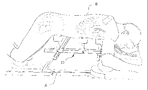
The frame for prone surgical positioning is designated generally
as 10 in the drawings. As shown in Fig. 1, the frame 10 is placed on
an operating table A and the patient H is positioned on the frame 10 in
the prone position, supported generally under the hips, the antero-
lateral aspects of the chest, and the upper chest, as set forth more
fully below. The operating table A may be a conventional operating
12
CA 02298088 2003-11-03
table, or it may be a radiographic operating table with a radiolucent
section below the patient's spine.
The frame 10 is shown more generally in Fig. 2. The frame has an
open, rectangular shaped base defined by a first lateral beam 12 , a
second lateral beam 14, and a pair of longitudinal beams 16. The beams
12, 14, and 16, are preferably square in cross section and may be made
from wood, aluminum, stainless steel, or other structural materials, as
is conventionally known in the art . In the preferred embodiment, the
beams 12, 14, and 16 are made from stainless steel and are hollow,
tubular, and capped at the ends . Exemplary dimensions of the beams in
the preferred embodiment may be a cross section measuring 1" by 1",
lateral beams 12 and 14 measuring about twenty inches, and longitudinal
beams 16 measuring about twenty-four inches . The base is positioned
' horizontally on the operating table A, the first lateral beam 12 being
aligned towards the head of the table A.
A pair of hollow sleeves 18 which are square in cross section and
which have an inside perimeter slightly larger than the outside
perimeter of the first lateral beam 12 are slidably disposed about the
beam 12 and temporarily secured to the beam 12 by thumbscrews 20. Each
sleeve 18 has a vertical post 22 extending at right angles to the .
sleeve 18. The posts 22 preferably are constructed from the same
material and have the same cross sectional shape and dimensions as the
13
CA 02298088 2003-11-03
lateral beam 12. An exemplary length for the vertical posts 22 is
about four inches . One end of the longitudinal beams 16 are preferably
fixedly attached to the top surface of the sleeves 18, as by welding,
in spaced apart relation from the vertical posts 22, each longitudinal
beam being positioned towards the outside of the frame 10, so that the
sleeve assemblies 18 are mirror images of each other.
Upper chest pads 24 are mounted on stainless steel platforms by
hook and loop fastening material (described below in conjunction with
the vertical posts shown in Figs. 3 and 4) at the top end of the
vertical posts 22. The upper chest pads 24 are preferably rectangular
in vertical cross section and may be made from any conventionally known
surgical pad material . An exemplary material which could be used is a
viscoelastic, polymeric material sold under the trade name Akton°
Polymer by Action Products, Inc. of Hagerstown, Maryland, product
number 40616, but custom sized, preferably to 4"L x 3"W x 3"H.
The frame 10 includes intermediate sleeves 26, as shown in Figs.
2 and 3. The intermediate sleeves 26 are hollow and preferably square
in cross section, having an inside perimeter slightly larger than the
outside perimeter of longitudinal beams 16. The sleeves 26 are
slidably mounted on the longitudinal beams 16 and temporarily secured.
by thumbscrews 28 which clamp the longitudinal beams 16 against the
opposing walls of the sleeves 26. A vertical post 30 is fixedly
14
CA 02298088 2003-11-03
mounted, as by welding, on the top wall of each sleeve 26 at a right
angle to the sleeve 26. The vertical posts 30 preferably have the same
size and construction as vertical posts 22. In the preferred
embodiment, vertical posts 30 are made from stainless steel, are hollow
and tubular, square in cross section, and measure 1" x 1" and four
inches long. Stainless steel pad mounting platforms 32 are mounted on
the top of the vertical posts 30.
Antero-lateral chest pads 34 are mounted to the platforms 32,
preferably by hook and loop fastening material 36 such as Velcro°
fixedly attached to the platforms 32 and the bottom of the pads 34.
Preferably in vertical cross section the antero-lateral chest pads
34 are shaped with a rectangular base lower section and a trapezoidal
upper section, having a side which slopes inward and downward towards
the open rectangular base defined by the lateral beams 12, 14 and
longitudinal beams 16, as seen most clearly in Fig. 2, and may be made
from any conventionally known surgical pad material. An exemplary
material which could be used is a viscoelastic, polymeric material sold
under the trade name Akton° Polymer by Action Products, Inc. of
Hagerstown, Maryland, product number 40622, but custom sized,
preferably to 6"L x 6 1/4"W x 5"H. The platforms 32 are preferably .
flat and rectangular, having a width approximately two inches shorter
CA 02298088 2003-11-03
than the width of the pads 34, permitting a two inch range of lateral
adjustment of the pads 34.
The frame 10 further includes a pair of transverse sleeves 38,
which are hollow, tubular, and have an inside perimeter slightly larger
than the outside perimeter of second lateral beam 14. The transverse
sleeves 38 are slidably mounted on the second lateral beam 14 and
temporarily secured by thumbscrews 40. Longitudinal sleeve 42 is
fixedly mounted, as by welding, to the top surface of transverse sleeve
38 at right angles to transverse sleeve 38. Longitudinal sleeves 42
are slidably mounted on longitudinal beams 16 and temporarily secured
by thumbscrews 44. It will be apparent to those skilled in the art
that, although sleeves 18, 26, 38, and 42 are shown in the drawings
being secured to the beams by thumbscrews, the sleeves 18, 26, 38, and
42 may be temporarily clamped or secured to the beams by a variety of
conventional clamping or locking mechanisms well known in the art.
Vertical posts 46, having the same construction and dimensions as
vertical posts 30, are mounted on the top wall of longitudinal sleeves
42 . Pad mounting platforms 48 are mounted on the top ends of posts 46.
Hip pads 50 are mounted to the platforms 48, preferably by hook
and loop fastening material 52 such as Velcro° fixedly attached to the
platforms 48 and the bottom of the pads 50. Preferably the size,
16
CA 02298088 2003-11-03
shape and material of hip pads 50 are identical to that of antero-
lateral chest pads 34.
Optionally, the frame 10 may include feet 54 positioned under the
ends of the f first 12 and second 14 lateral beams . The feet 54 should
be from a material resistant to sliding or skidding on the surface of
the table A, such as rubber or neoprene . The feet 54 may be removably
attached to the beams 12, 14, as is conventionally known in the art .
Advantageously, the feet 54 lift the frame 10 far enough above the
table that an x-ray cassette may slide under the frame so that plain
film x-rays may be taken intraoperatively.
It will be apparent from this construction that the longitudinal
beams 16 are disposed in a horizontal plane vertically superior to the
horizontal plane in which the lateral beams 12, 14 are disposed. It
will also be apparent that the lateral and longitudinal separation of
the pads may be adjusted by loosening the appropriate thumbscrews and
sliding the sleeves, thereby adjusting the size of the frame 10 to the
skeletal frame of the patient A.
In use, the frame 10 is assembled by sliding the first lateral
beam 12 through sleeves 18 and tightening thumbscrews 20, sliding
sleeves 26 onto longitudinal beams 16 and tightening thumbscrews 28,~
sliding sleeves 42 onto longitudinal beams 16 and tightening
thumbscrews 44, and sliding second lateral beam 14 into sleeves 38 and
17
CA 02298088 2003-11-03
tightening thumbscrews 40. The position of the pads 24, 34, and 50 are
adjusted to the patient B with the frame 10 inverted and the patient in
the supine position. The upper chest pads 24 should be positioned
below the second rib and above the nipple 1 ine or the f if th rib at the
sternoclavicular line, each pad 24 being disposed on opposite sides of
the patient' s B midline . The antero-lateral chest pads 34 should be
positioned below the fourth rib, not to extend below the costal margin
at the mid axillary line, each pad 34 being disposed on opposite sides
of the patient' s B midline . The hip pads 50 are placed on the anterior
aspect of the ilioinguinal region, each pad 50 being disposed on
opposite sides of the patient's H midline.
The frame 10 is placed on the table A and secured per facility
policy. The patient B is then rotated, positioned on the frame 10, and
secured. The patient's B head and upper arms are supported in
accordance with instructions of the anesthesiologist. The patient's B
knees are supported on knee pads, which may be elevated or lowered to
further decrease or increase flexion of the hips, respectively, if
desired for the particular surgery in hand. The frame 10 will
generally support the patient B is a prone position with the hips
flexed to less than 60°, a 30-30 flexion of the hips and knees being
preferable. It will be noted that positioning the longitudinal beams
16 at a fixed distance outside the vertical posts 22 will ordinarily
18
CA 02298088 2003-11-03
ensure that the longitudinal beams will not interfere with C-arm
fluoroscopy or radiography of the spine. After use, the frame 10 may
be conveniently collapsed and stored on a shelf . Advantageously, the
frame 10 is small enough and light enough to be manipulated by one
person.
The preferred embodiments of the invention provide a frame for use
with a conventional operating table for positioning a patient in a
prone position in which the curve of the spine during surgery
approaches the normal lordotic curve of the spine in the standing
position in order to facilitate surgical procedures involving
instrumentation or internal fixation of the spine. The abdomen is
pendulous to reduce hemorrhage, but intraoperative radiographs of the
spine, or C-arm fluoroscopy of the spine with a radiolucent table are
permitted. The position of the patient support pads is adjustable
1~ longitudinally and laterally in order to accommodate the different
skeletal sizes of children and adults. The frame supports the patient
at six locations and with a low profile for better positioning of the
spine for internal fixation. The frame may be disassembled for compact
storage and transport.
It is to be understood that the present invention is not limited .
to the embodiments described above, but encompasses any and all
embodiments within the scope of the following claims.
19