Note: Descriptions are shown in the official language in which they were submitted.
CA 02301924 2000-02-18
WO 99/09390 PCT/US98/16463
1
A HIGH QUALITY, CONTINUOUS THROUGHPUT, TISSUE
FIXATION-DEHYDRATION-FAT REMOVAL-IMPREGNATION
METHOD
BACKGROUND OF THE INVENTION
1. Field of the Invention
The present invention relates to the rapid, continuous flow,
processing of tissue for microscopic examination, from fixation to
impregnation.
2. Description of the Related Art
Conventional methods prepare tissues for histology by incubation
in separate solutions of phosphate-buffered 10% formaldehyde for
fixation, a series of increasing concentrations of ethanol for dehydration,
and xylene for clearing tissue of dehydration agent, prior to impregnation.
Because of the time required for this process, usually 8 hours or longer, it
is customary to complete these separate steps - fixation, dehydration,
clearing, and impregnation - overnight in automated mechanical
instruments designed for those tasks (see, for example, U.S. Pat. Nos.
3,892,197, 4,141,312, and 5,049, 510). A typical automated tissue
processor (TISSUE-TEK) requires more than eight hours and is
programmed to process batches of tissue samples as follows.
Sta. Solution Concen. Set Set P/V Agitation Vol.
Time Temp. ** of
(min) Sol.
I Buffered 10% 50 40 C On On 2.2-3.2
Formalin liters
2 Buffered 10% 50 40 C On On 2.2-3.2
Formalin liters
3 Alcohoi* 80% 50 40 C On On 2.2-3.2L
4 Alcohol 95% 50 40 C On On 2.2-3.2L
5 Alcohol 95% 50 40 C On On 2.2-3.2L
6 Alcohol 100% 50 40 C On On 2.2-3.2L
7 Alcohol 100% 50 40 C On On 2.2-3.2L
8 Alcohol 100% 50 40 C On On 2.2-3.2L
9 Xylene 100% 50 40 C On On 2.2-3.2L
10 Xylene 100% 50 40 C On On 2.2-3.2L
11 Paraffin 50 60 C On On 4
CA 02301924 2000-02-18
WO 99/09390 PCT/US98/16463
2
12 Paraffin 50 60 C On On 4
13 Paraffin 50 60 C On On 4
14 Paraffin 50 60 C On On 4
** - pressure/vacuum cycle
*- the alcohol used in most laboratories is a mixture of 90% ethyl, 5% methyl
and 5% isopropyl alcohol.
Such conventional methodology demands that the tissue specimens be sent
from the operating room, medical office or other sites, to a pathology
laboratory
on one day; the tissue specimens be prepared overnight; and the pathologist
render
a diagnosis based on microscopic examination of tissue sections the next day
at
the earliest, almost 24 hours after delivery of the specimen to the laboratory
(FIGURE 1). In addition to the minimum one-day delay in giving a surge6n the
benefit of a report from the pathologist, there are also problems associated
with
impeded work flow in the pathology laboratory necessitated by the requisite
batch
processing of specimens, the safety concerns that attend having instruments
operating overnight, the risk of possible instrument failures and the need to
monitor the instruments, and the waste of using large volumes of reagents for
such
processing when automated. Moreover, expensive measures are required to
prevent exposure of laboratory personnel to fumes and toxic substances
associated
with the reagents used in this process. Also, the large volumes of solvent
waste
and paraffin debris produced by conventional methodology pollute the
environment.
Conventional fixation and processing cause irreversible damage to the
structure of DNA and particularly RNA that limits the application of genetic
techniques for diagnosis and research. Consequently, most DNA and certainly
RNA analysis require special precautions with handling of material, such as
immediate freezing of fresh tissues, because retrospective genetic analysis is
impaired by conventional tissue processing techniques.
Histological diagnosis of a frozen section suffers from multiple
disadvantages in comparison to sections prepared from paraffin blocks: the
slide
prepared from a frozen section "does not possess . . . uniformity of quality";
"it is
technically more difficult for serial sections of the same specimen to be
CA 02301924 2000-02-18
WO 99/09390 PCT/US98/16463
3
examined"; "extreme caution must be exercised in cutting the specimen in order
to
ensure a sufficiently thin section and to avoid the possibility of damaging
details
of the specimen"; and all the slides must be prepared "while the tissue is in
the
initial frozen state" because, "[i)f the tissue is thawed and refrozen for
sectioning,
it is severely damaged" (U.S. Pat. No. 3,961,097).
There is an ever present interest in expediting tissue processing and
analysis for diagnostic purposes. Furthermore, recent healthcare focus has
been
directed to lessening the cost of various procedures including tissue
processing.
The costs of tissue processing are related to time, the space required for
preparation and analysis, reagents (both the amount required for processing
and
handling discard), and the number of personnel required. More importantly,
patients and their physicians depend on evaluation and diagnosis by the
pathologist to guide treatment. Reducing the amount of time needed to complete
tissue processing would lessen the anxiety experienced during the period
between
obtaining the specimen and delivering the pathologist's report to the surgeon.
Others have recognized the need to shorten the time required for tissue
processing, but they have made only modest improvements in the conventional
methods. To accelerate tissue processing, U.S. Pat. Nos. 4,656,047, 4,839,194,
and 5,244,787 use microwave energy; U.S. Pat. Nos. 3,961,097 and 5,089,288 use
ultrasonic energy; and U.S. Pat. No. 5,023,187 uses infrared energy. U.S. Pat.
No.
5,104,640 disclosed a non-aqueous composition of a fixative, a stabilizing
agent,
and a solubilizing agent that adheres a blood smear to a slide. However, the
aforementioned patents do not teach or suggest that the entire process of
preparing
diagnostic tissue slides could be accomplished in less than two hours,
starting
from fixation and ending with impregnation, with continuous throughput of
samples. The present invention provides such a process.
SUMMARY OF THE INVENTION
It is an object of the invention to provide compositions for tissue
processing and an apparatus and system for utilizing the same that reduces the
time required for tissue processing and analysis, and reduces the cost thereof
by
CA 02301924 2000-02-18
WO 99/09390 PCT/US98/16463
4
reducing time, the size of the laboratory facility, the volumes of reagents
used, and
the number of personnel required. This allows conversion of existing practice
to
rapid response surgical pathology for the patient undergoing an operation, and
may
even allow point-of-care diagnosis by the pathologist in the vicinity of the
operating room.
With regard to the processing and analysis of solid tissue, a tissue slice
must be on the order of 4 to 6 microns to be examined under a microscope,
whereas the thinnest slice of fresh tissue that can be obtained by cutting is
about 1
mm with the typical slice being on the order of 3 mm. In order to produce a
sufficiently thin slice from microscopic examination, it is necessary to
harden the
tissue so that a finer slice can be obtained, e.g., by sectioning with a
microtome.
The present invention greatly accelerates the tissue hardening process and
thus
turns the conventional overnight processing into a process which totals on the
order of 40 minutes. Thus, we have developed a simple, safe, low cost,
expeditious, and reliable method that permits preparation of impregnated
tissue
blocks suitable for microtome sectioning in less than two hours from the
moment
tissue is received in the pathology laboratory. This method allows continuous
flow of specimens, is adaptable to automation, precludes the need for formalin
and
xylene with their noxious fumes, allows standardization of tissue processing,
and
requires considerably smaller volumes of reagents than conventional methods.
Either fresh or previously fixed tissues can be processed by the present
invention.
In addition to the reduction in time required for tissue processing, the rapid
preparation of tissue by the present invention is capable of preserving tissue
structures and morphology that were lost with conventional methods.
Moreover, studies with tissues processed with the invention disclosed
herein indicate better preservation of DNA and particularly RNA extraction
than
with conventional processing methods. Thus, tissues obtained in hospitals and
other settings can be processed for both histologic and genetic studies soon
after
delivery to the laboratory, and archival material may be made available for
future
research and other applications. Improvements may be expected in the yield of
genetic material, the stability of the genetic material in archival form, the
size and
CA 02301924 2000-02-18
WO 99/09390 PCT/US98/16463
integrity of the genetic material, and reducing chemical modification of the
genetic
material in comparison to the prior art.
An object of the invention is to provide a method and an apparatus for
rapid processing of tissue for histology with continuous throughput. By
5 "continuous throughput," we mean accessing the system with additional
samples,
minutes apart. Therefore, at any given time there are samples of tissue in
different
stages of processing. In other words, with our method, there is continuous
throughput and flow of specimens along the various steps of tissue processing.
In
contrast with our method, batch processing is presently required because
conventional methodology takes eight hours or longer. Samples are placed in
automated instruments, which can not be access with additional samples until
the
entire instrument cycle is completed. All these tissue samples are at the same
stage of processing at any given step of the instrument cycle.
Yet another object of the invention is to provide non-aqueous reagents for
rapid, continuous flow processing of tissue for histology.
A further object of the invention is to eliminate the need for toxic
substances such as formalin and xylene in tissue processing.
In accordance with one aspect of the invention, a tissue specimen is fixed,
dehydrated, and fat is removed. A suitable admixture for use is a non-aqueous
solution comprised of fixative and dehydrating agents, preferably a ketone and
an
alcohol; the volume ratio of alcohol to ketone may be between about 1:1 to
about
3:1. The tissue specimen is incubated for about 25 nunutes or less, more
preferably for about 15 minutes or less, and even more preferably for about 5
minutes or less. Incubation is preferably between about 300C and 65 C, more
preferably between about 400C and 55 C, and most preferably between about
45 C and 500C.
Another aspect of the invention is fixation, dehydration, fat removal, and
clearing of a tissue specimen. A preferred solution in this aspect of the
invention
is alcohol and a clearant. This process may be accomplished in about 5 minutes
or
less.
CA 02301924 2000-02-18
WO 99/09390 PCT/US98/16463
6
In yet another aspect of the invention, a tissue specimen is cleared and
impregnated in a single solution comprised of a clearant and an impregnating
agent. Preferably, this process may be accomplished in about 5 minutes or
less.
Prior to sectioning, the impregnated tissue specimen may be embedded in the
impregnating agent.
A tissue specimen which has been fixed, dehydrated, and defatted may
then be impregnated in a wax solution. Consistent with dehydration of the
tissue
specimen, the wax solution is preferably as low as possible in water content.
Thus, the wax solution may be prepared prior to impregnation by heating the
wax
to evaporate any dissolved water and by degassing under reduced pressure.
Impregnation of the tissue specimen may take place under less than atmospheric
pressure and at elevated temperature to remove any solvents from the tissue
specimen and to draw the wax solution into the tissue specimen. Vacuum
decreases impregnation time by accelerating diffusion and reducing the
evaporation temperature of any solvents that may be present in the sample. The
wax solution may comprise degassed paraffin and/or mineral oil. Impregnation
of
the tissue specimen may be completed in about 15 minutes or less; preferably,
completed in about 10 minutes or less. Prior to sectioning, the impregnated
tissue
specimen may be embedded in the impregnating agent to form a tissue block.
Another embodiment of the invention is processing a tissue specimen from
fixation to impregnation in a series of solutions, at least some of which are
admixtures that perform more than one task at the same time: fixation,
dehydration, removal of fat, and impregnation. The admixture may include a
fixative, a dehydrating agent, and a fat solvent (e.g., ketone and alcohol).
Another
solution may include fixative, dehydrating agent, fat solvent, and clearant
(e.g.,
alcohol and xylene). Yet another solution may include a clearant and an
impregnating agent (e.g., xylene and paraffin). The tissue specimen may be
impregnated in a wax solution comprised of a mixture of different chain
lengths
(e.g., at room temperature, mineral oil which is liquid and paraffin which is
solid).
Preferably, an admixture contains at least two different chemicals (e.g., two
alcohols).
CA 02301924 2000-02-18
WO 99/09390 PCT/US98/16463
7
Processing time may be reduced by a non-aqueous admixture (e.g.,
fixative-dehydrating agent-fat solvent, fixative-dehydrating agent-fat solvent-
clearant, clearant-impregnating agent), microwave energy as a source to
achieve
uniform heating within the tissue specimen, and reducing the pressure by using
a
vacuum source. Diffusion of the solution into the tissue specimen and chemical
exchange may be promoted by mechanical agitation, heat, reduced pressure, or a
combination thereof.
The above steps may be accelerated by adding a fixative enhancer, a
surfactant, or both to the solutions used in the process. The fixative
enhancer may
be polyethylene glycol (PEG), mono- and dimethyleneglycol, propylene glycol,
polyvinyl pyrrolidone, or the like; the polymer used may be between about 100
and about 500 average molecular weight, preferably about 300 molecular weight.
The surfactant may be dimethyl sulfoxide (DMSO), polyoxyethylene sorbitan
esters (e.g., TWEEN 80), sodium dimethyl sulfosuccinate, mild household
detergents, or the like.
The fixative may be a ketone (e.g., acetone, methyl ethyl ketone), aldehyde
(e.g., acetylaldehyde, fonmaldehyde, glutaraldehyde, glyoxal), alcohol (e.g.,
methanol, ethanol, isopropanol), acetic acid, lead acetates and citrate,
mercuric
salts, chromic acid and its salts, picric acid, osmium tetroxide, or the like.
The tissue specimen may be dehydrated with methyl alcohol, isopropyl
alcohol, ethyl alcohol, propyl alcohol, butanol, isobutanol, ethyl butanol,
dioxane,
ethylene glycol, acetone, amyl alcohol, or the like.
Fat may be removed from the tissue specimen with an organic solvent such
as, for example, acetone, chloroform or xylene.
The clearant may be xylene, limonene, benzene, toluene, chloroform,
petroleum ether, carbon bisulfide, carbon tetrachloride, dioxane, clove oil,
cedar
oil, or the like.
The tissue specimen may be impregnated and/or embedded in paraffin,
mineral oil, non-water soluble waxes, celloidin, polyethylene glycols,
polyvinyl
alcohol, agar, gelatin, nitrocelluloses, methacrylate resins, epoxy resins,
other
plastic media, or the like.
CA 02301924 2000-02-18
WO 99/09390 PCTIUS98/16463
8
In the context of the invention, a "tissue specimen" is a piece of tissue that
may be processed by the methods disclosed herein. It may also refer to single
cells
from any biological fluid (e.g., ascites, blood, pleural exudate), or cell
suspensions
obtained from aspiration of solid organs or lavage of body cavities. Single
cells
may be pelleted by sedimentation or buoyant centrifugation prior to
processing.
The methods of the invention are specially suitable for tissue specimens in
which cell-cell contact, tissue organization, organ structure, or a
combination
thereof must be preserved. Such a specimen is a tissue slice preferably about
3
mm or less in its smallest dimension, more preferably about 2 mm or less, even
more preferably about 1.5 mm or less, and most preferably about 1 mm or less.
The tissue specimen may be fresh, partially fixed (e.g., fixation in 10%
formalin for 2-3 hours), or fixed (e.g., overnight fixation in 10% formalin or
any
other fixative). The above invention allows processing of a tissue specimen
from
fixation to impregnation in less than about two hours, preferably less than
about
90 minutes, more preferably less than about one hour, even more preferably
less
than about 45 minutes, and most preferably less than about 30 minutes. If the
tissue specimen is fixed or partially fixed, then the processing time may be
shortened accordingly. Tissue may be transported from the operating room to
the
pathology laboratory in an aqueous solution; such a transport solution may
consist
of equal volumes of an aqueous buffer and the non-aqueous admixture described
herein.
Following impregnation, the tissue specimen can be embedded to produce
a block. The agent used to embed the tissue specimen is preferably the same as
the material used for impregnation, but a different impregnating agent may
also be
used. The blocked tissue specimen can be mounted on a microtome to produce
tissue sections of between about 1 micron and about 50 microns, preferably
between about 2 microns and about 10 microns. The tissue sections may be
further processed for histochemical staining, antibody binding, in situ
nucleic acid
hybridization/amplification, or a combination thereof. The tissue specimens
are
then typically examined by microscopy, but other techniques for detecting
cellular
CA 02301924 2006-09-28
9
properties may be used to examine the processed tissue specimen (e.g.,
automated
cytometry, autoradiography, electrophoresis of nucleic acid).
The invention further provides a method of continuous tissue processing
comprising:
(a) providing a tissue specimen of up to 3 mm in thickness,
(b) fixing and dehydrating said tissue specimen by incubation in a series of
non-aqueous solutions with agitation and heating,
(c) impregnating said fixed and dehydrated tissue specimen under less than
atmospheric pressure, and
(d) repeating steps (b) and (c) at least once with another tissue specimen,
wherein at least one repetition of steps (b) and (c) takes about two hours or
less;
whereby tissue specimens are continuously processed by initiating step (b) for
a later-
processed tissue specimen before step (c) has been completed for an earlier-
processed
tissue specimen.
The invention further provides a method of continuous tissue processing for
histology comprising:
(a) slicing a solid tissue to provide tissue specimens with thickness between
I
mm and 3 mm, wherein a tissue specimen to be processed is not placed on a
slide for
histology until said tissue specimen has been hardened, impregnated, and
sectioned;
(b) hardening said tissue specimen by incubation in a series of non-aqueous
solutions with agitation and heating by microwave energy, wherein incubation
in a
first non-aqueous solution comprised of ketone and alcohol is then followed by
incubation in a second non-aqueous solution comprised of less concentrated
ketone
and more concentrated alcohol as compared to said first non-aqueous solution;
(c) impregnating said hardened tissue specimen in at least one wax solution, a
wax solution comprising a mixture of waxes having different melting points,
under
less than atmospheric pressure to provide a block comprised of said hardened
and
impregnated tissue specimen and wax, wherein hardening and impregnating a
tissue
specimen takes about two hours or less; and
(d) repeating steps (b) and (c) at least once with another tissue specimen;
whereby tissue specimens are continuously processed by initiating hardening of
a
later-processed tissue specimen before impregnation of an earlier-processed
tissue
specimen has been completed.
CA 02301924 2006-09-28
9a
The invention further provides an apparatus for continuous processing of
tissue specimens of up to 3 mm in thickness comprising:
(a) a microwave unit which sequentially fixes and dehydrates said tissue
specimens with a series of non-aqueous solutions in a vessel, and heats with
microwave energy and agitates said vessel of the microwave unit and its
contents;
wherein sources for the non-aqueous solutions are fluidly coupled to said
vessel of the
microwave unit to expose tissue specimens to said series of non-aqueous
solutions
and to drain said vessel of the microwave unit; and
(b) an impregnator unit which sequentially impregnates said processed tissue
specimens with a series of paraffin solutions in a vessel, and heats said
vessel of the
impregnator and its contents under less than atmospheric pressure during
impregnation;
wherein processing of a later tissue specimen in said microwave unit is
initiated
before impregnation of an earlier tissue specimen in said impregnator unit has
been
completed.
The invention further provides a tissue specimen or tissue section thereof
which has been fixed, dehydrated, and impregnated in a block of wax by the
above-
mentioned apparatus.
The invention further provides a solution for fixing and dehydrating tissue
used in the above-mentioned apparatus comprising an admixture of ketone and
alcohol, wherein the volume ratio of alcohol to ketone is between about 1:1 to
about
3:1.
The invention further provides a solution for fixing and dehydrating tissue
used in the above-mentioned apparatus selected from the group consisting of:
(a) 40%
acetone, 40% isopropyl alcohol, and 20% low molecular weight polyethylene
glycol
by volume; (b) 25% acetone, 55% isopropyl alcohol, 10% low molecular weight
polyethylene glycol, and 10% mineral oil by volume; (c) 10% acetone, 60%
isopropyl
alcohol, and 30% low molecular weight polyethylene glycol by volume; and (d)
25%
acetone, 55% isopropyl alcohol, and 20% low molecular weight polyethylene
glycol
by volume; wherein low molecular weight for a polymer is average molecular
weight
of about 300.
CA 02301924 2006-09-28
9b
The invention further provides a wax solution used in the above-mentioned
apparatus comprising mineral oil and paraffin which has been degassed and
dehydrated.
The invention further provides an apparatus for hardening and impregnating a
tissue specimen with thickness between 1 mm and 3 mm comprising:
(a) a microwave unit which hardens said tissue specimen with a non-aqueous
solution in a first vessel, and heats with microwave energy and agitates said
non-
aqueous solution in the first vessel, wherein said non-aqueous solution in the
first
vessel is comprised of ketone and alcohol;
(b) a microwave unit which hardens said tissue specimen with a non-aqueous
solution in a second vessel, and heats with microwave energy and agitates said
non-
aqueous solution in the second vessel, wherein said non-aqueous solution in
the
second vessel is comprised of less concentrated ketone and more concentrated
alcohol
as compared to said non-aqueous solution in the first vessel;
(c) a vacuum unit which impregnates said hardened tissue specimen with a
wax solution in a third vessel, and heats under less than atmospheric pressure
said
non-aqueous solution in the third vessel, wherein said wax solution in the
third vessel
is comprised of paraffin wax; and
(d) a vacuum unit which impregnates said hardened tissue specimen with a
wax solution in a fourth vessel, and heats under less than atmospheric
pressure said
non-aqueous solution in the fourth vessel, wherein said wax solution in the
fourth
vessel is comprised of more concentrated paraffin wax as compared to said wax
solution in the third vessel;
wherein tissue specimens are sequentially transferred through the first,
second, third
and fourth vessels after processing for at least about 15 minutes in each
vessel.
The invention further provides a tissue specimen or tissue section thereof
which has been hardened and impregnated in a block of wax by the above-
mentioned
apparatus.
The invention further provides a solution for fixing and dehydrating tissue
comprising an admixture of ketone and alcohol, wherein the volume ratio of
alcohol
to ketone is between about 1:1 to about 3:1.
CA 02301924 2006-09-28
9c
The invention further provides a solution for hardening tissue comprising (a)
to 40% acetone, (b) 40 to 60% isopropyl alcohol, (c) 0 to 30% polyethylene
glycol,
and (d) 0 to 20% mineral oil by volume.
The invention further provides a wax solution for impregnating tissue
5 comprising mineral oil and paraffin which has been degassed and dehydrated.
CA 02301924 2007-11-07
9d
BRIEF DESCRIPTION OF THE DRAWINGS
FIGURE 1 is a flow chart showing that almost 24 hours elapse between the
time a tissue specimen is obtained by a surgeon and the time a diagnosis by a
pathologist can be prepared from microscopic examination of sections of the
tissue;
FIGURE 2 is a schematic plan view of a tissue processing facility provided
in accordance with the invention. Equipment Legend: A. Vacumn Pump; B. Water
Bath; C. Microwave Oven; D. Microwave Oven; E. Microwave Oven; F. Vacumn
Pump; G. Water Bath; H. Counter Top Hood (small); I. Embeder; J. Parafin Bath;
K.
Slide Oven; L. Microt; M. Water Bath; N. Hot Plate; O. Stainer; P.
Imrnuno_Stainer
(IHC); Q. IHC Controls; R. Slip Cover.
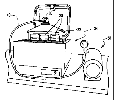
FIGURE 3 shows an exemplary shaker bath provided as a part of the
apparatus and system of the invention;
FIGURE 4 shows an exemplary microwave oven for use as a part of the
apparatus and system of the invention;
FIGURE 5 shows an exemplary paraffin bath provided as a part of the
apparatus and system of the invention;
FIGURE 6 is a schematic illustration of a microwave/impregnation unit
provided in accordance with an alternative embodiment of the invention;
FIGURE 7 is a schematic illustration of a slicing guide provided in
accordance with a further exemplary embodiment of the invention;
FIGURE 8 is a broken away view of a tissue clamp and slicing assembly
provided in accordance with a further embodiment of the invention;
FIGURE 9 is a schematic illustration of a tissue holder provided in
accordance with a further embodiment of the invention;
FIGURE 10 is a schematic illustration of a tissue cassette provided in
accordance with the invention for receiving small tissue samples;
FIGURE 1 I shows a tissue receiving and transporting jar with tissue
cassette in accordance with an embodiment of the invention; and
CA 02301924 2007-11-07
WO 99/09390 PCTIUS98/16463
FIGURES 12A-12B show agarose gel electrophoresis of DNA and RNA,
respectively, prepared from processed tissue specimens. In FIGURE 12A, lane I
contains molecular weight standards, lane 2 contains a diluted, sainple from a
tissue specimen processed according to the present invention, lanes 3-4
contain
5 DNA samples from tissue specimens processed according to the present
invention,
and lanes 5-6 contain DNA samples from tissue specimens processed according to
a conventional method. In FIGURE 12B: lanes 1, 4 and 6 are blanks, lanes 2-3
are
samples from tissue specimens processed according to a conventional method,
lane 5 contains an RNA sample from a tissue specimen processed according to
the
10 present invention, and lane 7 contains control RNA.
DETAILED DESCRIPTION OF THE INVENTION
A process and apparatus for rapid, continuous histological processing of
tissues is disclosed. The steps of fixation, dehydration, fat removal, and
impregnation can be performed in less than about two hours; this allows a
pathologist to evaluate samples shortly after receipt, perhaps while the
patient is
still in the operating or recovery room. Patient anxiety can be reduced by
reducing
the time required for pathological diagnosis. Rapid and continuous processing
is
accomplished by decreasing the thickness of tissue specimens, use of non-
aqueous
solutions composed of admixtures, solution exchange at elevated temperature
and
with agitation, uniform heating of tissues and solutions with microwave
radiation,
impregnation under vacuum pressure, or a combination thereof.
Fixation, dehydration, and removal of fat are required for the preparation
of tissue prior to impregnation. These steps are facilitated by trimming the
tissue
to a suitable size prior to processing, and using cassettes which hold such
tissue
blocks and allow their easy transfer between solutions for fixation,
dehydration,
removing fat, and impregnation.
Fixation initiates hardening of the tissue specimen, and may preserve cell
morphology by cross linking proteins and halting cellular degradation. Without
chemical fixation, endogenous enzymes will catabolize and lyse the cell, and
the
tissue microanatomy will be altered. Such fixatives may be a ketone, aldehyde,
CA 02301924 2000-02-18
WO 99/09390 PCT/US98/16463
11
alcohol, acetic acid, heavy metals, chromic acid, picric acid, or osmium
tetroxide.
Indications that fixation was inadequate can include: disassociation of tissue
structures, bubbles in tissue sections, poor and irregular staining, shrunken
cells,
clumping of cytoplasm, condensation and less distinct nuclear chromatin, and
autolysis/hemolysis of erythrocytes.
Dehydration removes water from the tissue specimen to promote
hardening. Replacement of water in the tissue specimen with a dehydrating
agent
also facilitates subsequent replacement of the dehydrating agent with material
used
for impregnation. This solution exchange is enhanced by using a volatile
solvent
for dehydration. The dehydrating agent may be low molecular weight alcohols,
ketones, dioxane, alkylene glycols, or polyalkylene glycols. Failure to
deliydrate
the specimen can lead to inadequate impregnation, poor ribbon formation during
sectioning, clefts in tissue sections, dissociation of structures, water
crystals in
tissue sections, and poor staining.
Fat in the tissue specimen is removed with a solvent because fat impairs
clearing and impregnation. Inadequate fat removal can result in spreading
artifacts
of tissue sections, wrinkling of tissue sections, and poor staining.
Optionally, the tissue specimen is cleared. The clearant extracts
dehydrating agent from the tissue specimen and reduces its opacity. Examples
of
clearants include xylene, limonene, benzene, toluene, chloroform, petroleum
ether,
carbon bisulfide, carbon tetrachloride, dioxane, clove oil, or cedar oil.
Finally, once the tissue specimen is suitably fixed and dehydrated, it is
hardened by impregnation with an agent such as wax, celloidin, polyalkylene
glycols, polyvinyl alcohols, agar, gelatin, nitrocelluloses, methacrylate
resins,
epoxy resins, or other plastics. Appropriate hardening of the tissue specimen
with
adequate preservation of cellular morphology is required prior to placing the
impregnated specimen in a block and obtaining ten micron or thinner sections
with
a microtome knife. Preferred impregnation materials are commercial wax
formulae, mixtures of waxes of different melting points (e.g., liquid mineral
oil
and solid paraffin), paraplast, bioloid, embedol, plastics and the like.
Paraffin has
been chosen for use in the examples herein because it is inexpensive, easy to
CA 02301924 2000-02-18
WO 99/09390 PCT/US98/16463
12
handle, and ribbon sectioning is facilitated by the coherence of structures
provided
by this material.
If processing of the tissue specimen is incomplete, the sections cut by the
microtome knife will appear cracked or "exploded". Tissue processing is deemed
a failure when one or more of the following problems is encountered: embedded
tissue blocks are too soft or too hard, sections fall out or show an amount of
compression different from the embedding agent, sections appear mushy, tissue
ribbons fail to form or are crooked, sections crumble or tear, erythrocytes
are
lysed, or clumping of cytoplasm, condensation of chromatin, basophilic
staining of
nucleoli, shrunken cells, spreading artifacts and moth-eaten effect.
For wax-impregnated sections on glass slides, the wax may be melted and
removed prior to staining or immunohistochemistry. The tissue section is
rehydrated and then analyzed as described below with stains or antibodies.
After
staining is completed or the histochemical reaction is developed, the slide
may be
coverslipped and viewed under a microscope. Alternatively, the stained or
antibody-decorated specimen may be studied with an instrument for cytometry.
The tissue blocks may be stored for archival purposes or retrospective
studies.
The present invention is compatible with preparation of nucleic acids,
DNA or RNA, from processed tissues. Thus, genetic study is possible for
specimens collected routinely in the clinical pathology laboratory. The
combined
power of these technologies will be great. Histological observations may be
correlated with genetics by analyzing one section by staining or
immunohistochen-iistry, and preparing nucleic acids from an adjacent section
for
genetic analysis. For example, diseased and normal regions of the same section
may be compared to detect genetic differences (e.g., mutations, levels of
transcription), disease progression may be characterized by comparing genetics
differences in samples taken at several time points, and tumor evolution may
be
assessed by following the accumulation of genetic differences from primary
cancer
to metastasis.
Many features distinguish the present invention: (a) thin slicing of the
tissues prior to processing; (b) continuous input of tissue specimens, and
CA 02301924 2000-02-18
WO 99/09390 PCT/US98/16463
13
continuous flow through the system; (c) elimination of water from solutions
(i.e.,
non-aqueous solutions); (d) fixation, dehydration, fat removal, clearing, and
impregnation of tissue performed with uniform heating (e.g., microwave
energy);
(e) admixture solutions to fix-dehydrate-remove fat, fix-dehydrate-remove fat-
clear, and clear-impregnate; and (f) impregnation of tissue under reduced
pressure
with degassed impregnating agent. These features make the present invention
simple, practical, easy to implement, and amenable to automation.
Hematoxylin-eosin staining is commonly used for histological study and
may be considered a standard for comparison by pathologists. In addition, the
present invention has been found to be compatible with other stains including
trichrome, reticulin, mucicarmine, and elastic stains as described in general
references such as Thompson (Selected Histochemical and Histopathological
Methods, C.C. Thomas, Springfield, Illinois, 1966), Sheehan and Hrapchak
(Theory and Practice of Histotechnology, C.V. Mosby, St. Louis, Missouri,
1973),
and Bancroft and Stevens (Theory and Practice of Histological Techniques,
Churchill Livingstone, New York, New York, 1982). Such staining procedures
would take between 30 minutes and several hours to complete, although rapid
staining procedures are available from Fisher Scientific that require only
five
minutes to accomplish.
Tissue may be obtained from an autopsy, a biopsy (e.g., endoscopic
biopsy), or from surgery. For cancer surgery, the ability to provide a
pathological
diagnosis from a stained tissue section will provide the surgeon with
information
that may be used prior to the patient's departure from the operating room. For
example, an indication from the pathologist that the cancer is confined to the
2 5 resected tissue may allow the surgeon to be conservative in treatment and
to
preserve neighboring healthy tissue. Alternatively, a finding by the
pathologist
that cancer is not confined to a resected organ would perniit more aggressive
surgical treatment while the patient was still in the operating room.
Over 20,000 samples of tissue have been successfully processed by the
3 0 present invention, including: brain, breast, carcinoma (e.g., bowel,
nasopharynx,
breast, lung, stomach), cartilage, heart, kidney, liver, lymphoma, meningioma,
CA 02301924 2000-02-18
WO 99/09390 PCT/US98/16463
14
placenta, prostate, thymus, tonsil, umbilical cord, and uterus. Mineralized
tissue
(e.g., bone, teeth) would require decalcification prior to processing by the
present
invention. For example, tissue may be decalcified with a hydrochloric
acid/ethylenediaminetetraacetic acid (EDTA) solution from Stephens Scientific
(Allegiance Healthcare Supply, catalog no. 1209-IA) according to the
manufacturer's instructions.
Tissue sections processed by the present invention may also be used in
immunohistochemistry. The present invention provides tissue specimens in which
antigen is recovered and preserved, the choice of fixative may be optimized
for
recovery and preservation of particular antigens. Non-specific binding sites
are
blocked, antigen is bound by specific antibody (i.e., the primary antibodyj,
and
non-bound antibody is removed. If labeled with a probe or signal generating
moiety, the primary antibody may be detected directly but it is preferred to
attach
the probe to a protein (e.g., a secondary antibody) that specifically binds
the
primary antibody. Secondary antibody may be raised against the heavy or light
chain constant region of the primary antibody. This amplifies the signal
generated
by an antigen-antibody conjugate because each primary antibody will bind many
secondary antibodies. Altematively, amplification may occur through other
specific interactions such as biotin-streptavidin. Antibody binding is
performed in
a small volume to reduce usage of expensive reagents and maintain a high
binding
rate; evaporation of this small volume is reduced by incubation in a humidity
chamber. The signal generating moiety is preferably an enzyme which is not
otherwise present in the tissue. For example, alkaline phosphatase and
horseradish peroxidase may be attached to the secondary antibody or conjugated
to
streptavidin. Substrates are available for these enzymes that generate a
chromogenic, fluorescent, or luminescent product that can be detected
visually.
The staining pattern for antigen may be used to localize expression of the
antigen in the context of cellular structures revealed by counterstaining.
Antigen
expression can identify cell or tissue type, developmental stage, tumor
prognostic
markers, degenerative metabolic processes, or infection by a pathogen.
CA 02301924 2007-11-07
WO 99/09390 PCT/US98/16463
Antigen-antibody binding may also be visualized witli radioactive,
fluorescence, or colloidal metal probes by autoradiography, epifluorescent
microscopy, or electron microscopy, respectively. Similar probes may be used
to
detect nucleic acid in the tissue section by in situ hybridization to identify
genetic
5 mutations or transcripts; alternatively, the nucleic acid (DNA or RNA) may
be
extracted from tissue sections and analyzed directly by blotting, or amplified
prior
to further genetic analysis.
Mutations may be germline and used to trace genetic predisposition of
disease, or mutations may be somatic and used to determine genetic alterations
in
10 disease pathogenesis. The disease may be a metabolic or neurologic
disorder,
malignancy, developmental defect, or caused by an infectious agent. The
present
invention preserves material for genetic analysis by a simple procedure and
room
temperature storage.
It is envisioned that the present invention will preserve tissue that yield
15 greater amounts of nucleic acid with a higher average molecular weight than
tissues processed by conventional processes.
In accordance with an exemplary system for tissue processing provided in
accordance with the present invention, a series of tissue processing stations
may
be provided, e.g., in a single tissue processing unit or area. By way of non-
limiting example, a suitable tissue processing facility is illustrated in
FIGURE 2.
The first step in the process, which may be carried out at the tissue
processing facility or elsewhere, is to prepare a suitable tissue sample for
hardening and ultimate examination. Typically, a slice of the tissue of
interest is
prepared. The finest slice possible is obtained, of about 1 to 3 mm and
preferably
1 to 2 mm in thickness. Processing time is proportional to the size of the
tissue
sample being processed. The tissue slice is placed in a tissue cassette in
which the
tissue is contained during the immediately following processing steps. The
tissue
cassette is next placed in a first solution provided in accordance with the
present
invention.
By way of example, the cassette 10 may be placed in a conventional beaker
12, having the first solution 14 therein, preferably by itself as the process
CA 02301924 2007-11-07
WO 99/09390 PCT/US98/16463
16
described is a substantially continuous one, or together with a limited number
of
otlier, similar tissue cassettes. The beaker 12 is tlien placed in a shaker
bath 16, as
illustrated in FIGURE 3, for gently agitating and heating the same. We have
used
a LAB-LINE/DUBNOFF incubator-shaker bath for this purpose. Rather than
water, as it is our goal to minimize moisture to which the tissue samples are
exposed and, in fact, ultimately to dehydrate the same, we have provided
glycerine
as the temperature conducting fluid 18 in the shaker bath 16. Glycerine has
the
advantage that it is an effective conductor of thermal energy but it does not
evaporate. Evaporation would undesirably increase the moisture of the
environment in which the tissue is processed, and would require periodic
replenishment . Because the glycerine neither needs replacement nor adds
moisture to the environment, it is most preferred. For this stage of the
process, the
tissue sample (in cassette 10) is disposed in the first solution, in the
shaker bath 18
for approximately 3-15 minutes.
Supplemental agitation is desirably also provided during the shaker-bath
step. Presently, an external pump (A) (FIGURE 2) is provided with a tube (not
shown) therefrom inserted into the solution beaker 12 or other receptacle for
bubbling and thus agitating its contents. An aeration diffusion nozzle or
plate may
be provided to provide for more uniform solution agitation as deemed necessary
or
desirable.
To ensure that the tissue cassette 10 and first solution containing beakers
12 remain upright and in a desired disposition, we have modified the
conventional
shaker-bath to provide transverse wires or stays 20, e.g., four wires,
defining, e.g.,
five longitudinal channels in which tissue cassette containing beakers 12 may
be
disposed. Thus, for example, sample containing beakers 12 may be regularly
added to the shaker-bath 18 and sufficiently processed tissue samples removed
in
tum therefrom for further processing as described hereinbelow, by adding new
samples on the left end of the shaker bath and removing sufficiently processed
samples from the right end thereof..
Next the tissue sample cassette 10 is exposed to a series of fluids while
simultaneously being agitated and subjected to microwave radiation. In the
CA 02301924 2007-11-07
WO 99/09390 PCT/US98/16463
17
currently proposed embodiment, three microwave units are provided, as shown in
FIGURE 2, each having a different solution in which the tissue sample
containing
cassette is submerged for a prescribed period. In the alternative, a single
source of
microwave energy could be provided. However, such would require sequential
placement of the respective solutions for receiving the tissue cassette. While
for a
single tissue sample such solution placement and replacement would not
significantly increase the duration of the tissue processing cycle, it can be
appreciated that the use of a single microwave that receives multiple
solutions,
may hinder the continuity of the process with respect to subsequent samples.
Indeed, where a series of microwave units are provided, as a given tissue
sample is
moved from one microwave to the next having the next solution, a subsequent
tissue sample can then be received in the first microwave unit. Thus,
providing a
unit for each of the respective solutions means that a subsequent tissue
sample
need not be held while all microwave processing steps of the proceeding sample
have been completed. It is to be understood, however, that with the noted
hindrance of continuity, the three microwave units illustrated could be
reduced to
two or even one. Likewise, other steps in the process may be combined or sub-
combined as deemed necessary or desirable from a balance of process continuity
versus a potential reduction in manpower, equipment, space requirements, etc.
An
exemplary such more compact unit is discussed in greater detail below, with
reference to FIGURE 6.
With reference now to FIGURE 4, an exemplary microwave unit 22 for
tissue processing is illustrated. For applying microwave radiation, we are
currently using laboratory microwave ovens obtained from Energy Beam Sciences,
Inc. We have used two microwave processor models, H-2800 and H-2500. Either
model or another, similar such system could be used. By way of example, a
Pyrex
or other clear microwaveable fluid receptacle 24 is utilized to hold
respectively
second, third and fourth solutions provided in accordance with the invention
in
each of the three microwave units (FIGURE 2). A temperature probe 26 is placed
in the solution to ensure that the temperature of the respective bath is
within the
desired range. Moreover, to provide for agitation, which accelerates the
tissue
CA 02301924 2007-11-07
WO 99/09390 PCT/US98/16463
18
processing, aeration is provided. The microwave units we have used include a
tube 28 for aeration. A single tube may be inserted into the bath, but for
more
uniform and complete agitation, it is most preferred to provide a diffusion
plate or
nozzle head (not shown in detail) in cooperation with the gas tube 28 for
diffusing
the agitating bubbles, e.g., across a substantial portion of the diameter of
the
solution receptacle for uniform agitation of the entire volume of solution.
Such
diffusion plates and nozzles are well known and can be provided, e.g., at the
base
of the solution receptacle.
Conventionally, paraffin is degassed as a part of the tissue processing
procedure. Degassing removes organic solvents from the paraffin. To enhance
this process, and to reuse the paraffin in the system we propose continuous
degassing. This is accomplished by maintaining the vacuum within the covered
Pyrex 32 at 640 mm. Hg.
Following the three sequential steps employing microwave radiation, the
tissue sample cassette(s) are placed in a paraffin bath, as shown in FIGURE 5.
Currently, we provide a paraffin bath comprising three paraffin bath stations
(beakers) 30 provided within a covered Pyrex jar 32. For the purpose of
temperature control, the Pyrex jar 32 is placed in, e.g., a Poly Science brand
water
bath 34. By applying a grease or the like to the internal edges of the flanges
on
both the lid and jar, an airtight coupling can be provided between the lid and
jar
and thus a vacuum can be pulled through a tooled hose connector 36 provided in
the lid. Suitable such Pyrex brand jars are available from Fisher Scientific.
We
have used Model No. 01-092-25. To create a vacuum within the Pyrex jar 32, a
conventional pressure/vacuum pump 38 is coupled to a tube 40 that is in turn
coupled to connector 36. A suitable such power operated pump is available from
Fisher Scientific and has for example a 100 psi max. Agitation is preferably
provided during the paraffin bath step, either through vibratory agitation;
ultrasound, or potentially via aeration.
Next the tissue sample must be embedded. For that purpose we use a
conventional Tissue-Tek embedding console system (I) (FIGURE 2) available
from Miles/Sakura, e.g. Model No. 4708.
CA 02301924 2007-11-07
WO 99/09390 PCT/US98/16463
19
The embedded tissue sample is then cut in a conventional manner witli a
microtome (L) (FIGURE 2) and floated (M) for placement, we use the Leitz 1512
Microtome, and the Lipshaw Electric Tissue Float Model 375.
After the slice is disposed on the slide, the slide is heated to remove the
paraffin. We have used the Isotemp Oven 300 series available from Fisher (K)
(FIGURE 2).
Next the slides are stained. To accelerate the staining process, we propose
to use an automated stainer (0) (FIGURE 2)to reduce the number of personnel
and time required. A non-continuous process could use the Sakura diversified
stainer DRS-601 which stains slides in batches; alternatively, a continuous
process
could use a Leica auto stainer XL which contains a dewaxing stage so that
separate incubation in an oven may be omitted. The fixed and stained tissue
sample is then covered, e.g. with the Tissue-Tek coverslipper, Manufacturer
No.
4764 (R) (FIGURE 2).
As described above, the system for carrying out the dehydration and
impregnation in accordance with the invention can be a series of discrete
units. In
the alternative, as also noted above, one or more steps can be carried out in
a
single processing component or unit. As also discussed above, the number of
units provided and the steps carried out by each unit impacts the continuity
of the
processing unit. Thus, in low volume environments, a single unit for carrying
out
a plurality of the tissue processing steps may be advantageous and will not
significantly impact continuity of tissue processing. In higher volume systems
environments, two or more units may be preferred.
An exemplary combined unit 42 is illustrated in FIGURE 6. The
combined unit 42 in fact includes two subunits; a microwave processor unit 44
and an impregnator unit 46. The microwave processor unit 44 is provided for
sequentially submerging the tissue being processed in solution A, solution B,
and
solution C, in each instance agitating the solution and exposing the tissue to
microwave energy. Thus, in the illustrated embodiment, a vessel 48 is provided
for receiving for example one or more trays 50 on which one or more tissue
cassettes 10 may be placed. The vessel 48 is fluidly coupled to a source of
each of
CA 02301924 2007-11-07
WO 99/09390 PCT/US98/16463
the solutions for tissue dehydration. Thus, once the tissue cassette(s) are
placed on
the respective tray(s) 50, solution A is conducted to the vessel 48 and
microwave
energy is applied thereto simultaneous to agitation via, for example, an
aeration
tube (not shown in FIGURE 6). After a sufficient time of exposure has passed,
5 solution A is drained and the tissue cassettes are preferably flushed either
with
solution B or with a combination of solution A and solution B so as to
substantially eliminate residual solution A. Solution B is then fed to the
vessel 48
whereupon microwave energy and agitation are again applied for a prescribed
period. At the conclusion of administration of solution B, solution B is
returned to
10 a storage vessel therefor and the tissue samples are flushed either with
solution C
or a combination of solution B and solution C. Thereafter, solution C is fcd
to the
vessel 48, agitation and microwave energy are applied, and ultimately solution
C
is drained. The tissue samples are then ready for impregnation.
In the illustrated embodiment impregnation is carried out in a second
15 subunit 46 of the assembly. This aliows impregnation to be carried out
while a
subsequent tissue sample(s) are subject to microwave energy application. If a
single unit is provided, then the vessel used for microwave processing can be
used
for impregnation however the microwave energy would not be applied thereto
during the impregnation steps.
20 In accordance with the proposed impregnation process, a series of paraffin
solutions, e.g., 3 or 4, are applied to the tissue cassettes disposed e.g. on
suitable
trays 52 in a vessel 54, to provide sequential paraffin baths to effect the
impregnation of the tissue sample as a final step in the tissue preparation
process.
In the impregnator subunit 46, the tissue samples are placed under a vacuum at
a
controlled elevated temperature. The tissue samples are preferably also
agitated
during this step with a magnetic stirrer, ultrasound, or air bubbler.
The remaining embedding, etc. steps of slide preparation are carried as
outlined above with reference to FIGURE 2.
In accordance with the invention, additional, specialized instruments and
apparatus have been developed to facilitate tissue processing in general and
in
CA 02301924 2007-11-07
WO 99/09390 PCT/US98/16463
21
accordance witli the invention, in particular. These specially designed
instruments
and apparatus are described herein below.
As noted above, it is difficult to cut a thin slice of a solid tissue sample.
On the other hand it is desirable, in terms of minimizing dehydration and
fixation
time, to have the tissue sliced as thinly as possible in advance of the
dehydration
process. To facilitate creation of a thin slice we have proposed three
instruments
to aid the pathologist. One, for convenience referred to herein as a slicing
guide
60, as illustrated in FIGURE 7, is in the form of a thin metal plate 62 on the
order
of, e.g., 1 to 2 mm in thickness, having a cutout 64 the width of, for
example, a
thumb nail (about 1 cm2). A stop 66 is defined at the end of the cutout or
notch 64
to serve as a knife or blade stop. To facilitate picking up the slicing guide
60 from
a flat surface or other cutting surface, a lip 68 may be provided at the end
of the
metal plate 62, remote from the cutting notch. To provide a thin slice of
tissue, a
larger segment of tissue is placed over the cutout or notch 64 so that a
portion
thereof is disposed in the notch. Pressure is then applied to the exposed
surface of
the tissue and a cutting instrument is placed against and slid horizontally
along the
slicing guide plate so as to sever the tissue disposed in the notch 64 from
the
remainder of the tissue. Engagement of the cutting blade with the blade stop
66
completes the cutting process and the bulk of the tissue, disposed above the
cut, is
placed aside. The remaining tissue, disposed in the slot, can then be placed
in a
suitable tissue cassette for dehydration and impregnation.
As can be appreciated, the slicing guide 60 facilitates the production of a
thin slice of tissue of generally uniform thickness which may be further
processed.
As another alternative for producing a thin tissue slice, we have proposed
to provide flat plates or blocks 70 at the end of an otherwise conventional
forceps
72, as schematically illustrated in FIGURE 8. The blocks may be permanently or
temporarily secured to the ends of the forceps. This provides rather large,
flat
clamping surfaces 74. The tissue to be cut may be placed between the clamping
blocks 70 and a sharp blade passed between the clamping blocks to slice the
tissue. By cutting closely to one of the two generally planar flat surfaces
74, a thin
tissue slice of generally uniform thickness can be provided. The parallel
rather
CA 02301924 2007-11-07
WO 99/09390 PCT/US98/16463
22
large flat surfaces provide uniform pressure distribution tlius liolding the
tissue in
position during the cutting process and then ensuring a uniform cut that
preferably
preserves the integrity of the tissue.
To hold the tissue in position during cutting we have also proposed a three
prong fork-like instrument 92, illustrated in FIGURE 9. In the illustrated
embodiment the prongs 94 are spaced from each other by approximately one
centimeter and each has a sharp, pointed tip 96 to facilitate penetration of
the
tissue with minimal disruption. By holding the tissue to a cutting board with
the
prongs 94 of the instrument 92, suitable slices of tissue can be obtained by
cutting
parallel to or between the prongs. In the illustrated embodiment, the
instrument
92 is characterized in that the prongs have a length on the order of 5-10 crim
to
accommodate a variety of specimens and a handle of about 8 centimeters in
length, itself spaced from the prongs by 2-4 centimeters, to facilitate
manipulation
of the instrument and a sure grip during cutting. We have found that the fork-
like
instrument 92 is particularly advantageous in obtaining sections from organs
such
as the intestine and gallbladder. Indeed, securing such specimens with prongs
94
prevents the various layers of tissue from sliding upon each other during the
cutting process.
We have also proposed to provide a tissue receiving unit and cassette for
2 0 use in the operating room, to facilitate transport of tissue, particularly
very small
segments of tissue, for example those obtained by needle biopsy. When such
biopsied tissue is put directly into, for example, a jar of suitable solution,
it can
often be difficult for the lab technician to retrieve the minute tissue sample
from
the jar and in particular to ensure that all biopsied tissue is retrieved.
Thus, as
illustrated in FIGURES 10 and 11, we have proposed to provide tissue cassettes
10' to the operating room for immediately receiving such minute tissue
samples.
To contain such tissue samples within the tissue cassette 10', we have
provided thin sheets of biopsy sponge material 80, which is an open cell
plastic
foam, at least one of which has a partial depth recess 82 defined therein to
provide,
together with the other biopsy sponge a compartment for receiving the biopsied
tissue. Thus, in the operating room the biopsied tissue can be disposed
CA 02301924 2007-11-07
WO 99/09390 PCT/US98/16463
23
immediately in the recessed portion 82 of one of the biopsy sponges 80 and the
tissue cassette 10' closed. To maintain the integrity of the tissue for
transport to
the processing lab, the tissue cassette 10' is placed within ajar of suitable
solution.
To facilitate retrieval of the cassette and to ensure that it is maintained
fully
submerged in the solution, we have provided a specimen jar 86 having a
columnar
support 88 projecting from the lid 90 and having structure at the tip thereof
90 for
coupling to complementary structure 84 on the tissue cassette 10'. FIGURE 11
shows the tissue cassette 10' attached by its top surface. However,
alternative
attachment points are possible such as the bottom surface or the hinged side
of the
cassette. Furthermore, two or more cassettes may be attached to the columnar
support 88.
Thus the tissue cassette 10' with the biopsied tissue therewithin can be
temporarily secured to the distal end of the columnar support 88 and inserted
into
a suitable solution for transport. At the tissue processing lab, the lid 90 is
removed from the jar 86 and the tissue cassette 10' removed from the column
88.
Any suitable fasteners such as velcro type fasteners, plastic snap lock, dove
tail
slide connectors or other cooperative engagement structure can be provided to
attach the tissue cassette 10' to the support column 88. The solution within
the
specimen jar 86 may be a transport (aqueous) solution or the first (non-
aqueous)
solution. It would be convenient to provide the specimen jar in the operating
room with the cassette attached to the outside of the jar and then to invert
the lid
so that the cassette is immersed in the solution within the jar after tissue
is placed
within the cassette.
The present invention will have many advantages over conventional
methods in the areas of the practice of pathology, patient care, biomedical
research, and education.
The availability of microscopic diagnosis of tissue samples within abotft 40
minutes to about 2 hours after receipt will allow rapid, or even real-time,
clinical
interaction between surgical intervention and pathological evaluation. This
may
bring about significant improvements in patient care by eliminating or
reducing to
CA 02301924 2000-02-18
WO 99/09390 PCT/US98/16463
24
a minimum patient anxiety during the wait for diagnosis of disease, prognosis,
and
planning for treatment.
Consequently, there will be a drastic reordering of the workflow in
pathology laboratories. Clinical laboratory space, pathological expertise, and
clerical and technical personnel will be utilized more efficiently. Continuous
workflow will improve accessibility and responsiveness of pathologists who
process and evaluate specimens, reduce the number of pathologists needed to
process and evaluate specimens, and may also improve medical education,
particularly the accessibility and responsiveness of residency programs.
The smaller volume of reagents will result in cost savings. Elimination of
formaldehyde and xylene and the diminished requirement for other hazardous
chemicals will provide benefits to the environment and increased safety in the
laboratory.
Standardization of tissue fixation and processing procedures will ease
comparison of specimens from different laboratories. Artifacts in histology
due to
the use of formaldehyde and/or prolonged processing will be eliminated; thus,
allowing more precise evaluation of microscopic morphology of normal and
diseased tissues. Similarly, antigen retrieval and staining will be improved.
For
genetic analysis, formaldehyde-induced DNA mutations will be eliminated and
extraction of nucleic acid from archival material may be enhanced. The
feasibility
of RNA studies from stored, fixed paraffin-embedded tissue opens unlimited
avenues for diagnostic and research applications.
All books, articles, applications, and patents cited in this specification are
incorporated herein by reference in their entirety.
The following examples are meant to be illustrative of the present
invention; however, the practice of the invention is not limited or restricted
in any
way by them.
EXAMPLES
EXAMPLE 1
Two mm thick or thinner slices of fresh or previously fixed tissue were
held in tissue cassettes and placed in a non-aqueous first solution of:
CA 02301924 2000-02-18
WO 99/09390 PCTIUS98/16463
40% isopropyl alcohol,
40% acetone,
20% polyethylene glycol (average molecular weight 300), and
1% dimethyl sulfoxide (DMSO) (i.e., 10 ml per liter of the above mixture).
5 Tissues samples were incubated for 15 min at a glycerin bath temperature
between 45 C and 50 C. The 400 mi solution for fixation was placed in a 500 ml
beaker in a water bath shaker (linear displacement of 5 cm/sec). Additional
agitation of the fixation solution was provided by bubbling with an air pump.
Fixation, dehydration, fat removal, clearing, and impregnation are
10 accomplished by sequential exposure of the tissue specimen to three
different
solutions (the second, third and fourth solutions described above), one in
each of
three microwave ovens from Energy Beam Sciences. A one liter solution of 70%
isopropyl alcohol and 30% polyethylene glycol (average molecular weight 300)
is
placed in the first oven (model H2800) in a 1500 ml beaker, the solution in
the
15 second oven (model H2800) consists of one liter of 70% isopropyl alcohol
and
30% xylene in a 1500 ml beaker, and the third oven (model H2500) contains a
solution of 1000 ml of xylene and 300 gm of paraffin in a 1500 ml beaker. Ten
ml
of DMSO per liter are added to these three solutions. Heating at 60 C by
microwave radiation is effected for 15 minutes in the first oven, and 5
minutes
20 each in the second and third ovens (75% power setting with a cycle of 2
seconds).
To continue paraffin impregnation after completion of the microwave
radiation steps, tissue sections were incubated in four 500 ml baths of molten
paraffin placed within a large dessicator filled with paraffin, and resting in
a
glycerin bath at 75 C. Tissue sections were transferred from one paraffin bath
to
25 the next at 3 minute intervals, for a total impregnation time of 12
minutes. Each 3
minute interval was measured from the time that the pressure reading is about
640
mm. Of Hg. No agitation was used during this step.
CA 02301924 2000-02-18
WO 99/09390 PCTIUS98/16463
26
EXAMPLE 2
Fixation, dehydration, fat removal, and paraffin impregnation of fresh or
fixed tissue sections, approximately 1 mm thick, was accomplished in 40
minutes
by exposing these tissue sections to four successive steps as follows.
Step 1.
In this example, the first solution consisted of:
60% isopropyl alcohol,
10% acetone,
30% polyethylene glycol (average molecular weight 300), and
dimethyl sulfoxide (DMSO) added at an approximate concentration of 1%
of the total volume. One liter of this solution suffices to fix 60 samples of
tissue
held in tissue cassettes. The samples were incubated at 55 C in a commercial
tissue microwave processor (H2500 or H2800, Energy Beam Sciences) for 5 min
each in a series of three baths containing the first solution (15 min total
incubation); agitation of the solution was obtained by bubbling to accelerate
solution exchange.
Step 2.
The samples were incubated in a solution of 70% isopropyl alcohol, 30%
acetone, and DMSO added at an approximate concentration of 1% at 60 C.
Samples were heated in a commercial tissue microwave processor (H2800, Energy
Beam Sciences) for 5 min each in two beakers containing the solution (10 min
total incubation), which were agitated by bubbling.
Step 3.
Following microwave irradiation, impregnation was initiated by incubation
in a wax solution of 25% mineral oil and 75% molten paraffin placed in a large
dessicator resting in a 60 C or 70 C glycerin bath, under a vacuum of about
200
mm of Hg, for 5 min. Paraffin was degassed prior to use as described in-
Example
1.
Step 4.
Impregnation was completed by incubation in four baths of molten paraffin
placed within a large dessicator resting in a glycerin bath at 75 C. Tissue
sections
CA 02301924 2000-02-18
WO 99/09390 PCT/US98/16463
27
were transferred from one paraffin bath to the next at 3 min intervals, for a
total
impregnation time of 12 min. Each 3 min interval was measured for the time
that
the pressure reading is about 640 mm of Hg.
In this example, 6 ml of a color indicator stock solution (10 gm methylene
blue in 1000 ml of isopropyl alcohol) was added to each of the solutions of
isopropyl alcohol and acetone. Tissue specimens acquire a blue tint that
facilitates
their handling during impregnation and handling; penetration of the tissue
specimen may also be monitored by observation of an even blue color throughout
the tissue specimen.
EXAMPLE 3
Fixation, dehydration, fat removal, and paraffin impregnation of fresh or
fixed tissue sections, up to about 1 to 2 mm thick, may be accomplished in 65
minutes as follows.
Step 1.
In this example, the first solution consists of:
40% isopropyl alcohol,
40% acetone,
20% polyethylene glycol (average molecular weight 300),
glacial acetic acid added at an approximate concentration of 0.5% of the
total volume, and
dimethyl sulfoxide (DMSO) added at an approximate concentration of 1%
of the total volume. One liter of this solution suffices to fix 60 samples of
tissue
held in tissue cassettes. The samples are incubated at 65 C in a commercial
tissue
microwave processor (H2500 or H2800, Energy Beam Sciences) for 15 min in a
1500 ml beaker containing the first solution; agitation of the solution is
obtained
by bubbling to accelerate solution exchange.
Step 2.
The samples are incubated in a solution of 55% isopropyl alcohol, 25%
acetone, 10% polyethylene glycol (average molecular weight 300), 10% low
viscosity mineral oil, glacial acetic acid added at an approximate
concentration of
CA 02301924 2000-02-18
WO 99/09390 PCT/US98/16463
28
0.5% of the total volume, and DMSO added at an approximate concentration of
1%. Samples are heated at 65 C in a commercial tissue microwave processor
(H2800, Energy Beam Sciences) for 15 min in a 1500 ml beaker containing the
solution, which is agitated by bubbling.
Step 3.
The samples are incubated in a solution of 55% isopropylic alcohol, 25%
acetone 20% low viscosity mineral oil, glacial acetic acid added at an
approximate
concentration of 0.5% of the total volume and DMSO added at an approximate
concentration of 1% of the total volume. Samples are heated at 65 C in a
commercial tissue microwave processor (H2800, Energy Beam Sciences ) for 5
minutes in a 1500 ml beaker containing the solution, which is agitated by
bubbling.
Step 4.
Following microwave irradiation, impregnation is initiated by incubation
in two baths of a wax solution of 30% low viscosity mineral oil and 70% molten
paraffin placed in a large dessicator resting in a 60 C glycerin bath, under a
vacuum of about 640 mm of Hg, for 5 min. in each bath.
Step 5.
Impregnation is completed by incubation in four baths of molten paraffin
placed within a large dessicator resting in a glycerin bath at about 75 C to
80 C
and a reduced pressure of about 640 mm of Hg, for 5 min each. Tissue sections
were transferred from one paraffin bath to the next at 5 min intervals, for a
total
impregnation time of 20 min. Each 5 min interval was measured for the time
that
the pressure reading is about 640 mm of Hg.
EXAMPLE 4: Detection of Antigen in Tissue Sections
Paraffin sections are cut on a microtome to a thickness of 3 microns,
placed in a water bath, and floated onto a glass slide. Paraffin was melted by
placing slides in either a 58 C oven for 30 minutes, or preferably in a 37 C
oven
for approximately 18 hours or overnight, and then dewaxed in a xylene bath for
10
minutes. Slides were rehydrated in decreasing ethanol solutions for 1 min each
CA 02301924 2000-02-18
WO 99/09390 PCT/US98/16463
29
(two baths of absolute, two baths of 95%, and one bath of 90%) and rinsed by
submerging in tap water for 2 min.
Endogenous peroxidase was blocked with a solution of 6% hydrogen
peroxide (H202) and methanol, or 35 nil of 6% H202 with 140 ml methanol,
incubated for 15 min. Slides were rinsed by submerging in tap water for 2 min
and PBS for 2 min, then dried.
Slides were transferred to a humidity chamber and normal horse serum
(NHS) was added to block for 10 nvn. Excess normal horse serum was decanted
from slides, and specific primary antibody was incubated for 30 min on the
tissue
section in a humidity chamber at room temperature. Slides were flushed with
PBS
with back and forth motion using a squeeze bottle, submerged in a PBS bath for
2
min, and excess PBS was dried off each slide. Linking solution (also known as
secondary antibody or biotinylated anti-rabbit or anti-mouse) was added to
each
tissue section and incubated for 25 min in a humidity chamber. Slides were
flushed with PBS using a squeeze bottle, submerged in a PBS bath for 2 min,
and
excess PBS was dried off each slide.
Signal was developed according to manufacturer's instructions (Vector
Laboratories). ABC solution was added to the tissue section and incubated for
25
min in humidity chamber. Slides were flushed with PBS in a squeeze bottle and
submerged in a rack in a PBS bath for 2 min. The rack was submerged in a bath
of DAB chromogen for 6 min, then submerged under running water to wash gently
for 4 min. Tissue sections were counterstained with hematoxylin (staining time
will depend on the age of the hematoxylin) from 15 sec to 90 sec. Slides were
washed under running water for 3 min to remove excess counterstain, dehydrated
in alcohol baths (about 10 sec in each) from 85% to 100%, cleaned in xylene,
and
coverslipped.
Better antigen reactivity has been shown for progesterone receptor, factor
VIII-related antigen, CD-3 1, CD-68, cytokeratin-7, chromogranin, and smooth
muscle antigen, probably because of better preservation of antigen.
CA 02301924 2000-02-18
WO 99/09390 PCTIUS98/16463
Reagents Catalog # Source
Microscope slides - snow coat X-TRA 00206 Surgipath
Elite ABC Kit (standard) PK-6100 Vector Labs.
Biotinylated anti-mouse IgG (H&L) BA-2000 Vector Labs.
Biotinylated anti-mouse IgM (H&L) BA-2020 Vector Labs.
Biotinylated anti-mouse/anti-rabbit IgG (H&L) BA-6000 Vector Labs.
Normal horse serum (NHS) S-2000 Vector Labs.
Diaminobenzidine tetrahydrochloride K3466 DAKO Corp.
Potassium phosphate (monobasic) 7100-500 NY Baxter Scientific
Sodium phosphate (dibasic) 7917-2.5 NY Baxter Scientific
Sodium chloride (AR Crystals) 7581-2.5 NY Baxter Scientific
30% Hydrogen peroxide 5240-500 NY Baxter Scientific
Xylene 8644-20 NY Baxter Scientific
Harris hematoxylin S-7735-3 Baxter Scientific
Methyl alcohol 3016-20 NY Baxter Scientific
95% Alcohol Florida Distillers
Absolute Ethyl Alcohol Florida Distillers
Antibodies, Dilutions and Incubation Times
Rabbit (R) Microwave (M) 30' Incubation
Mouse (MigG) Trypsin (T) 45' Incubation
Mouse (MigM) Protease (P) 90' Incubation
Goat (G) Fast Green (FG)
CA 02301924 2000-02-18
WO 99/09390 PCT/US98/16463
31
Abbrev. Antibody Special Incub. Linking
Procedure Time Sol.
(ACTH) Adrenocorticotropin Hormone 1:2000 30' R
(AACT) Alpha-I Antichymotrypsin 1:50000 30' R
(AAT) Alpha-1 Antitrypsin 1:2000 30' R
(ADENO) Adenoviurs 1:1000 30' MIgG
(AFP) Alpha Fetoprotein 1:2500 30' R
(AEI/3) Cytokeratin 1:200(M) 45' MIgG
(ALA) Alpha Lactalbuniin 1:600 30' R
(ACTIN) Actin Muscle 1:200 30' MIgG
(APP-A4) Anti-Alzheimer Precursor Protein 1:500(M) 45' MIgG
A4
(ASPE) Aspergillus 1:500 30' R
(AR) Androgen Receptor 1:20(M) 45' MIgG
(FG)
(BCA) B-Cell 1:200 30' MIgG
(bcl-2) Anti-Human Oncoprotein 1:100(M) 45' MIgG
(BerEp4) Human Epithelial Antigen 1:25 30' MIgG
(B72.3) TAG72 Tumor-Associated 1:100 30' MIgG
Glycoprotein 72
(BLA36) B Lymphocyte Antigen 1:100 30' MIgG
(CMV) Cytomegalovirus 1:50(P) 30' MIgG
(CHRG) Chromogranin 1:50 30' MIgG
(CALC) Calcitonin 1:2000 30' R
(CEA) Carcinoembryonic Antigen 1:6000 30' R
(CERb'B2) c-erbB-2 Oncogene Mabl 1:1500 90' R
CA 02301924 2000-02-18
WO 99/09390 PCT/US98/16463
32
Antibodies, Dilutions and Incubation Times
Abbrev. Antibody Special Incub. Linking
Procedure Time Sol.
(CATH) Cathepsin D 1:2000(M) 45'. R
(CAM 5.2) Cytokeratin 1:500(M) 45' R
(CK 7) Cytokeratin 1:200(M) 45' MIgG
(CK 20) Cytokeratin 1:25(M) 45' MIgG
(COLL IV) Collagen IV 1:25(P) 30' MIgG
(CA 125) Anti-Human CA 125 (MU) 1:20(M) 45' MIgG
(CD 30) Anti-Human Ki-1 Antigen 1:200(M) 45' MIgG
(BER-H2)
(ER) Estrogen Receptor 1:50(M)(FG) 45' MIgM
(FVIII) Von Willebrand Factor 1:50(P) 30' MIgM
(FSH) Follicle Stimulating Hormone 1:3000 30' R
(5 HT) Serotonin 1:50 30' MIgM
(FXIII) Anti-coagulation Factor 1:1200 30' R
(GAST) Gastrin 1:2000 30' MIgM
(GFAP) Glial Fibrillary Acidic Protein 1:1500 30' R
(GLUC) Glucagon 1:10000 30' R
(GH) Growth Hormone 1:5000 30' R
(GCDFP) Gross Cystic Disease Fluid 1:250 30' MIgM
Protein
(GRP) Gastrin-Releasing Peptide 1:1000 30' R
(HMWK) High Molecular Weight 1:10 45' MIgM
Keratin (34(3E12)
(Hbcore) Hepatitis B Core Antigen 1:5000 30' R
(HBsAg) Hepatitis B Surface Antigen 1:100 30' MIgM
(HSV I) Herpes Simplex Type I 1:10 30' R
(HSV II) Herpes Simplex Type II 1:10 30' R
(HCG) Human Chorionic 1:50000 30' R
Gonadotropin
(HPL) Human Placental Lactogen 1:100000 30' R
CA 02301924 2000-02-18
WO 99/09390 PCT/US98/16463
33
Antibodies, Dilutions and Incubation Times
Abbrev. Antibody Special Incub. Linking
Procedure Time Sol.
(HIST) Histoplasma 1:1000 30' R
(H.Pyl) Heliobacter pylori 1:500(M) 45'. R
(R-HCG) (3-Human Chorionic 1:10000 30' R
Gonadotropin
(IgA) Alpha Heavy Chain 1:400 30' R
(IgG) Gamma Heavy Chain 1:1000 30' R
(IgAs) Secretory Piece of IgA 1:200 30' R
(IgM) Mu Heavy Chain IgM 1:1000 30' R
(INS) Insulin 1:100 30' R
(Ki-67) Nuclear Antigen MIB-1 1:50(M)(FG) 45' MIgG
(K) Kappa Light Chain 1:200(M) 45' MIgG
(KERATIN) AEU3 CAM 1:50/1:500(M) 45' MIgG
(LCA) Leucocyte Common Antigen 1:50 30' MIgG
(Leu M I) Leu M 1 Antigen 1:200(M) 45' MIgM
(Leu 7) Leu 7 Antigen 1:50(M) 45' MIgM
(Lectin) Lectin 1:4000 USE INSTEAD
OF NHS
(Anti-Lectin) Anti-Lectin Antigen 1:10000 30' G
(LEA 135) Anti-Human Luminal 1:50 30' MIgG
Epithelial Antigen
(LH) Luteinizing Hormone 1:3000 30' R
(L) Lambda Light Chain 1:6000(M) 45' MIgG
(LMK-8) Low Molecular Weight 1:25(M) 45' MIgG
Keratin
(LIP-AS 105) Lipase 1:400 30' MIgG
(MCA) Myeloid Histiocyte Antigen 1:400(M) 45' MIgG
(MAC 387)
(MUR) Muramidase 1:2000 30' R
(MYOGL) Myoglobin 1:5000 30' R
(MAPH) Macrophage 1:50 30' MIgG
CA 02301924 2000-02-18
WO 99/09390 PCT/US98/16463
34
Antibodies, Dilutions and Incubation Times
Abbrev. Antibody Special Incub. Linking
Procedure Time Sol.
(MTLT) Metallothionein 1:50 30' MigG
(MEL) Melanoma HMB 45 1:50 30' MigG
(MAK 6) Anti-Cytokeratin 1:50(T) 90' MigG
(MBP) Myelin 1:500 30' R
(MESO) Mesothelial Antigen 1:500 30' MigM
(MAST-C) Mast Cell 1:2000(T) 30' MigG
(MPO) Myeloperoxidase 1:5000 30' R
(MGN) Myogenin 1:15 45' MigG
(NB) Neuroblastoma 1:200 90' MigG
(N-FIL) N-Filament (2F11) 1:250 30' MigG
(NSE) Neuron Specific Enolase 1:4000(M) 45' MigG
(PAMYL) Pancreatic Amylase 1:20 30' MigG
(PCP) Pneumocystis carinii 1:25 30' MigM
(PLAP) Placental Alkaline 1:800 30' R
Phosphatase
(PPP) Pancreatic Polypeptide 1:3000 30' R
(PTH) Parathyroid Hormone 1:250(M) 45' (RAT)
(PROL) Prolactin 1:500 30' R
(PAPH) Prostatic Acid Phosphatase 1:4000 30' R
(PML)(SV40) Progressive Multifocal 1:10000 30' R
Leucoencephalopathy
(PR) Progesterone Receptor 1:100(M) 45' R
(PR 1A6) Progesterone Receptor 1:50(M) 45' MigG
(PSA) Prostate Specific Antigen 1:750 30' R
(PCNA) Proliferating Cell Nuclear 1:100(M)(FG) 45' MigG
(PS2) PS2 Protein 1:1000 45' R
(P53) p53 Antigen 1:50(M)(FG) 45' MigG
CA 02301924 2000-02-18
WO 99/09390 PCT/US98/16463
Antibodies, Dilutions and Incubation Times
Abbrev. Antibody Special Incub. Linking
Procedure Time Sol.
(S 100 A) S 100 A Protein 1:3000 30'. R
(S 100) S 100 Protein 1:2000 30' R
(SOMAT) Somatostatin 1:3000 30' R
(SYNAP) Synaptophysin 1:800(M) 45' R
(SMA) Smooth Muscle Actin 1:100 30' MigG
(ocSR-1) Sarcomeric Actin 1:100 30' MigG
(TESTOS) Testosterone 1:250 30' R
(TGB) Thyroglobulin 1:20000 30' R
(TP-103) Treponema 1:50(T) 30' MigG
(TM) Thrombomodulin 1:50 30' MigG
(TSH) Thyroid Stimulating Hormone 1:2000 30' R
(TCA) T-Cell Antigen 1:800(M) 45' MigG
(TOXO) Toxoplasma 1:1000 30' R
(UBT) Ubiquitin 1:250 30' R
(VlP) Vasoactive intestinal peptide 1:1500 30' R
(VIM) Vimentin 1:800(M) 45' MigG
(VZV) Variecella-Zoster Virus 1:100 30' MigG
(WSKER) Wide Spectrum Keratin 1:500 30' R
5 EXAMPLE 5: DNA Extraction from Processed Tissue Sections
Two six micron tissue sections were placed in a.1.5 ml microfuge tube,
800 l xylene was added and mixed by vortexing, 400 l absolute ethanol was
added and mixed by vortexing, the tube was centrifuged for 5 minutes in a high
speed microfuge, and the supernatant was decanted. To the pellet, 800 ftl
absolute
10 ethanol was added and mixed by vortexing.
CA 02301924 2007-11-07
WO 99/09390 PCT/US98/16463
36
The supernatant was decanted after centrifugation as above, and 100 I of
a detergentlproteinase K solution (1 % NP40 or Triton X-100, 2.4 l of 2.5
mg/mi
proteinase K) was added to the pellet and incubated at 55 for one hour.
Proteinase K was inactivated by incubation at 95 for 10 min. Save the
supernatant containing DNA after centrifugation in the microfuge for 5 min.
This
material is ready for PCR. It should be precipitated and/or extracted further
if
Southern blotting is planned. More sections would be required to obtain enough
DNA for restriction analysis.
FIGURE 12A shows the quantity and quality of polymerase chain reaction
(PCR) amplified DNA are comparable between samples prepared according to the
present invention (Example 1) and by conventional tissue processing (Tissue-
Tek
VIP histoprocessor, Miles-Sakura, used according to manufacturer's
instructions).
EXAMPLE 6: RNA Extraction from Processed Tissue Sections
Ten sections (7 m each) of a paraffin block were cut using disposable
blades; the blocks were prepared according to the present invention and by
conventional tissue processing as described in Example 5. They were placed in
50
ml Falcon tubes, deparaffinized with 20 ml of xylene, and the remaining tissue
was then washed twice with absolute alcohol for 30 minutes. The tissue was
suspended at 0.5 g/mI in a solution containing 4M guanidinium thiocyanate, 25
mM Na citrate pH 7.0, 0.5% N-laurylsarcosine, and 0.1 M of 2-mercaptoethanol.
The solution was mixed by vortexing and DNA was sheared by passage through
an 18 to 22 gauge syringe needle.
The RNA-containing solution was carefully layered on 2.8 ml of 5.7 M
CsCI in several 5 ml centrifuge tubes (Sorvall), and RNA was sedimented by
centrifugation in an SW55Ti rotor at 35,000 rpm and I8 C for 14 hours in a
Beckman L8-53 ultracentrifuge. The top fraction was carefully removed to leave
an RNA pellet at the bottom of the tube. The pellet was resuspended with
ribonuclease-free water, and the Eppendorf tube was spun at 14,000 rpm for 10
min. The supernatant containing RNA was saved and the UV absorbance was
measurcd: extinction coefficient 1 ODzsc%rn is 40 g/ml RNA, OD26o/OD28o ratio
CA 02301924 2007-11-07
WO 99/09390 PCT/US98/16463
37
should be between about 1.8 and about 2Ø A total of 45 g RNA was extracted
from tissue specimens prepared according to the present invention whereas no
RNA was detectable from tissue specimens processed conventionally (FIGURE
]2B).
While the present invention has been described in connection with what is
presently considered to be practical and preferred embodiments, it is
understood
that the present invention is not to be liniited or restricted to the
disclosed
embodiments but, on the contrary, is intended to cover various modifications
and
equivalent arrangements included within the spirit and scope of the appended
claims.
Thus, it is to be understood that variations in the described invention will
be obvious to those skilled in the art without departing from the novel
aspects of
the present invention and such variations are intended to come within the
scope of
the claims below.