Note: Descriptions are shown in the official language in which they were submitted.
CA 02301942 2000-02-22
WO 99/10372 PCTNS98117670
MOLECULAR MIMETICS OF MENINGOCOCCAL B EPITOPES
The present invention pertains generally to bacterial pathogens. In
particular,
the invention relates to molecular mimetics of Neisseria meningitidis
serogroup B
{MenB) epitopes identified using anti-MenB antibodies that lack autoimmune
activity.
B~c~~ound of the Invention
-a
Neisseria meningitidis is a causative agent of bacterial meningitis and
sepsis.
Meningococci are divided into serological groups based on the immunological
characteristics of capsular and cell wall antigens. Currently recognized
serogroups
include A, B, C, D, W-135, X, Y, Z and 29E. The polysaccharides responsible
for
the serogroup specificity have been purified from several of these groups,
including
A, B, C, D, W-135 and Y.
N. meningitidis serogroup B ("MenB") accounts for approximately 50 percent
of bacterial meningitis in infants and children residing in the U.S. and
Europe. The
organism also causes fatal sepsis in young adults. In adolescents,
experimental
MenB vaccines consisting of outer membrane protein (OMP) vesicles have been
found to be approximately 50% protective. However, no protection has been
observed in vaccinated infants and children, the age groups at greatest risk
of disease.
Additionally, OMP vaccines are serotype- and subtype-specific, and the
dominant
MenB strains are subject to both geographic and temporal variation, limiting
the
usefulness of such vaccines.
Effective capsular polysaccharide-based vaccines have been developed
against meningococcal disease caused by serogroups A, C, Y and W135. However,
similar attempts to develop a MenB polysaccharide vaccine have failed due to
the
poor immunogenicity of the capsular MenB polysaccharide (termed "MenB PS"
herein). MenB PS is a homopolymer of (N-acetyl (a 2-->8) neuraminic acid.
Escherichia coli Kl has the identical capsular polysaccharide. Antibodies
elicited by
MenB PS cross-react with host polysialic acid (PSA). PSA is abundantly
expressed
CA 02301942 2000-02-22
WO 99/10372 PCT/US98/1?670
in fetal and newborn tissue, especially on neural cell adhesion molecules
{"NCAMs")
found in brain tissue. PSA is also found to a lesser extent in adult tissues
including
in kidney, heart and the olfactory nerve. Thus, most anti-MenB PS antibodies
are
also autoantibodies. Such antibodies therefore have the potential to adversely
affect
fetal development, or to lead to autoimmune disease.
MenB PS derivatives have been prepared in an attempt to circumvent the
poor immunogenicity of MenB PS. For example, C3-C$ N-acyl-substituted MenB PS
derivatives have been described. See, EP Publication No. 504,202 B, to
Jennings et
al. Similarly, U.S. Patent No. 4,727,136 to Jennings et al. describes an
N-propionylated MenB PS molecule, termed "NPr-MenB PS" herein. Mice
immunized with NPr-MenB PS glycoconjugates were reported to elicit high titers
of
IgG antibodies. Jennings et al. ( 1986) J. Immunol. 137:1708. In rabbits, two
distinct
populations of antibodies, purportedly associated with two different epitopes,
one
shared by native MenB PS and one unshared, were produced using the derivative.
Bactericidal activity was found in the antibody population that did not cross
react
with MenB PS. Jennings et al. (1987) J. Exp. Med. .166:1207. The identity of
the
bacterial surface epitope(s) reacting with the protective antibodies elicited
by this
conjugate remains unknown.
Peptides can serve as mimics of polysaccharides by binding to
polysaccharide-specific antibodies as well as to other polysaccharide binding
proteins. For example, concanavalin A (Con A), which binds to oligosaccharides
bearing terminal alpha-linked mannose or glucose residues, has been used to
select
peptide mimetics from random libraries of bacterial phage bearing short
peptide
sequences at the amino-terminus of the pIII coat protein. Oldenberg et al.
(1992)
Proc. Natl. Acad Sci. USA x:5393; Scott et al. (1992) Proc. Natl. Acad. Sci.
USA
$x:5398. Similarly, monoclonal antibodies have identified peptide mimetics of
a
carbohydrate present on the surface of adenocarcinoma cells from a phage
library.
Hoess et al. (1993) Gene 12$:43.
Peptides can also elicit polysaccharide-specific antibodies. For example,
Westerink et al. (1988) Infect. Immun. 66:1120, used a monoclonal antibody to
the N.
meningitides serogroup C ("MenC") capsular polysaccharide to elicit an anti-
idiotype
2
CA 02301942 2000-02-22
WO 99/10372 PCT/US98/17670
antibody. Mice immunized with the anti-idiotype antibody were protected
against-y
infection with a lethal dose of MenC bacteria. These experimenters
subsequently
demonstrated that a peptide fragment of a MenC anti-idiotype antibody elicited
serum anti-MenC antibodies and protected animals from bacteremia and death
after
lethal challenge with MenC bacteria. Westerink et al. (1995) Proc. Natl. Acad.
Sci.
USA 92:4021.
However, to date, no such approach has been taken with respect to MenB
vaccine development. It is readily apparent that the production of a safe and
effective vaccine against MenB would be particularly desirable.
~l~~~f the nvention
In commonly owned U.S. patent application, 08/925,002 filed on August 27,
1997, a number of functionally active antibodies directed against MenB PS
derivatives are described. These antibodies do not cross-react, or are
minimally
cross-reactive, with host tissues, and thus pose minimal risk of evoking
autoimmune
disease and are termed "non-autoreactive" herein. These non-autoreactive
antibodies
are used herein to identify molecular mimetics of unique MenB PS epitopes that
can
be used in vaccine compositions.
Accordingly, in one embodiment, the subject invention relates to molecular
mimetics of unique epitopes of MenB PS. These molecular mirnetics are
comprised
of novel compounds, identified using functionally active antibodies directed
against
MenB PS derivatives that do not cross-react, or are minimally cross-reactive,
with
host tissue. Such novel molecular mimetics are represented by the following
structure 1:
R=
O O
Rl -CHz IC N CH C~ R4 (1)
R3
wherein:
R, is -IVRSR~
RZ is -(CH2)P-R", wherein p is an integer from 0-8;
3
CA 02301942 2000-02-22
WO 99/10372 PCTNS98I17670
R3 H, 1-6C alkyl, aryl, alkyl-aryl, 1-6C alkenyl, and 1-6C alkynyl;
R4 is -NHZ, -NHOH, -NHNH2, -OH, -SH, or a multivalent linker moiety
selected from the group of amines such as -NH(CH2)qSH, amino acids, peptoids
and
peptides, wherein q is an integer from 1-5;
RS is R2, H or RS and R6 taken together form acarbocyclic or aryl ring, said
ring optionally containing up to two heteroatoms consisting of N, O and S;
R6 is -CO-(CHZ)m R" wherein m is an integer from 1-6;
- -N -(CH2)ri R9 or ~~~-R1o
wherein n is an integer from 0-5;
R8 is H, 1-3C alkyl and acyl;
R9 is -NHz, -NH-NHZ, -CONHZ, acyl, -COOH, -SH; -S-alkyl, -S- aryl,
sulfonic acid and sulfonamide, with the proviso that when n = 0, R9 is not -NH-
NH2;
R,o is H, 1-6C alkyl, halogen, OH, 1-6C alkoxy, acyl, amino, 1-SC
alkylamino, amide, -COOH, -SH, -S-alkyl, -S-aryl, sulfonic acid and
sulfonamide;
and
R" is a carbocyclic ring or an aryl, which is optionally substituted,
-CH=CH-(CHz)P CH3, -CF3, -OH, 1-6C alkoxy, acyl, amino, -N(CH3)2, -NH-NH2,
amide, -COOH, -SH; -S-alkyl, -S- aryl, sulfonic acid and sulfonamide.
In preferred embodiments, the molecular mimetic is represented by the
following structures:
x-
x
(2)
i
O (CH2)P O
r( ll l n
I N-CH2-C-N-CH-C-R4
~:i~. R3
O
wherein X is O, N, S or CHZ; R3 is H or alkyl; R, is -NH2, -NHOH, -NHIVIi2,
-OH or -SH, and p = 0-3;
4
CA 02301942 2000-02-22
WO 99/10372 PCT/US98/17670
x -"~
~x
. ..
H3C' %% \\ R p ( i H2)P p
N~'',~-CHz-C-NH-CH2-C-N-CH-C-R4
H3~ ----~~ R3
wherein X is O, N, S or CH2; R3 is H or alkyl; R, is -NH2, -NHOH, -NHNHZ,
-OH or -SH, and p = 0-3;
x
,,_j. (Q)
p~~\'O p (CHAP O
H3C'C,~/~ C-NH-CHZ-C-N-CH'C"'R4
R3
wherein X is O, N, S or CH2; R3 is H or alkyl; R4 is -NH2, -NHOH, -NHNHZ,
-OH or -SH, and p = 0-3;
:.
.x
p ~CH~p p
( H2)P
Ry-(CH~~N-CHz ~-N-'CHZ C-N-CH-C-R4
R~ R3
and
5
*rB
CA 02301942 2000-02-22
WO 99/10372 PCTNS98/17670
l i
.-
~ ~ X
(5B)
_.
\(~H2)P
( Hz)P
R9-(CH~r~N'CHi C-N-CHz C-N-CH'-'C-R4
Ra R3
wherein X is O, N, S or CH2; R3 is H or alkyl;R4 is -NH2, -NHOH, -NHNHz,
-OH or -SH; Ra is H or COCH3; p = 0-3; and
ci
R, is -COOH, -NH2, -NHN~iz or
-s cH~
F
.~-~~ ~H3
X (CH~~
~..~ cH (6A)
., I.: ,., ~H
(l Hz)P ~ (~H~a
Ry-(CHz)n N CHz-C-N-CHz-C-N-~Hz-C-R4
Ra R3
and
~H3
(CHz)~
I
CH
II
~H I (6B)
(CrHz)a ~ (~H~P
R9-(CH~ N'CH2 C-N! -CHz-C-N- i Hz-C-R4
Ra R3
6
CA 02301942 2000-02-22
WO 99/10372 PCT/US98/17670
wherein X is O, N, S or CH2; R, is H or alkyl;R4 is -NHZ, -NHOH, -NFiNHz,
-OH or -SH; R8 is H or COCH3; p = 0-3; and
- ct
R, is -COOH, -NH2, -NHNH2 or -s-cxs-~~
F
In another embodiment, the invention is directed to a composition,
comprising a molecular mimetic of a unique epitope of MenB, as described
above, in
combination with a pharmaceutically acceptable excipient.
In another embodiment, the subject invention is directed to a method for
preventing MenB and/or E. coli K1 disease in a mammalian subject comprising
administering an effective amount of the above composition to the subject.
In another embodiment, the invention is directed to a method for isolating a
molecular mimetic of a unique epitope of Neisseria meningitides serogroup B
(MenB), said method comprising:
(a) -providing a population of molecules comprising a putative molecular
mimetic of a unique epitope of MenB;
(b) contacting said population with an antibody directed against a Neisseria
meningitides serogroup B capsular polysaccharide {MenB PS) in an ELISA and is
not
autoreactive, wherein the contacting is earned out under conditions that allow
immunological binding between the antibody and the above described molecule,
if
present, to provide a complex; and
(c) separating the complexes form the non-bound molecules.
In another embodiment, the invention is directed to a method for detecting
Neisseria meningitides serogroup B and/or E. coli Kl antibodies in a
biological
sample comprising:
(a) providing a biological sample;
(b) reacting said biological sample with a molecular mimetic of the invention
under conditions which allow Neisseria meningitides serogroup B and/or E. coli
Kl
antibodies, when present in the biological sample, to bind to the molecular
mimetic
to form an antibody/mimetic complex; and
(c) detecting the presence or absence of the complex
7
*rB
CA 02301942 2000-02-22
WO 99/10372 PCT/US98/17670
thereby detecting the presence or absence of Neisseria meningitidis serogroup
B and/or E. coli Kl antibodies in the sample.
These and other embodiments of the present invention will readily occur to
those of ordinary skill in the art in view of the disclosure herein.
S
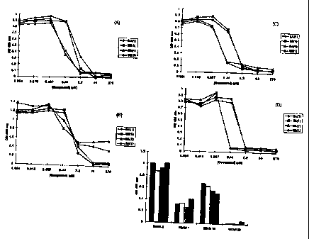
Figures lA-1D depict the concentration-dependent inhibition of (A) SEAM 3
{10 ,ug/ml), (B) SEAM 7 (lO,ugIml), (C) SEAM 18 (l0,ug/ml), (D) SEAM 30 {10
~cg/ml) binding to NPr-MenB PS by structure SA(1) (filled diamond), structure
SB(1)
(filled square), structure 6A(1) (filled triangle), and structure 6B(1)
(filled circle) in
an ELISA format.
Figure 2 depicts the binding of SEAM 3 (l0,uglml), SEAM 7 (10 ~cg/ml),
SEAM 18 (10 ~cg/ml), and SEAM 30 (10 ~cg/ml) to structure 5A(1} (filled bars),
structure SB(1} (open bars), structure 6A(1) (shaded bars), and structure
6B(1)
(cross-hatched bars) in an ELISA format.
Figure 3 depicts the cross-reactivity of SEAM antibodies with BSA
conjugates of structures SA(1), SB(1), 6A(1) and 6B(I). Particularly, Figure 3
depicts the binding of SEAM 3 (10 mg/ml}, SEAM 7 (10 mg/ml), SEAM 18 (10
mg/ml) and SEAM 30 (10 mglml) to SA(1) (filled bars), structure SB(1) (open
bars),
structure 6A(1) (shaded bars), and structure 6B(1) (cross-hatched bars).
Detailed Descrip io of the Invention
The practice of the present invention will employ, unless otherwise indicated,
conventional methods of immunology, microbiology, and molecular biology within
the skill of the art. Such techniques are explained fully in the literature.
See, e.g.,
Sambrook, et al. Molecular Cloning: A Laboratory Manual (2nd Edition, 1989);
Morrison and Boyd , Organic Chemistry (3rd Edition 1973); Carey and Sundberg,
Advanced Organic Chemistry (2nd Edition, 1985); Smith, M. B., Organic
Synthesis
(1994); Perbal, A Practical Guide to Molecular Cloning (1984); and Handbook of
Experimental Immunology, Vols. I-IV (D.M. Weir and C.C. Blackwell eds., 1986,
Blackwesll Scientific Publications).
8
CA 02301942 2000-02-22
WO 99110372 PCTIUS98/17670
As used in this specification and the appended claims, the singular forms "a,"
"an" and "the" include plural references unless the content clearly dictates
otherwise.
T Definitions
In describing the present invention, the following terms will be employed,
and are intended to be defined as indicated below.
As used herein, a "MenB PS derivative" refers to a molecule obtained by the
chemical modification of the native capsular polysaccharide of MenB. Such MenB
PS derivatives include, but are not limited to, MenB PS molecules which have
been
modified by the substitution of sialic acid residue N-acetyl groups of the
native
molecule with appropriate acyl groups, such as C3-CB, and higher, acyl groups
wherein the term "acyl group" encompasses any acylated linear, branched,
aliphatic
or aromatic molecule. A particularly preferred MenB PS derivative for use
herein
comprises the substitution of N-propionyl groups for N-acetyl groups of native
MenB PS (termed "NPr-MenB PS" herein}. Methods for synthesizing N-acyl-
substituted MenB PS derivatives, including NPr-MenB PS, are known in the art
and
described in e.g., U.S. Patent No. 4,727,136 to 3ennings et al. and EP
Publication No.
504,202 B, also to Jennings et al.
"Molecular mimetics" of MenB PS, or derivatives of MenB PS are molecules
that functionally mimic at least one "unique" epitope expressed on a MenB
bacteria.
A "unique epitope" is an epitope capable of eliciting the formation of
functionally
active (e.g., opsonic and/or complement-mediated bactericidal) anti-MenB
antibodies
that either are not cross-reactive with polysialic acid in host tissue and
hence lack
autoimmune activity, or are minimally cross-reactive. Such molecular mimetics
are
useful in vaccine compositions and in eliciting antibodies for diagnostic or
therapeutic applications, as described further below. Molecular mimetics
include,
but are not limited to: small organic compounds; nucleic acids and nucleic
acid
derivatives; saccharides or oligosaccharides; peptide mimetics including
peptides,
proteins, and derivatives thereof, such as peptides containing non-peptide
organic
moieties, synthetic peptides which may or may not contain amino acids and/or
peptide bonds, but retain the structural and functional features of a peptide
ligand;
pyrrolidines; peptoids and oligopeptoids which are molecules comprising N
9
CA 02301942 2000-02-22
WO 99/10372 PCT/US98/17670
substituted glycine, such as those described by Simon et al. (1992) Proc.
Natl. Acad.
Sci. USA $9:9367; and antibodies, including anti-idiotype antibodies. Methods
for
the identification and production of molecular mimetics are described more
fully
below.
The term "alkyl" as used herein refers to a branched or unbranched saturated
hydrocarbon group of 1 to 24 carbon atoms, such as methyl, ethyl, n-propyl,
isopropyl, n-butyl, isobutyl, t-butyl, octyl, decyl, tetradecyl, hexadecyl,
eicosyl,
tetracosyl and the like, as well as cycloalkyl groups such as cyclopentyl,
cyclohexyl
and the like. Preferred alkyl groups herein contain 1 to 12 carbon atoms, and
the
most preferred alkyl groups herein contain one to six carbon atoms. Further,
these
groups are optionally substituted with one or more, alkoxy, hydroxyl, amino,
amide,
halogen, vitro, acyl, carboxyl, thiol, sulfonic acids, sulfonamide, and the
like.
The term "alkenyl" as used herein refers to a branched or unbranched
hydrocarbon group of 2 to 24 carbon atoms containing at least one double bond,
such
as ethenyl, n-propenyl, isopropenyl, n-butenyl, isobutenyl, t-butenyl,
octenyl,
decenyl, tetradecenyl, hexadecenyl, eicosenyl, tetracosenyl, as well as cyclic
alkenyl
group of three to eight, preferably five or six, carbon atoms, and the like.
Preferred
alkenyl groups herein contain 1 to 12 carbon atoms, more preferably one to six
carbon atoms. Further, these groups are optionally substituted with one or
more,
alkyl, alkoxy, hydroxyl, amino, amide, halogen, vitro, acyl, carboxyl, thiol,
sulfonic
acids, sulfonamide, and the like.
The term "alkynyl" as used herein refers to a branched or unbranched
hydrocarbon group of 2 to 24 carbon atoms containing at least one triple bond,
such
as ethynyl, n-propynyl, isopropynyl, n-butynyl, isobutynyl, t-butynyl,
octynyl,
decynyl and the like. Preferred alkynyl groups herein contain 1 to 12 carbon
atoms,
preferably one to six carbon atoms. Further, these groups are optionally
substituted
as described above.
The term "aryl" as used herein refers to a mono-, bi-, tri- and tetra-cyclic
aromatic species containing five-, six- or seven-membered rings optionally
containing one or more heteroatoms such as N, O, and S. Examples include but
are
not limited to phenyl, benzyl, naphthyl, pyrrole, furan, thiophene, imidazole,
CA 02301942 2000-02-22
WO 99/10372 PCT/US98/17b70
oxazole, thiazole, pyrazole, pyridine, pyrimidine, purine, quinoline,
isoquinoline,
carbazole, phenanthrene, anthracene, benzopyrene, azulene, indole, indane, and
the
like. Further, these groups are optionally substituted as described above.
The term "alkyl-aryl" as used herein refers to an aryl ring attached to an
alkyl, wherein the terms alkyl and aryl are as defined above.
The term "carbocyclic ring" as used herein includes cylco-alkyl, -alkenyl, and
-alkynyl, as described above, optionally containing one or more heteroatoms
such as
N, O, and S. Further, these groups are optionally substituted as described
above.
The term "alkoxy" as used herein intends an alkyl group bound through a
single, terminal ether linkage; that is, an "alkoxy" group may be defined as -
OR
where R is alkyl as defined above. In a preferred embodiment, an alkoxy group
contains one to six, more preferably one to four, carbon atoms.
The term "acyl" is used in its conventional sense to refer to a molecular
substituent RCO- where R is alkyl as defined above. In a preferred embodiment,
an
acyl group contains alkyl containing one to six, more preferably one to four,
carbon
atoms.
The term "peptoid" as used herein encompasses oligomers of N-substituted
glycine and is used interchangeably with the term oligomeric N-substituted
glycines
(NS G).
The term "multivalent linker moiety" represents any suitable linker which
may be attached to any suitable or attachable moiety, including a ceramide or
a
protein or a peptide, including a multiple antigen peptide, and is preferably
a group
with a reactive group thereon which covalently allows it to bind to other
molecules,
such as molecular mimetics and the like.
The term "antibody" encompasses polyclonal and monoclonal antibody
preparations, as well as preparations including hybrid antibodies, altered
antibodies,
F(ab')2 fragments, Flab) molecules, Fv fragments, single chain fragment
variable
displayed on phage (scFv), single domain antibodies, chimeric antibodies and
functional fragments thereof which exhibit immunological binding properties of
the
parent antibody molecule.
As used herein, the term "monoclonal antibody" refers to an antibody
11
CA 02301942 2000-02-22
WO 99/10372 PCT/US98/17670
composition having a homogeneous antibody population. The term is not limited
by
the manner in which it is made. The term encompasses whole immunoglobulin
molecules, as well as Fab molecules, F(ab')Z fragments, Fv fragments, single
chain
fragment variable displayed on phage (scFv), and other molecules that exhibit
immunological binding properties of the parent monoclonal antibody molecule.
Methods of making polyclonal and monoclonal antibodies are known in the art
and
described more fully below.
An "antigen" is defined herein to include any substance that may be
specifically bound by an antibody molecule. An "immunogen" is an antigen that
is
capable of initiating lymphocyte activation resulting in an antigen-specific
immune
response.
By "epitope" is meant a site on an antigen to which specific B cells and T
cells respond. The term is also used interchangeably with "antigenic
determinant" or
"antigenic determinant site." B cell epitope sites on proteins,
polysaccharides, or
other biopolymers may be composed of moieties from different parts of the
macromolecule that have been brought together by folding. Epitopes of this
kind are
referred to as conformational or discontinuous epitopes, since the site is
composed of
segments the polymer that are discontinuous in the linear sequence but are
continuous in the folded conformation(s). Epitopes that are composed of single
segments of bioployrners or other molecules are termed continuous or linear
epitopes. T cell epitopes are generally restricted to linear peptides. A
peptide
epitope can comprise 3 or more amino acids in a spatial conformation unique to
the
epitope. Generally, an epitope consists of at least 5 such amino acids and,
more
usually, consists of at Least 8-10 such amino acids. Methods of determining
spatial
conformation of amino acids are known in the art and include, for example, x-
ray
crystallography and 2-dimensional nuclear magnetic resonance spectroscopy.
Furthermore, the identification of epitopes in a given protein is readily
accomplished
using techniques well known in the art. See, e.g., Geysen et al. (1984) Proc.
Natl.
Acad. Sci. USA $1:3998 (general method of rapidly synthesizing peptides to
determine the location of immunogenic epitopes in a given antigen); U.S.
Patent No.
4,708,871 (procedures for identifying and chemically synthesizing epitopes of
12
CA 02301942 2000-02-22
WO 99/10372 PCT/US9$/17b70
antigens); and Geysen et al. (1986) Molecular Immunology 2:709 (technique for -
identifying peptides with high affinity for a given antibody). Antibodies that
recognize the same epitope can be identified in a simple immunoassay showing
the
ability of one antibody to block the binding of another antibody to a target
antigen.
A "unique MenB epitope" is defined herein as an epitope present on a MenB
bacterium, wherein antibodies directed toward the epitope are capable of
binding
specifically to MenB and not cross-reacting, or minimally cross-reacting, with
sialic
acid residues present on the surface of host tissue. Immunogens containing or
mimicking one or more "unique MenB epitopes" are thus useful in vaccines for
prevention of MenB disease, and will not elicit an autoimmune response, or
pose
minimal risk of eliciting an autoimmune response.
An antibody displays "functional activity" against a MenB organism when
the antibody molecule exhibits complement-mediated bactericidal activity
and/or
opsonic activity against MenB as determined using the assays described herein.
An antibody specific for a "unique" MenB epitope "lacks autoimmune
activity," andlor is "not autoreactive" when the subject antibody does not
exhibit
cross-reactive immunological binding properties with polysialic acid in host
tissue as
determined using the binding assays described herein.
An antibody specific for a "unique" MenB epitope exhibits "minimal
autoreactivity," and/or is "minimally autoreactive" when the subject antibody
requires approximately ten times greater antibody concentration to exhibit
binding to
polysialic acid in host tissues, compared to a known cross-reactive auto
antibody
considered positive in the binding assays described herein.
As used herein, the terms "immunological binding," and "immunological
binding properties" refer to non-covalent interactions of the type which occur
between an immunoglobulin molecule and an antigen for which the immunoglobulin
is specific.
As used herein, a "biological sample" refers to a sample of tissue or fluid
isolated from a subject, including but not limited to, for example, blood,
plasma,
serum, fecal matter, urine, bone marrow, bile, spinal fluid, lymph fluid,
samples of
the skin, external secretions of the skin, respiratory, intestinal, and
genitourinary
13
CA 02301942 2000-02-22
WO 99/10372 PCT/US98I17670
tracts, tears, saliva, milk, blood cells, organs, biopsies and also samples of
in vitro-y
cell culture constituents including but not limited to conditioned media
resulting
from the growth of cells and tissues in culture medium, e.g., recombinant
cells, and
cell components.
As used herein, the terms "label" and "detectable label" refer to a molecule
capable of detection, including, but not limited to, radioactive isotopes,
fluorescers,
chemiluminescers, enzymes, enzyme substrates, enzyme cofactors, enzyme
inhibitors, chromophores, dyes, metal ions, metal sols, ligands (e.g., biotin
or
haptens) and the like. The term "fluorescer" refers to a substance or a
portion thereof
which is capable of exhibiting fluorescence in the detectable range.
Particular
examples of labels which may be used under the invention include fluorescein,
rhodamine, dansyl, umbelliferone, Texas red, luminol, NADPH and a-(3-
galactosidase.
The present invention is based on the discovery of molecular mimetics of
unique epitopes of Neisseria meningitides serogroup B (MenB), identified using
functional antibodies directed against MenB. The antibodies do not cross-
react, or
are minimally cross-reactive with polysialic acid in host tissue, and hence
the
antibodies have a lower risk of evoking autoimmune activity than antibodies
that are
highly cross-reactive with host tissue. The mimetics can be used as diagnostic
reagents andlor in compositions to prevent MenB and E. toll Kl disease.
As explained above, the native capsular polysaccharide of MenB, termed
"MenB PS" herein, is poorly immunogenic in humans and other mammalian
subjects. Furthermore, native MenB PS can elicit the production of
autoantibodies
and, hence, may be inappropriate for use in vaccine compositions. Thus, the
present
invention uses antibodies prepared against MenB PS derivatives. These
antibodies
were selected based on their ability to exhibit functional activity against
MenB
bacteria, wherein the functional activity is important in conferring
protection against
MenB disease. The antibodies were also selected on the basis of showing
minimal or
undetectable autoimmune activity.
14
CA 02301942 2000-02-22
WO 99110372 PCT/US98I17b70
More particularly, MenB PS derivatives were prepared for obtaining antibody
molecules used to identify the molecular mimetics of the present invention.
The
derivatives generally comprise C3-C8 acyl substitutions of sialic acid residue
N-acetyl
groups of the native molecule. Particularly preferred MenB PS derivatives
comprise
the substitution of N-propionyl groups for N-acetyl groups of native MenB PS
and
are termed "NPr-MenB PS" herein. Such derivatives and methods for synthesizing
the same are described in e.g., U.S. Patent No. 4,727,136 and EP Publication
No.
504,202 B, both to Jennings et al.
The C3-C8 acyl derivatives can be made by first treating native MenB
(obtained from e.g., N. meningitidis cultures) in the presence of a strong
base to
quantitatively remove the N-acetyl groups and to provide a reactive amine
group in
the sialic acid residue parts of the molecule. The deacylated MenB PS
fragments are
then N-acylated. For example, in the case of NPr-MenB PS, the deacylated
molecule
is N-propionylated using a source of propionyl groups such as propionic
anhydride
or propionyl chloride, as described in U.S. Patent No. 4,727,136 to Jennings
et al.
The extent of N-acylation can be determined using, for example, NMR
spectroscopy.
In general, reaction conditions are selected such that the extent of N-
acylation is at
least about 80%.
In order to increase the immunogenicity of the MenB PS derivatives, the
derivatives can be conjugated to a suitable carrier molecule to provide
glycoconjugates. Particularly, N-acylated MenB PS glycoconjugate preparatio$s
having well defned and controlled structural configurations can be formed from
intermediate sized N-acylated MenB oligosaccharides as described below.
Thus, a group of N-acylated MenB PS glycoconjugates, an example of which
is termed "CONJ-2" herein, can be prepared as follows. An N-acylated MenB PS
preparation, having substantially 100% N-acylated sialic acid residues, as
determined
by, e.g., NMR analysis, can be fragmented under mild acidic conditions to
provide a
population of oligosaccharide molecules of varying sizes. The fragmented
products
are size fractionated, using for example, standard ion exchange
chromatographic
techniques combined with e.g., stepwise salt gradients, to provide fractions
of N-
acylated MenB molecules of homogenous sizes. Fractions containing intermediate
CA 02301942 2000-02-22
WO 99/10372 PCTIUS98/17670
sized oligosaccharides e.g., with an average Dp of about 5 to about 22,
preferably 10
to about 20, and more particularly about 12 to about 18, are chemically end-
activated
at the non-reducing termini and conjugated to protein corners by a reductive
amination technique to provide the CONJ-2 glycoconjugates. Successful
conjugation can be determined by, e.g., gel filtration, and the final
saccharide to
protein ratio (w/w) assessed by colorimetric assay.
Glycoconjugates formed from MenB PS derivatives, such as the CONJ-2, are
then used herein to elicit the formation of anti-saccharide antibodies in an
immunized
host. A subset of such antibodies should bind to MenB bacteria, should not
cross-
react, or be minimally cross-reactive with host tissue sialic acid residues as
determined using the binding assays described herein. The antibodies can be
fully
characterized with respect to isotype, fine antigenic specificity, functional
activity
and cross-reactivity with host tissue.
For example, mammalian subjects, conveniently, standard laboratory animals
i S such as rodents and rabbits, can be immunized with compositions containing
the
glycoconjugates along with a suitable adjuvant to elicit the production of
polyclonal
sera. Groups of animals are generally immunized and boosted several times with
the
compositions. Antisera from immunized animals can be obtained, and polyclonal
sera that does not cross-react with host tissue can be obtained using in-situ
absorption or conventional affinity chromatography techniques. Successful
glycoconjugate antigens can be identified by their ability to elicit a
substantial IgG
anti-MenB PS derivative antibody response, characteristic of a T-cell
dependent
antigen. Conjugates that are found to be highly immunogenic and produce
predominantly IgG antibodies are particularly preferred for use in the methods
of the
present invention.
MenB PS derivatives that are capable of eliciting the formation of
bactericidal antisera are suitable for use in the production of monoclonal
antibodies.
More particularly, the process used to provide the various MenB PS derivative
conjugates is designed to produce superior immunogens presenting unique
saccharide-associated epitopes that mimic those found on the surface of MenB
organisms and are expressed minimally in the host. The MenB PS derivatives
16
CA 02301942 2000-02-22
WO 99110372 PCT/US98/17670
described herein are thus capable of eliciting the production of MenB-specific
antibodies that are used to search for mimetics of MenB polysaccharide
antigens that
will provide unique epitopes for anti-MenB vaccines.
Thus, in the practice of the invention, selected MenB derivatives are used to
provide monoclonal antibodies and functional equivalents thereof. The term
"functional equivalent" with respect to a particular monoclonal antibody, as
used
herein, means a molecule that: (a) cross-blocks an exemplified monoclonal
antibody;
(b} binds selectively to the MenB PS derivative or glycoconjugate in question;
(c)
does not cross-react, or minimally cross-reacts, with host PSA as determined
using
the binding assays described herein; and, optionally, activity (e.g.,
complement-
mediated bactericidal and/or opsonic activity) against MenB bacterial cells as
determined by standard assays described below. Further, as used herein with
regard
to a particular monoclonal antibody-producing hybridoma of the invention, the
term
"progeny" is intended to include all derivatives, issue, and offspring of the
parent
hybridoma that produce the monoclonal antibody produced by the parent,
regardless
of generation or karyotypic identity.
Monoclonal antibodies are prepared using standard techniques, well known in
the art, such as by the method of Kohler and Milstein, Nature (1975) 2:495, or
a
modification thereof, such as described by Buck et al. (1982) In Yitro 1$:377.
Typically, a mouse or rat is immunized with the MenB PS derivative conjugated
to a
protein carrier, boosted and the spleen (and optionally several large lymph
nodes)
removed and dissociated into single cells. If desired, the spleen cells may be
screened (after removal of non-specifically adherent cells) by applying a cell
suspension to a plate or well coated with the antigen. B-cells, expressing
membrane-
bound immunaglobulin specific for the antigen, will bind to the plate, and
will not be
rinsed away with the rest of the suspension. Resulting B-cells, or all
dissociated
spleen cells, are then induced to fuse with myeloma cells to form hybridomas.
Representative marine myeloma lines for use in the hybridizations include
those
available from the American Type Culture Collection (ATCC).
More particularly, somatic cell hybrids can be prepared by the method of
Buck et al., (supra), using the azaguanine resistant, non-secreting marine
myeloma
17
CA 02301942 2000-02-22
WO 99/10372 PCT/US98/17670
cell line P3X63-Ag8.653 (obtainable from the ATCC). The hybridoma cell lines
are
generally cloned by limiting dilution, and assayed for the production of
antibodies
which bind specifically to the immunizing antigen and which do not bind to
unrelated antigens. The selected monoclonal antibody-secreting hybridomas are
then
cultured either in vitro (e.g., in tissue culture bottles or hollow fiber
reactors), or in
vivo (e.g., as ascites in mice).
Hybridoma supernatant can be assayed for anti-MenB PS derivative-reactive
antibody using, for example, either solid phase ELISA or an indirect
immunofluorescence assay with the immunizing MenB PS derivative or with native
MenB PS (NAc-MenB PS). The selectivity of monoclonal antibodies secreted by
the
hybridomas can be assessed using competitive specific binding assays, such as
inhibition ELISA, or the like. For example, antibody molecules, either diluted
in
buffer, or buffer containing soluble MenB PS derivatives or NAc-MenB PS, are
reacted in an ELISA vessel in the presence of bound MenB PS derivatives. After
washing, bound antibody is detected by labeled anti-Ig (anti-IgM, IgG and IgA)
as
the secondary antibody. Antibodies that are inhibited by the soluble MenB PS
derivatives can be considered specific and, thus are selected for further
study
including, isotyping and additional screening for cross-reactivity, functional
activity,
and autoreactivity.
Specif cally, partially purified monoclonal antibody molecules can be
individually evaluated for their ability to bind to host cells which express
polysialic
acid residues on their cell surfaces. Such cells represent surrogate targets
for the
detection of antibodies that exhibit autoimmune activity. One target comprises
the
human neuroblastoma cell line, CHP-134, which expresses long chain a2-8
polysialic acid (NCAM) on its cell surface, as described by Livingston et al.
(1988)
J. Biol. Chem. ?.x:9443. Also, Granoff, D.M. et al. (1988) The J. of
Immunology
if 0:5028-5036 describe bactericidal monoclonal antibodies that define unique
MenB
PS epitopes that do not cross-react with human polysialic acid. Other suitable
targets
include, but are not limited to, newborn brain cells, tissues derived from
e.g., kidney,
heart and the olfactory nerve, cultured saphenous vein endothelial cells,
cytotoxic T
lymphocytes and natural killer (NK) cells. See, e.g., Brandon et al. (1993)
Intl. .l.
18
CA 02301942 2000-02-22
WO 99/10372 PCT/US98117670
Immunopathology and Pharmacology fi:77. Monoclonal antibody molecules
obtained from the hybridomas can be added to suitable test cell populations in
culture, and the potential binding of the monoclonals to the cellular targets
detected
and quantified directly using labeled monoclonals, or indirectly using an
appropriately labeled secondary reagent that reacts specifically with each
monoclonal
antibody (e.g., Staphylococcal Protein A and G and anti-murine antibody
molecules).
Antibodies that do not cross-react with test host tissue PSA or that display
minimal
reactivity are not considered autoreactive for purposes of the present
invention.
Thus, these antibodies are appropriate for further use. In addition, some
antibodies
that show binding with test tissue, which binding is not affected by pre-
treatment of
the test cells with neuraminidase, may also be appropriate for further use.
Autoreactivity of such antibodies is termed "indeterminate" herein.
Functional activity can be determined by assessing complement-mediated
bactericidal activity and/or opsonic activity. In particular, complement-
mediated
bactericidal activity of the antibodies can be evaluated using standard assays
such as
those described by Gold et al. (1970) Infect. Immun.1:479, Westerink et al.
(/988)
Infect. Immun. 5:1120, Mandrell et al. (1995) J. Infect. Dis. 172:1279, and
Granoff
et al. (1995) Clin. Diagn. Laboratory Immunol. 2:574. In these assays, N.
meningitides is reacted with a complement source as well as with the antibody
to be
tested. Bacterial counts are done at various sampling times. Those antibodies
that
demonstrate complement-mediated bactericidal activity, as demonstrated by a
minimum of a 50% reduction in viable bacterial cell counts determined after
sixty
minutes incubation with antibody and complement, as compared to colony counts
at
time zero, are considered to exhibit bactericidal activity for purposes of the
present
invention and are suitable for further use.
Complement-mediated bacteriolysis is thought to be the major mechanism
responsible for host protection against invasive Meningococcal disease.
However,
evidence also supports an important protective role for opsonization (see,
e.g.,
Bjerknes et al. (1995) Infect. Immun. x:160). Accordingly, the opsonic
activity of
the antibodies produced herein can be evaluated as a second measure, or as an
alternative measure, to assess functional activity. Results from opsonic
assays can be
i9
CA 02301942 2000-02-22
WO 99110372 PCT/US98117670
used to supplement bactericidal data, and to help in the selection of
antibodies
capable of conferring protection. Evaluation of opsonic activity is also
particularly
useful herein for the evaluation of the marine monoclonal antibodies of the
invention
which have an IgGI isotype. Marine igGl (in contrast to human IgGI) is
ineffective
S in activation of complement. Thus, marine IgGl antibodies do not activate
complement-mediated bacteriolysis of MenB in the above-described assays.
However, functional activity of IgGI anti-NPr-MenB PS monoclonal antibodies
can
be assessed by opsonization in the absence of complement.
A variety of opsonic assay methods are known in the art, and can be used to
evaluate functional activity of the monoclonal antibodies of the present
invention.
Such standard assays include those described by Sjursen et al. (1987) Acta
Path.
Microbiol. Immunol. Scand., Sec. C 9.:283, Halstensen et al. (1989) Scand J.
Infect.
Dis. 21.:267, Lehmann et al. (1991) APMIS 9:769, Halstensen et al. (1991) NIPH
Annals 14:157, Fredlund et al. (1992) APMIS 10:449, Guttormsen et al. (1992}
1 S Infect. Immun. 60:2777, Guttormsen et al. (1993) J. Infec. Dis. 162:1314,
Bjerknes et
al. (1995) Infect. Immun. 64:160, Hayrinen et al. (1995) J. Infect.
Dis.121:1481, de
Velasco et al. (1995) J. Infect. Dis.172:262, and Verheul, A.F.M. (1991)
"Meningococcal LPSDerived Oligosaccharide-Protein Conjugate Vaccines,
Immunochemical and Immunological Aspects," Thesis, Utrecht University, The
Netherlands, pp. 112-135.
Selected monoclonal antibodies of interest can be expanded in vitro, using
routine tissue culture methods, or in vivo, using mammalian subjects. For
example,
pristane-primed mice can be inoculated with log phase hybridoma cells in PBS
for
ascites production. Ascites fluid can be stored at -70°C prior to
further purification.
2S Antibody molecule fragments, e.g., F(ab')z, Fv, sFv and scFv molecules,
that
are capable of exhibiting immunological binding properties of the parent
monoclonal
antibody molecule can be produced using known techniques. mbar et al. (1972)
Proc. Nat. Acad. Sci. USA 62:2659; Hochman et al. (1976) Biochem 1:2706;
Ehrlich et al. (1980) Biochem 19:4091; Huston et al. (1988) Proc. Nat. Acad.
Sci.
USA $x(16):5879; and U.S. Patent Nos. 5,091,513 and 5,132,405, to Huston et
al.;
and 4,946,778, to Ladner et al.
CA 02301942 2000-02-22
WO 99/10372 PCTIUS98I17670
In the alternative, a phage-display system can be used to expand the
monoclonal antibody molecule populations in vitro. Saiki, et al. (1986) Nature
324:163; Scharf et al. (1986) Science 23.x:1076; U.S. Patent Nos. 4,683,195
and
4,683,202; Yang et al. (1995) JMoI Biol 254:392; Barbas, III et al. (1995)
Methods:
Comp. Meth Enzymol 8:94; Barbas, III et al. (199I) Proc Natl Acad Sci USA
$$:7978.
Once generated, the phage display library can be used to improve the
immunological binding affinity of the Fab molecules using known techniques.
See,
e.g., Figini et al. (1994) J. Mol. Biol. 232:68.
The coding sequences for the heavy and light chain portions of the Fab
molecules selected from the phage display library can be isolated or
synthesized, and
cloned into any suitable vector or replicon for expression. Any suitable
expression
system can be used, including, for example, bacterial, yeast, insect,
amphibian and
mammalian systems. Expression systems in bacteria include those described in
Chang et al. (1978) Nature 225:615, Goeddel et al. (1979) Nature 2$1:544,
Goeddel
et al. (1980) Nucleic Acids Res. $:4057, European Application No. EP 36,776,
U.S.
Patent No. 4,551,433, deBoer et al. (1983) Proc. Natl. Acad. Sci. USA X0:21-
25, and
Siebenlist et al. {I980) Cell20:269.
Expression systems in yeast include those described in Hinnen et al. {1978)
Proc. Natl. Acad. Sci. USA 25:1929, Ito et al. (1983) J. Bacteriol. 15.3:163,
Kurtz et
al. (1986) Mol. Cell. Biol. 6:142, Kunze et al. (1985) J. Basic Microbiol.
25:141,
Gleeson et al. {1986) J. Gen. Microbiol.132:3459, Roggenkamp et al. {1986)
Mol.
Gen. Genet. 2Q2:302, Das et al. (1984) J. Bacteriol.15$:1 I65, De Louvencourt
et al.
(1983) J. Bacteriol.15_4:737, Van den Berg et al. (1990) BiolTechnology $:135,
Kunze et al. (1985) J. Basic Microbiol. 25:141, Cregg et al. (1985) Mol. Cell.
Biol.
5:3376, U.S. Patent Nos. 4,837,148 and 4,929,555, Beach et al. (1981) Nature
3QQ:706, Davidow et al. (1985) Curr. Genet. 1Q:380, Gaillardin et al. (1985)
Curr.
Genet.1Q:49, Ballance et al. (1983) Biochem. Biophys. Res. Commun.112:284-289,
Tilburn et al. (1983) Gene 2f:205-221, Yelton et al. (1984) Proc. Natl. Acad.
Sci.
USA $1:1470-1474, Kelly et al. (1985} EMBO J. 4:475479; European Application
No. EP 244,234, and International Publication No. WO 91/00357.
21
CA 02301942 2000-02-22
WO 99/10372 PCT/US98/17670
Expression of heterologous genes in insects can be accomplished as described
in U.S. Patent No. 4,745,051, European Application Nos. EP 127,839 and EP
155,476, Vlak et al. (1988) J. Gen. Virol. 69:765-776, Miller et al. (1988)
Ann. Rev.
Microbiol. 42:177, Carbonell et al. (1988) Gene 23:409, Maeda et al. (1985)
Nature
316:592-594, Lebacq-Verheyden et al. (1988) Mol. Cell. Biol. $:3129, Smith et
al.
(1985) Proc. Natl. Acad. Sci. USA $2:8404, Miyajima et al. (1987) Gene 5$:273,
and
Martin et al. (1988) DNA 2:99. Numerous baculoviral strains and variants and
corresponding permissive insect host cells from hosts are described in Luckow
et al.
(1988) BiolTechnology 6:47-55, Miller et al. (1986) GENERIC ENGINEERING,
Setlow, J.K. et al. eds., Vol. 8, Plenum Publishing, pp. 277-279, and Maeda et
al.
(1985) Nature 316:592-594.
Mammalian expression can be accomplished as described in Dijkema et al.
(1985) EMBO J. 4:761, Gorman et al. (1982) Proc. Natl. Acad. Sci. USA 29:6777,
Boshart et al. (1985) Cell 41:521, and U.S. Patent No. 4,399,216. Other
features of
mammalian expression can be facilitated as described in Ham et al. (1979)
Meth.
Enz. 5_$:44, Barnes et al. (1980) Anal. Biochem.192:255, U.S. Patent Nos.
4,767,704,
4,657,866, 4,927,762, 4,560,655 and Reissued U.S. Patent No. RE 30,985, and in
International Publication Nos. WO 90/103430, WO 87/00195.
The anti-MenB antibodies of the present invention, described above, are
conveniently used as receptors to screen diverse molecular libraries in order
to
identify molecular mimetics of unique epitopes from MenB. Methods for
identifying
mimetics in molecular libraries generally involve the use of one or more of
the
following procedures: (1) affinity purification with an immobilized target
receptor;
(2) binding of a soluble receptor to tethered ligands; and (3) testing soluble
compounds directly in antigen competition assays or for biological activity.
Molecules screened for molecular mimics include but are not limited to small
organic compounds, combinatorial libraries of organic compounds, nucleic
acids,
nucleic acid derivatives, saccharides or oligosaccharides, peptoids, soluble
peptides,
peptides tethered on a solid phase, peptides displayed on bacterial phage
surface
proteins, bacterial surface proteins or antibodies, and/or peptides containing
non-
peptide organic moieties.
22
CA 02301942 2000-02-22
WO 99/10372 PCT/US98117670
For example, libraries of diverse molecular species can be made using -y
combinatorial organic synthesis. See, e.g., Gordon et al. (1994) J. Med. Chem.
32:/335. Examples include but are not limited to pyrrolidines; oligocarbamates
(Cho
et al. (1993) Science 261:1303); peptoids such as N-substituted glycine
polymers
{Simon et al. (1992) Proc. Natl. Acad. Sci. USA $9:9367); and vinylogous
polypeptides (Hagihara et al. (1992) J. Am. Chem. Soc. 114:6568).
A variety of approaches, known in the art, can be used to track the building
blocks as they are added during synthesis so that the history of individual
library
members can be determined. These approaches include addressable location on a
photolithographic chip (oligocarbamates), a deconvolution strategy in which
"hits"
are identified through recursive additions of monomers to partially
synthesized
libraries (peptoids, pyrrolidines, peptides), and coding combinatorial
libraries by the
separate synthesis of nucleotides (Nielsen et al. (1993) J. Am. Chem. Soc.
116: 9812)
or other organic moieties (Ohlmeyer et al. (1993) Proc. Natl. Acad. Sci. USA
20:10922) ("tags"). The coded tags associated with each library member can
then be
decoded after a mimetic has been selected. For example, nucleic acid tags can
be
decoded by DNA sequencing.
Peptoid combinatorial libraries are particularly useful for identifying
molecular rnirnetics of unique MenB epitopes. Peptoids are oligomers of N-
substituted glycine (Simon et al. (1992) Proc. Natl. Acad. Sci. USA .89:9367)
and can
be used to generate chemically diverse libraries of novel molecules. The
monomers
may incorporate t-butyl-based side-chain and 9- fluorenyl-methoxy-carbonyl a-
amine
protection. The assembly of monomers into peptoid oligomers can be performed,
for
example, on a solid phase using the "submonomer method" of Zuckermann et al.
{1992) J. Am. Chem. Soc.1.14:10646. In this method, syntheses are conducted
with
Rink amide polystyrene resin (Rink et al. (1987) Tetrahedron Lett. 2$:3787).
Resin-
bound amines are bromoacetylated by in situ activation of bromoacetic acid
with
diisopropyl-carbodiimide. Subsequently, the resin-bound bromoacetamides are
displaced by addition of an amine. The amines may incorporate t-butyl-based
protection of additional reactive groups. This two-step cycle is repeated
until the
desired number of monomers is added. The oligopeptide is then released from
the
23
CA 02301942 2000-02-22
WO 99110372 PCTIUS98I17670
resin by treatment with 95% trifluroacetic acid/5% water. The syntheses are
performed, preferably, using a robotic synthesizer. See, e.g., Zuckermann et
al.
(1992) Pept. Protein Res. 452:498; and Zuckermann et a1. (1996) Methods in
Enzymology 2~Z:437. In the alternative, oligomerization of the peptoid
monomers
may be performed by in situ activation by either benzotriazol-1-yloxytris
(pynrolidino)phosphonium hexafluorphosphate or bromotris(pyrrolidino)
phosphonium hexafluorophosphate. In this alternative method, the other steps
are
identical to conventional peptide synthesis using oc-(9- fluorenyl
methoxycarbonyl)
amino acids (see, e.g., Simon et al. (1992), supra).
Once the peptoid libraries are generated, they can be screened by, e.g.,
adding
the monoclonal antibodies of the present invention, along with various pools
of the
combinatorial peptoids, to wells of microtiter plates coated with MenB PS
derivatives or MenB bacteria, either alone or as glycoconjugates. After a
period of
incubation and a wash to remove unbound antibody, the presence of bound
antibody
is determined by standard ELISA assays. See, e.g., Harlow & Lane, Antibodies:
A
Laboratory Manual (1988), Cold Spring Harbor Laboratory, Cold Spring Harbor,
NY, 553. Wells that do not contain bound antibody indicate the presence of
peptoid
mimetics that bind to the antibody. The particular identities of the peptoid
mimetics
in the pools are determined by recursively adding back monomer units to
partially
synthesized members of the libraries. Zuckermann et al. (1994) J. Med. Chem.
32:2678. Other methods for identifying active compounds in pools of small
molecules include fractionating the pool by reverse phase HPLC or affinity
selection/mass spectroscopy (Nedved M. L. Et al (1996) Anal. Chem. 6$:4228).
Once putative molecular mimetics are identified, they are tested for their
ability to elicit functionally active (e.g., bactericidal and/or opsonic)
antibodies
which lack autoreactivity or have minimal autoreactivity, as described above.
Molecular mimetics that have these properties are appropriate for further use,
for
example, in vaccine compositions.
Molecular mimetics identified using the functionally active anti-MenB
antibodies of the invention can be used to generate antibody reagents for use
in
diagnostic assays. For example, antibodies reactive with the molecular
mimetics can
24
CA 02301942 2000-02-22
WO 99110372 PCT/US98l17670
be used to detect bacterial antigen in biological samples using
immunodiagnostic
techniques such as competition, direct reaction, or sandwich type assays. Such
assays include Western blots; agglutination tests; enzyme-labeled and mediated
immunoassays, such as ELISAs; biotin/avidin type assays; radioimmunoassays;
S immunoelectrophoresis; immunoprecipitation, and the like. The reactions
generally
include revealing labels such as fluorescent, chemiluminescent, radioactive,
enzymatic labels or dye molecules, or other methods for detecting the
formation of a
complex between the mimetic and the antibody or antibodies reacted therewith.
The aforementioned assays generally involve separation of unbound antibody
in a liquid phase from a solid phase support to which mimetic-antibody
complexes
are bound. Solid supports which can be used in the practice of the invention
include
substrates such as nitrocellulose (e.g., in membrane or microtiter well form};
polyvinylchloride (e.g., sheets or microtiter wells); polystyrene latex (e.g.,
beads or
microtiter plates); polyvinylidine fluoride; diazotized paper; nylon
membrane's;
activated beads, magnetically responsive beads, and the like.
Typically, a solid support is first reacted with a solid phase component
(e.g.,
one or more MenB molecular mimetics) under suitable binding conditions such
that
the component is sufficiently immobilized to the support. Sometimes,
immobilization of the mimetic to the support can be enhanced by first coupling
the
mimetic to a protein with better binding properties. Suitable coupling
proteins
include, but are not limited to, macromolecules such as serum albumins
including
bovine serum albumin (BSA), keyhole limpet hemocyanin, immunoglobulin
molecules, thyroglobulin, ovalbumin, and other proteins well known to those
skilled
in the art. Other molecules that can be used to bind the mimetics to the
support
include polysaccharides, polylactic acids, polyglycolic acids, polymeric amino
acids,
amino acid copolymers, and the like. Such molecules and methods of coupling
these
molecules to the antigens, are well known to those of ordinary skill in the
art. See,
e.g., Brinkley, M.A. Bioconjugate Chem. (1992) 3:2-13; Hashida et al., J.
Appl.
Biochem. (1984) 6:56-63; and Anjaneyulu and Staros, International J. of
Peptide and
Protein Res. (1987) 3.0:117-124.
After reacting the solid support with the solid phase component, any non-
CA 02301942 2000-02-22
WO 99110372 PCT/US98117b70
immobilized solid-phase components are removed from the support by washing,
and
the support-bound component is then contacted with a biological sample
suspected
of containing ligand moieties (e.g., MenB and/or E. coli Kl antibodies) under
suitable binding conditions. After washing to remove any non-bound ligand, a
secondary binder moiety is added under suitable binding conditions, wherein
the
secondary binder is capable of associating selectively with the bound ligand.
The
presence of the secondary binder can then be detected using techniques well
known
in the art.
More particularly, an ELISA method can be used, wherein the wells of a
microtiter plate are coated with a mimetic according to the present invention.
A
biological sample containing or suspected of containing anti-MenB or E. coli
Kl
immunoglobulin molecules is then added to the coated wells. After a period of
incubation sufficient to allow antibody binding to the immobilized mimetic,
the
plates) can be washed to remove unbound moieties and a detestably labeled
secondary binding molecule added. The secondary binding molecule is allowed to
react with any captured sample antibodies, the plate washed and the presence
of the
secondary binding molecule detected using methods well known in the art.
Thus, in one particular embodiment, the presence of bound MenB/E. coli Kl
antigen ligands from a biological sample can be readily detected using a
secondary
binder comprising an antibody directed against the antibody ligands. A number
of
anti-bovine irnmunogiobulin (Ig) molecules are known in the art which can be
readily conjugated to a detectable enzyme label, such as horseradish
peroxidase,
alkaline phosphatase or urease, using methods known to those of skill in the
art. An
appropriate enzyme substrate is then used to generate a detectable signal. In
other
related embodiments, competitive-type ELISA techniques can be practiced using
methods known to those skilled in the art.
Assays can also be conducted in solution, such that the mimetics and
antibodies specific for those mimetics form complexes under precipitating
conditions. In one particular embodiment, the mimetics can be attached to a
solid
phase particle (e.g., an agarose bead or the like) using coupling techniques
known in
the art, such as by direct chemical or indirect coupling. The mimetic-coated
particle
26
CA 02301942 2000-02-22
WO 99/10372 PCT/US98I17670
is then contacted under suitable binding conditions with a biological sample
suspected of containing antibodies for MenB and/or E. toll Kl. Cross-linking
between bound antibodies causes the formation of particle-mimetic-antibody
complex aggregates which can be precipitated and separated from the sample
using
washing andlor centrifugation. The reaction mixture can be analyzed to
determine
the presence or absence of antibody-mimetic complexes using any of a number of
standard methods, such as those immunodiagnostic methods described above.
In yet a further embodiment, an immunoaffinity matrix can be provided,
wherein a polyclonal population of antibodies from a biological sample
suspected of
containing MenB and/or E. toll K1 antibodies is immobilized to a substrate. In
this
regard, an initial affinity purification of the sample can be carried out
using
immobilized antigens. The resultant sample preparation will thus only contain
anti-
MenB and/or E. toll Kl moieties, avoiding potential nonspecific binding
properties
in the affinity support. A number of methods of immobilizing immunoglobulins
(either intact or in specific fragments) at high yield and good retention of
antigen
binding activity are known in the art. Not being limited by any particular
method,
immobilized protein A or protein G can be used to immobilize immunoglobulins.
Accordingly, once the immunoglobulin molecules have been immobilized to
provide an immunoaffinity matrix, labeled molecules are contacted with the
bound
antibodies under suitable binding conditions. After any non-specifically bound
mimetic has been washed from the immunoaffmity support, the presence of bound
antigen can be determined by assaying for label using methods known in the
art.
A particularly preferred method for diagnosing MenB and/or E. toll Kl
infection using the present invention involves the use of strip imrnunoblot
assay
(SIA) techniques, such as those known in the art which combine traditional
Western
and dot blotting techniques, e.g., the RIBA~ (Chiron Corp., Emeryville, CA)
test. In
these assays, one or more mimetics according to the present invention are
immobilized as individual, discrete bands on a membranous support test strip.
Visualization of anti-MenB and/or E. toll Kl reactivity in the biological
sample is
accomplished using anti-human IgG enzyme-conjugates in conjunction with a
colorimetric enzyme substrate. Internal controls, such as anti-human IgM and
human
27
CA 02301942 2000-02-22
WO 99/10372 PCTlUS98/17670
IgG, can also be present on the strip. The assay can be performed manually or
used
in an automated format.
The above-described assay reagents, including the mimetics of the invention
or antibodies thereto, can be provided in kits, with suitable instructions and
other
necessary reagents, in order to conduct immunoassays as described above. The
kit
can also contain, depending on the particular immunoassay used, suitable
labels and
other packaged reagents and materials (i.e. wash buffers and the like).
Standard
immunoassays, such as those described above, can be conducted using these
kits.
In addition, molecular mimetic compositions, unique (e.g., non-autoimmune)
Men B epitopes identified using the molecular mimetics can be used herein to
diagnose or prevent MenB disease in mammalian subjects. Particularly, vaccine
compositions of the molecular mimetics can be used for the prevention of MenB
disease in vaccinated subjects.
The vaccine compositions can comprise one or more of the molecular
mimetics or non-autoimmune epitopes of MenB. The vaccines may also be
administered in conjunction with other antigens and immunoregulatory agents,
for
example, immunoglobulins, cytokines, lymphokines, and chemokines, including
but
not limited to IL-2, modified IL-2 (cys125 -> ser125), GM-CSF, IL-12, y-
interferon,
IP-10, MIPl~i and RANTES.
The vaccines will generally include one or more "pharmaceutically
acceptable excipients or vehicles" such as water, saline, glycerol, ethanol,
etc.
Additionally, auxiliary substances, such as wetting or emulsifying agents, pH
buffering substances, and the like, may be present in such vehicles.
Adjuvants may also be used to enhance the effectiveness of the vaccines.
Adjuvants can be added directly to the vaccine compositions or can be
administered
separately, either concurrently with or shortly after, vaccine administration.
Such
adjuvants include, but are not limited to: (1) aluminum salts (alum), such as
aluminum hydroxide, aluminum phosphate, aluminum sulfate, etc.; (2) oil-in-
water
emulsion formulations {with or without other specific immunostimulating agents
such as muramyl peptides (see below) ar bacterial cell wall components), such
as for
example (a) MF59 (International Publication No. WO 90/14837), containing S%
28
CA 02301942 2000-02-22
WO 99110372 PCT/US98/17670
Squalene, 0.5% Tween 80, and 0.5% Span 85 (optionally containing various
amounts
of MTP-PE (see below), although not required) formulated into submicron
particles
using a microfluidizer such as Model 110Y microfluidizer (Microfluidics,
Newton,
MA), (b) SAF, containing 10% Squalane, 0.4% Tween 80, 5% pluronic-blocked
polymer L121, and thr-MDP (see below) either microfluidized into a submicron
emulsion or vortexed to generate a larger particle size emulsion, and (c)
RibiTM
adjuvant system (RAS), (Ribi Immunochem, Hamilton, MT) containing 2%
Squalene, 0.2% Tween 80, and one or more bacterial cell wall components from
the
group consisting of monophosphorylipid A (MPL), trehalose dimycolate (TDM),
and
cell wall skeleton (CWS), preferably MPL + CWS (DetoxTM); (3) saponin
adjuvants,
such as StimulonTM (Cambridge Bioscience, Worcester, MA) may be used or
particle
generated therefrom such as ISCOMs (immunostimulating complexes); (4) Freund's
Complete Adjuvant (FCA) and Freund's Incomplete Adjuvant (FICA); (5)
cytokines,
such as interleukins (IL-l, IL-2, etc.), macrophage colony stimulating factor
(M-
CSF), tumor necrosis factor (TNF), etc.; (6) detoxified mutants of a bacterial
ADP-
ribosylating toxin such as a cholera toxin (CT), a pertussis toxin (PT), or an
E. coli
heat-labile toxin (LT), particularly LT-K63 (where lysine is substituted for
the wild-
type amino acid at position 63) LT-R72 (where arginine is substituted for the
wild-
type amino acid at position 72), CT-S 109 (where serine is substituted for the
wild-
type amino acid at position 109), and PT-K9/G129 (where lysine is substituted
for
the wild-type amino acid at position 9 and glycine substituted at position
129) (see,
e.g., International Publication Nos. W093/13202 and W092119265); and (7) other
substances that act as immunostimulating agents to enhance the effectiveness
of the
composition.
Muramyl peptides include, but are not limited to, N-acetyl-muramyl-L-
threonyl-D-isoglutamine (thr-MDP), N-acetyl-normuramyl-L-alanyl-D-isogluatme
(nor-MDP), N-acetylmuramyl-L-alanyl-D-isogluatminyl-L-alanine-2-(f-2'-
dipalmitoyl-sn-glycero-3-huydroxyphosphoryloxy)- ethylamine (MTP-PE), etc.
In order to enhance the effectiveness of compositions formed from a
molecular mimetic, it may be necessary to conjugate the mimetic to a carnet
molecule. Such carrier molecules will not themselves induce the production of
29
CA 02301942 2000-02-22
WO 99/1fl372 PCT/US98117670
harmful antibodies. Suitable carriers are typically large, slowly metabolized
macromolecules such as proteins, polysaccharides, polylactic acids,
polyglycolic
acids, polymeric amino acids, amino acid copolymers, lipid aggregates (such as
oil
droplets or liposomes), inactive virus particles, CRM,9, (a nontoxic mutant
diphtheria toxin), and the like. Such earners are well known to those of
ordinary
skill in the art. The mimetic conjugates are selected for their ability to
express
epitopes that closely resemble those found on the surface of MenB bacterial
cells.
Suitable conjugates thus elicit the formation of antibodies that have
functional
activity against bacteria, and do not cross-react, or are minimally cross-
reactive with
polysialic acid in host tissue as determined using the binding assays
described herein.
Typically, the vaccine compositions are prepared as injectables, either as
liquid solutions or suspensions; solid forms suitable for solution in, or
suspension in,
liquid vehicles prior to injection may also be prepared. The preparation also
may be
emulsified or encapsulated in liposomes, or adsorbed to particles for enhanced
adjuvant effect, as discussed above.
The vaccines will comprise an effective amount of the molecular mimetic,
and any other of the above-mentioned components, as needed. By "an effective
amount" is meant an amount of a molecule which will induce an immunological
response in the individual to which it is administered and poses a minimal
risk of
stimulating an autoimmune response in the individual. Such a response will
generally result in the development in the subject of a secretory, cellular
and/or
antibody-mediated immune response to the vaccine. Usually, such a response
includes but is not limited to one or more of the following effects; the
production of
antibodies from any of the irnmunological classes, such as immunoglobulins A,
D, E,
G or M; the proliferation of B and T lymphocytes; the provision of activation,
growth and differentiation signals to immunological cells; expansion of helper
T cell,
suppressor T cell, and/or cytotoxic T cell and/or gd T cell populations.
Once formulated, the vaccines are conventionally administered parenterally,
e.g., by injection, either subcutaneously or intramuscularly. Additional
formulations
suitable for other modes of administration include oral and pulmonary
formulations,
suppositories, and transdermal applications. Dosage treatment may be a single
dose
CA 02301942 2000-02-22
WO 99110372 PCT/US98117670
schedule or a multiple dose schedule.
TTI~Rxp~imental
Below are examples of specific embodiments for carrying out the present
invention. The examples are offered for illustrative purposes only, and are
not
intended to limit the scope of the present invention in any way.
Efforts have been made to ensure accuracy with respect to numbers used
(e.g., amounts, temperatures, etc.), but some experimental error and deviation
should,
of course, be allowed for.
P_r~pa'ration of "sized" Cl~rcocon
An exemplary NPr-MenB oligosaccharide-tetanus toxoid conjugate vaccine,
hereinafter referred to as CONJ-2, was prepared as follows. The N-acetyl
groups of
MenB B polysaccharide were removed by heating the polysaccharide to
110°C in 2M
NaOH for 6 hours in the presence of NaBH4. The de-acetylated polysaccharide
was
exhaustively dialyzed in saturated sodium bicarbonate buffer then stirred with
an
excess of propionic anhydride for 12 hours at ambient temperature. The
solution was
exhaustively dialyzed in water and the N-propionylated meningococcal B (NPr-
MenB PS) polysaccharide was recovered by lyophilization.
For preparation of the conjugate vaccine, the NPr-MenB polysaccharide was
partially hydrolyzed in 10 mM sodium acetate at pH 5.5 at 50°C for 2
hours. The
resulting mixture of oligosaccharides was fractionated on Q-Sepharose.
Oligosaccharides having an average degree of polymerization (Dp) of 2-6 were
first
eluted with 100 mM NaCI and discarded. Intermediate-sized oligosaccharides
were
eluted with 500 mM NaCI. It was subsequently determined by analytical ion
exchange chromatography using a MonoQ column that the intermediate-sized
oligosaccharides ranged in size from Dp 13 to 20 (Mean = Dp 13).
A terminal aldehyde group was generated at the non-reducing end of the
intermediate-sized oligosaccharides by reacting them with 100 mM sodium
periodate
for 15-30 minutes at ambient temperature in the dark. Excess ethylene glycol
was
31
CA 02301942 2000-02-22
WO 99/10372 PCTIUS98/17670
used to quench the oxidative reaction and the product was desalted on a
Sephadex G-
25 column. The oligosaccharide-protein conjugate was prepared by stirring a
mixture of terminal aldehyde containing NPr MenB oligosaccharide with tetanus
toxoid (molar ratio of 200:1, respectively) in 0.75 M potassium phosphate
buffer, pH
9.0 with 40 mg/ml of sodium cyanoborohydride for one day at 40°C and
two days at
ambient temperature. The resultant NPr-MenB oiigosaccharide-tetanus toxoid
conjugate (CONJ-2) was finally purified by gel permeation chromatography on
Sephadex G-100 using SO mM sodium phosphate, pH 7.0, ISO mM sodium chloride
as the eluting buffer. Sialic acid and protein compositions of the conjugate
vaccine
were measured by the Svennerholm resorcinol reaction (Svennerholm, L. (1957)
Biochim. Biophys. Acta. 24:604) and Lowry assays, respectively. On a weight
basis,
the final saccharide-to-protein ratio of the CONJ-2 conjugates ranged from
0.10 to
0.25.
l~
The CONJ-2 glycoconjugate was characterized as follows. In order to
demonstrate covalence (e.g., establishing a covalent linkage between the NPr-
MenB
OS and the protein carrier), a number of physico-chemical techniques can be
used,
including: SDS-PAGE; Western Blot; Sephadex G-100 gel filtration; or the like.
For
the purposes of the present study, SDS-PAGE was used to establish covalent
attachment of the NPR-MenB OS/TT CONJ-2 glycoconjugates by revealing a shift
to higher molecular weight for the conjugate band as compared to the carrier
protein
band, per se. Western blot analysis of the CONJ-2 glycoconjugates demonstrated
covalence by the coincidence of positive immunoreactive signals for TT and NPr-
MenB PS with specific anti-TT and anti-NPr-MenB PS antisera.
Based on steric factors, the use of oligosaccharides instead of large
molecular
weight polysaccharides in the preparation of the CONJ-2 glycoconjugates allows
for
higher coupling efficiency of saccharide antigens onto the protein Garner
molecule.
The final saccharide-to-protein ratio of these NPr-MenB oligosaccharide-based
conjugates range from about 0.10 to 0.25 which corresponds to about 3 to S NPr-
32
CA 02301942 2000-02-22
WO 99/10372 PCT/US98117670
MenB oligosaccharide chains covalently bound per protein carrier. On a per
weight
basis, the CONJ-2 glycoconjugates appear to have a higher saccharide loading
than a
previously reported NPr-MenB polysaccharide-based conjugate (U.S. Patent No.
4,727,136) wherein CONJ-2 contains, on the average, about 7.5 to 18.8 times
more
saccharide (using 10,000 Daltons as the molecular weight of NPr-MenB PS).
In addition, constructing the CONJ-2 glycoconjugates to have substantially
homogenous-sized saccharide moieties of a well-defined intermediate chain
length
(e.g., average Dp of 10-20) is expected to result in glycoconjugates which
display
more consistent immunological behavior. Further, the selective end-activation
(e.g.,
selective introduction of the aldehyde group at the non-reducing terminus) of
the Q-
Sepharose chromatography-purified NPr-MenB oligosaccharides avoids the
possibility of cross-linked, heterogenous structures which could arise from
the use of
NPr-MenB PS molecules with "active" aldehyde groups introduced at both
termini.
In this regard, it is likely that bi-terminally activated PS (having aldehyde
groups at
both ends) could be derived from a periodate oxidation of N-acylated MenB PS
previously exposed to NaBH4 during the N-deacetylation procedure.
4 to 6 week old female CD1 mice were vaccinated by ip injection using a
composition containing an NPr-MenB OS/TT (CONJ-2) glycoconjugate antigen and
(except for the last booster injection) FCA. Vaccinations were administered at
one
month intervals for a total of 2 or 3 dosages (including the booster
immunization).
Three days prior to fusion, the primed animals were boosted with the NPr-MenB
OSITT (CONJ-2) glycoconjugate antigen in the absence of adjuvant. The final
volume of each dose was 0.1 ml, which contained 2.5 ,ug of sialic acid. After
the
booster injection, the animals were splenectomized and the spleen cells were
prepared for fusion with myeloma cells.
Approximately one week before fusion, non-secreting marine P3X63-
Ag8.653 myeloma cells (available from the ATCC under accession number ATCC-
1580-CRL), were expanded in complete RPMI-1640 medium with 25 mM HEPES
33
CA 02301942 2000-02-22
WO 99/10372 PC'T/US98/I7670
buffer and L-Glutamine (GIBCO BRL 041-02400). The cell cultures were assessed
periodically to monitor cell growth, cell numbers and to screen for
contamination. y
On the day of fusion, the spleen cells and the partner P3X63-Ag8.653
myelorna cells (Ag8 cells} were washed, harvested and mixed at a ratio of 5:1
(spleen
cells:myeloma cells). The cell fusions were performed at 37°C in the
presence of
50% polyethylene glycol (PEG). The resulting cell pellets were harvested and
plated
into 96 well flat-bottom cell culture plates (COSTAR 3596) and incubated under
suitable conditions (e.g., at 37°C in 5% COZ). After one day of
incubation, selective
medium containing hypoxanthine, aminopterin and thymidine {HAT) was added to
each well.
Hybridomas from wells containing growing cells and exhibiting about 10 to
25% confluence were selected for screening after about two weeks of incubation
in
the HAT selective medium. Selected hybridoma supernatants were screened using
a
solid phase avidin-biotinylated NPr-MenB PS based ELISA assay. Specificity of
antibody binding in the supernatants was determined using soluble NPr-MenB PS
as
the inhibitor. Negative controls included RPMI medium, Ag8 myeloma supernatant
and irrelevant monoclonal antibody preparations. Pooled polyclonal sera from
mice
immunized with the NPr-MenB OS/TT (CONJ-2} glycoconjugate was used as the
positive control. After overnight incubation with the supernatants, the
reaction wells
were washed and bound immunoglobulin was detected with alkaline phosphatase-
labelled polyvalent anti-marine immunoglobulins (IgG, IgA, IgM).
Candidate hybridomas were identified based on their demonstrated binding
affinity for NPr-MenB PS in the above-described ELISA assay. Hybridomas
secreting highly reactive antibody molecules were cloned by limiting dilution.
Particularly, candidate hybridoma cell lines were plated at 0.3, 1.0 and 3.0
cell/well
in Terasaki plates (NUNC) in 20 ,ul of cloninglexpansion medium (Complete RPMI-
1640 with iLd}. After two weeks, the cultures were visually inspected for
growth.
Frequency analysis was performed using the least squares method described by
Lefkovits et al. (1984) Immun. Today 6(9}:265. The ELISA assay used to
identify
reactive supernatant among the master wells was repeated to assess antibody
activity
on days 7 and 14. Selected clanes were then expanded and frozen for subsequent
use
34
CA 02301942 2000-02-22
WO 99/10372 PCT/US98/17670
in tissue culture and ascites production. A panel of 39 hybridomas was thus
produced, and the secreted monoclonal antibody molecules obtained therefrom
(termed "SEAM monoclonal antibodies," particularly, monoclonal antibodies
SEAM-1 through SEAM-24, SEAM-26, SEAM-28 through SEAM-31, SEAM-33
through SEAM-36, SEAM-38 through SEAM-42, and SEAM-48) were prepared for
further evaluation.
More particularly, selected monoclonal antibodies were produced either in
tissue culture, or in ascitic fluid using Pristane-primed 7 to 8 week old male
Balb/c
mice. Each animal subject was primed by i.p. injection with 0.5 ml Pristane
one
week prior to inoculation with hybridoma cells. Prior to inoculation, the
hybridoma
cell concentrations were adjusted to between 2.5 x 10⁶ and 3 x 10⁶ cells/ml
using
sterile PBS. The primed animals were injected i.p. with 1 ml of hybridoma
cells,
wherein each clonal cell Line was inoculated into three different mice. One to
two
weeks after inoculation, ascites fluid collection was started and continued
for a
period of approximately one week. The collected fluid was centrifuged at
ambient
temperature for 10 minutes at 2700 rpm (1500 x g). Supernatants were harvested
and
pellets discarded. The isolated ascites fluid was stored at 4°C over
the course of
collection, and fluid collected on different days was pooled, aliquoted and
frozen at
-70°C.
The concentrations of unpurified monoclonal antibodies were determined
using an ELISA capture assay and a radial immunodiffusion assay. Particularly,
a
capture ELISA procedure was used to determine the concentration of each of the
anti-NPr-MenB PS monoclonal antibodies. Microtiter plates (Immulon 2,
available
from Dynatech Laboratories, Inc.) containing 100 ,ul/well of affinity purified
rabbit
anti-marine IgG, IgM and IgA (H and L, Zymed) diluted to l ,ug/ml in 10 mM PBS
(pH 7.4) were incubated overnight at 4°C. After washing three times
with PBS, the
wells were filled with 250 ~cl of Blocking Buffer (PBS containing 1% bovine
serum
albumin (BSA) and 0.1 % sodium azide, pH 7.4) and incubated for 30 to 60
minutes
CA 02301942 2000-02-22
WO 99/10372 PCT/US98/17670
at ambient temperature to block nonspecific binding sites. The plates were
washed
three times with Washing Buffer (PBS containing 0.1% Tween 20 and 0.1% sodium
azide, pH 7.4). Antibodies to be tested were diluted in Diluting Buffer (PBS
containing 1 % B SA, 0.1 % Tween 20 and 0.1 % sodium azide, pH 7.4) and then
added
S at 100 ~1 per each well. The plates were covered and incubated overnight at
4°C.
Murine IgGl, IgG2b, IgG3 and IgM immunoglobulin standards (available from
Southern Biotechnology Associates), at concentrations ranging from 500 ng/ml
to 4
ng/ml, were used to construct standard curves for quantifying antibody
concentrations.
After incubation overnight, the wells were washed five times with cold
Washing Buffer and incubated for 3 hours at 4°C with 100 ,uUwell of
alkaline
phosphatase conjugated anti-murine IgG, IgM and IgA polyclonal antibodies (H
and
L, Zymed) that were diluted 1:2000 in Diluting Buffer. The plates were then
washed
with cold Washing Buffer, and 100 ,ul of freshly prepared substrate (p-
Nitrophenyl
phosphate, Sigma) diluted to 1 mg/ml in Substrate Buffer (1.0 M
diethanolamine, 0.5
mM MgCl2, pH 9.8) was added to each well. Absorbance values at 405 nm were
measured after approximately 30 minutes. Immunoglobulin concentrations of the
monoclonal antibody preparations were calculated from the standard curves.
Radial immunodiffusion assays were conducted as follows. Radial
immunodiffusion plates and reagents were obtained from The Binding Site
Limited
(Birmingham, England). The assay protocol was then based on the manufacturer's
specific instructions supplied with the RID kit. Briefly, calibrator antibody
supplied
with the kit was reconstituted with an appropriate amount of distilled water.
1:2 and
1:10 dilutions of calibrator antibody were prepared. Test samples can be
diluted in
1 % BSA if necessary. Aliquots of 10 ,ul (20 ~ul for IgA and IgG2a subclass
antibodies) for calibrator antibody (neat, 1:2, and 1:10 dilutions) and test
samples
were applied to separate wells on the plate and incubated for 120 hours at
room
temperature. The concentrations of the antibodies were determined by measuring
the
precipitation ring diameters and comparing these values to a reference table
included
with the RID kit.
The monoclonal antibodies from tissue culture or ascitic fluid were then
36
CA 02301942 2000-02-22
WO 99/10372 PCT/US98I17670
partially purified as follows. Tissue culture supernatant or ascites
containing the -y
monoclonals (200 ml or indicated volume) was added slowly to an equal volume
of
cold 100% saturated ammonium sulfate (SIGMA, Saint Louis, MO) while stirring
the solution gently. The monoclonal antibody and Ammonium sulfate mixture was
incubated overnight at 4°C. The following morning, the mixture was
stirred gently to
homogeneity and centrifuged at 5000 rpm in a Sorvall SS34 rotor for 30 minutes
at
4°C. After decanting the supernatant, an equal volume of 50% ammonium
sulfate
solution (i.e. same volume as the 100% saturated ammonium sulfate) was used to
wash and resuspend the pellet. The resulting mixture was centrifuged at 5000
rpm in
a Sorvall SS34 rotor for 30 minutes at 4°C. The supernatant was then
decanted and
drained.
For ascites, the pellet was reconstituted in 0.3 - 0.5 volumes of the starting
volume in PBS Buffer (50 mM sodium phosphate, 150 mM sodium chloride, pH
7.4). For tissue culture supernatant, the pellet was reconstituted in 0.1
volumes of
the starting volume of PBS Buffer. The reconstituted monoclonal antibody and
ammonium sulfate mixture was placed in a dialysis tubing (molecular weight cut
off
10,000-12,000) and allowed to dialyze in 4 L of PBS overnight. The PBS
solution
was changed 3 to 4 times over the following two days. Monoclonal antibody
molecules from the dialysis tubes were transferred into a syringe and sterile
filtered
through a 0.2 ,um membrane filter, and then stored at -20°C.
The partially purified monoclonal antibody preparations were then
characterized for: (a) immunoglobulin isotype, (b) concentration-dependent
binding
to NPr-MenB PS, (c) the ability of various NPr-MenB oligomers to inhibit
binding to
NPr-MenB PS, (d) cmss-reactivity with native MenB PS, (e) cross-reactivity
with
virulent strains of MenB, (fj complement-mediated bactericidal activity, (g)
opsonic
activity, and (h) autoreactivity as demonstrated by binding to a neuroblastoma
cell
line that expresses long chain a2-8 linked polysialic acid at the cell
surface. In these
experiments, the concentrations of monoclonal antibody were measured by the
capture ELISA and RID assay described above.
(al Iso i g of the Antibon 'es:
The isotypes of the monoclonal antibodies (heavy and light chains) were
37
*rB
CA 02301942 2000-02-22
WO 99110372 PCT/US98117670
determined by ELISA using the above-described protocol for the anti-NPr-MenB
PS
ELISA with the only difference that the secondary alkaline phosphatase-
conjugated
antibody was specific for IgG subclasses, IgM, IgA and x and ~, light chains.
A kit
was also used to isotype the antibody molecules. The kit consisted of typing
stick
substrates coated with goat antibodies specific for the different types of
immunoglobulin peptide chains. The kit provides a peroxidase-labelled species
specific for anti-marine immunoglobulin to detect the marine monoclonal
antibodies
bound to the goat antibodies on the substrate.
As depicted below in Table 1, the isotypic distribution among the 39
monoclonal antibodies was found to consist of one IgM and thirty-eight IgG
(eight
IgGI, five IgG2a, sixteen IgG2b, and nine IgG3). In addition, all antibody
molecules
had x light chains.
38
CA 02301942 2000-02-22
WO 99/10372 PCTIUS98117670
~ ~ z ~ ~ $ ~ $ $ ~ ~Z ~ ~ __
o Z Z Z
~a
_ '
~ $
U
r~ a
0
+ ~ ~ + ~ ~ $ w
$ + ~
QUO,
'd ~
r.
8 :3
a :~
w
+ + + o o + + + + + + + + + +
c
a
~
_
,
,
/~
~ +
L
rH
V
~ ~
~
z
x
:~'~z~
+ ~ $ +
Q ,
~'~
v~Z;~
a
_ +
+ +
~z~
x a sc ~e k x x ~e x x x x se x
'a '~ N N N N M N N N
C7 C7 C7 C7 C7 ~ C7 C7 ~ C7 ~ C7 C7 C7
O ~~~
O .-. oo O ~-~ tD oo CW1 N eat ~ ~n ~D O
.-, .--, N N N N N t~f .-, ... .-» .-1 .-~ cn
ri U ~,
d y~~ G7v
E. ~ ~,
39
CA 02301942 2000-02-22
WO 99/10372 PCT/US98117670
_..
O O O ~ ~ ~ ~ $
$ ~
a
,
O O O $ ~ O z O
~a
0
.., ... ....,..., .,..,...
~'a O O O O O O O O O
'O
O t
a~0.~.~v
~ O + ~~ + ~" + O O O O + + O O +
Opt
e~~.~
O
.W
ate.
~' ~
~
Q
1
~
~v
"""';,~0 0 0 0 0 0 0 0 0 0 0 0 0 00
,
~
Wxz
~~.a~ + ~ $ + + ~ ~ $ $ + ~ O O O O
z ;~
w o
v
~
'o
a
s
~'s~~
~z
M ct v1 t'~ 00 ,.~'" ~ N N ~ C't \C a
7
U ,~,
'p U ~r
r? ~t o
CA 02301942 2000-02-22
WO 99/10372 PCT/US98117670
N
o °'
o~~~ ~ ~ ~ ~ z ~ ~ ~ ~ _~
a~ ~ ~ O' y N
w ~ ~ ~ d ~.~ ~ v
... ;~ o
w dp cue! O H
OO OZ,~, ~p°p0~ ~~~ G
a1 ~ ° ° z ~ 6 r: .b ~
b~
~ty ~ .:3' M O.. O,
N ~" ~ a' ~ O ~ ar ~,'
0 0 0 .~ .~ Z ~ q b
~~~. ; o°
o ~o aw~
N .CS ~"
O .D ~ ~ D, ai 3 t7
ti'~v °°.°
~,~ fio ~ + + o + + 0 0 0 + ;ate ~ ~ ~ ~ .n -~ '~ ~ a.
d fi ~ N '" c, ,~ '~ °,.,' ~ :a ~o ~
aW, '~t. .wc ~ N ao.u v
~b ~ o ~ ° a, :~ ~
~+ ° d
~o~
a.>~~ + $ $ + + o c o o ~~~~ ~'~ ~ $b ~'3
w'~aw .t, ~ ~ ~
b'~o+ ~~~ o~ ~°
~p, ~~ ~ Z ~ ~ ~ ~ ~ ~ ~ ~'~~ o ~ 30 0~ ° a~
~z;~a~ N~ A °o~ °:0 5~
W °p~q~ ~~°N~~ ;~ n a io° .° o
~ ~ .~ '.d p a~ f" ~ .s
° ~::w.~ p -a ;~
-, °~'°.g ~~'° °'~ ='ssc
oG Z ~ .~ ~ 'a~' Q o ~ ~.y ~ .~ .,
t~A :o~
~ 0~'~.~ b o~
a a ~ Ng,~~b ~°~
se x ~e a. ,i~ ~ ~ $ c~ :~ ~ ~ '' ~
'~ ~ C7 ~ ~ c7 ~ c7 C7 C7 C7 ~ ~ 3 e~ o°~~ 'r' ~ ~ ~~",'° ,o a
o,
°.:.r
~~o'n°'°~°+ ~a.~
~ o. ~ ~ c~ q o~o ~
,°>, ,~ ~' o + ;~ ~~ ~ .n
~~MMM~'d'MMM~ ~W~OL~O_~'d~~'3.~0~
:~ .. ~ ..~:~ '3
°'"'~.at~~ o~i.: .s'~~
d y ~ ~ . ~ O p' p ~ w y
v°iA V O 00~,,
O
41
CA 02301942 2000-02-22
WO 99/10372 PCTIUS98/17670
(y) ('oncentration-Der~ende t Biading..Lo NPr-MenB PS.
A solid phase ELISA procedure was used to assess the concentration
dependent binding of the antibody molecules to NPr-MenB PS in the presence of
buffer alone or 25 ~cg/ml of a soluble NPr-MenB PS inhibitor. Biotinylated NPr-
MenB PS-ADH was prepared using the method of Sutton et a1. (1985) J. Immunol.
Methods $2:215. Microtiter plates (Immulon 2, available from Dynatech
Laboratories, Inc.) containing 100 ,ul/well of avidin (4 ,ug/ml Extr Avidin,
Sigma) in
mM PBS (pH 7.4) were incubated overnight at 4°C. After washing three
times
with PBS, 100 ,ul of biotinylated NPr-MenB PS in PBS was added to each well
and
10 incubated at 37°C for 2 hours. The plates were washed three times
with PBS, and the
wells were filled with 250 ~cl of Blocking Buffer and incubated for 30 to 60
minutes
at ambient temperature to block nonspecific binding sites.
After blocking, the plates were washed three times with Washing Buffer. 50
,ul aliquots of various dilutions of the monoclonals were added to wells of
replicate
plates containing either 50 ,ul of Diluting Buffer or 50 ,ul of Diluting
Buffer
containing 50 ,ug of soluble NPr-MenB PS per rnl (for a final inhibitor
concentration
of 25 ,ug/ml). The plates were then covered and incubated overnight at
4°C. On the
following day, the wells were washed five times with cold Washing Buffer and
then
incubated for 3 hours at 4°C with 100 ,ul/well of alkaline phosphatase
conjugated
anti-murine IgG, IgM and IgA polyclonal antibodies (Zymed) diluted 1:2000 in
Diluting Buffer. The plates were then washed with cold Washing Buffer, and 100
~1
of freshly prepared substrate (p-Nitrophenyl phosphate, Sigma) diluted to 1
mg/ml in
Substrate Buffer was added to each well. Absorbance values at 405 nm were
measured after approximately 30 minutes.
Table 1 summarizes the respective concentration ranges of antibody required
to yield an OD of 0.5 in an ELISA for each of the 39 SEAM monoclonal
antibodies.
The most likely explanation for the large heterogeneity in the values shown is
differences in antibody avidity to NPr-MenB PS.
~jl Inhibition of antibody Bin i g t~]Pr-MenB PS by Olig~omers:
A competitive solid phase ELISA procedure was used to assess the ability of
42
CA 02301942 2000-02-22
WO 99/10372 PCT/US98I17670
NPr-MenB oligomer inhibitors to inhibit binding of the monoclonal antibody
molecules to solid phase NPr-MenB PS. The assay was performed as described
above for the anti-NPr-MenB PS ELISA with the exception that the monoclonal
antibodies were pre-diluted to concentrations to yield an OD of 0.5 to 1. The
S monoclonal antibodies were added to wells of replica plates, each containing
one of
the following soluble inhibitors to yield a final inhibitor concentration of
25 ,ug/ml:
high molecular weight (HMW) NPr-MenB PS; or low molecular weight (LMW)
NPr-MenB OS (having an average Dp of 3.8).
The plates were covered and incubated overnight at 4°C. On the
following
day, the wells were washed five times with cold Washing Buffer and then
incubated
for 3 hours at 4°C with 100 ,ul/well of alkaline phosphatase conjugated
anti-murine
IgG, IgM and IgA polyclonal antibodies (Zymed) diluted 1:2000 in Diluting
Buffer.
The plates were then washed with cold Washing Buffer, and 100 ,ul of freshly
prepared substrate (p-Nitrophenyl phosphate, Sigma) diluted to 1 mglml in
Substrate
Buffer was added to each well. Absorbance values at 405 nm were measured after
approximately 30 minutes. Percent inhibition was calculated as compared to
binding
in the absence of inhibitor.
The HMW NPr-MenB PS inhibitor provided approximately 75% to 95%
inhibition in all monoclonal antibodies tested. Differences in fine antigenic
specificity in the monoclonal antibodies are evident from the different
respective
patterns of inhibition with the LMW inhibitor tested. For example, binding of
SEAM-3 and SEAM-18 to NPr-MenB PS is completely inhibited by the soluble
LMW inhibitor of NPr-MenB PS. In contrast, SEAM-2 and SEAM-16 are not
significantly inhibited by the oligomers (less than 20%). The results of LMW
NPr-
MenB OS inhibition for all of the monoclonal antibodies are depicted in Table
1. In
addition, as described below, other differences in the fine antigenic
specificity of the
monoclonals are evident by the differences observed in cross-reactivity to NAc-
MenB PS in ELISA and differences in binding to host polysialic acid.
~~~ rota-Reactivi>;r with NAc-MenBPB:
The monoclonal antibodies were evaluated for their ability to cross-react with
43
CA 02301942 2000-02-22
WO 99/10372 PCT/US98/I7670 -
the NAc-MenB polysaccharide as demonstrated by direct binding to NAc-MenB PS
in a solid phase ELISA format. The method used was similar to that described
above
for the NPr-MenB PS ELISA, with the exception that NAc-MenB PS-ADH was used
as the solid phase antigen instead of biotinylated NPr-MenB PS.
50 ,ul aliquots of various dilutions of the monoclonals were added to wells of
replicate plates containing either SO ,ul of Diluting Buffer or 50 ~cl of
Diluting Buffer
containing 50 ,ug of soluble NAc-MenB PS per ml (for a final inhibitor
concentration
of 25 ,ug/ml). The plates were then covered and incubated overnight at
4°C. On the
following day, the wells were washed five times with cold Washing Buffer and
then
incubated for 3 hours at 4°C with 100 ,ul/well of alkaline phosphatase
conjugated
anti-murine IgG, IgM and IgA polyclonal antibodies (Zymed) diluted 1:2000 in
Diluting Buffer. The plates were then washed with cold Washing Buffer, and 100
~l
of freshly prepared substrate (p-Nitrophenyl phosphate, Sigma) diluted to 1
mg/ml in
Substrate Buffer was added to each well. Absorbance values at 405 nm were
I S measured after approximately 30 minutes.
The cross-reactivity of each of the 46 monoclonal antibodies with the NAc-
MenB PS was scored over a range of (++) for highly cross reactive, to (0) for
non
cross-reactive. The results are depicted in Table 1. As can be seen, sixteen
of the
monoclonal antibodies cross-reacted with the NAc-MenB PS, and four minimally
cross reacted (~). Specificity of the cross-reactivity of these twenty
positive, or
weakly positive monoclonal preparations was confirmed by inhibition of binding
using soluble NAc-MenB PS. The 26 non cross-reactive monoclonal antibodies
showed no significant binding to solid phase NAc-MenB PS when tested at
antibody
concentrations up to 25 ,ug/ml.
(~.l Bacterial Binding:
The ability of the anti-N-Pr meningococcal B polysaccharide antibodies to
bind to the surface of pathogenic strains of N. meningitidis Group B was
determined
using flow cytometric detection of indirect immunofluorescence assay. Two
fully
encapsulated meningococcal B test organisms were used, strain 8047 (the strain
used
to measure bactericidal activity, see below) and NmB. A third unencapsulated
strain,
44
CA 02301942 2000-02-22
WO 99110372 PCT/US98117670
M7, which is a transposon-containing mutant of NmB (Stephens et al. (1991)
Infect.
& Immun. x:4097-4102) was used as a negative control for specificity of
antibody
binding to the capsular polysaccharide. Bacterial cells grown to mid-log phase
in
Mueller-Hinton broth and 0.25% glucose were harvested and resuspended in
Blocking Buffer at a density of ~10a cells per ml. The monoclonal antibodies
(concentration of 10 or 100 ,ug/ml) were then added and allowed to bind to the
cells
on ice for 2 hours. Following two washes with Blocking Buffer, the cells were
incubated with FITC-conjugated F(ab')Z fragment goat anti-mouse IgG (H+L)
(Jackson Immune Research, West Grove, PA), fixed with 0.25% formaldehyde in
PBS buffer, and analyzed by flow cytometry.
Positive control antibodies included rneningococcal- specific serotyping and
subtyping monoclonal antibodies (MN2C3B, MN16C13F4, RIVM, Bilthoven, the
Netherlands). The negative control consisted of a mouse IgG monoclonal
antibody
of irrelevant specificity.
As summarized in Table 1, twenty-four of the anti-N-Pr meningococcal B
polysaccharide antibodies showed evidence of bacterial binding when tested at
100
,uglml. Two additional antibodies showed evidence of minimal binding to both
encapsulated and non-encapsulated mutant strains. Bacterial binding of these
antibodies was scored as indeterminant (i).
(fl Complement-Me.~iated Bactericidal Activity:
A bactericidal assay was conducted using the methods described by Mandrell
et al. (1995) J. Infec. Dis.1Z2:1279, with the following modifications: the
organism
was grown in Mueller-Hinton broth containing 0.25% glucose; and serum diluting
buffer consisted of Gey's buffer instead of barbitol buffer. In several
experiments,
different sources of complement were used: these included two different infant
rabbit
serum pools (referred to as Rab C I and Rab C II) and human agammaglobulinemic
serum (referred to as Hu C).
The ability of each of the monoclonal antibodies to activate complement-
mediated bacterial lysis is reported in Table 1. There are examples of
bactericidal
antibodies that cross react with NAc-MenB PS by ELISA (e.g., SEAM-18, SEAM-
*rB
CA 02301942 2000-02-22
WO 99110372 PCT/US98/17670
30, and SEAM-35). There also are examples of bactericidal antibodies that show
no
cross-reactivity with NAc-MenB PS (e.g., SEAM-2, SEAM-5, SEAM-7, and SEAM-
8).
{g~ Opconic Activity:
Opsonic activity of the monoclonal antibodies can be measured by a variety
of established methods. Sjursen et al. (1987) Acta Path. Microbiol. ImmunoL
Scand., Sec. C 9:283, Halstensen et al. (1989) Scand. J. Infect. Dis. 21:267,
Lehmann et al. (1991) APMIS 93:769, Halstensen et al. (1991) NIPHAnnals
14:157,
Fredlund et al. (1992) APMIS 1Q0:449, Guttormsen et al. (1992) Infect. Immun.
60:2777, Guttormsen et al. (1993) J. Infec. Dis. 1f~7:1314, Bjerknes et al.
(1995)
Infect. Immun. 63:160, and Hayrinen et al. (1995) J. Infect. Dis. 171:1481.
In one opsonization assay, N. meningitidis freshly grown on GN agar plates
(Greiner Labortechniek, Greiner BV, Alphen a/d Rijn, Netherlands) at
37°C was used
to inoculate 8 ml of Mueller Hinton broth (Difco, Detroit, MI) to obtain an
initial OD
of 0.1. The bacteria were grown to log phase (660 nm absorbance of 0.75-0.85)
with
vigorous shaking. The cells were transferred to sterile plastic tubes with
caps and
centrifuged for 10 minutes at 3500 rpm.
Cells were fixed by adding 4 ml of 70% ethanol and incubating for at least 1
hour 4°C. The fixed cells were again pelleted by centrifugation for 10
minutes at
3500 rpm and resuspended in sterile phosphate buffered saline (PBS) to yield
an OD
of 1Ø The cell suspension (1.35 ml) was added to an eppendorf tube and
centrifuged for 5 minutes at 10,000 rpm. The supernatant was discarded, and
another
I.35 ml was added to the same tube followed by centrifugation to yield 1 x 10⁹
cells
per tube. A 1.0 mg/ml solution of fluorescein isothiocyanate (FITC) in PBS
(Sigma,
St. Louis, MO) was prepared and sonicated for 5 minutes, then centrifuged for
S
minutes at 10,000 rpm. The FITC-PBS solution (50 ~cl) was added to each tube
of
bacteria and then incubated for 1 hour at 37°C with slight agitation.
PBS {950 ,ul)
was added to each tube and centrifuged for 2 minutes at 10,000 rpm. The pellet
was
washed once with 1 ml of PBS and once with 1 ml of BSA-Hanks balanced salt
solution (BSA-HBBS). The FITC labelled meningococci were reconstituted in 1%
46
CA 02301942 2000-02-22
WO 99/10372 PCT/US98/17670
BSA-HBBS and divided into 100 ~cl aliquots which were stored at -20°C
until use in
the assay.
Human polymorphic nuclear cells (PMI~ were isolated from the peripheral
blood of healthy adults in heparin- containing tubes (Becton Dickinson,
Mountain
View, CA). A volume of 10 ml of blood was diluted with an equal amount of
phosphate buffered saline (PBS; pH 7.4) and layered on a Ficoll histopaque
gradient
consisting of 10 ml of Ficoll PaqueTM (Pharmacia, Uppsaila, Sweden) on top of
12
ml of histopaque (density 1.119, Sigma Diagnostics, St. Louis, MO). After
centrifugation at 400 x g for 20 minutes at room temperature, the PMN were
collected from the upper part of the histopaque and ice cold RPMI medium
(Roswell
Park Memorial Institute, NY) containing 1 % gelatin was added. Cells were
centrifuged at 250 x g and the residual erythrocytes were lysed by
resuspending the
cells in 9 ml of ice cold distilled water. After 1 minute, concentrated PBS
and
RPMI-gelatin was added to make the cell suspension isotonic. The PMN were
centrifuged and resuspended in RPMI medium to a density of 1 x 10'/ml. The
purity
and viability of the PMN was greater than 95%.
To a microtiter plate was added appropriate dilutions of monoclonal antibody
to be tested (diluted in BSA-HBBS), 5 ,ul of 10% human complement (in BSA-
HBBS), and 25 ,ul of FITC-labelled bacteria suspension to yield a total volume
of 50
,ul. Selected antibodies were tested without complement, and with up to three
different complement sources: normal pooled human serum; agammagiobulinemic
serum; and infant rabbit serum, varying the complement concentration from 1 to
10%. Each assay included a positive and negative antibody control, as well as
a
complement, non-opsonization and a cells-only control. The opsonization
reaction
was allowed to proceed for 30 minutes at 37°C on a shaker before
terminating the
reaction by placing the microtiter plate on ice.
Phagocyte cell suspension (50 ,ul) was added to a final concentration of 5 x
1 O6 cells/ml. This gives a ratio of bacteria to phagocytes of 10:1.
Phagocytosis was
allowed to proceed for 30 minutes at 37°C on a shaker, after which time
it was placed
on ice. Cold BSA-HBBS (100 ~cl) was added to each well. The plates were
centrifuged for 10 minutes at 1100 rpm. Supernatants were aspirated from the
wells
47
CA 02301942 2000-02-22
WO 99/10372 PCT/US98/17670
and the cells were washed twice more with 150 ,ul of cold BSA-HBBS. Cold BSA-
HBBS (150 ~cl) was then added, and the resulting cell suspensions were
transferred
to sterile tubes. A solution of 2% paraformaldehyde (Polysciences, Tnc.,
Warrington,
PA) in PBS was added to fix the cells. The samples were then analyzed by
indirect
florescence flow cytometry.
The results of the opsonization experiments for sixteen representative SEAM
monoclonal antibodies are reported in Table 1. All antibodies found to be
opsonic
were also bactericidal in the assay described above using at least one of the
complement sources. However, as can be seen in Table 1, there are examples of
antibodies that were bactericidal but not opsonic (see, e.g., SEAM-2, SEAM-5,
SEAM-7, SEAM-16, and SEAM-41).
(h~~ EvalLation of ALtoreactivihr:
Partially purified tissue culture supernatants containing the 39 SEAM
monoclonal antibodies were evaluated for autoreactivity to host polysialic
acid. In
one assay, the monoclonal antibodies were assessed for their ability to cross-
react
with the human neuroblastoma cell line CHP-134 (Livingston et al. (1988) J.
Biol.
Chem. 2,x:9443) using flow cytometric detection of indirect
immunofluorescence.
In this assay, the CHP-134 cells, which express long chain polysialic acid
(PSA)
associated with neuronal cell adhesion molecule (NCAM) on their surface, serve
as
cellular markers for human PSA antigens. In control experiments, nearly
confluent
cell cultures were collected in SO ml centrifuge tubes and centrifuged at 1000
x g.
After the supernatant was decanted, 5 ml of Blocking Buffer was added to
resuspend
the cells. The cells were then counted in a hemacytometer, and divided into
two
equal aliquots. One aliquot was incubated for 2 hours at ambient temperature
with
exoneuraminidase (10 units/108 cells, SIGMA Chemical Co., Saint Louis, MO);
the
other aliquot was treated identically but without enzyme. After incubation,
the cells
from each aliquot were distributed among individual reaction tubes so that
each tube
contained 106 cells. To wash the cells, 2 ml of Blocking Buffer was added to
each
reaction tube, the tubes centrifuged at 1000 rpm in a Sorvall RT-600B for 6
minutes
at 20°C, and the supernatant aspirated off. The washed cells were
incubated for 2
48
CA 02301942 2000-02-22
WO 99/10372 PCT/US98/I7670
hours in a total volume of 200 ul on ice with either no antibody, or the
indicated
concentration (usually 10 or 100 ~cg/ml) of the test antibody (i.e., SEAM
MAbs).
Control antibodies in the assay included: (1) an IgG monoclonal antibody of
irrelevant specificity (VIIG10, as a negative control); (2) an IgM anti-
polysialic acid
monoclonal antibody (2-1B, as a positive control); and (3) an anti-CD56
monoclonal
antibody specific for the protein backbone of NCAM (Immunotech, Marseille,
France). Blocking Buffer (2 ml) was added to each reaction tube, and the tubes
were
centrifuged at 1000 rpm in the Sorvall RT-600B for 6 minutes at 20°C.
Following
centrifugation, the supernatant was aspirated off and the cells incubated for
1 hour at
ambient temperature with 150 ,ul of fluorescein isothiocyanate (FITC)-
conjugated
Flab'}2 fragment goat anti-mouse IgG (H+L) (diluted to 4 ~cg/ml) (Jackson
Immune
Research, West Grove, PA). After washing with Blocking Buffer, 400 ,ul of
0.25%
formaldehyde in PBS buffer (50 mM sodium phosphate, pH 7.0, 150 mM sodium
chloride) was added to the cells, and the cells were analyzed by flow
cytometry using
a FACSCANTM cell sorter (Becton-Dickinson, Mountain View, CA).
All antibodies were tested at final concentrations of 10 and 100 ~cg/ml of
antibody in replicate, using untreated cells, and cells that had been pre-
treated with
neuraminidase. This treatment cleaves the surface polysialic acid and provides
a
control in the assay for specificity of antibody binding to polysialic acid.
In a typical
experiment, cells incubated without primary antibody, or with a control
monoclonal
antibody having an irrelevant antigenic specificity, show very little
fluorescence
(approximately 98% of the cells have < 10 units of fluorescence). In contrast,
virtually all cells treated with the anti-NAc MenB PS monoclonal antibody, 2-
1B,
fluoresce strongly. This fluorescence is decreased to control levels when the
antibody is incubated with cells that had been pre-treated with neuraminidase.
Similarly, cells treated with anti-CD56 fluoresce strongly. With this
antibody, the
fluorescence is unaffected by pre-treatment of the cells with neuraminidase
since the
CD56 determinant is located in the protein backbone of NCAM and is unaffected
by
the removal of polysialic acid with neuraminidase.
The SEAM-5 antibody gives no detectable binding when tested at 100 ~cg/ml,
and is considered as negative in this assay. The SEAM-35 antibody shows strong
49
CA 02301942 2000-02-22
WO 99/10372 PCTlUS98I17670
polysialic acid-specific binding when tested at 10 or 100 ,ug/ml, and is
considered -~
positive. A few anti-NPr MenB PS monoclonal antibodies show binding when
tested
at 100 ,ug/ml, but appear to be negative when tested at 10 ,ug/ml (e.g., SEAM-
12).
Such antibodies are considered minimally autoreactive for the purposes of this
application. A rare antibody appeared to have weak reactivity with the
neuroblastoma cell line that was unaffected by the by pre-treatment of the
cells with
neuraminidase (e.g., SEAM-7). The autoreactivity of such antibodies with
polysialic
acid was scored as indeterminant in the assay, and these antibodies were also
considered to have minimal autoreactivity to host PSA for purposes of this
application.
Table 1 summarizes the autoantibody activity of each antibody as determined
in this indirect fluorescence flow cytometry assay. Cross-reactivity with
polysialic
acid antigens expressed in CHP-134 cells was closely correlated with the cross-
reactivity of the antibodies with NAc-MenB PS in the ELISA assay. As shown in
I 5 Table 1, monoclonal antibodies that did not cross react with NAc-MenB PS
in the
ELISA also did not bind to CHP-134 cells, while all of the antibodies that
cross-
reacted with NAc-MenB PS in the ELISA also cross-reacted with PSA. This
correlation between the two assays was not unexpected since the polysaccharide
covalent structure of NAc-MenB PS and the host PSA is reported to be the same.
The following procedures were carried out in order to identify molecular
mimetics that interact with the SEAM monoclonal antibodies of the present
invention. Combinatorial synthetic molecules were synthesized according to the
procedures of Zuckerman et al described above. (See Zuckermann et al. (1996)
Methods in Enzymology 2frZ:437, and Zuckermann et al. (1992) J. Am. Chem. Soc.
114:10646). Molecular mimetics were identified by fractionating tehpool by
reverse-
phase HPLC. The pool 0200 ~cl or a 10 ,uM/molecule solution in DMSO) was
injected onto a Dynamax C18 reverse-phase column (Varian, Palo Alto, CA) and
CA 02301942 2000-02-22
WO 99/10372 PCTIUS98/17670
eluted with a alinear gradient of acetonitrile from 0% to 80% (v/v) in 0.1% .y
trifluoroacetic acid over a period of 60 min. The fractions were lyophilized
and
resuspended in 40 ,ul of DMSO or acetonitrile/water (1:1 ). Aliquots (5 ,ul)
of each
fraction were then tested by inhibiting binding of particular SEAM monoclonal
antibodies to Npr-MenB PS in ELISA. Molecules in fractions having inhibitory
activity were then identified by LCIMS (HP1100 LC/MSD, Hewlwtt-Packard, Palo
Alto, CA).
SEAM monoclonal antibodies (100 gel) diluted in PBS to a concentration
approximately equal to the concentration required to give an OD4os in 30 min
in the
standard ELISA assay described above, were added to wells of an NPr-MenB PS-
coated plate. Solutions (1-2 ,ul) of combinatorial synthetic molecules in DMSO
were
added to the monoclonal antibody solutions (final concentration of 1 ~M per
molecule in pool) and incubated at 4°C overnight. Controls included
wells with (1)
antibody alone, (2) buffer alone, and (3) antibody and buffer, each with 1-2
~1
DMSO. The plates were washed four times with PBS, and developed as described
previously. Pools were screened with SEAM monoclonal antibodies SEAM-2,
SEAM-3, SEAM-5, SEAM-12, SEAM-16, SEAM-18, and SEAM-28 and were
judged to be positive if the inhibition compared to antibody with DMSO was
>_80%
of binding observed in the absence of inhibitor.
The molecular mimetics of structure 1, as described above, were identified as
inhibiting binding of particular SEAM monoclonal antibodies to NPr-MenB PS in
ELISA assays. The ICso values (estimated concentration of the individual
compound
sufficient to inhibit 50% binding of SEAM monoclonal antibodies to NPr-MenB
PS)
were determined by serial dilution of the pools in the ELISA described above.
The
ICso concentrations of the molecular mimetics of the invention range from ~3
nM to
700 nM assuming a single active molecule in the pool.
Example
p~naration of A~olecular Mimetic Vacctine~ o i
Vaccine compositions containing smali molecules corresponding to the
above-described mimetics are prepared by non-covalent association, or covalent
51
CA 02301942 2000-02-22
WO 99/10372 PCT/US98/17670
linkage of the mimetic to carrier proteins. Hydrophobic or reactive groups are
added
to the mimetic to facilitate association of the smaller mimetic molecule to a
carrier
protein in a manner that preserves the ability of the mimetic to inhibit SEAM
monoclonal antibody binding to NPr-MenB PS in an ELISA.
pr~r ion o OMP Vesicles. OMP vesicles are prepared from the capsular-
deficient mutant strain of Neisseria meningitidis Group B (Strain M7), using a
combination of the techniques described by Lowell et al. (1988) J. Expt. Med.
.L62:658-663 and Zoilinger et al. (1979) J. Clan. Invest. 4:836-848. In brief,
bacteria
are grown in MH broth, pelleted by centrifugation and re-suspended in 15 ml
buffer
containing 0.05 M Tris-HCI, 0.15 M NaCI and 0.O1M EDTA (pH 7.4), and then
warmed to 56°C for 30 minutes. After cooling to room temperature, the
suspension
is sheared in a Polytron (Kinematica GmbH., Luzern, Switzerland) at full speed
for 3
minutes and then centrifuged at 16000 x g for 15 minutes. The resulting pellet
is
resuspended with 10 ml buffer (500 mM sodium chloride, 50 mM sodium
phosphate), and treated with S ml of Detergent Solution (10% sodium
deoxycholate
(DOC) (Calbiochem, La Jolla, CA), 0.15 M glycine (Biorad, Hercules, CA) and 30
mM ethylenediaminetetraacetic acid (EDTA) (SIGMA, Saint Louis, MO). The
suspension is centrifuged at 16,000 x g for 15 minutes. The supernatant is
then
collected and centrifuged at 100,000 x g for 2 hrs. A pellet containing the
outer
membrane protein preparation is resuspended in 10 ml of water and stored at
4°C.
The 10 ml suspension of outer membrane protein is retreated with 5 ml of the
Detergent Solution, and then warmed to 56°C for 30 minutes. After
cooling,
lipopolysaccharide (LPS) is removed from the outer membrane protein by
chromatography, 2 ml at a time, using a 2 cm x 20 cm Sephadex G-100 colurrm
(Pharmacia Fine Chemicals, Piscataway, N.J.) in a second detergent solution
(1%
DOC, 0.05 M glycine, and 0.005 M EDTA, pH 8.8). The peak fractions are
collected, warmed to 30°C and sterile-filtered through a 0.2 ~cm
membrane filter
directly into 4 volumes of cold, filter-sterilized ethanol. This mixture is
incubated at
4°C overnight. The resulting precipitate is collected by centrifugation
at 16,000 x g
for 10 minutes, and resuspended in 1 ml of sterile distilled water. The
resulting
OMP preparation is stored at -60°C.
52
CA 02301942 2000-02-22
WO 99/10372 PCTIUS98117670
A molecular mimetic containing a Lauroyl-GLY-GLY group is complexed to
OMP vesicles via hydrophobic interactions by combining equal amounts by weight
of the Lauroyl-GLY-GLY derivative and OMP vesicles solubilized in detergent
solution (1% DOC, 0.05 M glycine, and 0.005 M EDTA). The solution is placed in
a
dialysis tube with a molecular weight cut-off of 10,000 Daltons and dialyzed
against
3L of phosphate buffered saline (PBS, 50 mM sodium phosphate, pH 7.0, 150 mM
NaCl) in three buffer changes of 1L each over a period of 24 hours.
. Keyhole
Limpet Hemocyanin (KLH, e.g., Imject~, Pierce, Rockford, IL) is resuspended in
2
ml of distilled water to give a final concentration of 10 mg/ml. 200 ,ul of
this
solution is added to a 1.5 ml microcentrifuge tube. A heterobifunctional cross-
linking reagent (2 mg) containing an N-hydroxysuccinimide active ester and a
maleimide group such as Sulfo-SMCC~ (Pierce, Rockford, IL) is dissolved in 1
ml
of Conjugation Buffer (0.083 M sodium phosphate, 0.9 M sodium chloride, 0.1 M
EDTA, pH 7.2). 100 ,ul of the Sulfo-SMCC solution is immediately combined with
the KLH solution and allowed to react for 1 hour at ambient temperature. The
solution is applied to a 1.5 cm x 10 cm column of Sephadex G-25 equilibrated
with
Conjugation Buffer. The maleimide- activated KLH eluting in the void volume is
collected and retained. A molecule mimetic, as described above, containing a
cysteine amino acid is resuspended in 100 ,ul of DMSO and added to the
solution of
maleimide-activated KLH. The mixture is allowed to react at ambient
temperature
for 18 hours. The reaction is terminated by adding (3-mercapto ethanol to a
final
concentration of 5 mM. Finally, the hapten-carrier conjugate is purified by
gel
permeation chromatography on a 2 cm x 25 cm column of Sephadex G-25
equilibrated with PBS. The column is monitored by IJV absorbance at 280 nm.
Small molecule mimetics of NPr-MenB PS epitopes (Structures 5A(1),
5B(1), 6A{1) and 6B(2)) were synthesized as described above. The molecular
53
CA 02301942 2000-02-22
WO 99110372 PCTIUS98/17670
mimetics have the following structures:
J~ ~J
;~ ~ J
R
HzN'(CHz)a N-CHZ C-N-CH= C-N-CHZ C-NHz
CO
CH3
( ~- lsBcl,l
f ~ ~ w._
HzN'(CHz)~ N-CH2 C-'N-CHZ C-N-CHZ -C-NHz
CO
CH;
54
CA 02301942 2000-02-22
WO 99/10372 PCT/E1S98/17670
CH3
(CHI
cH [6A( 1 )]
~H
(~H~e
H2N-(CHs)' N-CHz-C-N-CHZ-C-N-CH2 C-NHZ
CO
CH3
1~
~H3
(CH2~7
CH
IIH ~ ''~' [6B(1)]
(GH2)a
HZN-(CH~,~ IN-CHZ-C-NI -CHz-C-N-CH2-C-NHZ
CO
t
CH3
$$
CA 02301942 2000-02-22
WO 99/10372 PCT/US98117670
A competitive solid phage ELISA procedure was used to assess the ability of
small molecule mimetics of NPr-MenB PS epitopes to inhibit binding of the
monoclonal antibody molecules to solid phase NPr-MenB PS. The assay was
performed as described above for the Inhibition of Antibody to NPr-MenB PS by
Oligomers ELISA, with the exception that the monoclonal antibodies were pre-
diluted to concentrations to yield and OD of 0.5 to 1 in PBS. The monoclonal
antibodies were then added to wells of replica plates, each containing 70 ,ul
of PBS
and 5 ~cl of inhibitor diluted in acetonitrile/water (1:1), which was added
prior to
adding 50 ~cl of the antibody solution. The inhibitor and antibody were
incubated
with the NPr-MenB PS ELISA plate at ambient temperature for 1 hr. The results
for
inhibition by four molecular mimetics of four SEAM antibodies is shown
graphically
in Figures lA-1D. Particularly, Figure 1 illustrates the concentration-
dependent
inhibition of (A) SEAM 3 (10 mg/ml); (B) SEAM 7 (10 mg/ml); (C) SEAM 18 (10
mg/ml); and (D) SEAM 30 (10 mg/ml) by structure SA{1), SB(1), 6A(1) and 6B(1).
l
The monoclonal antibodies were evaluated for their ability to cross-react with
the molecular mimetics as demonstrated by direct binding to the molecular
mimetics
in a solid phase ELISA format. The method used was similar to that described
above
for the NPr-MenB PS ELISA, with the following exceptions. The antibodies were
diluted in PBS and the plates were incubated at ambient temperature for 1 hr.
The
plates were washed five times with PBS, and the alkaline phosphatase
conjugated
anti-marine IgG, IgM, and IgA polyclonal antibodies (Zymed) were diluted in
PBS
and incubated at ambient temperature on the ELISA plates. Figure 2 depicts the
binding of SEAM 3 (10 mg/ml), SEAM 7 (10 mg/ml), SEAM 18 (10 mg/ml) and
SEAM 30 (10 mg/ml) to structures SA(1), SB(1}, 5A(1) and 6B(1).
56
CA 02301942 2000-02-22
WO 99/10372 PCT/US98/I7670
preparation of Bovill~' Semm Albumin Con'~lgates o
Strn ~rec SAI~ )~,~(1~,~(1)~~nd 6B(21
A solution of structure SA(1), SB(1), 6A(1) or 6B(2) {150 ul, ~50 mM) in
dimethyl sulfoxide was combined with 50 ~cl of 0.17 M MBS (Pierce Chemical
Co.,
Rockford, IL) in dimethyl sulfoxide. Approximately 20 ul of 1 M Hepes buffer
{Sigma), pH 8.0 was added to neutralize acid released from the reaction. After
1 hr
incubation at ambient temperature, 0.5 ml of 10 mg/ml BSA {Imject, Pierce) was
added. After incubation for 18 hrs at ambient temperature, the mixture was
purified
by passing it through a 10-DG gel filtration column (Bio-Rad, Richmond, CA)
equilibrated with PBS. Fractions (0.5 ml) were collected and tested for the
presence
of protein using a BCA protein assay (Pierce). The first three fractions
eluting from
the column that contained protein were combined.
f 7 C'rosc-reactivi ~r of SEAM antibodies with BSA con~.ugates of StroctLres
~A(l~~ll_),~A.{1 ) and 6B(21
The BSA conjugates of Structures SA(1),SB(1), 6A(1) and 6B(2) were
diluted 1:1000 in PBS and 100 ,ul of each was added to wells of microtiter
plates
(Immulon 2, available from Dynatech Laboratories, Ins.}. The plates were
incubated
overnight at 4°C. The wells were washed three times with PBS, filled
with 250 ~cl of
Blocking Buffer and incubated for 30 to 60 minutes at ambient temperature to
block
nonspecific binding sites. Then the plates were washed three times with
Washing
Buffer. Antibodies to be tested were diluted in PBS and then added 100 ~cl per
each
well. The plates were covered and incubated overnight at 4°C.
After incubation overnight, the wells were washed five times with PBS and
incubated for 1 hour at ambient temperature with 100 ,ul/well of alkaline
phosphatase
conjugated anti-murine IgG, IgM, and IgA polyclonal antibodies (Zymed) that
were
diluted 1:2000 in PBS. The plates were then washed with PBS, and 100 ,ul of
freshly
prepared substrate (p-Nitrophenyl phosphate, Sigma) diluted to 1 mg/ml in
Substrate
Buffer was added to each well. Absorbance values at 405 nm were measured after
approximately 30 minutes. Figure 3 depicts the binding of SEAM 3 (10 mg/ml),
57
CA 02301942 2000-02-22
WO 99/10372 PCT/US98117670
SEAM 7 {10 mglml), SEAM 18 (10 mg/ml) and SEAM 30 (10 mglml) to structures
SA(1), SB(1), 6A(1) and 6B{1) described above.
Thus, novel molecular mimetics of unique epitopes of MenB, and methods
for obtaining and using the same are disclosed. Although preferred embodiments
of
the subject invention have been described in some detail, it is understood
that
obvious variations can be made without departing from the spirit and the scope
of the
invention as defined by the appended claims.
D~r~ocit f Strains i lsefil in Practicing the Invention
Deposits of biologically pure cultures of the following hybridoma cell lines
were made with the American Type Culture Collection (ATCC), 12301 Parklawn
Drive, Rockville, Maryland. The accession numbers indicated were assigned
after
successful viability testing, and the requisite fees were paid. The deposits
were made
under the provisions of the Budapest Treaty on the international Recognition
of the
Deposit of Microorganisms for the Purpose of Patent Procedure and the
Regulations
thereunder (Budapest Treaty). This assures maintenance of viable cultures for
a
period of thirty (30) years from the date of deposit. The organisms will be
made
available by the ATCC under the terms of the Budapest Treaty, and subject to
an
agreement between Chiron Corporation and the ATCC, which assures permanent and
unrestricted availability of the progeny to one determined by the U.S.
Commissioner
of Patents and Trademarks to be entitled thereto according to 35 U.S.C. ~122
and the
Commissioner's rules pursuant thereto (including 37 C.F.R. ~1.12 with
particular
reference to 886 OG 638). Upon the granting of a patent, all restrictions on
the
availability to the public of the deposited cultures will be irrevocably
removed.
These deposits are provided merely as convenience to those of skill in the
art,
and are not an admission that a deposit is required under 35 U.S.C. ~112. The
nucleic acid sequences of these hybridomas, as well as the amino acid
sequences of
the antibody molecules encoded thereby, are incorporated herein by reference
and are
controlling in the event of any conflict with the description herein. A
license may be
required to make, use, or sell the deposited materials, and no such license is
hereby
58
CA 02301942 2000-02-22
WO 99/10372 PCTNS98/17670
granted. _-
~ D~ oci ATC No.
SEAM-3 August 16, 1996 HB-12170
SEAM-18 August 16, 1996 HB-12169
SEAM-2 July 30, 1997 CRL-12380
SEAM-12 July 30, 1997 CRL-12381
59