Note: Descriptions are shown in the official language in which they were submitted.
CA 02303072 2000-03-09
- 1 -
DESCRIPTION
MONOCLONAL ANTIBODY INDUCING APOPTOSIS
FOP-389-PCT
TECHNICAL FIELD
This invention relates to novel monoclonal
antibodies having the property of inducing apoptosis of
nucleated blood cells with Integrin Associated Protein
(IAP), as well as to their fragments, peptides and low
molecular compounds, and to hybridomas that produce the
monoclonal antibodies. The novel antibodies are useful as
therapeutic agents for myeloid leukemia and lymphoid
leukemia.
BACKGROUND ART
Granulocyte colony-stimulating factors, such as
recombinant granulocyte colony-stimulating factor (rG-CSF),
have been known in the prior art as humoral factors that
stimulate differentiation and proliferation of granulocytes.
Reports based on in vivo experiments with mice have shown
that administration of rG-CSF results in not only
accelerated myelopoiesis in bone marrow but also notable
extramedullary hemopoiesis in the spleen, and proliferation
of all hemopoietic precursor cells, including hemopoietic
stem cells, in the spleen. The mechanism of such
extramedullary hemopoiesis in the spleen has been believed
that stimulation by rG-CSF alters the hemopoietic
microenvironment of the spleen and promotes the hemopoiesis
supporting ability thereof, thus inducing hemopoiesis.
CA 02303072 2000-03-09
- 2 -
In order to elucidate the hemopoietic function in
the spleen, the present inventors have previously focused on
stromal cells of the spleen following repeated
administration of rG-CSF. The inventors have made efforts
to examine how the hemopoietic function is promoted by rG-
CSF via stromal cells, and have established a hemopoietic
stromal cell line (CF-1 cells) from mouse spleen by repeated
administration of rG-CSF. The inventors have studied the
hemopoiesis-supporting ability of the hemopoietic stromal
cells and confirmed the colony-stimulating activity in vitro
and the hemopoietic stem cell-supporting ability in vivo
[Blood, 80, 1914 (1992)].
However, while one cell line of the splenic
stromal cells has been established (CF-1 cells) and its
cytological characteristics have been studied, specific
antibodies that recognize the surface antigens of these
cells have never been prepared, nor have their
characteristics been elucidated yet in any way.
DISCLOSURE OF INVENTION
In light of the aforementioned findings relating
to splenic stromal cells and the results of prior research,
the present inventors have earnestly made further research
aiming at developing specific antibodies that can recognize
the splenic stromal cells, made efforts to prepare
monoclonal antibodies using the aforementioned splenic
stromal cell line as a sensitizing antigen, and finally
succeeded in obtaining novel monoclonal antibodies.
CA 02303072 2000-03-09
- 3 -
The inventors have further studied the properties
of the monoclonal antibodies obtained as above and found
that the monoclonal antibodies have the property of inducing
myeloid cell apoptosis. These monoclonal antibodies have
been designated "BMAP-1 antibody", which will be hereinafter
referred to as such.
The inventors have also examined the antigen
recognized by BMAP-1 antibody and found that it is mouse
Integrin Associated Protein (mouse IAP) (GenBank, Accession
Number 225524) by direct expression cloning.
The action of BMAP-1 antibodies has been studied
using recombinant cells into which the gene for mouse IAP
had been introduced. Specifically, the mouse IAP gene was
introduced into mouse Jurkat cells, which did not express
mouse IAP, by a conventional method to create a mouse IAP-
expressing cell line (recombinant Jurkat cells), and the
action of BMAP-1 antibody on the mouse IAP-expressing cells
has been investigated by MTS assay and DNA fragmentation by
using flow cytometry (Japanese Patent Application No. HEI 9-
67499).
It has been expected upon these findings that
monoclonal antibodies for the antigen of human Integrin
Associated Protein (hereinafter referred to as human IAP;
amino acid sequence and base sequence described in J. Cell
Biol., 123, 485-496, 1993; see also Journal of Cell Science,
108, 3419-3425, 1995) should have an effect of inducing
apoptosis of nucleated blood cells that express this antigen
CA 02303072 2000-03-09
- 4 -
(myeloid cells and lymphocytes), and the present inventors
have made efforts to prepare monoclonal antibodies for the
antigen of human Integrin Associated Protein and succeeded
in obtaining monoclonal antibodies that induce apoptosis of
human nucleated blood cells expressing this antigen.
In other words, it is an object of this invention
to provide novel monoclonal antibodies having the property
of inducing apoptosis of nucleated blood cells (myeloid
cells and lymphocytes) with human Integrin Associated
Protein (human IAP), and fragments thereof, as well as
hybridomas that produce the monoclonal antibodies.
These novel monoclonal antibodies are useful as
therapeutic agents for myeloid leukemia and lymphoid
leukemia.
The reported functions of Integrin Associated
Protein are the action of binding with the a chain of
integrin aV~3 to support binding between aV~3 and its ligand
vitronectin (J. Cell. Biol., 123, 485-496 (1993)), that of
inducing inflow of Ca2+ into the vascular endothelium upon
adhesion of neutrophils with the vascular endothelium (J.
Biol. Chem., 268, 19931-19934 (1993)), and that of
supporting migration of neutrophils through the vascular
endothelium (Proc. Natl. Acad. Sci. USA, 92, 3978-3982
(1995)), but no reports have been published on its function
relating to apoptosis of nucleated blood cells.
The monoclonal antibodies of the invention are
antibodies that specifically recognize human Integrin
CA 02303072 2000-03-09
- 5 -
Associated Protein. They therefore exhibit a function of
distinguishing and identifying human Integrin Associated
Protein.
In addition, the monoclonal antibodies of the
invention are antibodies that exhibit the property of
inducing apoptosis of nucleated blood cells (myeloid cells
and lymphocytes) with human Integrin Associated Protein.
Apoptosis is a phenomenon in which nuclear chromatin DNA is
cleaved into nucleosome units (known as a "ladder
formation"), resulting in death of the cell and which is
also referred to as cell suicide.
Monoclonal antibodies hitherto known to have the
property of inducing apoptosis of nucleated blood cells
(myeloid cells and lymphocytes) include anti-Fas antibody
(Cell, 66; 233-243, 1991), anti-CD43 antibody (Blood, 86,
502-511, 1995) and anti-HLA Class Ial Domain antibody
(Blood, 90, 726-735, 1997), but the property of inducing
apoptosis of nucleated blood cells by the Integrin
Associated Protein-recognizing antibodies of this invention
has never been known. The monoclonal antibodies of the
invention are therefore defined as encompassing any
monoclonal antibody capable of specifically recognizing
Integrin Associated Protein and having the property of
inducing apoptosis of nucleated blood cells (myeloid cells
and lymphocytes) with Integrin Associated Protein.
The antibodies of the invention are not limited
only to those that induce apoptosis of all nucleated blood
CA 02303072 2000-03-09
- 6 -
cells. They also include those that induce apoptosis of at
least one type of nucleated blood cells. Specifically, it
is sufficient in the case of myeloid leukemia to induce
apoptosis of at least myeloid cells.
More specifically, this invention provides
monoclonal antibodies that induce apoptosis of nucleated
blood cells having Integrin Associated Protein (IAP).
The invention further provides fragments, peptides
and low molecular compounds of monoclonal antibodies that
induce apoptosis of nucleated blood cells having Integrin
Associated Protein (IAP).
The invention still further provides hybridomas
that produce the monoclonal antibodies.
The invention still further provides an
antileukemic agent that contains a substance that binds to
IAP and promotes the action of IAP to induce apoptosis of
nucleated blood cells.
The invention still further provides an
antileukemic agent characterized in that the substance is a
monoclonal antibody.
The invention still further provides an
antileukemic agent characterized in that the substance is a
fragment, a peptide or a low molecular compound of the
monoclonal antibodies.
BRIEF DESCRIPTION OF DRAWINGS
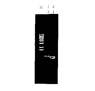
Fig. 1 is an electrophoresis pattern showing a
band for human IAP amplified by PCR using cDNA prepared from
CA 02303072 2000-03-09
- 7 -
mRNA of HL-60 cell line. From left are shown a molecular
weight marker (M), human IAP (1) and ~-actin (2).
Fig. 2 is a graph showing the level of expression
of human IAP by L1210 cells that have expressed human IAP,
using anti-CD47 antibody. The peak represents L1210 cells
transfected with only pCOSl gene as a control.
Fig. 3 is another graph showing the level of
expression of human IAP by L1210 cells that have expressed
human IAP, using anti-CD47 antibody. The peak shows that
human IAP expression has definitely increased in L1210 cells
transfected with the human IAP gene.
Fig. 4 is a graph showing antibody titers in
immunized mice. The left peak represents intact L1210
cells. The right peak represents L1210 cells transfected
with human IAP, showing that the serum of the mouse
subjected to cell fusion clearly recognizes human IAP.
Fig. 5 is a bar graph showing the results of a
growth inhibition experiment (Jurkat cells) using a
hybridoma culture supernatant.
Fig. 6 is a bar graph showing the results of a
growth inhibition experiment (ARH77 cells) using a hybridoma
culture supernatant.
Fig. 7 is a graph showing the apoptosis-inducing
effect on Jurkat cells by a culture supernatant (as analyzed
by PI staining), which is the result for an 8G2 culture
supernatant used as a control. R1 indicates the percentage
(~) of apoptosis, which is 7.43.
CA 02303072 2000-03-09
- $ -
Fig. 8 is a graph showing the apoptosis-inducing
effect on Jurkat cells by a culture supernatant (as analyzed
by PI staining), which is the result for 7D2-E3. R1
indicates the percentage (~) of apoptosis, which is 9.84.
Fig. 9 is a graph showing the apoptosis-inducing
effect on Jurkat cells by a culture supernatant (as analyzed
by PI staining), which is the result for 11C8. R1 indicates
the percentage (~) of apoptosis, which is 15.32.
Fig. 10 is a graph showing the apoptosis-inducing
effect on HL-60 cells by a culture supernatant (as analyzed
by PI staining), which is the result for an 8G2 culture
supernatant used as a control. M1 indicates the percentage
(~) of apoptosis, which is 6.94.
Fig. 11 is a graph showing the apoptosis-inducing
effect on HL-60 cells by a culture supernatant (as analyzed
by PI staining), which is the result for 11C8. Ml indicates
the percentage (~) of apoptosis, which is 12.16.
Fig. 12A is a monochrome photomicrograph showing
the result of apoptosis analysis (TUNEL method) in a
coculturing system with KM-102 and HL-60 cells, using 9C5
culture supernatant as a control. The apoptotic cells are
stained black or brown. The nuclear staining was
accomplished with Methyl Green, and the magnification is
100x.
Fig. 12B is a color photomicrograph showing the
result of apoptosis analysis (TUNEL method) in a coculturing
system with KM-102 and HL-60 cells, using 9C5 culture
CA 02303072 2000-03-09
_ 9 _
supernatant as a control. The apoptotic cells are stained
black or brown. The nuclear staining was accomplished with
Methyl Green, and the magnification is 100x.
Fig. 13A is a monochrome photomicrograph showing
the result of apoptosis analysis (TUNEL method) in a
coculturing system with KM-102 and HL-60 cells, using 11C8
culture supernatant. More TUNEL-positive cells are seen
than in Fig. 12. The apoptotic cells are stained black or
brown. The nuclear staining was accomplished with Methyl
Green, and the magnification is 100x.
Fig. 13B is a color photomicrograph showing the
result of apoptosis analysis (TUNEL method) in a coculturing
system with KM-102 and HL-60 cells, using 11C8 culture
supernatant. More TUNEL-positive cells are seen than in
Fig. 12. The apoptotic cells are stained black or brown.
The nuclear staining was accomplished with Methyl Green, and
the magnification is 100x.
Fig. 14 is an electrophoresis pattern showing the
results of SDS-PAGE analysis of IgG purified from hybridoma
lines 7D2-E3 and 11C8. Shown are molecular weight markers
(M, M'), mouse IgG (authentic sample) under non-reducing
conditions (1), 7D2-E3 (2), 11C8 (3), mouse IgG (authentic
sample) under reducing conditions (4), 7D2-E3 (5) and 11C8
(6).
Fig. 15 shows the results of analysis of CD47
expression by flow cytometry, using HL-60 cells.
CA 02303072 2000-03-09
-10-
Fig. 16 shows the results of analysis of CD47
expression by flow cytometry, using Jurkat cells.
Fig. 17 shows results for mIgG (10 ~g/ml) as a
control to demonstrate its apoptosis-inducing effect on
L1210 cells transfected with the human IAP gene (L1210-hIAP)
(incubation for 72 hours).
Fig. 18 shows the apoptosis-inducing effect of
MABL-1 (10 ~,g/ml) on L1210 cells transfected with the human
IAP gene (incubation for 72 hours).
Fig. 19 shows the apoptosis-inducing effect of
MABL-2 (10 ~,g/ml) on L1210 cells transfected with the human
IAP gene (incubation for 72 hours).
Fig. 20 shows results for mIgG (10 ~,g/ml) as a
control to demonstrate its apoptosis-inducing effect on
Jurkat cells (incubation for 48 hours).
Fig. 21 shows the apoptosis-inducing effect of
MABL-1 (10 ~,g/ml) on Jurkat cells (incubation for 48 hours).
Fig. 22 shows the apoptosis-inducing effect of
MABL-2 (10 ~,g/ml) on Jurkat cells (incubation for 48 hours).
Fig. 23 shows results for mIgG (10 ~,g/ml) as a
control to demonstrate its apoptosis-inducing effect on
L1210 cells transfected with the human IAP gene introduced
therein (L1210-hIAP) (incubation for 72 hours).
Fig. 24 shows the apoptosis-inducing effect of
MABL-2 Fab fragments (10 ~,g/ml) on L1210 cells transfected
with the human IAP gene.
CA 02303072 2000-03-09
-11-
Fig. 25 is an SDS electrophoresis pattern for
MABL-2 antibody Fab fragments.
Fig. 26 shows a notably extended survival period
upon treatment with MABL-2.
Fig. 27 shows the results of ELISA for Example
5(2).
Fig. 28 shows a notably extended survival period
upon treatment with MABL-2 F(ab')2 fragments.
Fig. 29 is an SDS electrophoresis pattern for
MABL-1 antibody and MABL-2 antibody F(ab')2 fragments.
Fig. 30 shows that human IgG levels of mouse serum
were decreased significantly in the groups treated with
MABL-1 and MABL-2, which indicates anti-tumor effects of
these antibodies.
BEST MODE FOR CARRYING OUT THE INVENTION
Preparation of Monoclonal Antibody
The monoclonal antibodies of this invention can
generally be prepared in the following manner. That is,
monoclonal antibodies of the invention may be obtained, for
example, by using human Integrin Associated Protein as the
sensitizing antigen, immunizing animals with the antigen by
an immunization method known in the art, performing cell
fusion by a cell fusion method known in the art and cloning
by a cloning method known in the art.
More specifically, a preferable method of
preparing monoclonal antibodies of the invention is, for
example, a method wherein recombinant cells of the mouse
CA 02303072 2000-03-09
-12-
leukemia cell line L1210 that express human Integrin
Associated Protein are used as the sensitizing antigen,
plasma cells (immunocytes) of a mammal immunized with the
sensitizing antigen are fused with myeloma cells of mammals
such as mice, the resulting fused cells (hybridomas) are
cloned, the clones producing the antibodies of the invention
that recognize the aforementioned cell line are selected
from the resulting clones and cultured, and the target
antibodies are obtained.
The above method is merely one possible example of
the invention and, for example, the sensitizing antigen is
not limited to the aforementioned L1210 recombinant cells
but may also be human Integrin Associated Protein (IAP)
itself, or human IAP in soluble form; the target monoclonal
antibodies that induce apoptosis of nucleated blood cells
(myeloid cells and lymphocytes) can be prepared in the same
manner as in the L1210 recombinant cells mentioned above.
The phage display method may also be used to
prepare a target monoclonal antibody from a cDNA library for
the antibody.
The mammals to be immunized with the sensitizing
antigen in the method of preparing the monoclonal antibodies
are not particularly limited, but they are preferably
selected in consideration of their compatibility with the
myeloma cells used for cell fusion, acrd mice, rats, hamsters
and the like are general suitable.
CA 02303072 2000-03-09
-13-
The immunization is preferably accomplished by a
standard method. For example, the human Integrin Associated
Protein-expressing L1210 recombinant cells are administered
to the animal by intraperitoneal injection or the like.
More specifically, an appropriate dilution or suspension
with PBS or physiological saline is preferably administered
to the animal a few times at 10-day intervals. The
immunocytes used are preferably spleen cells extracted after
the final administration of the cells.
The mammalian myeloma cells used as the parent
cells for fusion with the immunocytes may be any of various
cell lines known in the art, for example, P3 (P3X63Ag8.653)
[J. Immunol., 123, 1548 (1978)], P3-U1 [Current Topics in
Microbiology and Immunology, 81, 1-7 (1978)], NS-1 [Eur. J.
Immunol., 6, 511-519 (1976)], MPC-11 [Cell, 8, 405-415
(1976)], Sp2/0-Agl4 [Nature, 276, 269-270 (1978)], FO [J.
Immunol. Meth., 35, 1-21 (1980)], 5194 [J. Exp. Med., 148,
313-323 (1978)] and 8210 [Nature, 277, 131-133 (1979)].
The cell fusion between the immunocytes and
myeloma cells may be performed basically according to a
conventional method, such as the method of Milstein et al.
[Methods Enzymol., 73, 3-46 (1981)].
More specifically, the cell fusion is carried out,
for example, in a common nutrient medium in the presence of
a fusion promoter. For example, the fusion promoter used
may be polyethylene glycol (PEG), Sendai virus (HVJ) or the
like, and, if desired, an adjuvant such as dimethyl
CA 02303072 2000-03-09
-14-
sulfoxide may also be added appropriately in order to
increase fusion efficiency. The immunocytes are used
preferably in the amount of 1-10 times as much as myeloma
cells. The medium used for the cell fusion may be, for
example, RPMI-1640 medium, MEM medium and the like, which
are suitable for growth of myeloma cell lines, or other
media commonly used for such cell culturing, and it may also
be used in combination with a serum supplement such as fetal
bovine serum (FBS).
The cell fusion is carried out by thoroughly
mixing prescribed amounts of the immunocytes and myeloma
cells in the medium, adding a solution of PEG preheated to
about 37°C, the PEG having an average molecular weight of
approximately 1,000-6,000, for example, to the medium
usually at a concentration of about 30-60~ (W/V), and
mixing. A suitable medium is then successively added, and
the supernatant obtained by centrifugation is removed. This
procedure is repeated to produce the target hybridomas.
The hybridomas are selected by culturing in a
common selection medium, such as HAT medium (a medium
containing hypoxanthine, aminopterin and thymidine).
Culturing in the HAT medium is continued for a sufficient
time to allow death of all the cells other than the target
hybridomas (all the non-fused cells), which is usually from
a few days to a few weeks. The usual limiting dilution
method is then employed for screening and monocloning of
hybridomas producing the target antibodies.
CA 02303072 2000-03-09
-15-
The hybridomas prepared in this manner that
produce the monoclonal antibodies of the invention may be
subcultured in common medium, and may be placed in long-term
storage in liquid nitrogen.
In order to obtain the monoclonal antibodies of
the invention from the hybridomas, any suitable methods may
be employed, such as a method wherein the hybridomas may be
cultured according to standard methods and the antibodies
may be obtained from the culture supernatants; or
alternatively, a method wherein the hybridomas may be
administered to a compatible mammal for proliferation and
then the antibodies may be obtained from the ascites fluid
thereof. The former method is suitable for obtaining highly
pure antibodies, while the latter method is more suited for
mass production of antibodies.
The antibodies obtained by the aforementioned
methods can be highly purified by utilizing standard
purification methods such as salting-out, gel filtration,
affinity chromatography, or the like.
Monoclonal Antibody Fragments
The monoclonal antibodies of this invention may be
the complete antibodies described above, or fragments
thereof. That is, they may be any fragments of a monoclonal
antibody of the invention that specifically recognize human
Integrin Associated Protein and induce apoptosis of
nucleated blood cells (myeloid cells and lymphocytes) having
human Integrin Associated Protein. Such fragments include
CA 02303072 2000-03-09
-16-
Fab, F(ab')z, Fab', etc. These fragments can be prepared by
digestion with an enzyme such as papain, pepsin, ficin or
the like. The properties of the obtained fragments can be
confirmed in the same manner as described above.
Peptides and Low Molecular Compounds Having the Same
Function as the Monoclonal Antibodies
The monoclonal antibodies described above, which
recognize human Integrin Associated Protein and induce
apoptosis of nucleated blood cells, also encompass peptides
and low molecular compounds that likewise recognize IAP and
induce apoptosis of nucleated blood cells.
Properties of Monoclonal Antibodies of the Invention
As specifically described in the following
Examples, the monoclonal antibodies of the invention
specifically recognize human Integrin Associated Protein.
The monoclonal antibodies of the invention also
induce apoptosis of nucleated blood cells (myeloid cells and
lymphocytes) with human Integrin Associated Protein.
These properties can be utilized to obtain useful
therapeutic agents in the field of treatment for myeloid
leukemia and lymphoid leukemia.
Thus, it will be readily appreciated that the
construction of specific systems involving the use of the
monoclonal antibodies of the invention, as antibodies to
specifically recognize an antigen that causes apoptosis of
nucleated blood cells, for distinction and identification of
the antigens, or the use of the unique properties of the
CA 02303072 2000-03-09
-17-
monoclonal antibodies as therapeutic agents for myeloid
leukemia and lymphoid leukemia, as well as any modifications
and applications of the system, are also within the scope of
this invention insofar as they can be carried out by
applying standard methods that are obvious to those skilled
in the art.
Antileukemic Agents
An antileukemic agent according to this invention
is based on the fact that the action of IAP is promoted by
binding of an antibody or the like of the invention. While
there are no particular limitations on the dose of the
antibody of the invention, it is preferably in the range of
5 ~,g to 500 mg/kg.
EXAMPLES
This invention will now be explained in greater
detail by way of the following examples; however, the
invention is not to be limited to these examples.
Example 1 (Monoclonal Antibody Preparation)
(1) Sensitizing antigen and immunization method
Antigen sensitization was accomplished using a
recombinant cell line as the sensitizing antigen, which was
the L1210 cells transfected with human IAP gene and highly
expressed the product. L1210 is obtained from the DBA
mouse-derived leukemia cell line (ATCC No. CCL-219, J. Natl.
Cancer Inst. 10:179-192, 1949).
The human IAP gene was amplified by PCR using a
primer with a human IAP-specific sequence (sense primer:
CA 02303072 2000-03-09
-18-
GCAAGCTTATGTGGCCCCTGGTAGCG, antisense primer:
GCGGCCGCTCAGTTATTCCTAGGAGG) and cDNA prepared from mRNA of
HL-60 cell line (Clontech laboratories, Inc.) as the
template (Fig. 1).
The PCR product was subcloned into a cloning
vector pGEM-T (Promega Corporation) and used to transform E.
coli JM109 (Takara Shuzo Co., Ltd.), and after confirming
the nucleotide sequence of the insert DNA with a DNA
sequences (373A DNA Sequences, available from ABI), it was
subcloned with an expression vector pCOSl.
Expression vector pCOSl is a derivative of pEF-BOS
(Nucleic Acids Research, 18, 5322, 1990), and it is a vector
obtained by subcloning the neomycin resistant gene using
human elongation factor-la as a promoter/enhancer. This
human IAP-subcloned expression vector was used for gene
introduction into L1210 cell line with DMRIE-C (GIBCO/BRL),
selection was performed with Geneticin (final concentration:
1 mg/ml, available from GIBCO/BRL), and the gene-introduced
L1210 cells were cloned by the limiting dilution method.
The antigen expression of the obtained clones was
examined using human IAP-recognizing anti-CD47 antibody
(PharMingen), and the clones with high levels of expression
were selected as antigen-sensitized cells (Figs. 2, 3). For
culturing of the recombinant L1210 cells, 10~ fetal bovine
serum (FBS, available from Moregate Inc.) and Iscove's-
Modified Dulbecco's Medium (IMDM) (GIBCO/BRL) were used as
CA 02303072 2000-03-09
-19-
the medium, and the cells were subcultured in a 5~ COZ
incubator at a temperature of 37°C.
The immunized animals used were DBA/2 mice (bred
by Charles River, Japan), which were of the same strain as
the L1210 cells. The human Integrin Associated Protein
(IAP) gene-transfected L1210 cells, used for antigen
sensitization, were incubated for about 30 min with
mitomycin C (Kyowa Hakko Kogyo Co., Ltd.) at a concentration
of 200 ~g/ml, and after suspending growth of the cells,
mitomycin C was thoroughly washed off prior to suspension in
PBS.
The cells were intraperitoneally injected into the
mice three times at intervals of about 10 days, at
approximately 5 x 10⁶ cells each time. After the third
immunization, blood was taken from the eye socket, the serum
was diluted 50-fold with PBS containing 1~ BSA, and binding
between the diluted serum and the recombinant L1210 cells
used for antigen sensitization was confirmed with a FACScan
(Becton Dickinson and Company) (Fig. 4); the mouse having
the best antiserum activity was subjected to a booster
immunization with intraperitoneal injection of 1 x 10' cells
5 days after the fourth immunization. Four days after the
final immunization, the mouse was sacrificed and the spleen
extracted.
(2) Cell fusion
After thinly slicing the spleen extracted from the
mouse, the dissociated spleen cells were centrifuged and
CA 02303072 2000-03-09
-20-
then suspended in IMDM medium, allowed to float, and
thoroughly rinsed. Separately, the mouse myeloma cell line
P3-U1 [Current Topics in Microbiology and Immunology, 81, 1-
7 (1978)] was cultured in IMDM medium containing 10~ fetal
bovine serum (FBS, available from Moregate Inc.), and after
rinsing similarly with the IMDM medium, the 1 x 10' cells
were placed in a centrifuge tube in admixture with 5 x 10'
cells of the spleen cells and subjected to cell fusion
according to a standard method [Clin. Exp. Immunol., 42,
458-462 (1980)], using polyethylene glycol 4000 (Nakarai
Chemical Co., Ltd.).
The resulting fused cells were then suspended in
IMDM medium containing 10~ FBS and a fused cell growth
stimulating agent (BM-Condimed H1, available from Boehringer
Mannheim Biochemicals) and dispensed into a 96-well plate
for culturing at 37°C in a 5~ COZ incubator. On the
following day, the cells were placed in the HAT selection
medium and then the 10~ FBS/IMDM medium containing the
growth-stimulating agent, and culturing was continued to
sustain growth.
In order to examine the effect of the culture
supernatant of these fused cells on leukemia cell lines, the
medium for fused cells was replaced with IMDM medium
containing 10~ FBS, and culturing was continued to sustain
growth.
(3) Screening
CA 02303072 2000-03-09
-21-
The following screening was performed using the
culture supernatant of the aforementioned fused cells.
[1] Primary screening
Cells of a mouse spleen stromal cell line (CF-1
cells) transfected with the human Integrin Associated
Protein (IAP) gene (recombinant cells into which the same
plasmid was subcloned as the plasmid used to prepare the
human IAP-expressing L1210 cells used for antigen
sensitization) were seeded in a 96-well plate at 1 x 10⁴
cells per well and cultured overnight, and then fixed with
2~ PLP (periodate-lysine-paraformaldehyde) to prepare an
ELISA plate. After rinsing, the plate was subjected to
blocking for 1 h at room temperature using a l~ BSA
solution, and after further rinsing, 50 ul of the culture
supernatant of each hybridoma was added for incubation at
room temperature for one hour.
After rinsing, anti-mouse IgG+A+M (H+L) (Zymed
Laboratories Inc.) labeled with alkaline phosphatase was
added prior to incubation at room temperature for 1 h.
After rinsing, SIGMA 104 substrate (Sigma-Aldrich
Corporation) was added to provide a final concentration of 1
mg/ml, incubation was continued at room temperature, and the
specific activity was measured with a microplate reader
(Model 3550, available from BioRad Laboratories Inc.).
As a result, appearance of hybridomas was
confirmed in 2089 wells among the hybridomas seeded in 2880
wells, with 187 wells being positive in the primary
CA 02303072 2000-03-09
-22-
screening. 50 ~,1 each of Mouse IgGl as a negative control
and anti-human CD47 antibody (BD PharMingen) as a positive
control were added at a concentration of 3 ~,g/ml,
respectively, prior to incubation at room temperature for 1
h.
[2] Secondary screening
The clones judged as positive in the primary
screening were subjected to an ELISA system using human
Integrin Associated Protein (IAP)-expressing CF-1 cells,
where the negative control was CF-1 cells transfected with
only the expression vector pCOSl, in order to screen whether
the antibodies produced by the hybridomas would specifically
recognize human IAP.
As a result, the positive was confirmed for 21 of
the 187 wells found to be positive in the primary screening.
Table 1 shows the specific binding of human IAP with 7D2 and
11C8 as representative examples among these, in terms of the
absorbance in ELISA.
CA 02303072 2000-03-09
-23-
(Table 1) ELISA analysis of specific binding of hybridoma
culture supernatants with human IAP
Table 1
<Raw data> PBS ahCD47 7D2 11C8
3 ~,g /ml
CFl-pCOSl 0.185 0.160 0.189 0.149
CF1-hIAP-55-8 0.192 0.456 0.568 0.812
<Subtracted> PBS ahCD47 7D2 11C8
3 ~,g/ml
Specific binding 0.007 0.296 0.379 0.663
[3] Tertiary screening
The clones judged to be positive in the secondary
screening were subjected to a growth inhibition test using
Jurkat cells (human T cell lymphoma line) and ARH77 cells
(human myeloma cell line). 100 ~,l of the Jurkat cells at 5
x 103 cells per well and the ARH77 cells at 1 x 10⁴ cells per
well were seeded in each well of a 96-well plate, and 5 or
10 ~,1 of culture supernatant of the hybridoma clones were
added to the cell suspensions. After culturing for about 2
days, the cell numbers were measured by MTS assay. As a
control, 5 or 10 ul each of IMDM medium containing 10~ FBS
and culture supernatants of clones that were negative in the
primary screening (8G2 and 9C5) were added.
CA 02303072 2000-03-09
-24-
Figs. 5 and 6 show the results of the growth
inhibition effect of four representative clones, 11C8, 7D2-
E3 (subclone of 7D2), 13F1 and 2F12.
(4) Antibody properties
[1] The immunoglobulin types of the culture
supernatants of 11C8, 7D2-E3, 13F1 and 2F12 were examined
using an ELISA system.
Specifically, human Integrin Associated Protein
(IAP)-expressing CF-1 cells were seeded into a 96-well plate
to prepare an ELISA plate, and then 50 ~,1 of each culture
supernatant was added, alkaline phosphatase-labeled anti-
mouse IgG antibody (Zymed Laboratories Inc.) or anti-mouse
IgM antibody (Biosource Intl., Inc.) were reacted therewith
as secondary antibodies, and the activity was measured with
a microplate reader. As a result, 11C8 and 7D2-E3 were
confirmed to be IgG, while 13F1 and 2F12 were confirmed to
be IgM.
[2] The DNA fragmentation of the two clones 11C8 and
7D2-E3 among the four clones described above was analyzed by
flow cytometry (FACScan, available from Becton, Dickinson
and Company) using Jurkat cells and HL-60 cells. The Jurkat
cells were used for 11C8 and 7D2-E3, and the HL-60 cells
were used for 11C8.
The Jurkat cells and HL-60 cells were seeded in a
12-well plate at 4 x 10⁴ cells per well/2 ml, respectively,
and 200 ~,1 of the culture supernatants of 7D2-E3 and 11C8
were added. The cells were cultured for 2 days, and
CA 02303072 2000-03-09
-25-
measured. As a control, 8G2 culture supernatant was added
in an equal volume. The cells were recovered from the
culturing plate and a cell pellet was fixed under 200 x g
for 60 minutes at 4°C in 2 ml of chilled 70~ ethanol. The
cells were then centrifuged, rinsed in 1 ml of PBS and
resuspended in 0.5 ml of PBS. To a 0.5 ml sample of the
cells, 0.5 ml of RNAse (Type I-A, Sigma-Aldrich Corporation,
St. Louis, M0, USA; 1 mg/ml in PBS) was added, and these
were mixed with a 1 ml propidium iodide solution (PI, Sigma,
100 ~,g/ml in PBS). The mixed cells were incubated for 60
min in a darkroom at 37°C, and then kept in the darkroom at
4°C and measured by flow cytometry.
As shown in Figs. 7-9 and 10-11, the culture
supernatants of 7D2-E3 and 11C8 increase a proportion of
apoptosis cells of Jurkat cells and the culture supernatant
of 11C8 increases a proportion of apoptosis cells of HL-60
cells, respectively.
[3] The culture supernatants of 11C8 were used in a
coculturing system with HL-60 cells using a feeder layer of
cells of the human myeloid stromal cell line KM102, to
determine whether these culture supernatants induce
apoptosis of HL-60 cells.
Specifically, KM102 cells were seeded in a 2-well
Lab-Tek Chamber Slide (Nalge Nunc Intl. Corporation) and
brought to a sub-confluent state, 1 x 10⁵ cells of HL-60
cells were seeded thereon and cultured for about one day,
and then the non-attached HL-60 cells were removed. The
CA 02303072 2000-03-09
-26-
aforementioned culture supernatants were simultaneously
added to provide a final concentration of 10~ and the cells
were cultured for 2 days. After culturing, the cells were
fixed with 10~ formalin and the apoptosis-induced HL-60
cells were detected by the TUNEL method (ApopTag Plus
available from Oncor Inc.). As shown in Figs. 12 and 13,
the culture supernatant of 11C8 more increases apoptosis
cells of HL-60 cells than the culture supernatant of 9C5
does, which is the culture supernatant of the human IAP non-
reacting hybridoma clone used as the control.
(5) Antibody purification
For purification of the antibodies produced by
hybridomas, the cell lines of the IgG-producing clones 7D2-
E3 and 11C8 among the above hybridoma lines were
intraperitoneally injected into pristane-administered
BALB/c/AnNCrj mice (male, available from Charles River,
Japan) according to a standard method. After several weeks,
the ascites fluid produced was taken and the antibodies were
separated and purified by standard methods. Specifically,
the antibodies were purified from the obtained ascites fluid
by a Polos Protein A plastic column (Perceptive Biosystems
Inc.) and dialyzed with PBS (Dulbecco Inc.), and bands were
confirmed with SDS-PAGE analysis. As shown in Fig. 14,
electrophoresis using an authentic sample of mouse IgG
(Cappel Inc.) as a control confirmed bands for the IgG of
clones 7D2-E3 and 11C8 at the same positions as the
CA 02303072 2000-03-09
-27-
authentic sample mouse IgG, under both non-reducing
conditions and reducing conditions.
In this example, the human Integrin Associated
Protein (IAP)-expressing L1210 cells were used as the
sensitizing antigen for illustrative purposes, but it is
also possible to prepare monoclonal antibodies in the same
manner using other human IAP-expressing cells or human IAP
itself, and to prepare monoclonal antibodies from an
antibody library using the phage display method; this
invention is not limited to the aforementioned monoclonal
antibodies but encompasses all monoclonal antibodies with
properties similar thereto and all hybridomas that produce
those monoclonal antibodies.
Furthermore, the invention of these monoclonal
antibodies also includes humanized antibodies, human
antibodies, chimeric antibodies, single-chain antibodies,
primatized antibodies and antibody fragments obtained by
digesting the antibodies with various enzymes (papain,
pepsin, ficin, etc.).
The hybridomas producing the monoclonal anti-human
Integrin Associated Protein (IAP) antibodies of the
invention are novel fused cells created from DBA mice spleen
cells and the mouse myeloma cell line P3-U1 as the parent
cells; anti-IAP antibody (mouse hybridoma 11C8-F8 (subclone
of 11C8), designated as "MABL-1") was deposited as FERM BP-
6100 and anti-IAP antibody (mouse hybridoma 7D2-E3 (subclone
of 7D2), designated as "MABL-2") as FERM BP-6101 on
CA 02303072 2000-03-09
-28-
September 1, 1997 with the National Institute of Bioscience
and Human Technology, Agency of Industrial Science and
Technology, Ministry of International Trade and Industry,
located at 1-3 Higashi 1-chome, Tsukuba-shi, Ibaraki-ken,
Japan, as an authorized depository for public
microorganisms.
Example 2 (Subclass identification of MABL-1 and MABL-2
antibodies)
In order to identify the subclasses of MABL-1 and
MABL-2 antibodies obtained above, 500 ~1 each of MABL-1 and
MABL-2 adjusted to 100 ng/ml was spotted on an Isotyping Kit
(Stratagene), by which MABL-1 was shown to be IgGl, x and
MABL-2 was shown to be IgG2a, x.
Example 3 (Human IAP-expressing human leukemia cells)
IAP expression in different human leukemia cell
lines was detected by flow cytometry with human IAP-
recognizing anti-CD47 antibody (a commercially available
product). This antibody was used for the detection because
human IAP is believed to be identical to CD47 (Biochem. J.,
304, 525-530, 1994). The cell lines used were Jurkat and
HL-60 cells (K562 cells, ARH77 cells, Raji cells and CMK
cells). The cells were used at 2 x 10⁵ cells per sample,
the anti-CD47 antibody was incubated with the cells at a
final concentration of 5 ~ug/ml, and the secondary antibody
used was FITC-labeled anti-mouse IgG antibody (Becton
Dickinson and Company). Mouse IgGl antibody (Zymed
Laboratories Inc.) was used as a control. The results of
CA 02303072 2000-03-09
-29-
the flow cytometry as shown in Fig. 15 (HL-60) and Fig. 16
(Jurkat) confirmed that both cell lines expressed IAP.
Example 4 (Apoptotic effect in vitro)
(1) The apoptosis-inducing activity of the MABL-1 and
MABL-2 antibodies on L1210 cells transfected with human IAP
gene, Jurkat cells and HL-60 cells were examined using
Annexin-V (Boehringer Mannheim). The results of analysis
with Annexin-V are shown in Figs. 17-22, wherein the dots in
the lower left region indicate the live cells, those in the
lower right region indicate apoptotic cells, and those in
the upper right region indicate necrotic cells. The
antibodies used were mouse IgG (Zymed Laboratories Inc.) as
a control and MABL-1 and MABL-2 at 10 ~,g/ml, and after 4 x
103 cells of L1210 cells transfected with the human IAP gene
were incubated for 72 h and 6 x 10⁴ cells of the Jurkat
cells were incubated for 48 h, they were analyzed with
Annexin-V. Cell death was observed, as shown in Figs. 17-
22. For the HL-60 cells, 10 ~.g/ml of MABL-1 was used, and
analysis with Annexin-V at 1 x 10⁵ cells likewise revealed
cell death.
(2) The apoptosis-inducing activity of MABL-2 antibody
Fab fragments on L1210 cells transfected with human IAP gene
was examined. Specifically, L1210 cells transfected with
human IAP gene were cultured at 4 x 10³ cells, and MABL-2
antibody Fab fragments and mouse IgG as a control were used
at a concentration of 10 ~,g/ml. The cells were incubated
for 72 h and measured with Annexin-V. As a result,
CA 02303072 2000-03-09
-30-
considerable cell death was observed (Figs. 23, 24). The
MABL-2 antibody Fab fragments used for the experiment were
obtained by digesting the antibody with papain (Pierce
Laboratories, Inc.) and purifying it. The MABL-2 antibody
Fab fragments were analyzed by SDS electrophoresis (Fig.
25).
Example 5 (Investigation of apoptosis in vivo)
(1) Drug efficacy of MABL-1 and MABL-2 (whole IgG)
Human IAP-expressing KPMM2 cells (human myeloma
cell line) were transplanted into SCID mice, and on the 10th
day after transplantation, MABL-1 and MARL-2 (whole IgG)
were administered by single intravenous injection in a dose
each of 5 ~,g/head and 50 ~ug/head, respectively (n=5); on the
28th day after KPMM2 transplantation, the human IgG levels
derived from KPMM2 were measured by ELISA, and the
disappearance was confirmed. The survival period was also
examined. The results showed marked suppression of blood
levels of human IgG in the groups treated with MABL-1 and
MABL-2, which represented the anti-tumor effect (Fig. 30).
The survival period was also shown to be notably lengthened
(Fig. 26).
(2) Drug efficacy of MABL-1 and MABL-2 (F(ab')z)
F(ab')z fragments prepared by digestion of the
MABL-1 and MABL-2 antibodies with pepsin and purification
with Protein A (Pierce laboratories, Inc.) were used to
examine the anti-tumor effect except the cytotoxic effect
via the Fc regions. Specifically, human IAP-expressing
CA 02303072 2000-03-09
-31-
KPMM2 cells (human myeloma cell line) were transplanted into
SCID mice, and MABL-1 and MABL-2 F(ab')Z fragments were
intravenously administered to the groups in a dose of 100
~,g/head on the 6th and 10th days after transplanting, and to
the groups in a dose each of 10 and 30 ~ug/head on the 6th,
8th and 10th days after transplantation, respectively; the
human IgG levels derived from KPMM2 were measured by ELISA
on the 30th day after transplantation (Fig. 27). The
survival period was also examined up to 90 days after
transplanting. As a result, a notable suppressing effect on
human IgG levels in the blood was found in the groups
treated with MABL-1 and MABL-2, which represented the anti-
tumor effect. The survival period was also considerably
lengthened (Fig. 28). Fig. 29 shows the SDS electrophoresis
pattern for the F(ab')z fragments of MABL-1 antibody and
MABL-2 antibody.
INDUSTRIAL APPLICABILITY
The monoclonal antibodies of this invention are
antibodies that specifically recognize human Integrin
Associated Protein, and the antigens that induce apoptosis
of nucleated blood cells having human Integrin Associated
Protein. Accordingly, they are useful as antibodies that
recognize human Integrin Associated Protein for its
distinction and identification, while also having an action
of inducing apoptosis of nucleated blood cells; these
properties can be utilized to prepare useful therapeutic
CA 02303072 2000-03-09
-32-
agents in the field of treatment for myeloid leukemia and
lymphoid leukemia.
CA 02303072 2000-03-09
- 1 -
SEQUENCE LISTING
<110> CHUGAI SEIYAKU KABUSHIKI KAISHA
<120> Monoclonal antibody inducing apoptosis
<130> CGS98-03PCT
<160> 2
<210> 1
<211> 26
<212> DNA
<213> Artificial Sequence
<220>
<223> PCR primer
<400> 1
GCAAGCTTAT GTGGCCCCTG GTAGCG 26
<210> 2
<211> 26
<212> DNA
<213> Artificial Sequence
<220>
CA 02303072 2000-03-09
- 2 -
<230> PCR primer
<400> 2
GCGGCCGCTC AGTTATTCCT AGGAGG 26