Note: Descriptions are shown in the official language in which they were submitted.
CA 02303157 2000-03-09
WO.Q9/14595 PCT/US98/19418
DEVICE FOR RECEIVING AND PROCESSING A SAMPLE
BACKGROUND OF THE INVENTION
1. Field of the Invention
This invention relates to the field of diagnostic assays and the collection
and
processing of samples therefor.
The ability to measure quantitatively a wide variety of physiologically active
compounds, both naturally occurring and synthetic, has become of increasing
importance, both as an adjunct to diagnosis and therapy. The medical industry
has
become increasingly dependent upon the ability to measure various entities in
physiological fluids in order to be able to determine the health status of an
individual, dosage level for drugs, use of illegal drugs, genomic sequences
and the
like. Thus, the capability of taking a physiological sample and rapidly
analyzing for
a particular component has made medical therapies more efficient and
increasingly
successful.
For the most part diagnostic assays of physiological fluids or biological
samples for one or more analytes have required clinical laboratory
determinations
although there has been an increasing focus on being able to carry out assay
determinations in the doctor's once and in the home. Numerous systems have
been
developed in efforts to try to address the various problems associated with
analyses
carried out in the clinical laboratory.
There is substantial interest in providing for protocols and devices which are
simple, easy to manipulate, and reduce the opportunity for operator failure.
The ideal
situation would be collection of an unmeasured sample in a container, which is
then
sealed. Subsequently, the sample could then be introduced into an assay device
without opening the sealed container and without the need for accurately
measuring
the sample. The device into which the sample is introduced provides for
precise
CA 02303157 2000-03-09
WD 99/14595 PCT/US98/19418
2
measurement of the sample to be analyzed, which is important in obtaining a
quantitative result.
In many instances blood is a source of a sample to diagnose a patient's health
or to monitor the efficacy of drugs that have been administered to the
patient. Blood
as a source for the determination of these parameters has many deficiencies
when
used directly or even when diluted with buffer. These deficiencies include:
rapid
coagulation, the presence of a large number of light absorbing and fluorescent
substances, variations in composition, susceptibility to changes in relation
to reagents
used in assays, and variations In the presence or absence of oxygen. These
properties complicate the use of blood as a sample for diagnostic purposes.
Various
techniques have been employed to avoid these problems, e.g., high dilution,
addition
of anticoagulants, separation of blood into plasma and its cellular
components, and
the like. During such manipulations great care must be taken to avoid lysis of
red
blood cells to avoid the release of hemoglobin, which can interfere with
diagnostic
assays. Despite the problems associated with the use of blood as the sample
medium, in many instances, blood is the only source that provides the
information of
interest. Therefore, identifying ways of using whole blood, while diminishing
the
interference from its constituents is highly desirable. There is, therefore,
substantial
interest in devising new approaches for using and manipulating blood for
diagnostic
purposes.
Thus, the use of whole blood in diagnostic assays is not unusual in the
medical field. When the volume of blood needed to perform the test becomes
greater
than a few drops, a blood collection container such as a vacuum tube or
syringe is
used. The subsequent delivery of the sample into the assay requires the
transfer of
blood from the collection container to an assay device. The transfer increases
the
risk of both hazardous contact to the clinician as well as alteration of the
specimen.
Also, in some circumstances, it is desirable to preprocess the blood sample
such as
by removal of cells from whole blood, lysing cells in whole blood, and so
forth.
CA 02303157 2000-03-09
WO 99/14595 PCTNS98/19418
3
Certain devices are known for the collection of a sample for a qualitative
determination of an analyte of interest. As may be appreciated, the
considerations
for collection of a sample for a quantitative determination of an analyte are
much
different. In general, for a qualitative result the collection of a precise
amount of a
sample is not a consideration.
One area of particular interest in analyses employing whole blood samples is
the assessment of platelet function. The role of platelets in mammalian
physiology is
extraordinarily diverse, but their primary role is in promoting thrombus
formation.
In many situations, an evaluation of the ability of blood to clot is desired,
a
parameter that is frequently controlled by the ability of platelets to adhere
and/or
aggregate. Thus, one may wish to assess the adhesive functions of platelets.
For
example, one may wish to know whether to administer drugs that will block, or
promote, clot formation , or one may need to detect deficiencies in platelet
function
prior to surgical procedures. In other instances one may be interested in
evaluating
the effectiveness of a platelet inhibitor that is being tested as a new drug
or is being
used as approved clinical treatment in a patient.
Platelets are known to aggregate under a variety of conditions and in the
presence of a number of different reagents. Platelet aggregation is a term
used to
describe the binding of platelets to one another. The phenomenon can be
induced by
adding aggregation inducing agents to platelet rich plasma (PRP) or to whole
blood.
Platelet aggregation in vitro depends upon the ability of platelets to bind
fibrinogen
to their surfaces after activation by an aggregation inducing agent such as
ADP or
collagen.
Platelets play a critical role in the maintenance of normal homeostasis. When
exposed to a damaged blood vessel, platelets will adhere to exposed sub
endothelial
matrix. Following the initial adhesion, various factors released at the site
of injury
such as thrombin, ADP and collagen activate the platelets. Once platelets are
CA 02303157 2000-03-09
WO_99/14595 PCr/US9g~19418
4
activated, a conformational change occurs in the platelet glycoproteln
GPIIb/IIIa
receptor allowing it to bind fibrinogen and/or von Willebrand factor.
It is this binding of the multivalent fibrinogen and/or von Willebrand factor
molecules by GPIIb/IIIa receptors on adjacent platelets that results in the
recruitment
of additional platelets to the site of injury and their aggregation to form a
hemostatic
plug or thrombus
The success of aspirin, and more recently ticlopidine, in treating, and
preventing ischemic complications of thrombosis has stimulated the search for
more
potent agents. New agents that block platelet GPIIb/IIIa receptors are being
developed for use as antithrombotic agents, including peptides and
peptidomimetics,
based on the arginine-glycine-aspartic acid (RGD) and related cell recognition
sequences. A recombinant murine/human chimeric antibody Fab fragment
(c7E3Fab, abciximab, ReoProTM) c7E3 has been approved in Europe and the United
States for use as adjunctive therapy in high risk angioplasty. Moreover, one
additional trial (EPILOG), was stopped early by the Data and Safety Monitoring
Boards because of the greater than expected benefit of 61% reduction in
thrombotic
events with c7E3 in the full range of patients undergoing coronary
angioplasty.
However, the benefit of GPIIb/IIIa blockers has been accompanied by an
increased
risk of bleeding. A number of other agents are currently in early and advanced
trials,
including agents that are orally active.
Intrinsic differences in pharmacokinetics and pharmacodynamics among the
GPIIb/IIIa antagonists may affect the dose required to achieve a safe
therapeutic
antiplatelet effect. Beyond these overall differences in the drugs, however,
interindividual variations in drug excretion and metabolism may have an impact
on
optimal drug dosing. Prolonged therapy, either with parenteral or oral agents,
is
likely to magnify the importance of interindividual differences, especially
with pro
drugs and low molecular weight agents that rely on renal excretion or hepatic
metabolism.
CA 02303157 2000-03-09
W0..99/14595 PCT/IJS98/19418
Direct measurement of GPIIb/IIIa receptor blockade has been reported for
only a few GPllb/IIIa antagonists. An assay to measure GPIIb/IIIa receptor
blockade
by c7E3 Fab, based upon inhibition of platelet binding of radiolabelled c7E3
has
been used to correlate GPIIb/IIIa receptor blockade, inhibition of platelet
5 aggregation, prolongation of the bleeding time, and antithrombotic e~cacy in
animal
models. Based on these results, the target level for coronary artery
angioplasty was
defined as >80% GPIIb/IIIa receptor blockade, and this level of blockade has
proved
efficacious in three separate Phase III studies in humans.
Available assays for evaluating GPIIb/IIIa receptor blockade, including
platelet aggregation, bleeding time, thromboelastography, clot retraction,
radiolabelled antibody binding, and flow cytometry are time consuming, require
standardization, or require specialized equipment.
In vitro platelet aggregation is the laboratory method used to assess the in
vivo
ability of platelets to form the aggregates leading to a primary hemostatic
plug. In
this technique an aggregating agent such as ADP or collagen is added to whole
blood
or PRP and aggregation of platelets monitored. Platelet aggregometry is a
diagnostic
tool that can provide insights difficult to obtain by other techniques, thus
aiding in
patient diagnosis and selection of therapy. Current methods of monitoring
platelet
aggregation require expensive, laboratory dedicated instruments that are not
easily
portable and require standardization to ensure accurate quantitative results.
In
addition, unless performed using whole blood, results are unlikely to be
available for
several hours.
Currently there are two detection methods used in instruments with FDA
clearance for performing platelet aggregometry: optical and impedance
measurements. Optical detection of platelet aggregation is based on the
observation
that, as platelets aggregate into large clumps, there is an increase in light
transmittance. Different aggregation-inducing agents stimulate different
pathways of
activation and different patterns of aggregation are observed. The main
drawback of
CA 02303157 2000-03-09
WO 99/14595 PCTNS98/19418
6
the optical method is that it must be performed on PRP, necessitating the
separation
of platelets from red blood cells and adjustment of the platelet count to a
standardized value.
Impedance detection can be used to test anti coagulated blood with no need to
isolate platelets from other components of the blood, although in many cases
the
sample is diluted before testing. The method detects aggregation by passing a
very
small electric current between two electrodes immersed in a sample of blood
(or
PRP) and measuring electrical impedance between the electrodes. During initial
contact with the blood or PRP, the electrodes become coated with a monolayer
of
platelets. If no aggregating agent is added, no further interactions occur
between the
platelets and the electrodes and electrical impedance remains constant. When
an
aggregation inducing agent is added, platelets aggregate on the electrodes and
there is
an increase in impedance.
The CHRONO LOG Model 530 and Model 540 use the optical method for
PRP and the impedance method for whole blood aggregometry. The impedance
method has been shown to be substantially equivalent to the optical method for
measuring platelet aggregation in PRP.
Various photometers are commercially available for measuring the light
absorbance of liquid samples in microtitration plates or other sample holding
vessels.
Examples of such equipment are the MR 600 Microplate Reader (Dynatech
Laboratories, Inc., Alexandria, Va.), and the Vmax Kinetic Microplate Reader
(Molecular Devices, Palo Alto, CA).
It is desirable to have a rapid and simple platelet function assay including
the
ability to transfer blood to be tested from a collection container to an assay
device
without opening the collection container. In the setting of coronary
angioplasty, it is
desirable to have a platelet aggregation assay that could be conducted at the
same
time as the activated clotting time (ACT), which is performed to assess the
adequacy
CA 02303157 2000-03-09
WO 99/14595 PCT/US98/19418
7
of heparinization. During chronic infusions of GPIIb/IIIa antagonism, or with
chronic
oral therapy, periodic monitoring may also be desirable. In certain
circumstances, as
for example, prior to surgery or an invasive procedure, it may be desirable to
rapidly
determine whether the effect of the GPIIb/IIIa antagonist has worn off
suffciently to
allow the surgery or procedure to be performed without further interventions
to
reverse the effect of the GPIIb/IIIa inhibitor. Finally, in the event of
bleeding
complications, a rapid measure of platelet function may be helpful in
determining
whether the bleeding is due to a high or toxic level of platelet inhibition.
The level of
platelet inhibition may also be helpful in guiding whether to reverse the drug
effect
with platelet transfusions or look for other causes of bleeding.
2. Previous Disclosures
O'Brien, J.R., Nature 202:1188 (1964) discloses aggregation studies of 2 mL
plasma samples placed in a cuvette in an EEL titrometer or electrophotometer.
Each
sample is treated individually and aggregation is said to occur when the
optical
transmission increases.
Mills, D.C.B., and Roberts, G.C.K., .J. Physiol. 193:443 453 (1967) disclose
platelet aggregation measurements in a modified EEL Long Cell Absorptiometer
(Evans Electroselenium Ltd., Halstead, Essex, U.K.). The measurements are
taken on
a 1 mL plasma sample, stirred from below by a magnetic stirrer while
continuous
recordings are made.
Michal, F., and Born, G.V.R., Nature New Biol. 23I 220 (1971) disclose a
modification of the traditional optical method of measuring aggregation which
permits the simultaneous measurement of scattered and transmitted light. This
method encompasses a modification of the cuvette chamber of an aggregometer to
allow for the measurement of light scattered at right angles to the incident
light
beam. In the aggregometer the incident light illuminates a suspension of
platelets
which are kept in rapid motion by a magnet rotating in the bottom of the glass
tube at
CA 02303157 2000-03-09
WO 99/14595 PCT/US98/19418
8
1,000 rpm. The sample volume is about 1 mL and the optical density is read
individually for each sample which is kept in a water jacketed environment at
37°C.
Fratontoni, et al., in U.S. Patent No. 5,325,925 disclose the use of an
agitated
microtiter plate to assess aggregation in PRP.
Shaw, et aL, in U.S. Patent No. 5,427,913 disclose a method for determining
platelet function in PRP by contacting the platelets in suspension with an
immobilized extracellular matrix protein while applying mechanical stimulus to
the
platelets, and determining the platelet activation produced by various
indicia.
Coagulation monitors are known for the analysis of whole blood. U.S. Patent
No. 3,695,842 describes a method of analyzing the transformation of a liquid,
e.g.
blood, to a gelatinous or solid mass. The coagulation system uses a vacutainer
with
all the necessary reagents, as well as a ferromagnetic component. Once the
blood
sample has been drawn into the vacutainer, it is placed into the instrument in
an
inclined manner. This procedure makes the ferromagnetic component sit at the
bottom of the tube in close proximity to a magnetic reed switch. As the sample
is
rotated, gravity ensures that the component remains close to the reed switch.
However, as the blood starts to clot, viscosity increases to the point where
the
component starts to rotate with the blood sample. The reed switch is thus
activated,
enabling a coagulation time to be estimated.
Hillman, et al., disclose a unit use cartridge in which dry reagents are
placed
into the analyzer which is then heated to 37°C before a drop of brood
is introduced.
The sample is mixed with the reagent by capillary draw. The detection
mechanism is
based on laser light passing through the sample. Blood cells moving along the
flow
path yield a speckled pattern specific to unclotted blood. When the blood
clots,
movement ceases producing a pattern specific to clotted blood. Several patents
disclose aspects of this technology and are described further below.
CA 02303157 2000-03-09
WO 99/14595 PCT/US98/19418
9
U.S. Patent No. 4,756,884 discloses the component parts of the cartridge
technology, which is based on capillary draw, including certain antibodies and
reagents for blood clotting.
U.S. Patent No. 4,948,961 discloses the components and method of use of an
optical simulator cartridge used with the above instrument. U. S. Patent No.
4,963,498 discloses a method of obtaining chemical information from the
capillary
draw cartridge. U.S. Patent No. 5,004,923 discloses optical features by which
the
above. instrument interrogates the cartridge
Shenaq and Saleem, in "Effective Hemostasis in Cardiac Surgery," Eds:
Ellison, N. and Jobes, D.R., Saunder & Co. (1988), utilize a sonic probe that
is
inserted into a cuvette containing the sample and reagents. The sonic probe
responds
to clot formation in the cuvette and, thus, can be used to measure the
coagulation
time.
PCT application WO 89/06803 describes a unit use cartridge having two
capillary tubes that simultaneously draw blood from a single finger stick. The
design
allows for duplicate measurement or two different measurements based on
different
reagent coatings. Blood coagulation is measured by charges in light
permeability
through the capillary tube.
U.S. Patent No. 5,110,727 describes another format based on the use of
magnetic particles mixed into a dry reagent contained within a flat capillary
chamber.
An applied oscillating magnetic field from the instrument causes the particles
to
oscillate once the reagent has dissolved in the blood. This motion is
monitored
optically. When the blood clots, the particles become entrapped and motion is
diminished. Fibrinolysis assays are performed by monitoring the reverse
process
{See, Oberhardt, et al., Clin. Chem. (1992) 37:520).
Machado, et al., J. Acoust. Soc. Am (1991) 90:1749 describe another method
of detecting coagulation based on ultrasonic scattering from 200 micron glass
CA 02303157 2000-03-09
WO 99/14595 PCT1US98/19418
spheres suspended in a blood sample. Amplitude and phase changes of the
scattered
waves are used to detect coagulation.
Varon, et al., in U.S. Patent No. 5,523,238, describe a method for determining
platelet function by introducing a blood sample into a vessel having a flat
bottom, the
5 inner surface of which is covered with a substrate capable of inducing
platelet
adhesion thereto and aggregation; rotating the preparation inside the vessel,
inducing
shear forces at the surface which aggregate the platelets; and determining
parameters
of the adhered blood platelets, such as number of adhered platelets, aggregate
size,
aggregate morphology, total area covered by the aggregates, and distribution
of
10 adhered platelets or aggregates on the surface.
Coller, in U.S. Patent No. 5,763,199 and U.S. patent application Serial No.
08/754,773, describes a method for the analysis of whole blood for GPiIb/IIIa
receptor activity. The disclosed method relies upon the visual observation of
platelet
mediated agglutination of fibrinogen coated beads.
The disclosure of all the aforementioned patents and publications is
incorporated herein by reference.
SUMMARY OF THE INVENTION
One aspect of the invention concerns a device for receiving and processing a
sample. The device comprises a sample receiving element adapted to establish
fluid
communication with and receive a sample directly from a sample container. The
sample receiving element also allows for introduction of a sample into the
device. A
first chamber is in fluid communication with the sample receiving element. One
or
more second chambers are in fluid communication with the first chamber. The
device also comprises first and second ports. The first port provides for
venting the
device. The second port provides for establishing communication between the
CA 02303157 2000-03-09
W0 99/14595 PCT/US98/19418
11
device and means for moving the sample from the sample receiving element to
the
first chamber and for moving the sample from the first chamber to the one or
more
second chambers. The device may also include means for controlling the precise
amount of the sample introduced into each of the second chambers. The first
chamber and/or one or more of the second chambers are adapted for processing
the
sample.
Another embodiment of the present invention also concerns a device for
receiving and processing a sample. In this embodiment a sample receiving
element
has an input port that provides for direct sealing connection and
establishment of
fluid communication with a sample container such that sample in the container
is
capable of introduction into the device. A first chamber is in fluid
communication
with the input port. One or more second chambers are each respectively in
fluid
communication with the first chamber. The second chambers contain one or more
reagents for processing the sample. The device has first and second ports. The
first
port provides for connecting the device with a pressure varying apparatus for
alternately increasing and decreasing pressure in the device. The second port
provides for venting the device. The device also includes means for permitting
air to
escape from the one or more second chambers and for sealing the one or more
second chambers when a predetermined amount of the sample fills the one or
more
second chambers.
Another embodiment of a device in accordance with the present invention has
an input port comprising a needle for piercing a sample container and allowing
introduction of a sample suspected of containing an analyte directly into the
device.
A first chamber is in fluid communication with the input port by means of a
first
channel connecting the input port and the first chamber. A manifold is in
fluid
communication with the first chamber. One or more second chambers are each
respectively in fluid communication with the manifold by means of a second
channel
between the manifold and the respective second chamber. One or more of the
CA 02303157 2000-03-09
WO 99/14595 PCT/US98/194t8
12
second chambers contain one or more reagents for conducting an assay. A first
port
is included for connecting the device with a pressure varying apparatus for
alternately increasing and decreasing pressure in the device. The device also
includes a second port for venting the device. The device further comprises a
vent
plug for each of the one or more second chambers for permitting air to vent
from the
one or more second chambers and sealing the one or more second chambers when
the sample reaches the vent plug.
Another embodiment of the present invention is a kit for processing a sample.
The kit comprises in packaged combination a device as described above and
reagents
for processing the sample other than reagents that are included in the device
itself.
Another embodiment of the present invention is a method for receiving and
processing a sample. In the method a sample is applied to a sample receiving
element of a device as described above. The first port is connected to a
pressure
varying apparatus. Pressure in the device is adjusted to cause the sample to
flow
from the sample receiving element to the first chamber. After a holding period
pressure is again adjusted in the device to cause the sample to flow from the
first
chamber to the one or more of the second chambers where the sample is
processed.
BRIEF DESCRIPTION OF THE DRAWINGS
Fig. 1 is a diagrammatic plan view of a device according to the subject
invention.
Fig. 2 is a plan view of an assembled device of Fig. 1.
Fig. 3 is a diagrammatic plan view of the device of Fig. 2 without a cover
plate.
Fig. 4 is an exploded view of the device of Fig. 1.
CA 02303157 2000-03-09
WO 99/14595 PCT/US98/19418
13
Fig. 5 is diagrammatic plan view of an alternative embodiment of a device in
accordance with the present invention.
Fig. 6 is a schematic diagram of one embodiment of an instrument that may be
employed in conjunction with a device in accordance with the present
invention.
DETAILED DESCRIPTION OF THE INVENTION
The present invention provides for the collection in a sealed container of a
sample to be analyzed and transferring the sample into a device that can be
used in
conjunction with an assay apparatus for analysis of the sample and can then be
easily
disposed of if desired. The transfer can be accomplished without opening the
sealed
container, thus avoiding exposing the clinician to undue risks and avoiding
placing
the blood in an environment that may be detrimental to its ability to function
as a
reliable source for a determination of an analyte. The device permits the
measuring
of precise amounts of the sample into chambers for analysis. The sample can be
conditioned with reagents and analyzed in the device of the present invention,
which
can be disposable.
Before proceeding further with a detailed description of the present
invention,
a number of terms as used herein are defined.
Sample =- any solution, synthetic or natural, containing an analyte, including
body fluids such as, for example, whole blood, blood fractions such as serum
and
plasma, synovial fluid, cerebro-spinal fluid, amniotic fluid, semen, cervical
mucus,
sputum, saliva, gingival fluid, urine, and the like, and aqueous or water
soluble
solutions of natural or synthetic compounds, particularly, compounds that are
potential therapeutic drugs, and it is desired to determine if the compound
binds to a
specific receptor. The amount of the sample depends on the nature of the
sample and
the analyte contained therein. For fluid samples such as whole blood, saliva,
urine
and the like the amount of the sample is usually about 0.1 to 10 ml, more
usually,
CA 02303157 2000-03-09
WO_99/14595 PCT/US98/19418
14
about 1.8 to 4.5 ml. The term "sample" includes unprocessed samples directly
from
a patient or samples that have been pretreated and prepared in any convenient
medium although an aqueous medium is preferred.
As mentioned above, the sample may be preprocessed prior to placing the
sample in a sample container. Such preprocessing may include lysing of cells
in the
sample, releasing an analyte from binding materials in a sample, absorbing
unwanted
materials by affinity matrices, and so forth. Reagents for lysing cells in the
sample
include, for example, ammonium chloride, sodium chloride, detergents such as
Triton X-100, Zwittergen and the like. The amount of the lysing reagent
applied to
the sample is generally su~cient to bring about the level of lysing desired
and is
usually about 0.01 to 10% by weight.
Other preprocessing reagents include precipitation reagents, affinity matrices
with antibodies or antigens or lectins and so forth. The amount of such a
reagent
employed is generally sufficient to achieve the desired result and the reagent
is
applied in a manner similar to that described above for the stabilization
reagent.
Analyte -- the substance to be determined. The analyte may be any chemical
entity and includes ligands and receptors, where the ligand and the receptor
are
defined as members of a specific binding pair that have an affinity or avidity
for each
other. The ligand may be a hapten or antigen, where haptens generally range
from
about 100 to 5000 molecular weight and include drugs of abuse such as cocaine,
marijuana, etc., and therapeutic drugs such as cyclosporin, theophylline,
dilantia,
antibiotics such as amikacin, tobramycin, anticonvulsants, etc., and the like.
Antigens generally range from about 2500 molecular weight to any upper limit
such
as, e.g., one million or more, and include cancer antigens such as PSA, CEA,
AFP,
CA19.9, etc., cardiac markers such as myoglobin, CKMB, etc., and so forth.
Analytes may include naturally occurring ligands and receptors, synthetic
compounds, pollutants, contaminants, microorganisms, e.g., viruses such as HIV
virus, herpes virus (HSV) and the like, unicellular organisms, etc., blood
components
CA 02303157 2000-03-09
WO._99/I4595 PCT/US98/19418
such as platelets, and the like and receptors thereon, blood proteins such as
hemoglobin Alc, HLA, and the like, surface membrane proteins, cytokines,
interferons, hormones, growth factors, etc. Receptors may be naturally
occurring or
synthetic, for the most part being proteins, such as immunoglobulins,
fragments
5 thereof, particularly monovalent fragments of immunoglobulins, e.g., Fab,
Fv, etc.,
enzymes, naturally occurring receptors, e.g., T-cell receptors, hormone
receptors,
surface membrane receptors, lectins, etc. Other specific binding pairs include
nucleic acids, e.g., DNA and RNA. For disclosure of certain specific ligands
and
receptors, see U.S. Patent No. 3,996,345, columns 10-17, which disclosure is
10 incorporated herein by reference. Analyte specific assays have been used to
detect
antibodies produced in response to infection, components of pathogenic agents,
levels of drugs, hormones, and enzymes, etc. In addition to medicine,
immunoassays
and other related assays have also found numerous applications in
manufacturing
industries, for example, the detection of food contaminants.
15 Specific binding -- the specific recognition of one of two different
molecules
for the other compared to substantially less recognition of other molecules.
Specific binding molecule -- one of two different molecules having an area on
the surface or in a cavity that specifically binds to, and is thereby defined
as,
complementary with a particular spatial and polar organization of the other
molecule.
The members of the specific binding pair may be an antibody and antigen,
antibody
and hapten, ligand and receptor, and so forth.
Receptor -- that part of a molecule capable of recognizing and binding to an
epitope.
Ligand -- a molecule having an epitope to which a receptor can bind; a
receptor may be a ligand for another receptor.
Label or reporter molecule -- a chemical entity capable of being detected by a
suitable detection means, including, but not limited to, spectrophotometric,
CA 02303157 2000-03-09
WO 99/14595 PC'f/US98/194I8
16
chemiluminescent, immunochemical, or radiochemical means. The reporter
molecule can be conjugated to another molecule such as a ligand or an antibody
by
procedures well-known in the art. Typically, the reporter molecule contains a
functional group suitable for attachment to the ligand or antibody. The
functional
groups suitable for attaching the reporter group are usually activated esters
or
alkylating agents. Details of techniques for attaching reporter groups are
well known
in the art. See, for example, Matthews, et al., Anal. Biochem. (1985) 151:205-
209
and Engelhardt, et al., European Patent Application No. 0302175.
Reporter molecules are members of a signal producing system capable of
being detected directly or through a specif c binding reaction to produce a
detectable
signal. The reporter molecule can be isotopic or nonisotopic, usually
nonisotopic,
and can be a catalyst, dye, fluorescent molecule, chemiluminescent molecule,
coenzyme, enzyme, substrate, radioactive group, certain particles such as
carbon and
the like.
As mentioned above, the reporter molecule is a member of a signal producing
system, which may have one or more components, at least one of which is the
reporter molecule. The signal producing system includes all of the reagents
required
to produce a measurable signal. Other components of the signal producing
system
can include substrates, coenzymes, enhancers, activators, chemiluminescent
compounds, cofactors, inhibitors, scavengers, specific binding substances, and
the
like.
As mentioned above, the present device is used for collecting a sample to be
analyzed in a sealed container and transferring such sample into an assay
device that
can be used in conjunction with an assay apparatus for analysis of the sample.
The
transfer of sample into the present device is carried out without opening the
sealed
container. The present device also provides for the measuring of precise
amounts of
the sample into chambers for analysis.
CA 02303157 2000-03-09
W0.99/14595 PCTNS98/19418
17
One aspect of the invention concerns a device for receiving and processing a
sample. The device comprises a sample receiving element adapted to establish
fluid
communication with and receive a sample directly from a sample container. The
sample container is usually a container in which the sample to be processed is
collected. The sample container may be in any form such as a syringe, test
tube,
cuvette, vial, cartridge and the like. For blood samples the sample container
may
conveniently be a Vacutainer ~ container, a syringe and so forth. Suitable
materials
for fabrication of the sample container are glass, plastic and the like. In
general, any
material may be used that does not react with, or otherwise cause detrimental
effects
on, the sample or any solvents in which the sample is dissolved or suspended.
The
sample container may not necessarily be a container in which sample is
collected.
For example, the sample container may be a container in which a sample is
placed
after collection and pre-processing such as to remove debris, filter cells,
add diluents
and so forth.
An appropriate element is included as part of the sample container for
attachment to the sample receiving element of a device in accordance with the
present invention. For instance, if the sample receiving element of the
present device
includes a needle or other piercing element, the sample container comprises a
corresponding element capable of being pierced such as a septum, membrane, and
the like. Alternatively, the sample receiving element and the sample container
can
have other mating elements that provide for sealed fluid communication between
the
instant device and the sample container. For example, the sample container may
include a piercing element and the sample receiving element may comprise a
corresponding septum. The primary principle involved is that sample can be
transferred from the sample container to the present device without opening
the
sample container. Other suitable mating elements include luer fittings and
other
mechanical sealing connections.
CA 02303157 2000-03-09
WO 99/14595 PCT/US98/19418
18
The sample container may include one or more other features depending on
the nature of the sample and its processing. For example, separation elements
such
as filters, membranes and the like may be included. As mentioned above, in
addition
to establishing fluid communication between the sample container and the
present
device, the sample receiving element also.allows for introduction of a sample
into the
device. A filter element may be employed for removing particles and other
debris
from the sample. In one embodiment, where it desired to analyze serum or
plasma,
the filter element can provide for the efficient removal of red blood cells
from a
whole blood sample so as to provide a serum or plasma sample substantially
free of
interfering red blood cells or hemoglobin or metabolic or degradation products
thereof. The filter element can also be used to remove particles and other
unwanted
materials from other types of samples, such as urine and the like.
The sample receiving element may be of any suitable design, preferably a
design that provides for holding the sample container when the latter is
secured to the
instant device. Conveniently, the sample receiving element may be a recess,
such as
a well or the like, in a housing. In such a configuration part or all of the
sample
container can be secured in the well. The recess may include friction elements
for
securing the sample container in the well. The friction elements may take the
form
of circumferential ribs, longitudinal ribs, spring fingers and so forth. As
described
above, the sample receiving element also comprises a component for
establishing
sealed fluid communication with the sample container.
One convenient design for the sample receiving element is a needle assembly
comprising a needle and a needle holding means that is attached to the base of
the
bottom inside wall of a well. The needle holding means generally comprises a
cylindrical passageway in a housing in which the needle can be mounted. The
device may be manufactured with the needle secured in the housing. On the
other
hand, the needle can be secured in the housing prior to use. The needle is
usually
constructed from metal tubing and is usually about 26 to 16 gage. The
dimensions of
CA 02303157 2000-03-09
WO 99/14595 PCT/US98/19418
19
the needle are about 10 to 15 mm, preferably, about 13 mm in length and about
1 to
1.5 mm, preferably, about 1.3 mm, in outside diameter, and about 0.5 to 1 mm,
preferably, about 0.75 mm, in inside diameter. The needle holding means can be
of
any convenient size and shape as long as it holds the needle to permit ready
piercing
of the sample container. The needle holding means generally has a bore
therethrough to provide access of the sample to the device. The needle
assembly
may include a cover for the needle portion to protect both the needle and the
user.
At least one first chamber is in fluid communication with the sample receiving
element. Fluid communication may occur through a channel or capillary between
the
sample receiving element and the first chamber. Generally, the size of the
channel or
capillary is about 0. I mm to about 3 mm, more usually, 0.8 to 1.3 mm, in
diameter.
The size of the first chamber is dependent on the nature of the sample, the
suspected
concentration of any analyte to be determined, sample heating time and so
forth.
Generally, the first chamber is about 0.1 to 5 ml, usually about 0.6 to about
2 ml.
The first chamber serves as a staging area for the sample to be processed. In
the first chamber the sample may be processed such as by incubation at a
particular
temperature or temperatures, exposure to certain processing agents such as,
e.g.,
enzymes, reagents, activators, inhibitors, lysing agents contained in the
first chamber,
and so forth. It is desirable that the communication between the sample
receiving
element and the first chamber occur at a point, the fill point, in the first
chamber that
provides maximum separation between the sample input point into the first
chamber
and the point at which pressure is adjusted in the first chamber. Furthermore,
preferably, the fill point is also remote from the point at which fluid exits
the first
chamber (exit point). In a preferred embodiment the fill point is at or near
the top
portion of the first chamber and the exit point is at~or near the bottom of
the first
chamber. Such a configuration maximizes the avoidance of premature filling of
the
first chamber. Also included is means for preventing premature movement of
fluid
CA 02303157 2000-03-09
W0 99/14595 PC'f/US98/19418
out of the first chamber. Exemplary of such means are valves either passive or
active
by external means, resistive elements, capillary stop junctions, and the like.
The device also includes one or more second chambers that are in fluid
communication with the first chamber. Fluid communication may occur through a
5 channel or capillary between each of the second chambers and the first
chamber.
Generally, the size of the channel or capillary is about 0.1 mm to about 3 mm,
more
usually, 0.8 to 1.3 mm, in diameter. Generally, the first chamber is about 0.1
to 5 ml,
usually about 0.6 to about 2 ml.
A detector can be included between the first chamber and the second
10 chambers to monitor movement of the sample into the second chambers prior
to the
desired time. Premature filling of the second chambers can be detected in this
fashion. The detector may take the form of a transmissive or reflective
optical
sensor, ultrasonic detector and the like. This may also be used as means for
accurate
measurement of the start time to fill.
15 The second chambers are used for conducting further processing of the
sample. For example, the second chambers can contain various reagents for
conducting an assay. A mixing means may be included in the second chambers for
mixing the reagents with the sample introduced into the second chambers. A
suitable
mixing means is a mixing ball or the like. The mixing ball may be made from
20 material susceptible to magnetic influence, such as ferrous material and
the like, and
caused to move at an appropriate time by application of a magnetic field. In a
preferred embodiment the second chambers are constructed so that the results
of an
assay may be read either visually or mechanically. Accordingly, the second
chambers are usually optically transmissive so that signals generated in an
assay may
be read, for example, when the present device is inserted into an appropriate
instrument.
CA 02303157 2000-03-09
WO 99/14595 PCT/US98/19418
21
The device also comprises first and second ports. The first port provides for
venting the device. The port can be adapted so that it readily connects to a
valve for
controlling the outlet of air or other gas from the device. In general, the
valve permits
flow only in one direction. Suitable valves include check valves, solenoid
valves,
shuttle valves and so forth. Usually, the first port is adapted for ready
connection to
a valve by mating means such as a compliant fitting, luer style fitting and
the like.
The corresponding mating means from the venting valve is generally found at
the end
of a channel, capillary, or other tubing. The venting valve and its capillary
may be
part of an apparatus or instrument in which the present device is inserted.
The second port provides for establishing communication between the device
and means for moving the sample from the sample receiving element to the first
chamber and for moving the sample from the first chamber to the one or more
second
chambers. One such means is alternately increasing and decreasing pressure in
the
device. In one embodiment such means comprises a capillary or channel that is
branched and forms a loop at one end, thus creating a pneumatic circuit. The
second
port is at the end of a channel or capillary leading from the first chamber,
preferably,
from a point adjacent the top of the first chamber and opposite the fill point
to
provide optimum filling of the first chamber and transfer to the second
chambers at
an appropriate point in time. Included within the loop are two-three way
valves
separated by an intervening air pump, which may be, for example, a diaphragm
pump, piston, rotary vane pump and the like. If a reversible pump is used,
only one
two-way valve is required. The valves are connected to the air pump and to the
capillary and are configured such that in one position air may be pumped into
the
device to create pressure and in another position air may be pumped out of the
device
to create a vacuum. The pneumatic circuit conveniently may be part of an
apparatus
in which the present device is inserted. Usually, the second port is adapted
for ready
connection to the capillary from the pneumatic circuit by mating means such as
compliant coupling, luer style fitting and so forth. The corresponding mating
means
CA 02303157 2000-03-09
WO 99/14595 PCT/US98/19418
22
from the pneumatic circuit is generally found at the end of a channel,
capillary, or
other tubing.
When the present device comprises more than one second chamber, the
channel leading from the first chamber to the second chambers is interrupted
by a
first manifold. The position of the manifold is generally after the detector
mentioned
above, if such is included in the device. The size of the manifold is
dependent upon
the volume of the sample to be moved to the~second chambers. Usually, the
first
manifold is about 0.1 to about 3 mm, in diameter, more usually, 0.8 to 1.3 mm.
The
cross-sectional area of the first manifold may be varied to maintain proper
flow
characteristics.
Each of the second chambers is connected to the first manifold by a channel
or capillary, which connects to the second chamber, preferably, at a point
adjacent
the bottom of the second chamber. This channel or capillary is configured to
prevent
any reagents or other materials in the second chambers from contaminating the
common areas of the device, particularly, the first manifold mentioned above.
To
this end, the channels or capillaries may have an S- shape so as to form a J-
trap to
prevent migration of reagent. Generally, the size of the channels or
capillaries is
about 0.1 to about 3mm, more usually, 0.5 to 1 mm, in diameter.
Each of the second chambers is in fluid communication with an exit port that
is part of a second manifold. Usually, each second chamber is connected to its
respective exit port by means of a channel or capillary. Generally, the size
of the
channel or capillary is about 0.1 to about 2 mm, more usually, 0.3 to 0.7 mm,
in
diameter. One end of the channel or capillary is usually connected adjacent to
the
top of the second chamber.
Also included as part of the device is means for controlling the precise
amount
of the sample introduced into each of the second chambers. In one embodiment
this
is achieved by having the point of connection for the channel or capillary
connected
CA 02303157 2000-03-09
WO 99/14595 PCTNS98119418
23
to the exit port configured with respect to the fill point so that all air
within the
second chamber is forced out through the exit port. Optimally, such a
configuration
is achieved by positioning the point of connection diagonally opposed to the
fill
point. Furthermore, the shape of the second chamber may be chosen to optimize
removal of air during filling and controlling the precise amount of sample
introduced
into the second chambers. To this end, the shape of the second chamber can be
selected so that the top of the second chamber slopes upwardly to the point of
connection of the channel or capillary leading to the second manifold. The
shape of
the second chambers, therefore, may be rhomboidal, triangular and the like. In
this
way precise filling of the second chambers can be realized, which is important
for
obtaining accurate, reproducible quantitative results in assays.
Each exit port allows air to escape from the second chamber as the chamber
fills with sample. The exit port is designed to permit air, but not liquid, to
escape
from the second chamber. This effect may be achieved in a number of ways. For
example, the exit port may be fitted with a material that permits air to pass
through
but, when liquid contacts the material, a seal is formed. Such materials
include, by
way of illustration and not limitation, porous polymer, e.g., Porex XM-1374 ~
(from
Porex Technologies, Inc., hydrophobic membranes such as, e.g., Gore-Tex~ (from
W.L. Gore & Associates, Inc.), and the like. Typically, the size of the exit
ports is
about 0.1 to 0.2 mm, usually about 0.3 to about 0.7mm. Other means for
achieving
the above effect include solenoid valves and optical sensors to close off
ports. The
exit port may also include a detector for detecting when the second chambers
are
filled. Such detectors include, for example, reflective and transmissive
optical
detectors, ultrasonic detectors and the like.
An embodiment of a device in accordance with the present invention is
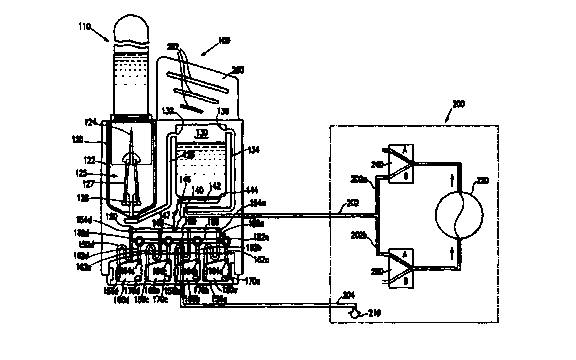
depicted in Fig. 1 by way of illustration and not limitation. Device 100 is
shown
with a sample container 110 mated with a sample receiving element 120, which
comprises well 122. An input needle 124 is part of a needle assembly 125,
which
CA 02303157 2000-03-09
WO 99/14595 PCT/US98/19418
24
comprises needle 124, needle holder 127 and base 128 affixed to the bottom
inside
wall of well 122. Both needle holder 127 and base 128 comprise a longitudinal
bore
to provide for fluid to enter device 100. Needle 124 is in fluid communication
with a
first chamber 130 by means of a first channel I26, which at one end is
connected to
base 128 of needle assembly 125 and at the other end to an upper part 132 of
first
chamber 130.
A second channel 134 is connected at one end 136 to first chamber 130 and
terminates at the other end at port 140. A third channel 142 provides fluid
communication between first chamber 130 and first manifold 150. Accordingly,
142
is connected at its one end 144 to the base of 130 and at its other end 146 to
indicator
147. The purpose of indicator 147 is to monitor flow out of first chamber 130
so that
premature leakage of fluid from first chamber 130 may be detected. Indicator
147 is
connected at point 148 to first manifold 150, thus lying between first
manifold 150
and third channel 142 and forming part of the fluid communication between
first
chamber 130 and first manifold 150.
Fourth channels 152 ( 152a, 152b, 152c and 152d) provide fluid
communication between first manifold 150 and second chambers 160. The device
depicted has second chambers 160a, 160b, 160c and 160d. Lying within second
chambers 160 are mixing balls 170 (170a, 170b, 170c and 170d, respectively).
Fourth channels 152 connect at one end 154 ( 154a, 154b, 154c and 154d,
respectively) to first manifold 150 and at the other end 156 (156a, 156b, 156c
and
156d, respectively) to the bottom left of second chambers 160. Diagonally
across
from I56 are fifth channels 162 connected at one end 164 to the top right
corner of
160 and at the other end to second manifold I80 at vent plugs 182 (182a, 182b,
182c
and 182d, respectively). Second manifold 180 contains vent port 190.
Device 100 depicted in Fig. 1 is shown in conjunction with pneumatic circuit
200 wherein communication is established between device 100 and pneumatic
circuit
200. To achieve such communication, port 140 is connected to sixth channel 202
CA 02303157 2000-03-09
WQ_ X9/14595 PGT/US98/19418
and vent port 190 is connected to seventh channel 204, which ternlinates at
check
valve 210. Sixth channel 202 provides for fluid communication between port 140
and pump 220. Sixth channel 202 branches to give 202a and 202b. Three way
valve
240 lies between 202 and pump 220 along 202a and three way valve 260 lies
5 between 202 and pump 220 along 202b. The three-way valves 240 and 260 each
have positions A and B: When valves 240 and 260 are in position A, valve 240
is
open to the atmosphere and valve 260 is on line with channel 202. Conversely,
when
valves 240 and 260 are in position B, valve 260 is open to the atmosphere and
valve
240 is ~on line with channel 202.
10 The device depicted in Fig. 1 also includes holder panel 280 for gripping
the
device. Holder panel 280 has slots 282, which provide a firmer gripping means
as
the device is manipulated to secure sample container 110 and to place device
100 in
a suitable instrument for connection to a pressure. varying apparatus and/or
to read
the results of an assay.
15 As mentioned above, the device of this invention is generally useful for
the
analysis of fluid samples, particularly of physiological fluid samples.
In the embodiment of Fig. 1, a sannple in removable sample container 110 (not
a part of the invention) is inverted and mounted in well 122 of sample
receiving
element 120. The top of the sample container has a septum, which is pierced by
20 needle assembly 124 so that fluid may flow into device 100. To induce flow
of
sample into the device, negative pressure is applied to first chamber 130 via
port
140, which is shown attached to exemplary pneumatic circuit 200. As mentioned
above, pneumatic circuit 200 comprises an air pump 220 and two three way
valves
240 and 260. In use, the valves 240 and 260 are set at positions A to remove
air from
25 the first chamber 130 and draw sample into first chamber 130. Sample is
then
transferred to second chambers 160 by applying positive pressure to first
chamber
130 through port 140. This is accomplished by switching the three way valves
240
and 260 to position B. Fluid enters second chambers 160 via first manifold
150.
CA 02303157 2000-03-09
WO 99/14595 PCT/US98/19418
26
Second chambers 160 optionally contain reagents and mixing balls 170. Fluid
fills
each of second chambers 160 up to vent plugs I80. Vent plugs 180 permit
passage of
air but not of liquid. Air passes through vent port 190 and out check valve
210.
The device may be fabricated from individual injection molded parts or by
any other convenient process. The device may be fabricated from a material
that is
not reactive with the sample to be analyzed or the processing reagents
employed.
Furthermore, the material must be able to withstand the temperatures employed
in a
processing of the sample. In general, any material may be used that does not
react
with, ar otherwise cause detrimental effects on, the sample or any solvents in
which
the sample is dissolved or suspended. Suitable materials for the manufacture
of the
present device include, for example, polystyrene, acryIonitrile-butadiene-
styrene
(ABS), styrene-acrylonitrile (SAN), polyethylene terephthalate (PET),
polycarbonate
and so forth.
For further understanding of fabrication of a device in accordance with the
present invention, by way of example and not limitation, reference is made to
Figs.
2-4. The device depicted is that shown in Fig. 1. There are four individual
parts for
this embodiment of the present device, namely, housing assembly plate 300,
second
chamber assembly plate 302, cover plate 304 and rear plate 306. Housing
assembly
plate 300 includes well 322; which is preformed in housing assembly plate 300.
Also preformed in the bottom of well 322 is base 328 and needle holder 327,
which
are part of needle assembly 325. Needle 324 may be secured in needle holder
327.
First channel 326, first chamber 330, second channel 334 including first port
340,
third channel 342, indicator 347, first manifold 350, fourth channels 352
(352x,
352b, 352c and 352d), fii~h channels 362 (362x, 362b, 362c and 362d), second
manifold 380, vent port 390, and vent plug recesses 381 (381a, 381b, 381c and
381d)
are all included in housing plate 300. Vent plugs 382 (382a, 382b, 382c and
382d)
are placed in vent plug recesses 381. Second chamber plate 302 comprises a
cover
for second chambers 360 (360a, 360b, 360c and 360d). Second chambers 360
CA 02303157 2000-03-09
WO 99/14595 PCT/US98/19418
27
having appropriate openings for aligning with fourth channels 352 at ends 356
(356a,
356b, 356c and 356d). Second chamber plate 302 has appropriate openings for
aligning with fifth channels 362 at ends 364 (364a, 364b, 364c and 364d).
Housing
plate 300 includes a recessed area for inserting second chamber plate 302,
which is
placed in device 300 so that the openings in the second chambers align with
ends 356
and 364. Mixing balls 370 are placed in the second chambers of second chamber
plate 302 prior to welding to housing plate 300. Plate 302 is then welded to
secure
it to housing plate 300. Finally, cover plate 304 and rear plate 306 are
welded into
position on housing plate 300, thereby completing the manufacture of the
device 100.
The primary factor in determining the size of the device is the ease of use of
such device. The device should not be so large or so small as to be cumbersome
or
difficult to use. Furthermore, the size of the device should be such that it
is easily
manipulated to insert the sample container and to insert the device into an
apparatus
that has the aforementioned pneumatic circuit as well as a reading means for
determining the result of an assay.
Another embodiment of a device in accordance with the present invention is
depicted in Fig. 5. In the device of Fig. 5 fluid is drawn through sample
inlet port
424 into first chamber 430 via application of vacuum at port 440. Dam 441
prevents
direct flow into the vacuum port 440. The application of positive pressure to
port 440
forces fluid into second chambers 460, with mixing balls 470, through fill
ports 480,
which are in fluid communication with first chamber 430 by means of a manifold
(not shown. Vent plugs 456 prevent overflow of fluid while permitting passage
of
air. Overflow well 482 is connected to check valve connection 484 and to vent
plugs
456 by means of manifold 458.
As mentioned above, the device may include one or more reagents for
processing the sample. The nature of the reagents for processing the sample
will
depend on the type of processing to be carried out.I Such processing reagents
may
include reagents for stabilizing and/or preserving the sample or the analyte
contained
CA 02303157 2000-03-09
WO 99/14595 PCT/US98/19418
28
therein and may be included in the first chamber. If the sample is to be
subjected to
an assay, the nature of the reagents depends on the nature of the assay to be
conducted. For example, if the sample is to be analyzed by conducting an
immunoassay, the second chamber may include an antibody reagent.
The reagents for processing a sample may include one or more stabilization
reagents and/or preservatives for stabilizing and preserving the sample and/or
the
analyte, applied to the device. Examples of stabilization reagents are
chelating
compounds such as ethylenediaminetetraacetic acid, water soluble polymers such
as
polyethylene glycol, polyvinyl pyrrolidine, polyvinyl alcohol, and the like,
protease
inhibitors such as aprotinin, phenyl methyl sulfonyl fluoride (PMSF), and the
like.
The amount of stabilization reagent employed is, in general, that which would
be
effective in bringing about the desired stabilization. The stabilization
reagent may be
present in an amount of about 0.01 to 2% by weight or more. The stabilization
reagent is usually in the form of a buffer containing one or more of the
stabilization
reagents. Suitable buffers may be any convenient buffer, generally a
substantially
dilute buffer, which may include phosphate, saline, tris, MOPS, borate,
carbonate, or
the like. Usually, the buffered solution will be at a pH in the range of about
4 to 9.
The buffer concentration is generally from about 10 to 50 mM, preferably,
about 15
to 25 mM.
The processing reagents may also include one or more reagents for preserving
the sample applied to the device such as to prevent bacterial, fungal and
other
contamination, e.g., bactericides, antibiotics, fungicides and the like. Such
reagents
include, for example, sucrose, polyvinyl alcohol, polyvinyl pyrrolidone,
dextran,
sodium azide, gentamicin, Proclin 300~ (Supelco, Bellefonte, PA} and so forth.
The
amount of the preservation reagents employed is about 1 to 20 weight percent,
more
usually from about 2 to 10 weight percent, and the reagent is applied in a
manner
similar to that described above for the stabilization reagent.
CA 02303157 2000-03-09
WO_ 99/14595 PC'T/US98/19418
29
The processing reagents may include one or more reagents for releasing an
analyte from binding proteins and the like that might be present in the
sample. Such
reagents depend on the nature of the analyte and include, for example, sodium
hydroxide, tetrachlorothyronine salicylate, 8-amino-1-naphthalenesulforuc
acid, 2-
S hydroxy-4-methoxybenzophenone-5-sulfonic acid, etc. (see EPA 0 133 464),
Nonidet P 40 ~ (NP40, from Fluka Chemie AG; Switzerland), Tween 20 and the
like. The amount of the analyte-releasing reagent employed is about 0.01 to 2
% by
weight and the reagent is applied in a manner similar to that described above
for the
stabilization reagent.
As mentioned above, the sample may be analyzed by any convenient method.
Assays include, by way of illustration and not limitation, agglutination
assays,
precipitation assays, nephelometric assays, turbidimetric assays,
immunoassays,
coagulation assays, and so forth. The assays may involve members of a specific
binding pair such as antigens, antibodies, receptors, and so forth. Such
assays
include immunoassays, receptor binding assays, coagulation assays,
agglutination
assays and the like. Detection of an assay result depends on the signal
producing
system chosen, example of which are set forth above. For example, where the
label
is a fluorescent label, signal is detected with a fluorometer, and so forth.
For enzyme
labels, the signal is often detected spectrophotometrically. Exemplary of
assays
employing enzyme labels are the EMIT~ assay described in U.S. Patent No.
3,817,837, the disclosure of which is incorporated herein by reference, the
CEDIA
assay, and so forth.
While the embodiments of Figs. 1 and 5 have been illustrated having four and
three second chambers, respectively, there is no inherent limitation upon the
number
of chambers, which usually will be between one and four. In addition, there
may be
more than one first chamber usually connected in series in fluid communication
through channels or capillaries in a manner similar to that described for the
first and
second chambers. Generally, the last of the first chambers is in direct fluid
CA 02303157 2000-03-09
W0.99/14595 PCT/US98/19418
communication with the first manifold. Likewise, the illustrated embodiments
are
equipped with magnetic mixing means, which may be substituted as desired with
alternative motive means. It is contemplated that the observation of the
sample in the
assessment chamber will be by optical means, principally via fluorescence or
5 infrared absorption.
An example, by way of illustration and not limitation, of an instrument into
which the present device may be employed is depicted in Fig. 6. The instrument
includes a turbidimetric-based optical detection system that measures
aggregation as
an increase in light transmittance. Due to the ratio of bead size to the
measurement
10 wavelength, the light scattering is primarily forward (Mie) scatter. As a
result, the
chambers of the present device are illuminated by a narrow bandwidth emitter
with
detectors, mounted in direct opposition, to collect the in-coming light. The
optical
detector converts the light into an electrical current that is input into a
transimpedance amplifier and converted to a voltage, which is the measured
signal.
15 This instrument is AC powered and is based on an embedded PC architecture.
The
instrument controls the assay sequencing, establishes and maintains the assay
temperature, controls the reagent-sample mixing for the required duration,
determines the result, displays result and status information to the user, and
performs
self diagnostics. The instrument supports bar code data entry, printing of
teat results
20 to an external printer, and an RS-485 interface to interconnect to a
laboratory
network. The instrument has four independent optical detection channels
comprised
of narrow band emitters and high gain broadband detectors. Each detector
output is
A/. D converted at a rate of up to 16 Hz. The assay mixing is controlled by a
programmable clock-driven solenoid that provides uniform mixing across all
four
25 channels. The temperature of the sample is controlled by a closed-loop
feedback
design utilizing. a precision thermistor and a resistive heater element.
The following description of a platelet function assay utilizing the device of
the present invention is provided by way of illustration and not limitation.
As
CA 02303157 2000-03-09
W0.9_9/14595 PCT/US98/19418
31
mentioned above, the present device has broad application to the processing of
many
different types of samples without intervening opening of sample containers.
An
example of a platelet function assay using the device of this invention is
similar to
the optical platelet aggregation assay of Coller, supra, involving the use of
fibrinogen
coated microparticles and activating agents. As in optical aggregation, the
activated
platelets complex with soluble fibrinogen, fibrinogen akeady bound to the
surface of
another platelet and fibrinogen coated or bound to the microparticles. As a
consequence of the latter, the microparticles coagglutinate with the
platelets, forming
aggregates of sufficient size so as to be detectable. By varying the type and
concentration of activating agent, results substantially equivalent to the
platelet
aggregometer can be obtained.
In the embodiment of Coller, supra, a 70 pl sample of blood plus
anticoagulant is added to a borosilicate tube containing a buffer with 0.05 mM
calcium chloride, a blue bead suspension (20 pl fibrinogen coated beads, 3
ptn), and
an activating peptide [5-l0E,t1 (iso S)FLLRN-NH2, 2Ea,M final concentration].
After
the tube is capped and mixed, the blood is rocked on an end-to-end tube mixer
and
viewed for the presence or absence of bead agglutination. The agglutinated
beads are
readily seen in the stream of blood as the tube is tilted back and forth, and
the extent
of agglutination is rated from O+ (no agglutination) to 4+ (extensive
agglutination).
The assay conditions were designed to yield an end point at 120 seconds in
order to
satisfy the practical needs for a rapid determination desired in a clinical
setting. This
method, while entirely satisfactory for its intended use, is however
subjective and
therefore operator dependent due to the method of assessing and reporting the
results. Further, it is desirable to have the ability to generate a permanent,
quantified
record of various platelet functions.
Assays using turbidimetric methods are usually conducted by first minimizing
the concentration of red blood cells (RBC's). This is necessary because RBC's
constitute about 45% of the volume of whole blood, and therefore, their
absorption
CA 02303157 2000-03-09
WO 99/14595 PC"T/US98/19418
32
and scattering characteristics have a significant impact on transmitted light.
Two
common methods of reducing the effects of red blood cells on turbidimetric
measurements are 1) lysis of the red blood cells and dilution of the resulting
solution
and 2) mechanical separation of the red blood cells from the sample by
centrifugation or filtration through a porous membrane. Certain assays,
however,
depend upon the integrity of the whole blood to achieve an accurate
measurement.
For example, with an assay intended to measure platelet aggregation, Iysing of
RBC's
is not acceptable since the lysed RBC's release ADP, a potent platelet
activator.
Similarly, the use of a porous membrane to filter the RBC's can result in loss
of
platelets, which adversely affects the measurement of platelet aggregation.
Filtering
RBC's may also cause hemolysis, again releasing the potent platelet activator
ADP.
Two problems must be overcome to perform an accurate turbidimetric assay
of analyte concentration or functional behavior in whole blood with intact
RBC's.
First, the method must compensate for the error in optical density readings
due to the
concurrent change in the oxygenation state of the RBC's in the whole blood
sample.
Second, the agglutination media (e.g., beads) must have high light absorption
characteristics in comparison to RBC's at the measurement wavelength such that
agglutination of the light absorbing media results in a detectable change in
optical
density.
The present invention addresses these problems via the provision of a system
consisting of a stand alone monitor and disposable test cartridge based on
microbead
agglutination technology. The system does not require platelet isolation. The
assay
requires only a small amount of whole blood and provides quantitative results
within
a few minutes after blood is drawn.
The assay is based on the principle that fibrinogen coated microparticles
exhibit a visible agglutination reaction in whole blood in the presence of
activated
platelets with normal GPIIb/IIIa receptors. Blockade of the GPIIb/IIIa sites
by c7E3
antibody or other agents can be detected by inhibition of microbead
agglutination.
CA 02303157 2000-03-09
WO._99/14595 PCT/US98/19418
33
The assay, unlike other activated coagulation assays, is only minimally
influenced by
the anticoagulant effect of heparin and is believed to primarily reflect
GPIIb/IIIa
status, unless there is severe thrombocytopenia or serious qualitative
platelet
dysfunction. The presence of normal plasma levels of fibrinogen (~ 2-4 pg/ml)
also
does not greatly influence the assay because of preferential interaction of
the
platelets with the immobilized fibrinogen. In practice, the assay requires the
presence
of an agglutination medium, preferably GPIIb/IIIa receptor land coated
microparticles, a platelet activating agent, means for observing the
aggregation of the
microparticles, and means for recording, compiling, and displaying the
results. Each
of these is discussed more fully below.
Receptor Ligands:
A GPIIb/IIIa receptor ligand is a small organic molecule, polypeptide,
protein,
monoclonal antibody or nucleic acid that binds, complexes or interacts with
GPIIb/IIIa receptors on the platelet surface. Platelet mediated aggregation of
the
microparticles results when the GPIIb/IIIa receptors on the surface of
platelets bind,
complex or otherwise interact with the GPIIb/IIIa receptor ligands on the
particles or
beads. Typical GPIIb/IIIa ligands include fibrinogen, monoclonal antibody 10E5
(Coller, et al., J. Clip. Invest. 72:325 (1983)), monoclonal antibody c7E3
(The EPIC
Investigators, N.E. Journal of Med., 330:956 (1994)), von Willebrand factor,
fibronectin, vitronectin and other ligands that have an arginine glycine-
aspartic acid
(RGD) sequence or other peptides or peptidomimetics that mimic this sequence
(Cook, et al., Drugs of the Future 19:135 (1994)). RGD functionally equivalent
ligands include gamma chain peptides, peptidomimetics and cyclic peptides with
activity about the same as an RGD ligand surface through a suitable spacer.
Examples of suitable ligands are disclosed in Beer, et al., Blood 79:117 (
1992), the
contents of which are incorporated herein by reference.. Suitable GPIIb/IIIa
receptor
ligands include the peptide (glycine)n arginine glycine aspartic acid, wherein
n is an
integer from 2 to 20. The polyglycine portion of the ligand serves as a spacer
and is
CA 02303157 2000-03-09
W0.~9/14595 PCT/US98/19418
34
covalently bound to the surface of the polymeric bead via the N terminal amino
group. While the RGD sequence may participate in the binding of platelets, a
gamma
chain sequence forming a molecular mimic of the RGD sequence may be more
important in the binding of fibrinogen to platelets (Coller, Platelet
Morphology,
Biochemistry, and Function, 1175). Optionally, an additional amino acid or
oligopeptide that does not significantly interfere with the binding of
arginine glycine
aspartic acid to the GPIIb/IIIa receptor may be bound to the C terminus of
aspartic
acid by means of a peptide bond. In one embodiment, the GPIIb/IIIa receptor
ligand
comprises (Glycine)9_,1-arginine glycine aspartic acid phenylalanine.
Alternatively,
the spacer portion of the ligand can comprise any moiety which causes the
arginine
glycine aspartic acid sequence to extend out from the surface of the
microparticle
sufficiently to allow binding between the ligand and GPIIb/IIIa receptors on
the
surface of platelets and does not significantly interfere with the ability of
arginine
glycine aspartic acid to bind with GPIIb/IIIa. Examples of suitable moieties
include
alkyl groups and polyglycol groups.
Activating Agents:
The thrombin receptor is a transmembrane protein that is present in platelets
(Vu, et al., Cell 64:1057 ( 1992)). A thrombin receptor activator, as defined
herein, is
a peptide, protein, antibody or small organic molecule that induces platelet
activation
via the thrombin receptor, i. e., which increases the rate of agglutination
when
platelets whose GPIIb/IIIa receptors are not blocked when the platelets are
combined
with a GPIIb/IIIa receptor ligand bound to solid surfaces. A suitable peptide
is any
peptide of appropriate sequence and size to activate platelets, as described
above.
The peptide can comprise thrombin, or a portion thereof, such that the amino
acid
sequence of the peptide or peptide mimic result in activation of the
platelets. Vu, et
al., identified the amino acid sequence of the thrombin receptor and proposed
a
mechanism of thrombin receptor activation. Thrombin cleaves the thrombin
receptor
protein, releasing a short receptor fragment and leaving a new amino terminal
CA 02303157 2000-03-09
WO_99/14595 PCT/US98/19418
peptide on the platelet surface. The new amino terminal peptide activates the
receptor by functioning as a tethered ligand that interacts with another
region of the
receptor to induce activation signals. A fourteen amino acid peptide (T-14)
corresponding to the new N-terminus of the cleaved receptor protein is capable
of
5 aggregating platelets directly without prior thrombin cleavage (Vu, et al).
However,
the entire peptide is not required for activity because an eleven amino acid
peptide
(T-11 ) lacking the three C terminal amino acids of T-14 is twice as potent as
T-14
(see Coller, et al., Biochemistry 31:11713 ( 1992), the contents of which are
hereby
incorporated by reference). A peptide comprising the first five or six amino
acids has
10 also been shown to be active. (Vassallo, et al., J. Biol. Chem. 267: 6081
(1992), Hui,
et al., Biochem. Biophys. Res Commun. 184:790 (1992),
Sabo, et al., Biochem. Biophys. Res. Cammun. 188:604 ( 1992) and
Scarborough, et al., J. Biol. Chem. 267:13146(1992).
The N-terminal serine group of the thrombin receptor activating peptides is
15 essential to their ability to induce platelet aggregation,. This conclusion
is based on
the observation that acetylation of the N terminal serine of T-I 1 results in
loss of
aggregating ability. In addition, T-11 and T-14 lose their ability to
accelerate
aggregation when incubated in plasma because the plasma component
aminopeptidase M cleaves the N-terminal amino acid. The presence of
20 aminopeptidase M in whole blood can result in variability in the amount of
time
required for agglutination of the beads in the assay. Acetylation of the N-
terminus of
the thrombin receptor peptide ligand is the traditional method for producing a
peptide
that resists cleavage by aminopeptidase M, but acetylation of this ligand
eliminates
receptor activator activity.
25 The variability in the time required for agglutination can be avoided by
carrying out the assay under conditions wherein the cleavage of the N-terminal
serine
of the thrombin receptor activating peptide is suppressed. This can be
accomplished
by employing a thrombin receptor activating peptide (TRAP) that is resistant
to
CA 02303157 2000-03-09
WO 99/14595 PCTNS98/19418
36
degradation by aminopeptidase M. The rate of microbead agglutination is more
rapid
and reproducible if the platelets are activated. A variety of TRAPs are
disclosed and
claimed in Coller and Prestwich, U.S. Patent No. 5,455,228, the relevant
disclosure
of which is incorporated herein by reference. These peptide derivatives are
resistant
to cleavage and inactivation by plasma aminopeptidase M, thus eliminating a
potential source of variability. The level of GPIIb/IIIa receptor activation,
and
therefore the fibrinogen binding capacity of the platelets, is dependent upon
the
amount of added activating peptide. A preferred thrombin receptor activating
peptide
that is resistant to degradation by aminopeptidase M is racemic isoSer-Phe-Leu-
Leu-
Arg-Asn. This peptide is hereinafter referred to as T-6'. See Coller and
Prestwich,
U.S. Patent No 5,455,228, and Coller, et al., J. Biol. Chem. 268:20741
(1993}), the
contents of which are incorporated into this application by reference}. Other
suitable
thrombin receptor activating peptides that are resistant to aminopeptidase M
include
peptides comprising an N-terminus having the amino acid sequence of T-6', such
that
the peptide is resistant to aminopeptidase M degradation and retains
sufficient
platelet activating activity, as described above. Alternatively, the
variability can be
avoided by including an inhibitor of aminopeptidase M in the assay. A suitable
inhibitor of aminopeptidase M is amastatin, which has been shown to enhance
platelet aggregation in the presence of aminopeptidase M (Coller, et al.,
Biochemistry 31:11713 (1992)).
Other platelet activators can be used in place of the thrombin receptor
activating peptides described above. For example, adenosine diphosphate (ADP},
collagen, ristocetin, botrocetin, epinephrine, arachidonic acid and its
metabolites
including thromboxane A2, platelet activating factor, plasnnin, serotonin,
vasopressin, tissue plasminogen activator, streptokinase and immune complexes
can
be added, alone or in combination with other platelet activators, to increase
the rate
of agglutination of the beads in the assay of the present invention.
Additionally,
application of high levels of shear stress, and artificial surfaces such as
those used
CA 02303157 2000-03-09
WO 99/14595 PCT/US98/19418
37
clinically for prosthetic materials can also activate platelets (Coller,
Platelet
Morphology, Biochemistry, and Function, 1185)
Agglutination Media:
The agglutination media may be any suitable solid surface bearing a receptor
ligand. Preferably the surface is a small polymeric bead or microparticle to
which a
GPIIb/IIIa receptor ligand is covalently bound or absorbed. The polymeric
microparticles can be virtually any shape, but are generally spherical with
uniform
diameters ranging from about 0.1 pxn to about 50 ~.un in diameter. Preferred
diameters are from about 1 pm to about 10 ~n in diameter, most preferably
about 6
pm. The composition of the particle may be any convenient composition, such as
bioglas, organic polymers, e.g. polyacrylonitrile, polystyrene, polycarbonate,
polymethacrylate, combinations thereof, or the like, or other material which
absorbs
in the infrared or can be made to do so with infrared absorbing dyes. For the
most
part the particle composition without the dye will not absorb significantly in
the
infrared region of interest, usually absorbing less than about 25% of the
total light
absorbed in that region compared to the particle doped with the infrared
absorbing
dye. Also, there will be many regions in the visual region in which the
particle
composition will be substantially transparent, as distinguished from carbon or
colloid
particles which do not transmit light over the visual and infrared region.
Usually, at
least 50 weight %, preferably at least about 75 weight %, will be of a size or
diameter within the range indicated. The particles may be modified in a
variety of
ways. The particles may be chemically activated by having functional groups
present
on the surface of the particles, or be coated with a compound, e.g. protein,
which
may serve to substantially irreversibly (under the conditions of the
processing and
assay) bind to the dye. The coating compound may be the binding component,
which
will be involved in the aggregation of the particles, or other compound,
usually being
a protein. Alternatively, depending on the nature of the particles, the
particles may
not have chemically active groups, but rather provide binding by adsorption.
In
CA 02303157 2000-03-09
WO 99/14595 PCT/US98/I9418
38
addition, infrared absorbing dyes which are stable under the conditions of
formation
of the particles, e.g. extrusion, may be mixed with the polymer prior to
particle
formation and the particle formed with the dye distributed throughout the
particle.
The particles are loaded with a dye which absorbs in the infrared. Various
S dyes have been reported as useful in this absorption range. See, for
example, Fabian,
et al., Chem. Rev. ( 1992) 92:1197 1226. These dyes include bacteriochlorin,
bacteriochlorophytin, meropolymethine dyes, benzoannulenes, vinylogous
porphyries, polymethine dyes, cyanines and merocyanines, and the like. The
particular dye which is selected is one of convenience, availability,
stability,
compatibility with the particle, and the like. Specific dyes of interest
include dyes of
the class of phthalocyanines, napthalocyanines, metaled napthalocyanine dyes,
and
modified natural bacterochlorines. Specific example dyes include IR 140, 1,1'
Diethyl 4,4' dicarbocyanine iodide, 1,1' Diethyl 2,2' quinotncarbocyanine
iodide,
Vanadyl,10,17,24 tetra tert butyl 1, 8, I5, 22, 25 tetrakis(dimethylamino)
29H,31H
phthalocyanine, [RA800 (from Exciton), ProJet 830NP (from Zeneca). These dyes
may be incorporated directly into the particle itself, through polymerization
or
passive adsorption. Alternatively, the dyes may be linked to the bead in
combination
with the binding component, such that they do not leach from the surface. The
dyes
will adsorb light in the range of about 750 to 900 nm, particularly in the
range of
about 750 to 850 nm.. For samples with high levels of red blood cells, the
light will
be at about 800nm ~ 10 nm, which is the isobestic point for oxyhemoglobin and
reduced hemoglobin. The amount of the dye employed with the particles will
vary
with the extinction coefficient of the dye in the light range of interest, the
required
sensitivity of the assay, the size of the particles, the mode of binding of
the dye to the
particles, compatibility of the dye with the particle matrix, and the like.
Usually,
loading will be in the range of about 1 to 20 weight percent, more usually 5
to IS
weight percent"
CA 02303157 2000-03-09
WO_ 99/14595 PCT/US98/19418
39
For example, the polymeric mieroparticle may be polyacrylonitrile beads with
N-hydroxysuccinimide ester groups on their surface (e.g., Matrex 102 beads
from
Amicon Corporation) (Coller, Blood, 55:169 ( 1980)). The N-hydroxysuccinnmide
ester groups allow coupling of the N-terminus of a peptide, protein or
monoclonal
antibody to the surface of the bead. Alternatively, the microparticle can be
carboxylated polystyrene beads (Polysciences Inc.). The surface carboxyl
groups of
this bead can be coupled to the N-terminus of the protein, peptide or
monoclonal
antibody by means of a carbodiimide coupling. The beads are preferably colored
to
render the results of the agglutination reaction easier to interpret. In a
preferred
embodiment, the beads are adapted to absorb light in the infrared region.
To eliminate effects of red blood cell oxygenation, an IR source of
wavelength of about 800 nm is preferred since oxyhemoglobin and
deoxyhemoglobin
have the same optical absorption coefficient at 805 nm. Assay dependence upon
the
variable state of red blood cell oxygenation is thereby eliminated. Further,
the
hemoglobin isobestic point at 805 nm has the lowest absorption coefficient
between
300 nm and 1000 nm. This results in the widest possible differential between
light
absorption by the red blood cells versus that by the agglutination medium
(beads).
The agglutination medium is selected to have high absorption at ~ 800 nm.
The ratio between the agglutination medium absorption coefficient and whole
blood
absorption coefficient should preferably be greater than about 4:1 at 800 nm.
The
absorption ratio for a particular assay is a function of both the absorption
coefficient
of the agglutination medium and the concentration of the agglutination medium
in
the assay sample.
The IR absorbing particles preferably have the following properties:
absorption peak around 800 to 810 nm; reasonably broad half widths (>75 nm)
around the absorption peak; little or no fluorescence (high fluorescence
requires the
use of a high pass optical filter), and a molar extinction coe~cient of about
30,000.
CA 02303157 2000-03-09
WO 99/14595 PCTNS98/19418
Four categories of IR absorbing particles are useful in the assay: latex
particles dyed with IR dyes, Dl dye sole, carbon black particles, and
liposomes with
entrapped IR absorbers. The particles are preferably spherical beads about 6
dun in
diameter.
5 Generally, an hydrophobic IR dye soluble in organic solvents is preferred.
Latex particles are dyed with the IR dye according to established methods
using dyes
which absorb in the visible range at about 800 nm (see Lee Bangs, Uniform
Latex
Particles). Latex offers the advantage of size uniformity. Water insoluble
dyes may
be induced to form colloids (sols, sub micron or larger particles) when a
solution of
10 the dye in a water miscible solvent is added to water. Carbon black absorbs
well in
the IR region of the spectrum and can be dispersed in aqueous solutions by
sonication. Liposomes with entrapped IR absorbers, e.g., a water soluble IR
dye, can
be tailored to the desired sizes and fibrinogen bound to the surface via an
anchor
compound, such as pahnitoyl chloride.
15 Preferably, the concentration of beads is adjusted so that the
platelet/bead
ratio is from about 1.9 to about 2.8. A GPIIb/IIIa ligand may be covalently or
ionically coupled to the bead, or the ligand may be simply coated on the bead.
In one
embodiment the ligand bearing beads are lyophilized. A representative
formulation
for Iyophilization is about 10 pg/ml beads, 75 ~g/ml fibrinogen, and 200 pg/ml
20 bovine serum albumin.
The time for fluxing may be varied widely, usually being at least about 1 sec.
and not more than about 5 min., usually not more than about 2 min., and
preferably
for about 5 sec. to 1 min.
The particular manner of agitation is not critical, so long as it provides for
25 thorough mixing, without preventing the formation of aggregates. If
desired, mild
agitation may be maintained during the course of the assay, again insuring
that there
CA 02303157 2000-03-09
WO Q9/14595 PCT/US98/19418
41
is homogenous distribution of the particles and any other particulate matter,
while
insuring that aggregation is not impeded.
The temperature for the assay may be varied widely, depending upon the
nature of the component of interest. Conveniently, ambient temperatures may be
employed, although elevated temperatures which can be controlled and
maintained
are preferred. Where nucleic acids are involved, the temperature may be
elevated, so
as to enhance the degree of stringency of hybridization. Thus, the temperature
may
vary from about 15 to 90°C, where with other than nucleic acids, the
temperature
will generally vary from about 25 to 40°C. Usually, with nucleic acids
the
temperature will generally be in the range from about 20 to 90°C, more
usually in the
range of about 30 to 85°C.
The time for the assay will depend upon the manner in which the
measurement is taken. Where zero time is carefully controlled, one may take
one or
two measurements at different time intervals to determine the absolute
infrared
transmission at the time intervals or determine the rate of formation of the
aggregation. Alternatively, one may take a plurality of measurements over the
time
course of the assay and analyze the slope beginning at a fixed time from the
time of
mixing. The data may be analyzed by any convenient means, particularly using
an
algorithm which can manipulate the data in relation to calibrators and/or
controls.
The total time of the readings from the zero time (time of mixing), may range
from
about 10 sec. to 5 min., more usually about 30 sec. to 5 min., and preferably
about
sec. to 2 min.
Usually, the result will be compared to a calibrator, which may be performed
concomitantly or have been performed previously or may be provided as a
standard
25 curve. The calibrators will vary depending upon the nature of the component
of
interest. Samples having known amounts of the component of interest may be
prepared and performed in the assay and the results charted so as to be able
to
translate the measurement obtained with the sample to the standard. In some
CA 02303157 2000-03-09
WO 99/14595 PCT/US98/19418
42
instances controls will be used, where the base value may vary depending on
the
source of the sample. The particular control will be associated with the
sample and
the component of interest.
Where platelet aggregation is to be measured, because of interest in the
platelet status of an individual, which may be the natural status or the
status resulting
from administration of a drug, the sample will be in effect whole blood, which
has
been subjected to less than about 50%, preferably less than about 20%
dilution.
The whole blood is drawn desirably in the substantial absence of air.
Conveniently, a ~acutainer is employed for capturing and holding the blood
sample.
The Vacutainer desirably includes a small volume of a solution of sodium
citrate
generally in the range of about 3 5% sodium citrate having a volume in the
range of
about 0.05 to 0.5 ml. The blood sample should be obtained from an extremity
free of
peripheral venous infusions. Conveniently, the needle should be at least about
21
gauge.
The first tube which is withdrawn is discarded, the second tube or subsequent
tubes being used. Mild agitation by simply gently inverting the Vacutainer is
employed to insure the mixture of the anticoagulant with the sample. The
sample in
each container may range from about 1 to 10 ml, more usually from about 1 to 8
ml,
conveniently from about 1 to 5 ml. The sample should not be stored for an
unduly
long period, generally storage before the assay should not exceed 1 hour.
The container containing the whole blood is then secured to a device of the
present invention as described above, an example of which is shown in Fig. 1.
The
second chambers contain the particles that have been coated with fibrinogen as
well
as various agents that serve to activate the platelets. Illustrative agents
include iso
TRAP (See U.S. Patent No. 5,455,228), TRAP, ADP, collagen, thrombin,
ristocetin,
or any combination thereof. Any convenient activator may be employed. Iso TRAP
is employed at a concentration in the range of about 1 to 5, preferably about
2
CA 02303157 2000-03-09
WO 99/14595 PCTNS98/I9418
43
prnol/L. The activating 'agent may be incorporated with the bead reagent to
which the
blood sample is added. The beads and other reagents may be dry, so as to not
dilute
the sample, although in some instances a small amount of liquid may be
present,
desirably less than about 25% of the volume of the sample.
The particles are conveniently polystyrene particles of a size in the range of
about 2 to 8 microns, which have been coated with fibrinogen by passive
adsorption
or by covalent linkage in accordance with conventional ways. Generally, the
weight
of fibrinogen to the weight of particles volt be in the range of about 1:1000
to 1:10.
The amount of beads should provide a ratio between the agglutination media
absorption coefTlcient and whole blood absorption coefficient of greater than
about
4:1 at 800 nm, generally, not more than about 10:1 at 800 nm. The optimal
absorption ratio may be achieved by configuring both the light absorbing
characteristics of the agglutination media and the concentration of the
agglutination
media in the assay sample.
As mentioned above, the sample container is secured to the present device.
The sample container is inverted and inserted into the well of the device of
Fig. 1
whereupon the needle therein pierces the septum of the sample container. The
device is then inserted into an instrument that provides both a pneumatic
circuit and a
reading function as mentioned above.
Sample is moved into the first chamber as described above by adjusting the
pressure in the device using the pneumatic circuit. Generally, the volume of
the .
sample transferred into the first chamber is about 0.1 to 2 ml. The blood in
the first
chamber is incubated at 37°C for 2 minutes. Then, the blood is moved
into the
second chambers as described above where it mixes with the particles and other
reagents.
The mixture of citrates, whole blood, particles and activating agent is gently
agitated by causing the mixing ball contained in the second chambers to
magnetically
CA 02303157 2000-03-09
WO 99/14595 PCT/IJS98/19418
44
activate. Mixing insures homogeneity and the mild agitation is continued so as
to
maintain homogeneity without impeding aggregation formation. The temperature
for
the medium will be maintained at a constant temperature. After a short time,
generally under 30 sec., usually under about 10 sec., readings are begun by
illuminating the sample with light at about 800 nm. The total time for the
readings
will generally be under about 5 min., usually 3 min. When one is determining
the
rate of change to determine the change in slope with time, the number of data
points
per second may range from about 0.01 to 100, more usually from about 1 to S0.
Thus, one reagent may then be combined with the particles coated with
fibrinogen in
the same manner as the sample. If desired, the buffered medium may be
augmented
with blood constituents, such as red blood cells, serum albumin,
immunoglobulins,
or other significant constituent of blood, which does not participate in the
aggregation of the particles.. A convenient buffer medium is HEPES sodium
chloride
buffer comprising from 1-5 mg/ml protein, e.g., BSA.
After the sample has been combined with the reagents, desirably it will be
heated to a temperature above room temperature, but below interference with
the
assay, so as to insure that the temperature can be controlled without
adversely
affecting the assay result. Desirably, the temperature should be at least
25°,
preferably in the range of 30 to 40°C, more preferably about
37°C.
Another aspect of the present invention is a kit for processing a sample. The
kit comprises in packaged combination a device as described above. The kit may
also include, in predetermined amounts, reagents for processing a sample other
than
those included in the device. The kit may also include a sample collection
container.
Where the device is for conducting an assay, reagents for conducting an assay
for an
analyte in a sample may be included. The relative amounts of reagents may be
varied widely to provide for concentrations in solution of the reagents which
substantially optimize the sensitivity of an assay. Where appropriate, the
present
device and reagents can be placed in an air-tight package in order to maintain
the
CA 02303157 2000-03-09
WO 99/14595 PCT/US98/19418
activity of any reagents. The package may be, for example, a bag, pouch, or
the like
fabricated from a material that is substantially non-permeable to moisture.
Such
materials include, by way of example and not limitation, plastic, aluminum
foil, and
the like. For blood samples the kit may also include an article for piercing a
person's
5 skin, disinfectant or sterilizing pads and so forth. It will be evident that
the nature of
additional articles to be included in the kit with the device of the present
invention
are dependent, among others, on the type of sample being collected. These
articles
will be known to the skilled person.
For platelet function assays, kits can be provided comprising some or all of
10 the reagents which find use in such assay. The kit will have the particles
for use with
the component of interest. In addition, neutralizing immunoglobulins may be
provided for removing inhibitor in a sample to serve as a control. Calibrators
may be
provided providing particles with the appropriate binding component mixed with
any
other reagents associated with the assay and, if desired, a source of the
component of
15 interest, either in measured amounts or in bulk. For platelet aggregation,
a
combination of thrombin and uncoated particles may be supplied. Also, of
convenience, would be Vacutainers comprising 0.1 to 1 ml of 1-SM
anticoagulant,
e.g. sodium citrate. Of particular Interest for the kit is a container
containing one or
more of the appropriate reagents in order to reduce the manipulative steps for
the
20 assay. For example, a container, such as a cuvette, may be provided
containing the
particles and, as appropriate, other reagents for the assay.
EXAMPLES
The following examples are offered by way of illustration and not by way of
25 limitation. Parts and percentages are by weight unless otherwise indicated.
Temperatures are in degrees Centigrade {°C) unless indicated
otherwise. The
following preparations and examples illustrate the invention but are not
intended to
limit its scope.
CA 02303157 2000-03-09
WO 99/14595 PGT/US98/1941$
46
EXAMPLE 1.
Preparation of IR particles coated with hFg.
Water (8.72 mL) and 1M sodium phosphate at pH7.2 (0.2 mL) were first
fluxed in a 1 SOL conical tube. Purified hFg (82.5 Nl at 40 mg/mL, Enzyme
Research Laboratories) was then added and -gently mixed. This was followed by
a
bolus addition of I mL of a 10% particle suspension of beads (Matxex, 100 mg
solids/mL) and quickly mixed. The mix was incubated on a rocker at room
temperature for about 2 hours, after which it was centrifuged to remove the
supernatant and washed untie the storage buffer twice. The final particle
concentration was 1 to 1.5% in storage buffer.
EXAMPLE 2.
Preparation of dye coated particles.
IR140 (a cationic IR dye from Lambdochrome Laser Dyes) was dissolved in
minimal volume of methylene chloride to make a solution about 5 mg/mL in IR
140.
2 Propanol (38 parts) was then added to 9 parts of the above solution,
followed by 53
parts of 20 mM sodium phosphate at pH7.5 to make up the final dye solution.
Polystyrene latex beads with carboxyl functional groups, passively coated with
human fibrinogen (hFg) as taught in Example 1, were washed, pelleted and
finally
mixed with the above dye solution ( 1 mL per 10 mg of latex) at room
temperature for
5 minutes. The mix was then diluted with 10 volumes of 20 mM sodium phosphate
at
pH7.5 containing 1 mg/mL BSA(storage buffer) prior to centrifugation to
mvnimize
aggregation. The resulting pellets were then washed with the storage buffer to
yield
the final dyed beads. Further dye loading using the process of Example 2 may
be
accomplished. If the application is such that the desired substance on the
bead is not
hFg, agents such as SMCC can be used to modify hFg for later attachment of
sulfliydryl containing or sulfhydryl modified substances.
CA 02303157 2000-03-09
WO 99/14595 PCTNS98/19418
47
It is evident from the above results that a simple device is provided. The
user
is only required to place a sample container, into which a sample has been
collected,
in the present device to secure it to an input port. The present device is
then placed
into an apparatus for assisting in introducing the sample into the device and
assaying
the sample. The device is easily manipulated without risk to the operator and
without risk of compromising the sample. The device provides for precise
measurement of the sample into chambers wherein the sample is analyzed and
quantitative results are obtained.
All publications and patent applications cited in this specification are
wherein
incorporated by reference as if each individual publication or patent
application were
specifically and individually indicated to be incorporated by reference.
Although the foregoing invention has been described in some detail by way of
illustration and example for purposes of clarity of understanding, it will be
readily
apparent to those of ordinary skill in the art in Iight of the teachings of
this invention
that certain changes and modifications may be made thereto without departing
from
the spirit or scope of the appended claims.