Note: Descriptions are shown in the official language in which they were submitted.
CA 02303557 2000-03-14
WO 99/17673 PCT/US98/20798
ROTATING ELECTROSURGICAL BLADE
FOR CORNEAL RESHAPING
FIELD OF INVENTION
The present invention relates to the field of correcting refractive errors of
the eye, and more particularly to corneal electrosurgery.
DESCRIPTION OF THE RELATED ART
Anomalies in the overall shape of the eye can cause visual disorders.
Hyperopia ("farsightedness") occurs when the front-to-back distance in the
eyeball is too
short. In such a case, parallel rays originating greater than 20 feet from the
eye focus
behind the retina. In contrast, when the front-to-back distance of the eyeball
is tvo long,
myopia ("nearsightedness") occurs and the focus of parallel rays entering the
eye occurs
in front of the retina. Astigmatism is a condition which occurs when the
parallel rays of
light do not focus to a single point within the eye, but rather have a
variable focus due to
the fact that the cornea refracts light in a different meridian at different
distances. Some
degree of astigmatism is normal, but where it is pronounced, the astigmatism
must be
corrected. Hyperopia, myopia, and astigmatism are usually corrected by glasses
or
contact lenses.
Another method for correcting those disorders is by reshaping the corneal
surface through an operative procedure. Such methods include radial keratotomy
(see,
e.g., U.S. Patent Nos. 4,815,463 and 4,688,570) and laser corneal ablation
(see, e.g.,
U.S. Patent No. x,941,093). Other surgical techniques involve scraping or
cutting the
exterior corneal surface. Lieberman (U.S. Patent No. 4,807,623) employs a pair
of
angled cutting blades that are rotated around the corneal center to excise an
annular
wedge from the cornea to correct refractive errors. Khmer, et al. (U.S. Patent
No.
5,318,044) provides curved rotating blades that scrape the corneal surface to
correct
refractive errors. That patent is incorporated by reference herein.
Some other corneal reshaping techniques do not involve surgery, but
rather apply a radio frequency electrical signal to remove corneal tissue
noninvasively.
SUBSTITUTE SHEET ( rule 26 )
CA 02303557 2000-03-14
WO 99/17673 PCTNS98/20798
One technique, conductive keratoplasty, described in U.S. Patent No.
5,533,999, issued
to Hood, et al. , applies an RF current directly to symmetrical spots on the
cornea. This
technique heats the corneal tissue to shrink and steepen the tissue in order
to correct
hyperopia and astigmatism. Similarly, both Doss, et al. (U.S. Patent No.
4,326,529)
and Doss (U.S. Patent No. 4,381,007) employ an electrode that is placed near
but not
physically touching the anterior corneal surface. An electrically conductive
coolant is
placed over the corneal surface and circulated around the electrode as RF
energy is
applied through the electrode. The RF apparently heats various stroma within
the
cornea and thereby reshapes it as a biological response to the heat generated
by the RF.
Two related patents, Dobrogowski, et al. (tJ.S. Patent No. 5,025,811)
and Latina, et al. (U.S. Patent No. 5;174,304) illustrate noninvasive methods
for focal
transcleral destruction of living human eye tissue. In general, these devices
and their
underlying procedures involve the use of electric currents for ablating eye
tissue,
particularly the ciliary process. No mention of corneal reshaping is made.
These
references also relate to the application of a DC signal to the eye employing
an ionic
solution within the electrosurgical probe. The use of RF is not disclosed.
Further, the
ablation process is performed by repeatedly applying the probe to 10-30 spots
around the
circumference of the eye.
SUMMARY OF THE INVENTION
The present invention provides a rotatable electrosurgical apparatus for
reprofiling a cornea. The apparatus includes one or more electrosurgical
electrodes that
extend radially outward from a center point. The electrodes are shaped to
reform at
least a portion of an anterior surface of the cornea. The electrodes are
disposed on an
electrode support, which is rotatable. The electrodes project from the bottom
of a rotary
handle, which rotates the electrodes about a central visual axis of the
cornea. The rotary
handle has a hollow bore and a viewing port. The apparatus includes a support
base
having a base ring for positioning on the eye. The base ring can hold a
solution against
the eye to even out irregularities in the cornea.
2
SUBSTITUTE SHEET ( rule 26 )
CA 02303557 2000-03-14
WO 99/17673 PCT/US98/20798
The apparatus may include pads to exert pressure on the cornea to cause
the cornea to bulge in desired areas. These bulged areas are more easily
modified by the
electrodes when energized. The pressure pads take different forms depending
upon
whether they are used for the correction of myopia, hyperopia or astigmatism.
DETAILED DESCRIPTION OF THE INVENTION
The present invention provides a rotary electrosurgical blade assembly for
corneal reshaping and a related method. In the following description, numerous
details
are set forth in order to enable a thorough understanding of the present
invention.
However, it will be understood by those of ordinary skill in the art that
these specific
details are not required in order to practice the invention. Further, well-
known
elements, devices, process steps and the like are not set forth in detail in
order to avoid
obscuring the present invention.
Prior to explaining the details of the inventive procedures and devices, a
short explanation of the physiology of the eye is provided.
Figure 1 shows a horizontal cross-section of the eye with the globe 11 of
the eye resembling a sphere with an anterior bulged spherical portion
representing the
cornea 12.
The globe 11 of the eye consists of three concentric coverings enclosing
the various transparent media through which the light must pass before
reaching the
light-sensitive retina 18. The outermost covering is a fibrous protective
portion, the
posterior five-sixths of which is white and opaque and called the sclera 13,
and
sometimes referred to as the white of the eye where visible to the front. The
anterior
one-sixth of this outer layer is the transparent cornea 12.
A middle covering is mainly vascular and nutritive in furiction and is
made up of the choroid, ciliary body 16, and iris 17. The choroid generally
functions to
maintain the retina 18. The ciliary body 16 is involved in suspending the lens
21 and
accommodation of the lens. The iris 17 is the most anterior portion of the
middle
covering of the eye and is arranged in a frontal plane. It is a thin circular
disc similar in
function to the diaphragm of a camera, and is perforate near its center by a
circular
aperture called the pupil 19. The size of the pupil varies to regulate the
amount of light
3
SUBSTITUTE SHEET ( rule 26 )
CA 02303557 2000-03-14
WO 99/17673 PCT/US98/20798
which reaches the retina 18. The iris divides the space between the cornea 12
and the
lens 21 into an anterior chamber 22 and the posterior chamber 23. The
innermost
portion of covering is the retina 18, consisting of nerve elements which form
the true
receptive portion for visual impressions.
The retina 18 is a part of the brain arising as an outgrowth from the fore-
brain, with the optic nerve 24 serving as a fiber tract connecting the retina
part of the
brain with the fore-brain. A layer of rods and cones, lying just beneath a
pigmented
epithelium on the anterior wall of the retina serve as visual cells or
photoreceptors which
transform physical energy (light) into nerve impulses.
The vitreous body 26 is a transparent gelatinous mass which fills the
posterior four-fifths of the globe 11. At its sides it supports the ciliary
body 016 and the
retina 18. A frontal saucer-shaped depression houses the lens.
The lens 21 of the eye is a transparent bi-convex body of crystalline
appearance placed between the iris 17 and vitreous body 26. Its axial diameter
varies
markedly with accommodation. A ciliary zonule 27, consisting of transparent
fibers
passing between the ciliary body 16 and lens 21 serves to hold the lens 21 in
position
and enables the ciliary muscle to act on it.
Referring again to the cornea 12, this outermost fibrous transparent
coating resembles a watch glass. Its curvature is somewhat greater than the
rest of the
globe and is ideally spherical in nature. However, often it is more curved in
one
meridian than another, giving rise to astigmatism. Most of the refraction of
the eye
takes place through the cornea.
Figure 2 is a more detailed drawing of the anterior portion of the globe
showing the various layers of the cornea 12 making up the epithelium 31. An
anterior
limiting lamella 33, referred to as Bowman's membrane or layer, is positioned
between
the epithelium 31 and the stroma 32 of the cornea. The term "corneal mass"
refers to
the various stroma 32 between Bowman's layer 33 and Descemet's membrane 34.
The
corneal stroma 32 are made up of lamellae having bands of fibrils parallel to
each other
and crossing the whole of the cornea. While most of the fibrous bands are
parallel to
the surface, some are oblique, especially anteriorly. A posterior limiting
lamella 34 is
4
SUBSTITUTE SHEET ( rule 26 )
CA 02303557 2000-03-14
WO 99/17673 PCT/US98I10798
referred to as Descemet's membrane. It is a strong membrane shaiply defined
from the
stroma 32 and resistant to pathological processes of the cornea.
The endothelium 36 is the most posterior layer of the cornea and consists
of a single layer of cells and function to maintain transparency of the cornea
12. These
epithelial cells are rich in glycogen, enzymes and acetylcholine and their
activity
regulates the transport of water and electrolytes through the lamellae of the
cornea 12.
The limbus 37 is the transition zone between the conjunctiva 38 and sclera on
the one
hand and the cornea I2 on the other.
In general, there are two distinct electrosurgical delivery probe types: the
monopolar probe and the bipolar probe. An in-between electrosurgical
configuration
applicable to this invention also exists and is known as sesquipolar. In each
instance,
some section of the human body is used to complete a circuit between one pole
and the
other. In the monopolar probe device, there is a single active contact which
is inserted
or otherwise contacted with the human body and it is the site at which some
body
activity, e.g., desiccation, ablation, necrosis, fulguration, or the like,
takes place. To
complete the circuit in a monopolar device, there must be another contact
which is
inactive and placed against the body in a location remote from the active
contact. By
"inactive" is meant that only an insignificant temperature rise occurs at that
contact
point. One such method of ensuring that the inactive electrode is in fact
"inactive" is to
make it quite large in area. This causes the current to spread over a large
area for
completion of the circuit.
A bipolar electrode typically has two equal-area active electrodes
contained in the same electrode probe-handle structure. This symmetric bipolar
electrode design produces a significant temperature rise at both electrodes.
In a monopolar or sesquipolar configuration, only one electrode has an
area of tissue contact producing a significant temperature rise. Unlike the
monopolar
configuration, however, the sesquipolar return electrode is not so remote,
thereby
limiting current flow through the body to the nearby return electrode. The
return
electrode area in the sesquipolar configuration electrode is usually at least
three times the
area of the active electrode and produces little or no tissue effect. For
ocular surgery,
SUBSTITUTE SHEET ( ruie 26 )
CA 02303557 2000-03-14
WO 99/17673 PCTNS98/20798
the sesquipolar return electrode may be located on a non-remote region of the
body,
such as on the sclera or on a shaved area at the back of the patient's head.
There are a variety of effects that may occur depending upon the
electrosurgical mode desired. For instance, there are both high temperature
and low
temperature desiccation effects when the active electrosurgical probe
contacts) are used
to promote tissue desiccation. The resistance of the tissue in contact with
the active
probe electrode obviously varies with the tissue temperature and water
content. A low
temperature desiccation effect involves heating such that the temperature-time
product
causes tissue necrosis with little immediate denaturation or discoloration of
the tissue.
There is a transient decrease in local tissue impedance with little drying of
tissue. In
high temperature desiccation, there are significant increases in local tissue
impedance
and also in local tissue desiccation.
In the ablation mode, the electrosurgical energy density delivered largely
causes the tissue near the probe contact to vaporize. The temperature at the
electrode/tissue interface is increased significantly past the point of steam
formation.
The effect of electrical resistance varies during a specific radio frequency
cycle and
although there is sparking, carbonization is not usually significant and the
effects of the
device are relatively rapid.
Electrosurgical ablation and cutting produce an effect where a thin layer
of tissue is vaporized (cutting) or where a larger section of tissue is
vaporized (ablation).
The line between "cutting" and "ablation" is not always clear.
Blended mode is essentially a combination of the cutting and coagulation
(desiccation) modes. In blended mode, cutting or ablation with hemostasis is
achieved.
The present invention employs electrosurgical ablation to reprofile the
anterior surface of the cornea 301. For techniques that employ electrosurgery
to modify
the cornea from below the surface, please refer to U.S. Patent Application
Ser. Nos.
08/194,207, 08/513,589, and 08/698,985, all of which are incorporated by
reference
herein.
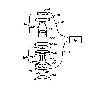
Figure 3A illustrates a rotatable electrosurgical apparatus of the present
invention, where the basic parts of the assembly are shown in an exploded
view. In one
embodiment, the components of the assembly include a generally cylindrical
support
6
SUBSTITUTE SHEET ( ruie 26 )
CA 02303557 2000-03-14
WO 99/17673 PCT/US98/20798
base 300 having an annular base ring 302 and a cylindrical bore 304 extending
through
the support base. The base ring 302 may be implemented as a circumcomeal
vacuum
ring. A vacuum hose 306 connects the vacuum ring 302 to a vacuum pump (not
shown).
The vacuum ring 302 is configured so that it meets with and seals to the front
of the eye,
rendering the support base 300 relatively immobile when the support base 300
is applied
to the front of the eye and a suitable vacuum is applied to the vacuum hose.
A rotary handle 305 having an electrosurgical blade assembly 307 is
adapted for insertion into the support base 300. The rotary handle 305 has a
hollow bore
309 to allow viewing of the corneal surface during operation of the apparatus.
The side of
the bore is also open to provide a viewing port 313. The inner diameter of the
handle bore
309 is at least large enough so that the surgeon can see the blade assembly
307 by looking
down into the bore 309. The bore 309 is desirably a length such that the ratio
of the bore's
length to its diameter is between 0.25:1 and 15:1; specifically between 0.4:1
and 1:1, at
least about 1:1 and less than about 3 :1; or at least 3 :1 up to about 15:1.
Preferably, the
ratio is about 2.5:1. This sizing allows easy manipulation by the surgeon.
Figure 3B illustrates a bottom view of the base ring 302 to show its
internal structure when implemented as a vacuum ring. The vacuum ring 302
comprises
an inner wall 308 having an inner diameter that allows the outer diameter of
the rotary
handle tube 311 to fit into the base ring 302.
The outer vacuum ring wall 310 forms the outside of the base ring 302.
Interior to the vacuum ring 302 may be one or more ridges 312 which extend
down to the
corneal surface when the support base 300 is attached to the eye. These ridges
312 may be
made of conducti ~e material, whereas the surrounding support base structure,
such as the
inner wall and outer wall, are made of insulative material. The ridges may be
coupled to
an electrosurgical generator. Using this configuration, the ridges may act as
return
electrodes when operating in sesquipolar mode. These return electrodes may be
positioned
to rest on the sclera 314 or translimbal region of the eye.
Figure 3C shows an alternative arrangement of return electrodes
comprising radial vanes 316 that extend downward through the vacuum ring 302
to make
contact with the sclera 314 or translimbal region.
7
SUBSTITUTE SHEET ( ruie 26 )
CA 02303557 2000-03-14
WO 99/17673 PGT/US98/20798
Similar vacuum ring configurations for other purposes are described in U.S.
Patent No. 5,403,335, issued to Loomas et al., and assigned to the assignee of
the present
invention. That patent is incorporated by reference herein.
Alternatively, the support base 300 can rest on the sclera 314 without use of
a vacuum ring. In its place, a base ring 302 of resilient material can be used
as a substitute
for the hollow annular vacuum ring. As another alternative, the bottom of the
base ring
302 can be serrated to hold the ring in place.
- The support base 300 may include two standoffs 318, shown here as one
behind the other on opposite sides of the support base 300. The standoffs 318
are topped
by a support ring 320. The support ring 320 may have an inner diameter greater
than or
equal to that of the base ring 302. As shown in Figare 3D the support ring 320
may be
threaded and screwed into a calibrated micrometer-like adjustment ring 322,
similar to that
used in the Kilmer '044 patent. A collar 324 of the handle 305 rests on top of
the
adjustment ring 322. By rotating the adjustment ring 322, the adjustment ring
322 controls
the axial depth of the blades 307.
Because only two thin standoffs 318 are employed to support the
adjustment ring 322, the surgeon is provided with a relatively large viewing
port area to
allow observation of the operational steps taking place at the corneal
surface. The base 300
may have substantially more open area than closed area to maximize visibility.
As an
alternative, the support base may not include the standoffs 318 and support
ring 320. The
base ring 302 alone may serve as a guide for the handle to increase viewing
area. Further,
the entire support base may be omitted when performing the surgical procedure.
In that
case, the surgeon essentially performs the operation "free hand."
w
The electrode blade assembly 307 is coupled through one lead 326 to an
electrosurgical generator 328 so as to act as an active electrode. In a
sesquipolar
configuration, the other lead 330 of the generator 328 may be coupled to
return
electrodes 312 or 316 disposed on the bottom of the support base 300, as shown
in
Figures 3B and 3C. The return electrodes 312 or 316 rest on the scleral
portion 314 of
the eye. Alternatively, in a monopolar configuration, the return electrode may
be placed
elsewhere on the patient's body.
8
SUBSTITUTE SHEET ( rule 26 )
CA 02303557 2000-03-14
WO 99/17673 PCT/US98/20798
When using the complete support base as a guide, the surgeon positions
the support base 300 on the eye so that it is centered over the central visual
axis of the
cornea 301. A vacuum is applied to hold the base in place if the vacuum ring
embodiment is employed. The surgeon inserts the rotary handle 305 into the
support base
300 so that the collar 324 rests on the adjustment ring 322. The surgeon
rotates the
adjustment ring 322 so that the electrode blade assembly 307 contacts the
cornea 301.
Because the invention employs electrical energy, the blades 307 need only
lightly touch
the corneal surface.
Alternatively, the blades 307 may be positioned near the corneal surface
without touching the surface when a conducting medium such as saline is
present. For this
purpose, the blades 307 may be placed within a range of approximately 50-500
microns
from the eye. It is the electrical contact, not the mechanical contact,
between the blades
and the cornea that achieve modification of the corneal surface. Initial
electrical contact
may be indicated by a continuity tester, as is well known in the art. The
proper distance to
achieve local conduction between the blades and the cornea can instead be
determined by
the surgeon by energizing and slowly lowering the energized blade assembly 307
towards
the cornea while viewing the effects on the corneal surface 301. To aid in
blade
placement, the distance from the cornea may be measured with a traveling
scale, such as an
electronic dial caliper manufactured by Mitsutoyo, Inc. The scale can be
zeroed when the
blades touch the cornea.
The surgeon energizes the rotary blade assembly 307 with an RF current
from the generator 328 to achieve volume modification of the cornea 301.
Preferably,
the procedure should be performed while the eye is bathed in a solution, such
as saline,
in order to even out irregularities in the tissue caused by uneven hydration
of corneal
tissue. The solution is held in the bore 332 of the base ring 302, and does
not leak
because of the tight fit between the base ring 302 and the eye.
The current employed by the present invention to achieve volume
modification is typically a radio frequency current approximately on the order
of 500
KHz or more. Additionally, the RF energy is often delivered in a pulsed or a
continuous, non-pulsed operation depending on the exact effects desired. For
further
information concerning the electrical characteristics of electrosurgical
waveforms, and
9
SUBSTITUTE SHEET ( rule 26 )
CA 02303557 2000-03-14
WO 99/17673 PCT/US98120798
electrosurgery in general, please refer to J.A. Pearce, Electrosurgery, John
Wiley &
Sons, 1986; U.S. Patent No. 4,438,766 issued to Bowers; the SSE2K
Electrosurgical
Generator Service and Instruction Manuals (1982, 1980), the SSE2L
Electrosurgical
Generator Instruction Manual (1991), and the Force 2 Electrosurgical Generator
Instruction Manual (1993), Valleylab. All of these references are incorporated
by
reference herein.
The rotary blades 307 may be energized by a common electrosurgical
generator such as the Force 2, manufactured by Valleylab, Inc. The generator
328
includes settings for providing the appropriate electrosurgical waveforms for
cutting,
coagulation or blended modes. The wave shape for each mode is specified in the
Valleylab generator manual. Cutting or ablation is performed with a S lOKHz
continuous sinusoid. Coagulation (desiccation) employs a S lOKHz damped
sinusoidal
burst with a repetition frequency of 3lKHz. In blended modes, the generator
outputs a
S lOKHz sinusoidal burst at various duty cycles recurring at 3lKHz. Those
skilled in the
art will recognize that the present invention is not limited to the
generators, particular
wave shapes or electrical characteristics disclosed herein.
The blades 307 initially may be energized at a low power setting (e.g., 0-
watts) for approximately 1-5 seconds or longer. During energization of the
blades, the
surgeon rotates the blade assembly 307 and observes the volume reduction
process to
ensure that tissue is being safely removed or shrunk from the proper corneal
regions.
Typically, this observation may be performed through an ophthalmic microscope
commonly used in opthalmological surgical procedures. The observation is
conducted
through the viewing ports or by removing the entire apparatus after each
iteration of the
procedure.
After completion of the corneal volume reduction step, the support base
300 and rotary handle assembly 305 are removed and the curvature of the
corneal
surface is then measured. One common method for measuring corneal curvature
employs the Placido ring technique embodied in the Corneal Topography System
manufactured by Eyesys of Houston, Texas. Curvature may also be measured using
the
technique described in allowed U.S. Patent Application Ser. No. 08/200,241,
assigned
to the assignee of the present invention, and incorporated by reference
herein. The
SUBSTITUTE SHEET ( rule 26
CA 02303557 2000-03-14
WO 99/17673 PGT/US98/x0798
procedure may be repeated if insufficient correction has occurred. When
repeating the
procedure, the surgeon may increase the output power to reduce a greater
volume of
tissue until the desired effect is achieved. The surgeon may also lower the
blades 307 by
adjusting the adjustment ring 322.
Figures 4-10 illustrate side and bottom views of various configurations of
the rotary blade assembly. Figure 4 illustrates an embodiment of a single
blade
assembly for correction of myopia. A single active blade electrode 400 is
disposed on
an insulating electrode blade support 401 and extends radially outward from a
center
point 402. The broken lines of the bottom views of Figures 4-10 illustrate the
full
circles that can be swept by the blades and blade supports of those figures.
In Figure 4,
the electrode is shaped to flatten the central portion of the anterior surface
of the cornea
301. By rotating the electrode 400 in ablation mode, a surgeon may modify the
volume
of the central corneal region in order to correct myopia.
Selecting the proper blade shape for the desired correction is relatively
easy using well-known relationships between the radius of corneal curvature
and
refractivity. The patient is given an eye exam to determine the degree of
correction
necessary. The refractive power correction is then correlated to a desired
radius of
corneal curvature, as is known in the art. A blade, such as that of Figure 4,
is chosen
with this radius to reform the cornea to the correct radius. Blade selection
may be
refined by conforming the blade shape to the shape determined by known
topographical
techniques as necessary for proper correction.
Figure 5 illustrates a single blade embodiment for the correction of
hyperopia. An active electrode 500 is disposed on an insulating blade support
501 and
extends radially outward from a center point 502. The active electrode 500 is
disposed
near the periphery of the rotary blade assembly 307. When the blade 500 is
rotated by
the surgeon in ablation mode, the blade removes an annulus of corneal tissue
in order to
steepen the central corneal region so as to correct hyperopia.
Generally, the blade electrode of Figure 4 is rotated 360 degrees to
correct myopia. Similarly, the blade electrode 500 of Figure 5 is rotated 360
degrees to
correct hyperopia. Those skilled in the art will recognize that the blades can
be rotated
over smaller angular sectors in order to vary the correction of refractive
error. For
I1
SUBSTITUTE SHEET ( rule 26 )
CA 02303557 2000-03-14
WO 99/17673 PCTNS98/20798
example, the blades of any of the embodiments described herein may be rotated
through
various angular sectors to correct astigmatism.
Figure 6 illustrates side and bottom views of a dual blade embodiment of
the blade assembly 307 for correcting myopia. The assembly 307 includes two
active
electrodes 600 and 602 disposed on an insulating blade support 603 along a
curved
diameter line 604 passing through a center point 606. Each of the blade
electrodes 600
and 602 is curved to reform the shape of the central corneal region to correct
myopia.
The blade electrodes 600 and 602 may be separated by an insulator 608. The
blades 600
and 602 may be electrically coupled together by a wire (not shown) in the
rotary handle.
The wire itself is connected to the active lead of the generator.
Alternatively, one
integrated conducting blade electrode (not shown) that is symmetric about the
center
point may replace the two separate electrodes 600 and 602.
The blade assembly 307 may also fit into an annular peripheral pressure
pad 610, which is shown in cross-section in the side view of Figure 6. The
insulative
pad is placed inside the bore 332 of the base ring 302, and allowed to move
freely in the
axial direction. The pad 610 may include a vertical groove on its outer side
to accept a
pin (not shown) in the base 302 so that the pad is fixed in the direction of
rotation, but
still allowed to move in the axial direction. Alternatively, the pad 610 may
be mounted
to the interior of the tube 311. The pad may rotate with the tube 311 or
loosely placed
in the tube 311 so that it is held in place on the eye while the tube 311
rotates. When
the peripheral pad 610 is applied to the peripheral area on or near the
cornea, the central
corneal region bulges to provide a more well-defined region for ablation.
Those skilled
in the art will recognize that the peripheral pad may be employed with any of
the blade
assemblies described herein for modifying tissue near the center of the
cornea.
The blade support 603 is mounted to the interior of the tube 311 of the
rotary handle 305, for example, by thin brackets 605, so that the blade
support 603 (and
the blades 600 and 602) rotates as the handle 305 is rotated. (Generally, all
blade
assemblies described herein are mounted to the interior of the tube 311.)
The brackets 605 act as a stop to prevent upward movement of the pad
610. Thus, by using pads of different heights, the relationship between the
bottom of
the pad 610 and the edge of the blades 600 and 602 may be adjusted. This, in
turn,
12
SUBSTITUTE SHEET ( ruie 26 )
CA 02303557 2000-03-14
WO 99/17673 PCT/US98/20798
adjusts the size of the corneal bulge when the assembly is placed on the eye,
thereby
giving a different resulting corneal curvature for the same blade. That is,
the higher the
bulge, the deeper the resulting tissue modification.
This dual blade configuration allows the surgeon to ablate a 360 degree
region by rotating the assembly 307 through only 180 degrees because each
blade ablates
half of the total 360 degree region. Similarly, the blade assembly can be
reproduced and
orthogonally combined so that the electrodes are separated by 90 degrees.
Further
combinations can be made for smaller angular separations. Those skilled in the
art will
recognize that any of the blade assemblies 307 disclosed herein may be
combined in this
manner.
To effectively achieve multiplexing, each blade can also be independently
energized to provide a higher current density per blade for the same amount of
power.
For example, the surgeon can rotate the dual blade assembly in one direction
with only
one blade energized, and then rotate the assembly back in the other direction
with only
the other blade energized.
Figure 7 illustrates a dual blade assembly 307 for correcting hyperopia.
Blades 700 and 702 are disposed on an insulating blade support 703 along a
diameter
line 704 passing through a center point 706. The blade electrodes 700 and 702
may be
electrically coupled together in the same manner as in Figure 6. The blade
assembly
may also include an insulative central pressure pad 708. The pad extends
slightly
below, about 0. lmm, the portion of the blade support 703 adjacent the pad
708. The
blade support 703 is mounted on the rotary handle 305 so that the blade
support (and the
blades) rotate as the handle is rotated. The pad 708 is rotatably coupled to
the blade
support so that when the blade assembly 307 is applied to the eye, the pad 708
is held
stationary against the cornea 301 by friction as the blade support 703 swivels
around the
pad 708 when the handle 305 is rotated. Alternatively the pad 708 may be fixed
to the
handle 305. When the pad 708 is applied to the central area of the cornea, the
peripheral corneal surface bulges to provide a more well-defined region for
ablation.
The size of the bulge is governed by the relative distance between the bottom
of the pad
708 and the edge of the blades 700 and 702. Those skilled in the art will
recognize that
13
SUBSTITUTE SHEET ( rute 26 )
CA 02303557 2000-03-14
WO 99/17673 PGTNS98/20798
a central pressure pad may be employed in any of the blade assemblies
described herein
for modifying tissue outside the center area of the cornea.
Figure 8 illustrates another embodiment of the dual blade myopic
correction assembly 307. In this embodiment, the active electrode assembly is
divided
into four active electrodes 800, 802, 804 and 806. The electrodes are
separated by
insulative portions 808, 810 and 812, respectively, of a blade support 814.
The
electrodes 802 and 804 may be electrically coupled to each other to form a
first set of
coupled electrodes, and electrodes 800 and 806 may be electrically coupled
together to
form a second set of coupled electrodes. The four electrodes of this
embodiment are
configured to have effectively the same blade area for contact with the cornea
as the two
electrodes of the embodiment of Figure 6.
By employing this configuration, the sets of electrodes can be energized
independently of each other using a simple switching circuit between the
generator and
the blades. For example, the surgeon can ablate the central corneal region
with the first
set of coupled electrodes through a given angular sector using a given axial
pressure and
power setting. Then, the surgeon can ablate a concentric region with the
second set of
coupled electrodes through the same or another angular sector using the same
or a
different axial pressure and the same or different power. In this manner, the
surgical
procedure is effectively multiplexed.
Figure 9 illustrates another embodiment of the dual blade assembly for
hyperopic correction. This embodiment features four blades 900, 902, 904 and
906
mounted on an insulating blade support 912. The blades 902 and 904 may be
electrically coupled to form a first coupled set of electrodes, and electrodes
900 and 906
w
may be electrically coupled to each other to form a second set of coupled
electrodes.
Electrodes 900 and 902 are separated by an insulative portion 908 of the blade
support
912. Electrodes 904 and 906 are separated by an insulative portion 910. These
blades
may be operated by the surgeon in a manner similar to that described with
respect to
Figure 8, and may include a central pressure pad (not shown) such as that
illustrated in
Figure 8.
Figure 10 illustrates a combination electrode blade assembly 307. This
embodiment includes eight blade electrodes 1000, 1002, 1004, 1006, 1008, 1010,
1012,
14
SUBSTITUTE SHEET ( rule 26 )
CA 02303557 2000-03-14
WO 99/17673 PCT/US98/Z0798
and 1014, separated by insulative portions 1016, 1018, 1020, 1022 and 1024,
respectively, disposed on a blade support 1026. These electrodes may be
electrically
coupled in any manner and energized in any sequence to correct myopia,
hyperopia,
astigmatism or any other error correction desired by the surgeon.
As mentioned above, the present invention may be employed to treat
astigmatism. Referring to Figure 11, astigmatism occurs, generally, when the
curvature
of the anterior surface is not uniform along the circumference of the cornea,
resulting in
a steep axis 1100 and a flat axis 1101 along perpendicular meridians. The
steeper axis is
known as the axis of astigmatism 1100. A butterfly or figure-8-shaped region
1103
about the astigmatic axis 1100 is steeper than the surrounding region 1102 of
the cornea.
To correct astigmatism, the region 1103 must be flattened to cause the cornea
to become
reasonably symmetrical and more spherical in shape.
Blade assemblies such as those shown in Figures 6 and 8 may be employed
to flatten the steepened region 1103 along the astigmatic axis 1100. Using
those blade
assemblies, a surgeon would not rotate the assemblies through a full 360
degree angle,
but rather would only rotate them through angular sectors A and B to ablate
the
steepened tissue.
Figures 12-14 illustrate pressure pads that may be employed in the
correction of astigmatism. The pads create bulges in the corneal regions
adjacent the
point of contact between the pads and the cornea. By making those regions more
prominent, the pads make it easier for the surgeon to ensure that the correct
areas of the
cornea are modified.
Figure 12 illustrates a bottom view of a central astigmatic pressure pad
1200 similar to the central pressure pad of Figure 7 along with a blade 1202
and a blade
support 1204. The pad 1200 is rotatably coupled to the blade support 1204.
Unlike the
pad of Figure 7, this pad 1200 does not apply a uniform disc of pressure to
the central
corneal region. Instead, the pad has a butterfly shape to complement the steep
butterfly
region 1103 of the astigmatic cornea. The pad 1200 is applied to the flatter
regions near
the corneal center in order to cause the steep areas near the center to bulge.
A first axis
1206 of the pad 1200 is applied to the flat corneal axis 1101. Wings 1208 of
the pad
limit rotation of the blade to the angular sectors A and B about the steep
astigmatic axis
SUBSTITUTE SHEET ( rule 26 )
CA 02303557 2000-03-14
WO 99/17673 PCTNS98/20798
1100. The dashed lines indicate the limit angular sector swept by the blades
1202 and
blade support 1204.
Figures 13A and 13B illustrate a side cross-sectional view and a bottom
view, respectively, of an annular peripheral astigmatic pressure pad 1300
similar to the
peripheral pad of Figure 6, along with a blade 1302 and a blade support 1304.
The pad
1300 is mounted to the base ring 302. However, unlike the pad of Figure 6,
this pad
does not circumscribe a complete 360 degree annulus. Instead, the pad 1300 is
shaped
so that no pressure is applied to the angular sectors A and B, thereby causing
those
regions to bulge when pressure is applied. The pad comprises first and second
annular
segments or wings 1306 and 1308, respectively. Figure 13C is a side view (not
sectional) of Figure 13A rotated 90 degrees to show the side of wing 1308. A
first axis
1310 is disposed along the flat corneal axis 1101. The annular segments limit
rotation of
the blade 1302 to the angular sectors A and B about the steep astigmatic axis
1100. The
pad 1300 also allows the blade to contact the center of the cornea.
Figures 14A and 14B illustrate a variation of Figures 13A and 13B,
wherein pressure is applied not only to a peripheral annular region, but also
to the
central corneal region in which the corneal surface is relatively flat. The
pad 1400 is
mounted to the base ring 302. The pad 1400 comprises first and second wings
1402 and
1404, respectively. Figure 14C is a side view (not sectional) of Figure 14A
rotated 90
degrees to show wing 1404. A first axis 1406 is disposed along the flat axis
1101. The
wings apply pressure to both the central and peripheral corneal regions to
limit rotation.
As a result, the corneal surface bulges in the angular sectors A and B, almost
as if a
combination of the central astigmatic and peripheral astigmatic pressure pads
were
applied.
Of course, any of the pad configurations disclosed herein may be varied to
cause different corneal regions to bulge.
As apparent from the discussion above, the present invention exhibits
advantages over prior art mechanical techniques. Because the electrical blade
assembly
requires only light or no mechanical contact, the invention does not
traumatize the
corneal surface and provides a more controlled tissue removal procedure than
mechanical methods. When a mechanical blade scrapes a cornea, tissue in the
path of
16
SUBSTITUTE SHEET ( rule 26 )
CA 02303557 2000-03-14
WO 99/17673 PCT/US98/20798
the advancing blade can bulge, leading to a possible gash in the bulge or
other
non-uniformity in the surface modification. Further, debris resulting from
mechanical
scraping in the path of the advancing blade can jam the blade, also leading to
non-uniformities. In contrast, electrical ablation by the blade assembly of
the present
invention vaporizes tissue cleanly in the path of the blades.
While the invention has been described with reference to numerous specific
details, one of ordinary skill in the art will recognize that the invention
can be embodied
in other specific forms without departing from the spirit of the invention.
Further, all
patents, applications and other references cited herein are incorporated by
reference
herein. One of ordinary skill in the art will understand that the invention is
not to be
limited by the foregoing illustrative details, but rather is to be defined by
the appended
claims.
w
17
SUBSTITUTE SHEET ( rule 26 )