Note: Descriptions are shown in the official language in which they were submitted.
CA 02305163 2003-05-08
M1N1:A'fURE S1'E(:"rf.'RC~MI~,'hER S~'STEM
Technical F~i~ld
This invention relates to the in situ diagnosis of tissue and organs tlwough
the
use of interventional spectrometry.
Backyound.lyliynati~a~r
Illumination of tissue can induce endogenous tissue; 'luorescence, also known
as autofluorescence. The spectrum emitted by tissue autoflue~rescence can b
characteristic of a tissue's underlying condition. Fox example, when
illuminated with
370nm light, the spectrun~r emitted from normal mucosa diff~;rs ti~om that of
an
adenoma. Tissue autofluorescence spectrometry can thus be employed to diagnose
cancerous conditions such as adenoma. G)tlner cotaditions that can be
identified by
tissue autofluorescence iveludc arteriosclerosis.
Tissue fluorescence rr~ay be based on intrinsic prohorcic;s of the tissue, or
on
the differential uptake of a fluorophore administered before the spectrometry
is
performed.
Interventional tissue autofluorescence spectrometry is known in the art.
Currently known devices locate the spectrometer at the proximal end ofthe
interventional device, i.e. outside the patient. These devices rely on fiber
optic
bundles to transmit light between the analysis site and the externally-located
spectrometer. The limitations inherent in ~.ymnl~loyir~g fiber <°rptic
bundles are threefold.
First, they are expensive. Second, they are stiff, lacking flexibility and
maneuverability. Third, they are large, re~iriring a relatively large
di~unEter to transmit
the necessary amount of light to and trom the <analysis site. Currently known
interventional spectrometry devices axe thus lirlrited to arse: in relatively
large and
straight passages, such as the gastrointestinal tract.
CA 02305163 2003-05-08
'7 -
Summa~pf tl~~;";Invent~o
S This invention relates to an intervention.al clevic~~.~ with a spectrometer
at its
distal end. The spectrometer can be used to perform an in vi~lo analysis of a
tissue's
fluorescence characteristics, which c<zrr be used i~z diagnosing conditions
such as
cancer.
It is an object of this inven~tion~ to place a sp~~trcamet~~r at the distal
end of an
interventional device with a small enough form factor to be. useful in
diagnosing a
large variety of tissues and organs ari .situ.
It is a further object of this invention to pr°ovide a means of
communication
between the. distal and proximal ends o:k'the ir~te~rveratior~al device that
is flexible and
narrow, thus allowing the device to be used in a variety of passageways
throughout
the body. It is a further object of the invention that the means oI'communica
ion be
inexpensive, such as a copp~;r wire.
The spectrometer comprises a source unit fo:r enwitting light at: a wavelength
sufficient to induce fluorescence of tissue, and a plurality of sensors, each
sensor
capable of detecting light at a wavelength at which tla~, tissue fluores~;es;
and at least
one electrical conduit extending From the spectrometer, within? a~od along the
length of
the device, and to the proximal end.
In one embodiment, the source unit comprises a light source. The light source
can be monochromatic or polychromatic. In one embodiment, a tungsten-halogen
light is employed as a polychromatic light source. If a holyclzromatic light
sc:~urc;e is
used, a bandpass filter may he attached. The bandpass filter znay allow one or
more
frequencies to pass through. The frequer~c;i~;s errritteci by the sou ce unit
are selected to
provide data diagnostic of a tissue's condition. In one extzbodin~ent, the
source unit
emits light at a frequency of43Snrrz. In other ernbodinzc:nts, the source unit
rrzay emit
light at a frequency of 420nm, 49t>nm, or any combio.~rtion thereof'.
CA 02305163 2003-05-08
_ '~,~a~
Similarly, the frequencies measured lay the sensors a~°e selected to
provide data
diagnostic of a tissue's condition. Tn one er~al:~radiment, tl~e
slsectrc>rn~;te~° comprises
nvo sensors, which measure tight at wavelengths of 3?()nm anci 440r~m,
respectively.
Another object of this invention is to ir~inimize the waste heat generated by
the
spectrometer. In one embodiment, the source. ~~t~it emits ~?t>0 ~w or less. In
another
embodiment, the surface of the distal end of the interverzti«na1 device does
not exceed
a temperature of 40
CA 02305163 2000-04-04
WO 99/18844 PCT/US98/21100
-3-
degrees Celsius after 30 seconds of continuous operation. In one embodiment of
the invention,
the source unit is activated in brief pulses in order to keep heat down to a
minimum.
FIG. 1 depicts a side-looking embodiment of a spectrometer comprising a 435nm
LED
s and two sensors.
FIG. 2A is an exploded cross-sectional side view of a clinically-sized end-
looking device,
with the cross section being taken along line A-A in FIG. 2B.
FIG. 2B is an end of view of the device of FIG. 2A.
FIG. 3 shows the distal end of the clinically-sized device of FIGS. 2A and 2B.
~o FIG. 4 depicts an electronics block diagram for the clinically-sized device
of FIGS. 2A
and 2B.
FIG. 5 depicts the emission spectrum of a tungsten-halogen lamp.
FIG. 6 depicts the excitation intensity of a filtered tungsten-halogen lamp.
FIG. 7 depicts a PIN photodiode response as a function of wavelength.
~s FIG. 8A depicts the wavelengths of light let through a 370nm bandpass
filter.
. FIG. 8B depicts the wavelengths of light let through a 400nm bandpass
filter.
FIG. 9 depicts a testbed apparatus.
FIG. 10 depicts the on-channel and off channel sensitivity of the system
depicted in FIG.
9.
20 FIG. 11 depicts the spectral response of the coumarin fluorophore to 300nm
light.
FIG. 12 depicts the spectral response of the PBD fluorophore to 300nm light.
FIG. 13 depicts an excitation source response test setup.
FIG. 14 depicts an excitation source inrush and steady-state characteristics.
FIG. 15 depicts the exterior temperature of a probe during and after source
excitation.
25 FIG. 16 depicts two possible geometric configurations for a pair of
sensors.
FIG. 17 depicts an excitation source radiation pattern
FIG. 18 depicts the spatial response of a sensor pair in a coplanar
configuration.
FIG. 19 depicts the spatial response of a sensor pair in an angled
configuration.
FIG. 20 depicts apparatus for measuring the power output of an excitation
source.
so FIG. 21 depicts apparatus for measuring sensor efficiency.
CA 02305163 2000-04-04
WO 99/18844 PCT/US98/Z1100
_4_
In one embodiment, depicted in FIGURE 1, the spectrometer 100 is contained in
a
housing 110 with a diameter of 9.3F (0.128 inches) and a wall thickness of
0.015 inches. This
embodiment employs as its light source a LED 200 which emits light at a
frequency of 435nm.
This embodiment further employs two PIN photodiodes as sensors 150 and 160,
disposed or~
either side of the LED 200. Attached to each sensor 150 and 160 is a bandpass
filter 170 and 180
that lets through 370nm and 440nm, respectively. The LED and sensors are
disposed along the
longitudinal axis of the housing 110, and face in a direction perpendicular to
the longitudinal
axis. In a preferred embodiment, the sensors are angled inward towards the LED
200. The
to housing 110 is transparent, and is designed to minimize attenuation of both
excitation and
emitted energy. In a further preferred embodiment, the LED 200 and the PIN
photodiodes 150
and 160 are made with single layer construction. In yet another embodiment,
the LED 200 is a
LEDtronics model 435.
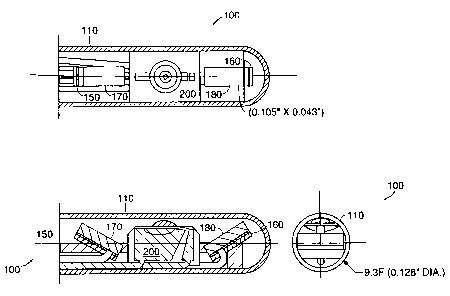
In another embodiment, depicted in FIGURES 2A and 2B, the spectrometer 100 is
contained in a housing 110 with a diameter of 0.625 inches, and an overall
length of 8 inches. In
this embodiment, the light source 120 is a tungsten-halogen bulb 130 with a
bichromatic filter
140 attached. The bichromatic filter 140 only lets through light with
wavelengths of 420nm and
490nm. This embodiment employs two PIN photodiodes 150 and 160 as sensors.
Attached to
each sensor is a bandpass filter 1?0 and 180 that lets through 370nm and
440nm, respectively.
2o The light source 120 is disposed along the longitudinal axis of the housing
110 and faces the
distal end of the housing 110. Similarly, the sensors 1 SO and 160 face the
distal end of the
housing, and are disposed on either side of the longitudinal axis. An end cap
190 covers the
distal end of the housing. The end cap is designed to minimize attenuation of
both excitation and
emitted energy. in a preferred embodiment, the sensors are angled inward about
30 degrees
z5 towards the longitudinal axis.
In FIGURE 3, the sensors 150 and 160, their filters 170 and 180, as well as
the light
source 120 are visible through the end cap 190.
FIGURE 4 depicts an electronics block diagram for the embodiment depicted in
FIGURE
2 and FIGURE 3. In this embodiment, the test sample 400 fluoresces at
wavelengths of 440nm
so and 370nm when illuminated by 300nm light from light source 120. Filters
170 and 180 are
attached to PIN photodiodes 150 and 160, respectively. Bandpass filters 170
and 180 let through
CA 02305163 2000-04-04
WO 99/18844 PGT/US98/21100
-5-
light of 440nm and 370nm, respectively. PIN photodiodes 150 and 160 emit an
electrical signal
in response to light. The strength of their signals is proportional to the
intensity of the light
shining on them. These electrical signals are sent through low pass filters
410 and 420. These
filters remove 60Hz electrical signals, and serve to increase the signal-to-
noise ratio of the output
of the PIN photodiodes 150 and 160. The signals are next sent to amplifiers
430 and 440, and
combined into a comparator decision process 450. Depending on the signals'
relative intensities,
the comparator decision process 450 indicates either result A 460 or result B
470.
In an embodiment of the comparator decision process 450, colonic tissue is
diagnosed for
adenoma. The colon is illuminated with 325nm light, and tissue
autofluorescence readings are
~ o taken at 460nm and 680nm. A numeric result, C, is calculated according to
the following
formula, C = A * (tissue autofluorescence at 460nm) + B * (tissue
autofluorescence at 680nm),
where A and B are constants set according to the relative autofluorescent
characteristics of
normal and adenomous tissue. If C is above some threshold value, T, then the
tissue is
diagnosed as an adenoma.
~ s In a preferred embodiment of this invention, the light source operates in
the "blue" region
of the visible spectrum, emitting light at a wavelength or wavelengths
selected from a region
between 400nm and 490nm.
For the purposes of tissue autofluorescence spectrometry, a light source
emitting light at a
wavelength of 300nm is desirable. FIGURE 5 depicts the output spectrum of a
tungsten-halogen
Zo lamp. The units along x-axis 500 represent the wavelength of the light
emitted by the light
source in nanometers. The units along the y-axis 510 represent the intensity
of the light in a.u.
The spectrum indicates that the lamp emits a useful amount of light in the
300nm range.
FIGURE 6 depicts output spectra of a tungsten-halogen lamp with a bichromatic
filter
attached. The units along the x-axis 600 represent the wavelength of the light
emitted by the
2s light source in manometers. The units along the y-axis 610 represent the
intensity of the light in
a.u. Emission curve 620 depicts the output spectrum when 7V is applied.
Emission curve 630
depicts the output spectrum when 6V is applied. Emission curve 640 depicts the
output spectrum
when 5V is applied. The intensity of the spectrum varies as a result of the
voltage used. A large
increase in light output at 300nm is observed when the voltage is increased
from 5V to 7V.
so For the purposes of this invention, it is necessary that the sensors are
able to respond to
the light at wavelengths at which the tissues to be examined autofluoresce.
FIGURE 7 depicts
CA 02305163 2000-04-04
WO 99/18844 PCT/US98/21100
-6-
the spectrum response of a PIN photodiode. The units along x-axis 700
represent the wavelength
of light input into the sensor in nanometers. The units along the y-axis 710
represent the
response of the photodiode in A/W. As evidenced from the response curve 720,
the PIN
photodiode reacts to a broad spectrum of light.
For the purposes of this invention it is further necessary that a sensor
responds only to
specific wavelengths of light, and not respond to light outside its designated
wavelength.
FIGURE 8A and FIGURE 8B depict two photoresponse curves of a PIN photodiode.
The units
along the x-axes 800 and 820 represent the wavelength of the light input into
the sensor in
nanometers. The y-axes 810 and 830 represent the transmission in a.u. FIGURE
8A depicts the
~o photoresponse curve of a PIN photodiode with a 370nm bandpass filter
attached. Similarly,
FIGURE 8B depicts the photoresponse curve of a PIN photodiode with a 400nm
bandpass filter
attached. As evidenced by photoresponse curve 840, the PIN photodiode with a
370nm bandpass
filter attached responds only to a narrow range of wavelengths centered around
370nm.
Response to wavelengths outside of this range is essentially zero. Response
curve 850 depicts
analogous results for the 400nm bandpass filter.
FIGURE 9 depicts a test fixture used to analyze the sensitivity and
specificity of the
response of the filtered PIN photodiodes. A sample fluorescin is placed in a
cuvette 900. A DC
power supply powers filtered light source 120. Filtered light source 120
illuminates the sample
fluorescin with 300nm light. The sample fluorescin, fluoresces in response to
the 300nm light.
zo Photodiode assemblies 910 and 920 emit electrical signals in response to
light of 370nm and
440nm, respectively. These electrical signals are sent to channel amplifiers
960 and 970, where
the intensities of the electrical signals are read. A fiber optic bundle 930
provides access for an
external spectrometer (not shown) to corroborate results. The light source
120, the cuvette 900
and the photodiode assemblies 910 and 920 are all enclosed in a light-tight
metal enclosure 950.
zs FIGURE 10 depicts the response of each photodiode assembly to each
fluorophore. The
units along the x-axis 1040 represent fluorophore concentration as a
percentage in solution. The
units along the y-axis 1050 represent the response of the photodiodes to the
light in
nanoamperes. Response curve 1000 depicts the response of the test fixture's
440nm channel
amplifier to coumarin, a 460nm fluorophore. Response curve 1010 depicts the
response of the
so test fixture's 370nm channel amplifier to PDB, a 370nm fluorophore.
Response curve 1020
depicts the response of the test fixture's 440nm channel amplifier to PDB, a
370nm fluorophore.
CA 02305163 2000-04-04
WO 99/18844 PCT/US98/21100
-7-
Response curve 1030 depicts the response of the test fixture's 370nm channel
amplifier to
coumarin, a 460nm fluorophore. Intensity of coumarin fluorescence at decreases
at higher
concentrations due to self absorption. These results indicate that each sensor
responds to its
selected wavelength with a high degree of sensitivity and specificity.
FIGURE 11 depicts the emission spectrum of a 0.1 % mixture of the fluorescin
coumarin
to 300nm light. The units along the x-axis 1100 represent the wavelength of
the light emitted in
nanometers. The units along the y-axis 1110 represent the intensity of
fluorescence in counts.
These results indicate that the majority of coumarin's fluorescence is emitted
at wavelengths
around 460nm. FIGURE 12 depicts the emission spectrum of a 0.1 % mixture of
the fluorescin
~o PBD to 300nm light. The units along the x-axis 1200 represent the
wavelength of the light
emitted in nanometers. The units along the y-axis 1210 represent the intensity
of fluorescence in
counts. These results indicate that the majority of PBD's fluorescence is
emitted at wavelengths
around 370nm.
FIGURE 13 depicts the testing apparatus used to analyze inrush and steady
state response
~ s of the light source 120 to the application of power. The light source 120
is powered by a DC
power supply 940 set at 2.0 amps and 37 volts. One channel of an oscilloscope
1300 is placed
across a 25 ohm resistor 1310 placed between power supply 940 and light source
120.
Photodiode 150 emits an electrical signal in response to the light output by
light source 120. The
electrical signal is then sent to an amplifier 960 and then to another channel
of oscilloscope 1300.
zo Light source 120 and photodiode 150 are enclosed in a light tight container
950. The signals on
the two channels of the oscilloscope 1300 are analyzed to compare light output
to power input.
FIGURE 14 depicts the results of these tests. Response curve 1400 depicts the
intensity of the
Iight emitted by the light source 120. Response curve 1410 depicts the current
supplied to the
light source 120. From these tests, it was determined that the spectrometer
would require a
is power supply of 10.64 watts, and that it took 400 milliseconds from the
application of power for
the light source to reach full intensity.
For in vivo use, surface temperature needs to be moderate. FIGURE 15 depicts
probe
surface temperature as a function of time of operation. The units along the x-
axis 1500 represent
time in seconds. The units along the y-axis 1510 represent the surface
temperature of the probe
ao in degrees Celsius. To obtain these measurements, a J-type thermocouple
(Omega Engineering,
Inc., Stamford, CT) model STC-GG-J-20-36 was attached to the exterior surface
of the
CA 02305163 2000-04-04
WO 99/18844 PCT/US98/21100
_g_
embodiment depicted in FIGURE 3. The tungsten-halogen bulb 130 of this
embodiment has
been demonstrated to generate a surface temperature of no more than 40 degrees
Celsius after 30
seconds of continuous operation. To prevent an undue increase in surface
temperature, the light
source I20 can be operated intermittently or with short excitation times.
The spatial characteristics of the sensors effect the sensitivity of the
spectrometer.
FIGURE 16 depicts two possible spatial configurations for an array pair 1600
of sensors.
FIGURE 16(a) depicts the array pair I600 as coplanar, while FIGURE 16(b)
depicts the array
pair 1600 angled inwards toward the light source (not shown). FIGURE 17
depicts the radiation
pattern of the excitation source. FIGURE 18 depicts the response pattern for
an array pair in a
~o coplanar configuration. FIGURE 19 depicts the response pattern for an array
pair in an inwardly
angled configuration. These results indicate that angling the array pair may
improve system
sensitivity.
The spectrometer must be able to operate within certain parameters so as not
to cause
tissue damage. For example, it is desirable to keep the surface temperature of
the spectrometer to
~s a minimum. In order to minimize waste heat generated by the spectrometer,
it is therefore
desirable to obtain fluorescence readings with the minimal amount of
excitation energy.
FIGURE 20 depicts apparatus used to measure the power output of an excitation
energy source,
such as a light source. In this apparatus, the light source 120 and its filter
140 is attached to a
Newport radiometer head 1720 by means of an adapter 1700. The detector 1710
measures the
zo power output of the light source 120. In a preferred embodiment of the
spectrometer, the sensors
are able to obtain fluorescence readings using an excitation energy as low as
200 pW.
FIGURE 21 depicts a test fixture used to measure the response and effciency of
the PIN
photodiodes. A baseline value was first obtained by shining light source 120
onto reflectance
standard 1800 and measuring reflected light using an advanced PhotonX detector
1820. The
2s reflectance standard was then replaced with an uncalibrated fluorescence
standard and the
unfiltered photodiode 1820 was replaced with a photodiode with a bandpass
filter centered at
442nm. A fluorescence signal of 4.1 nW was recorded, which is about 10% of the
reflected
signal from a white target.
While certain embodiments have been used to illustrate the invention, it will
be
so recognized by those skilled in the art that various modifications can be
made therein without
departing from the scope of the invention as claimed.