Note: Descriptions are shown in the official language in which they were submitted.
CA 02305898 2000-04-10
WO 99118845 PCT/US98/21450
METHOD FOR MEASURING TISSUE MORPHOLOGY
GOVERNMENT SUPPORT
The invention was supported, in whole or in part, by a
Grants No. P41RR02594 and CA53717 from the National
Institutes For Health. The Government has certain rights
in the invention.
RELATED APPLICATIONS
This is a continuation-in-part of U.S. Serial No.
08/948,734 filed on October 10, 1997, the entire contents
of which is incorporated herein by reference.
BACKGROUND OF THE INVENTION
Methods for diagnosis of cancer at an early stage are
essential for cancer prevention and therapy. Many types of
cancers grow from epithelial tissues, which cover inner and
SUBSTITUTE SHEET ( rute 26 )
CA 02305898 2000-04-10
WO 99118845 PCT/US98/Z1450
-2-
outer surfaces of the human body. Many of these, for
example cancer in gastrointestinal tract, progress through
the stage of dysplasia. Dysplasia can be defined as
neoplastic tissue which is not malignant yet, but is
considered to be a precursor of malignancy. If diagnosed
at this stage, most tumors are curable. In the case of
gastrointestinal tumors, current methods of diagnosis are
based on endoscopy. However, dysplastic tissue is
frequently not endoscopically apparent. Thus, detection of
dysplasia in the gastrointestinal tract and other sites
often relies on sporadic sampling for this "invisible~~
malignant precursor. However, sporadic biopsies have a
high probability of missing dysplastic changes. In some
cases the biopsy procedure is impossible.
Efforts toward a substitution for standard biopsies
have been made in order to provide accurate diagnosis of
cancerous tissue in different organs in vivo and in real
time. For this purpose, optical techniques that are non-
invasive do not require tissue removal and can be performed
in-vivo. Such methods provide information at the
microscopic level and can thus provide for the search for
very small sites which are likely to be missed by standard
biopsies. While most human organs can be diagnosed by
means of optical techniques, they are particularly
applicable to the tissues in human body lumene, since they
are easily accessible by optical probes, which can be
SUBSTITUTE SHEET ( rule 26 )
CA 02305898 2000-04-10
WO 99/18845 PCTIUS98/21450
-3-
inserted into one of the channels of a conventional
endoscopic tube.
SUM1HARY OF THE INVENTION
The present invention relates to the uae of light to
determine physical characteristics of a structured layer of
material, and in particular certain qualitative information
regarding the morphology of tissue structures using
scattered light. Both backscattered and transillumination
methods can be used, depending upon the thickness of the
material and the size and distribution of the structure
being measured. Examples of properties of materials that
can be measured include surface roughness, parasity,
cytometer measurements, or any material in which changes in
the refractive index of a material correspond to changes in
structures. This type of scattering spectroscopy can be
differentiated from absorption spectroscopy which is unable
to quantitatively measure particle morphology.
Despite extensive investigations, no reliable optical
technique to diagnose dysplasia in-vivo is known. One of
the difficulties resides in the fact that dysplastic
changes are limited to the uppermost epithelial layer,
which can be as thin as 20um, a ane cell layer that is
nearly transparent to optical radiation.
Tissue in the gastrointestinal tract, for example
(other hollow organs share the same features also), is
covered by a layer of cells called epithelium (from 20um
SUBSTITUTE SHEET ( rude 26 )
CA 02305898 2000-04-10
WO 99118845 PCTIUS98/Z1450
-4-
to 300um thick depending on the part of the tract)
supported by relatively acellular and highly vascular loose
connective tissue, lamina propria, which can be up to
50oum in thickness and contains a network of collagen and
elastic fibers, and variety of white blood cell types.
Beneath the lamina propria there is a muscular layer,
muscularis mucosae, (up to 400um thick) and another layer
of moderately dense connective tissue called submucosa
(400-600um thick) containing many small blood vessels and
abundant collagen and elastic fibers. The overall
thickness of those layers is about lmm. Since a
characteristic penetration depth of optical radiation into
biological tissue does not usually exceed lmm, for a
preferred embodiment it is sufficient to limit measurements
of tissue by those layers.
Adenocarcinoma of the esophagus arises in metaplastic
columnar epithelial cells in the esophagus, termed
"Barrett's esophagus", which is a complication of long-
standing gastrointestinal reflex. In this condition, the
distal squamous epithelium is replaced by columnar
epithelium consisting of a one cell layer which resembles
that found in the intestines. Barrett's esophagus is
frequently associated with dysplasia which later can
progress to cancer. Trials of endoscopic surveillance of
patients with Barrett's esophagus have not resulted in a
reduction of esophageal cancer mortality. The most likely
explanation is that dysplasia occurring 'in the esophagus
SUBSTITUTE SHEET ( rule 26
CA 02305898 2000-04-10
WO 99/18845 PCTIUS98/Z1450
-5-
cannot be seen with standard endoscopic imaging and
sporadic biopsy sampling is necessary. This procedure can
sample only about 0.3% of the tissue at risk. Thus, there
is tremendous potential for sampling error.
The application of optical techniques to diagnose
dysplasia in Barrett's esophagus is limited by the fact
that the primary alterations in the tissue occur in the
epithelium which is one cell thick (-20-30um) while
fluorescence or reflectance spectra are mostly formed in
deeper tissue layers. One of the most prominent features
of a dysplastic epithelium is the presence of enlarged,
hyperchromatic, and crowded nuclei. In fact, these changes
ire nuclei size and spatial distribution are the main
markers used by a pathologist to diagnose a tissue specimen
as being dysplastic. No significant changes in other
tissue layers is observed. Unfortunately, epithelium does
not contain strong absorbers or fluorophores, and the
thickness of the epithelium is relatively small and thus
negligible. These make epithelium diagnosis in Barrett's
esophagus to be a difficult problem.
Diffuse reflectance spectroscopy can provide
quantitative biochemical and morphological information for
the analysis of biological tissue epithelium and the
detection of precancerous lesions. Diffuse reflectance
spectra were collected from adenomatous colon polyps
(cancer precursors) and normal colonic mucosa of patients
undergoing colonoscopy. The data were analyzed using an
SUBSTITUTE SHEET ( rule 26 )
CA 02305898 2000-04-10
WO 99/18845 PG"T/US98/21450
-6-
analytical light diffusion system, which was measured on a
physical tissue model composed of polystyrene beads and
hemoglobin. Four parameters were obtained: hemoglobin
concentration, hemoglobin oxygen saturation, effective
scatterer density, and effective scatterer size. Normal
and adenomatous tissue sites exhibited differences in
hemoglobin concentration and effective scatterer size.
Thus, diffuse reflectance can be used to obtain tissue
biochemical and morphological information in vivo.
A preferred embodiment of the present invention
relates to a system of measuring a fine structure component
in backscattered light from mucosal tissue which is
periodic in wavelength. This structure is ordinarily
masked by a diffusive background, which must be removed to
render it observable. The origin of this component is due
to light which is Mie-scattered by surface epithelial cell
nuclei. By analyzing the amplitude and frequency of the
periodic structure, the density and size distribution of
these nuclei can be extracted. These quantities are
important indicators of neoplastic precancerous changes in
biological tissue, and the technique can thus provide a
useful tool for observing such changes in patients
undergoing endoscopy.
The light that is incident on the thin layer at the
tissue surface is not completely randomized. In this thin
region the details of the elastic scattering process can be
preserved. Mucosal tissues, which line the hollow organs
SUBSTITUTE SHEET ( rule 26 )
CA 02305898 2000-04-10
WO 99/18845 PCT/US98I21450
of the body, generally consist of a thin surface layer of
epithelial cells supported by underlying, relatively
acelluiar connective tissue. In healthy tissues the
epithelium often consists of a single, well-organized layer
of cells with an endface diameter of about 10-20 a m and a
height of about 25 a m. In cancerous and pre-cancerous
(dysplastic) epithelium cells proliferate, the cellular
layer often thickens and becomes more tightly packed, and
the cell nuclei enlarge and appear darker (hyperchromatic)
when stained. This may indicate increased nucleic acid
density, hence increased refractive index.
A preferred embodiment of the invention utilizes a
broadband light source to illuminate the region of interest
in the tissue with optical radiation in the range between
350 and 700 nm. A fiber optic probe can be used to deliver
and/or collect radiation from the tissue. The system can
be used during endoscopy of a patient to optically survey a
body lumen within the patient and thereby eliminate the
need for removal~of tissue for biopsy, or alternatively,
can be used to aid in locating tissue suitable for biopsy.
Backscattered light is preferably collected over a
small collection angle of between 2° and 12°, preferably in
the range between 3° and 8°. When using an optical fiber
system to collect the scattered light fibers having a
numerical aperture between 0.05 and 0.22, and preferably
between 0.07 and 0.1 can be used. Collection angles within
SUBSTITUTE SHEET ( rule 26 )
CA 02305898 2000-04-10
WO 99/18845 PCTIUS98I21450
_g_
this range reduce the level of background light without
loss of the periodic component in the returning light.
BRIEF DESCRIPTION OF THE DRAWINGS
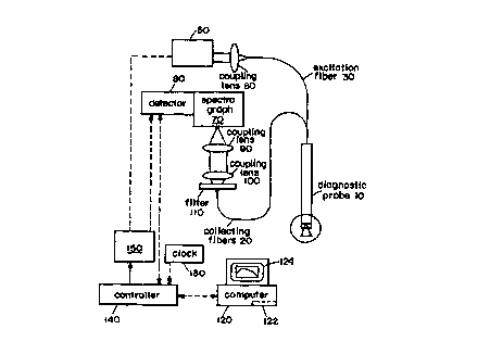
Figure 1 is a schematic diagram of a fiber optic probe
in accordance with the invention.
Figure 2 is an enlarged view of the distal end of an
endoscope in accordance with the invention.
Figures 3A, 3B and 3C illustrate reflectance spectra
from cell monolayers for normal colon cells (R - 0.46);
T84 cells (1t - 0.38);~(c) BaSO, diffusing plate (R - 1.0)
Figure 4 illustrates nuclear size distributions from
data of Figures 3A and 3B, respectively for normal colon
cells; and T84 cells respectively. In each case, the solid
line is the distribution extracted from the data, and the
dashed line is the distribution measured using light
microscopy.
Figures 5A, 5B and 5C are reflectance spectra from
Barretts~ esophagus for diffuse reflectance from a normal
site (solid line), a dysplastic site (dashed line), and the
model fit (thick solid line); for corresponding fine
structures; and of resulting nuclear size distributions,
respectively.
Figure 6 graphically illustrates a comparison of
samples analyzed by standard pathology and the optical
methods in accordance with the invention.
SUBSTITUTE SHEET ( rule 2G
CA 02305898 2000-04-10
WO 99118845 PCT/US98/21450
_g_
Figure 7 is a system used for in vitro tissue analysis
in accordance with the invention.
Figure 8 is a process flow diagram illustrating a
method of performing an optical diagnosis of tissue in
accordance with the invention.
Figure 9 is the molar extinction coefficient spectra
(per heme group) of the oxygenated (thin line0 and
deoxygenated (thick line) hemoglobin (21). Note the
characteristic peaks at 415, 542, and 577 nm
{oxyhemoglobin-Hb02) and at 430 and 555 nm
(deoxyhemoglobin-Hb).
Figure l0 is the reduced scattering cross section
spectra Q' (1~), calculated using Mie theory. Results are
shown for four different diameters, d" (0.2, 0.6, 1.0, and
2.0 ~cm). The slope of the spectra is inversely related to
the diameter. A refractive index of 1.4 was assumed for
the scattering particles, and 1.36 for the surrounding
medium.
Figure 11 shows the Diffuse reflectance spectra
measured on physical tissue models (thick line)
corresponding to four different Hb concentrations: (a) 0.0
mg/dL, (b) 50 mg/dL, (c) 125 mg/dL, (d) 250 mg/dL. The
analytical model predictions using the same optical
parameters employed in the preparation of the physical
tissue models are also shown (thin line), with agreement
between the analytical and the tissue model being very
good.
SUBSTITUTE SHEET ( ruie 26 )
CA 02305898 2000-04-10
WO 99/18845 PCT/US98I21450
-10-
Figure 12 shows the typical normal and adenomatous
polyp spectra (thick line), and best fits to. the data using
the model (thin line). Mie theory was used to approximate
the reduced scattering coefficient used for the model fits
(see Figure 13).
Figure 13 shows the scattering spectra obtained from
the data shown in Figure 12 {thin line), and best fits
using Mie theory (thick line). The Mie theory fits assign
an effective scatterer size d$ to the reduced scattering
spectra. The polyp is characterized by a larger effective
scatterer size (ds = 1.5 um) as compared to normal mucosa
( d$ = 0 . 3 5 ~Cm) .
Figure 14A-14D show parameters obtained from data
analysis: {A) total Hb concentration. Cue, (H) Hb oxygen
saturation, a (C) effective scatterer density, pe, and (D)
effective scatterer size, d,. The largest difference
between normal mucosa (squares) and adenomatous polyps
(stars) is observed in the total Hb concentration.
Figure 15 is a binary plot of the total Hb
concentration Cue, vs. the effective scatterer size d9,. The
normal data tend to form a well defined cluster, while the
adenomatous polyp data are marked by wider variation.
The foregoing and other objects, features and
advantages of the invention will be apparent from the
following more particular description of preferred
embodiments thereof, as illustrated in the accompanying
drawings in which like reference characters refer to the
SUBSTITUTE SHEET ( rule ZG )
CA 02305898 2000-04-10
WO 99/18845 PCT/US98lZ1450
-I1-
same parts throughout the different views. The drawings
are not necessarily to scale, emphasis instead being placed
upon illustrating the principles of the invention.
DETAILED DESCRIPTION OF THE INVENTION
A preferred embodiment of the invention involves the
use of a fiber optic system to deliver and collect light
from a region of interest to measure one or more physical
characteristics of a surface layer. Such a system is
illustrated in Figure 1. This system can include a light
source 50 such as a broadband source, a fiber optic device
10 for delivery and/or collection of light from the tissue,
a detector system 80 that detects the scattered light from
the tissue, a computer 120 having a memory 122 that
analyzes and stores the detected spectra, and a display 124
that displays the results of the measurement. A lens 60
can be used to couple light from the source 50 into the
excitation fiber 30 of the probe 10. A filter 110 and lens
system 90,100 can be used to efficiently couple collected
light to a spectrograph 70. A controller 140 connected to
the data processing system 120 can be connected to a clock
and a pulser 150 that controls the light source 50.
The distal end 15 of the probe 10 is illustrated in
Figure 2 where the central excitation fiber 30 is
surrounded by six peripheral collection fiber 20. The
distal end of the device can be enclosed in an optical
shield 25 such as that described in U.S. Patent No.
SUBSTITUTE SHEET ( rule 2~ )
CA 02305898 2000-04-10
WO 99/18845 PGT/US98/~1450
-12-
5,199,431, the entire contents of which is incorporated
herein by reference. Other endoscopic devices can be used
such as an optical needle as described in the above
referenced patent or as described in U.S. Patent No.
5,280,788, the entire contents of which is also
incorporated herein by reference.
The collection fibers 20 preferably have a numerical
aperture in the range of 0.05 to 0.22 in order to provide a
desired collection angle from the material being measured.
This aids in reducing background that is removed from
scattering spectrum without loss of the periodic component.
The collection fibers can also be replaced or
supplemented by a distally mounted imaging sensor 35 such
as a charged coupled device or CMOS imager. The sensor has
a pixellated structure that is sensitive to the different
colors contained in the scattering spectrum being recorded.
Further details regarding the use of a distally mounted
sensor can be found in U.S. Serial No. 08/745,509 filed on
November 12, 1996, the entire contents of which is
incorporated herein by reference.
The backscattered light collected with this system can
be analyzed to determine certain physical characteristics
of epithelial tissue. The relationship between the
collected light and the physical characteristics to be
determined using this light can be described as follows.
Epithelial nuclei can be represented as spheroidal Mie
scatterers with a refractive index higher than that of the
SUBSTITUTE SHEET ( ruie 26 )
CA 02305898 2000-04-10
wo ~nssas pcrn,rs9sniaso
-13-
surrounding cytoplasm. Normal nuclei have a characteristic
diameter 1=4-7um. In contrast, dysplastic nuclei can be
as large as 20 u~a in size, occupying almost the entire
cell volume. Thus, in the visible range, the wavelength
h « I, and the component of light scattered by the nuclei
will exhibit a periodicity with wavelength, the details of
which are determined by the nuclear size distribution. The
Van de~Hulst approximation can be used to describe the
optical scattering cross section of the nuclei:
~f(~,,1) - 1 ~lZ 1 _ sin(28W) + sin(a W) 2 ~ ( 1
2 ~I~, ~I~.
where 8= ~rln~(n-1) , with n~ the refractive index of
cytoplasm and n the refractive index of the nuclei relative
to that of cytoplasm.
When a beam of light is incident on an epithelial
layer of tissue, a portion of this light is backscattered
from the epithelial nuclei, while the remainder is
transmitted to deeper tissue layers, where it undergoes
multiple scattering and becomes randomized. All of the
diffusive light which is not absorbed in the tissue
eventually returns to the surface, passing once more
through the epithelium, where it is again subject to
scattering from the cell nuclei. Thus, the emerging light
consists of a large diffusive background plus the component
of forward scattered and backscattered light from the
nuclei in the epithelial layer. For a thin slab of
SUBSTITUTE SHEET ( rule 2G )
CA 02305898 2000-04-10
WO 99/1$845 PCTIUS98I21450
-14-
epithelial tissue containing nuclei with size distribution
N(I) (number of nuclei per unit area (mm~) and per unit
interval of nuclear diameter (um)), the approximate
solution of the transport equation for the reflectance
R(1~) collected by an optical probe with acceptance solid
angle SZ~ is given by the following expression:
R(~,} a rt~~ + 1 1 ~, ~~l) \le~~'' s~~p~~''s' s~~~nr +\Id ~~''s,~z ~~n~ ( 2 )
d ( ~ ~flo
where Ii ( tl~. s> ) is the intensity of the incident light
delivered in solid angle 52;, Id(~,,s) is the intensity of the
light emerging from the underlying tissue, and
(1(s)~f~= jt(s)ds for any function I (s) and solid angel ~2,
n
with s a unit vector pointing outward from the tissue
surface in an arbitrary direction. The quantity
R(~,) =~Id(~,,s)~n~ I ~l,(~.,s)~~~ is the reflectance of the
diffusive background. The optical distance
W
z(~,) _ ~Qs(~.,1)N{1)dl and scattering phase function
0
p(~,,s,s')= 1 jp(~,,l,s,s')Crf(~,,1)N(I)dl both depend on N(1) ; for a
To
sphere, p(~,,l,s,s') is determined by Mie theory. The first
term in Eq. (2) describes the attenuation of the diffusive
SUBSTITUTE SHEET ( rule 2b )
CA 02305898 2000-04-10
WO 99/18845 PCT/US98121450
-15-
background, and the terms in brackets describe
backscattering of the incident light and forward scattering
of diffusive background by the epithelial cell nuclei,
respectively.
For small S'2~ the forward scattering term in Eq. (2)
can be expanded in T ( 1~) . Thus, ~~Id(~.,s ~~=x~ . l ~Id(~,,s)~o~ a fo + f,r
l r,,
O.
with zo =~rl2JlZN(l~dl. It is found numerically that fl«fa and
0
that fo and fl are approximately independent of wavelength
in the range of interest (~,~;o = 360 to ~,~u = 685nm). Similarly,
for the backscattering term
~~I~(.~,-s)p~~.'s s~~~n,~n' l~Id(~,'s)~n~ =bo-b, r/ro. Note that in the
forward scattering contribution the first order term
oscillates in phase with r(~,~ as required by the optical
theorem, whereas for the backscattering contribution it is
out of phase. Thus, Eq. (2) reduces to
R~~) = a s~'~~ + (1- a '~s~ l~ + bo ~" (.f -b~ ) ~~~) ,
R (~) zo (~)
which shows that the epithelial nuclei introduce a periodic
fine structure component into the reflectance with a
wavelength dependence similar to that of the corresponding
scattering cross section. Its periodicity is approximately
proportional to nuclear diameter, and its amplitude is a
SUBSTITUTE SHEET ( rule 26 )
CA 02305898 2000-04-10
WO 99118845 PGT/US98/21450
-16-
function of the size and number of nuclei in the epithelial
layer. These quantities can be determined by analyzing the
reflectance, R (1~) .
As example of the effects described by Eq. (2),
elastic light scattering from normal T84 tumor human
colonic cell monolayers (10 and 15 sites respectively) was
measured and analyzed. The cells, approximately l5um
long, were affixed to glass slides in buffer solution and
placed on top of a BaS04 diffusing (and highly reflective)
plate. The BaSO, plate was used to approximate the diffuse
reflectance from underlying tissue. The diameters of the
normal cell nuclei generally ranged from 5 to 7 um and
those of the tumor cells from 7 to l6y,m.
An optical fiber probe was used to deliver white light
from a xenon arc flashlamp to the samples and collect the
return reflectance signal, as shown in Figure 1. The probe
tip, 1 mrn in diameter, consisted of a central delivery
fiber surrounded by six collection fibers, all of which
were covered with a 1 mm thick quartz optical shield. The
fibers were 200 urn core fused silica, NA=0.22
~5~~=S2~=~dVA2~. To eliminate specular reflection, the probe
was beveled at 17° to the normal. At the proximal end the
collection fibers were arranged in a line and imaged onto
the input slit of a spectrograph. A diode array detector
recorded the reflectance spectra from 360 to 685 nm.
SUBSTITUTE SHEET ( rule 26
CA 02305898 2000-04-10
WO 99118845 PCTIUS98/Z1450
-17-
Figures 3A and 38 show the normalized reflectance
R (1.) / rt'(?~) from normal and T84 tumor cell samples,
respectively. Distinct spectral features are apparent.
For comparison, the reflectance spectrum from the BaS04
plate by itself is also shown in Figure 3C. This spectrum
lacks structure and shows no prominent features.
To obtain information about the nuclear size
distribution from the reflectance data, Eq.(3) needs to be
inverted. The nuclear size distribution, N(1), can then be
obtained from the Fourier transform of the periodic
component of the opt ical distance z - zo = (I - R(~,)~ / q. The
parameter q = 1 - bo - fo +2 (bl - fl) is associated with
forward and backward scattering, and depends on the probe
geometry and the angular distribution of the incident and
reflected light. In this particular example q=0.15. By
introducing the effective wavenumber k=2lla~ (n-1) /~,-ko , and
ko =2ms~(n-1)l ~,",~,K=2nre~(n-1)~~,n;;" -~,-";~~ and we obtain,
K
N(1} = q~ 2 f R~~~ -1 e~ (k + ko )dk. ( 4 )
0
Equation (4) was used to analyze the data. In order to
remove spurious oscillations, N(1) was further processed by
convolving it with a Gaussian filtering function. The
solid curves in Figure 4 show the resulting
SUBSTITUTE SHEET ( ruie 26 )
CA 02305898 2000-04-10
WO 99/18845 PCT/US98/21450
18-
nuclear size distributions of the normal and T84 cell
monolayer samples extracted from the spectra of Figures 3A
and 3B. A nucleus-to-cytoplasm relative refractive index
of n=1.06 and cytoplasm refractive index of n~=1.36 were
used. The dashed curves show the corresponding size
distributions, measured morphometrically via light
microscopy. The size distributions can be approximated by
Gaussian distributions. The parameters for those are
presented in Table 1. The extracted and measured
distributions are in good agreement for both normal and T84
cell samples.
Normal Cells Tumor T84 Cells
Mean Standard Mean Standard
Diameter Deviation Diameter Deviation
(gym) (~Cm) (gym)
(!gym)
Microscopy -.6 ~0.5 10.2 2.0
Spectroscopy6.2 0.45 10.1 2.2
The periodic fine structure in diffuse reflectance of
esophagus and colon mucosa of human subjects can be
measured during gastroenterological endoscopy procedures.
In the case of Harretts' esophagus, in which the epithelium
consists of a thin monolayer of columnar cells. similar to
those used in the cell culture experiments, data were
collected as in the cell culture studies. The optical
fiber probe is inserted into the biopsy channel of the
endoscope and brought into contact with the tissue surface.
SUBSTITUTE SHEET ( ruie 26 )
CA 02305898 2000-04-10
WO 99/18845 PGTlUS98/21450
-19-
The methods described herein can also be used to measure
structural properties of other GI tissue, tissues in the
oral cavity, the cervix, the bladder, and skin.
The fine structure component, which is the scattering
signature of the cell nuclei, is typically less then 5% of
the total signal and is ordinarily masked by the background
of diffusely scattered light from underlying tissue, which
itself exhibits spectral features due to absorption and
scattering, as shown in Figure 5A. Its spectral features
are dominated by the characteristic absorption bands of
hemoglobin and collagen scattering. In order to observe
the fine structure, this background must be removed. The
absorption length, ~Q', ranges from 0.5 to 250 mm as the
wavelength is varied, and the effective scattering length
(~,,')-1 ranges from 0.1 to 1 mm. Thus, both scattering and
absorption have to be taken into account in subtracting or
removing the background signal.
To represent the background light incident on the
tissue is assumed to be exponentially attenuated, and that
at any given depth, z, an amount of light proportional to
the reduced scattering coefficient ,uQ is scattered back
towards the surface and further exponentially attenuated.
Since light attenuation depends on both scattering and
absorption, the attenuation coefficient is assumed to be
the sum of absorption coefficient a and effective
a
scattering coefficient ~.,~°~=~i~u,' . The parameter (3 was
SUBSTITUTE SHEET ( ruie 26 )
CA 02305898 2000-04-10
wo ~nss4s Pcnus9ani4so
-20-
determined by comparison with Monte Carlo simulations and
more accurate models of light transport, and was found to
be p~0.0'7. Since light only penetrates ~1 mm into the
tissue, most of the diffusely scattered return light is
confined to the mucosal layer.
The tissue is thereby represented as a two layer
medium and neglected diffusely reflected light from the
Lower layer. The following approximate expression for the
diffusive light from underlying tissue impinging on the
epithelial cell layer is then obtained:
_ _ c~~
la('~~s) = F'(S)~Ir(~~s~nr 1 expf (fps +~~f~a)~J (5)
1 + c( fta I acs )
with F(s) being a Lambertian function describing the
angular dependence of light emerging from mucosal layer, L
a parameter representing the thickness of the mucosal
layer, and L a parameter representing the thickness of the
mucosal layer, and c, the concentration of hemoglobin,
which we find to be the main absorber relative to that of
collagen, which is responsible for light scattering.
Because both oxygenated and deoxygenated hemoglobin are
present, the total hemoglobin absorption is represented as
,uQ=(I-a)~t~~~+afto'~°~~ with oxygen saturation parameter
a(0 <_ a S 1).
Figure 5A shows the reflectance spectra from two
Barretts~ esophagus tissue sites, both independently
SUBSTITUTE SHEET ( rule 2G )
CA 02305898 2000-04-10
WO 99/18845 PCT/US981ZI450
-21-
diagnosed by standard pathological analysis to indicate (1)
normal and (2) precancerous (i.e. low grade dysplasia). As
can be seen, the differences in these unprocessed spectra
are small. To analyze them, Eq.(5.) was first fit to the
broad features of the data by varying the parameters c, a
and L. As seen in Figure 5A, the resulting fits are quite
accurate. After removing this diffuse background structure
by calculating R(A)/R(a), the periodic fine structure is
seen in Figure 5B. Note that the fine structure from the
dysplastic tissue site exhibits higher frequency content
than that from the normal site. Equation (4) was then
employed to extract the respective nuclear size
distributions, yielding Figure 5C. The difference between
normal and dysplastic tissue sites is evident. The
distribution of nuclei from the dysplastic site is much
broader than that from the normal site and the peak
diameter is shifted from -.7 ~m to about -.10 um. In
addition, both the relative number of large nuclei (>l0um)
and the total number of nuclei are significantly increased.
Based on computer analysis, the uncertainty of the
above method in extracting nuclear size information is
estimated to be from 5% to 30%, depending on the noise
level and accuracy of the model. The distributions were
calculated using the same refractive index for both normal
and dysplastic nuclei. This is not entirely correct,
inasmuch as in stained histological sections dysplastic
nuclei appear hyperchromatic, which maybe indicative of an
SUBSTITUTE SHEET ( rule 26 )
CA 02305898 2000-04-10
WO 99/18845 PCT/US98l21450
-22-
increase in refractive index. Thus, the relative number of
large nuclei in the distributions measured from dysplastic
sites may be slightly overestimated.
The ability to measure nuclear size distribution in
vivo has valuable applications in clinical medicine.
Enlarged nuclei are primary indicators of cancer, dysplasia
and cell regeneration. In addition, measurement of nuclei
of different size can provide information about the
presence of particular cells, and can thus serve, for
example, as an indicator of inflammatory response of
biological tissue. This suggests that different
morphology/pathology in the mucosal Iayer gives rise to
distinct patterns of nuclear distributions.
The physical characteristics that have been found to
be useful to differentiate between Barrett's non-dysplastic
and dysplastic epithelium were the total number of nuclei
and the percentage of large nuclei ( I > 10 u~). A
comparison of pathological analysis of samples with the
optical analysis thereof provided the plot (total number of
nuclei vs. percentage of nuclei with a diameter large that
10 um) in Figure 6. From those 50 sites, the cumulative
sensitivity and specificity, for this analysis were 83% and
100% respectively. The study had a positive predictive
value is 100%. The points indicated by n's were either
normal or inflamed and those indicated by d's were
displastic. A percentage in the range of 20-30% was found
SUBSTITUTE SHEET ( rule 26 )
CA 02305898 2000-04-10
WO 99/18845 PCTIUS98I~1450
-23 -
to be an accurate diagnostic for this type, with 25% being
used in this particular example.
A preferred embodiment of a spectrograph system 300
employed for the collection of backscattered spectral data
from excised tissue samples using a spectrograph and a
charge coupled device (CCD), CMOS or other integrated solid
state imaging array is illustrated in Figure 7.
System 300 can use a broadband light source or tunable
laser 314 for irradiating a sample 46. Source 314
generates a beam 316 which is directed by mirror 318
through focusing optics 320 to impinge on sample 46 mounted
on a scattering substrate 325 and behind a transparent
window 321. The beam was focused on the sample at an angle
of incidence. The collection angle 330 can be determined
by an aperture or collimator 340 and is between 2 and 12
degrees, preferably between 3 and 8 degrees.
A portion of the scattered light 322 emitted by sample
46 was collected by collecting optics 324 a small angle
relative to the incident light. In another preferred
embodiment the angle of incidence and collection can be
along a single common axis. Collecting optics 324
collimates and F/matches the collected light for the
spectrograph 310. Prior to entering the entrance slit of
the spectrograph 310, the collected light was passed
through a series of filters 326 which attenuated the
unwanted scattered component of the collected light.
SUBSTITUTE SHEET ( rule 25 )
CA 02305898 2000-04-10
WO 99/1$845 PCTIUS98IZ1450
-24-
Figure 8 illustrates generally a process 400 for
collecting and analyzing the scattering spectrum from a
material of interest such as tissue. The method can be
performed both is vitro using a microscopy system or a
color imaging system as shown in Figure 7, or in vi vo on a
patient. The illuminating light 402 from a source can use
radiation in the range of 300nm-1200nm, including the
infrared range. After collecting 404 and detecting 406
radiation, the diffuse background 408 can be removed and
the desired characteristics calculated 410. These results
can be used to provide a diagnosis of the region of
interest.
A preferred embodiment of the present invention
employs an analytical method based on data collection with
an optical fiber probe with fixed delivery and collection
geometry. Data analysis uses light diffusion, but rather
than directly apply the model to analyze tissue spectra, a
correspondence is established between the diffuse
reflectance spectra and a physical tissue representation
composed of scatterers and absorbers with known optical
properties. By analyzing spectra from this tissue model
using the analytical formulation, a calibration system is
provided which is simple to invert and accurately predicts
the concentrations of the scatterers and absorbers. Once
the calibration is established, the method is applied to
tissue spectra.
SUBSTITUTE SHEET ( ruie 26 )
CA 02305898 2000-04-10
WO 99/18845 PCTIUS98/21450
-25-
The following relates to the application of spectral
analysis to mucosal surfaces of tissues in vivo. In this
example adenomatous colon polyps are measured, which are
precursors of colon cancer. These polyps are a form of
colonic dysplasia and are histologically similar to
visually undetectable flat dysplasia, and are readily
detectable. The results are correlated with standard
histological examination, and can be used for early
detection of disease and, moat importantly, exemplify how
this spectroscopic method can be applied in vivo to obtain
morphological and biochemical information.
Diffuse reflectance spectra were collected in vivo
from adenomatous polyps on 13 patients undergoing routine
colonoscopy. Data were collected simultaneously with
multi-excitation fluorescence spectra: a xenon-arc
flashlamp with 10 acs pulse duration and an average input
energy of 4Jj/pulse was used as white light source. An
imaging spectrograph dispersed the collected, light and a
gated diode array detector was employed for light detection
in the 360-685 nm spectral range. A 12 ,sec gate
synchronized with the lamp pulse was used to minimize
background from endoscopic illumination. The detector was
controlled by a PC notebook computer, where the data were
transferred and stored.
Light was delivered and collected using an optical
fiber probe which was advanced through the accessory
channel of the colonoscope and brought into contact with
SUBSTITUTE SHEET ( rule 26 )
CA 02305898 2000-04-10
WO 99/18845 PGTIUS98l21450
-26-
the tissue. The probe consisted of a central optical fiber
for light delivery, surrounded by six concentric fibers for
light collection. All fibers had 200 ~cm core diameter and
NA=0.22. The probe tip was fitted with a quartz shield
approximately 1.5 mm in length and diameter, which provided
a fixed delivery/collection geometry with uniform circular
delivery and collection spots in the form of overlapping
cones with approximate radii of rd=0.35 mm and r~=0.55 mm,
respectively. The tip was beveled at an angle of 17
degrees, to eliminate unwanted specular reflections from
the shield/air interface.
The diffuse reflectance spectrum of a 20% by volume
BaS04 powder suspension was used as a reference, to take
into account the spectral characteristics and overall
intensity of the xenon lamp. The probe was immersed in the
suspension and a reference diffuse reflectance spectrum was
recorded prior to collection of each data set. Tissue
spectra were calibrated by dividing by this reference
spectrum. Diffuse reflectance spectra were measured from a
few different sites on every adenomatous polyp and from
corresponding sites in the surrounding normal mucosa.
Single-pulse excitation was used in order to avoid motional
artifacts. The polyps were than removed and examined
histologically, while the normal mucosa sites were not
biopsied.
To represent diffuse reflectance, biological tissue
was approximated as being a homogeneous semi-infinite
SUBSTITUTE SHEET ( rule 26 )
CA 02305898 2000-04-10
WO 99/18845 PCTIUS98IZ1450
-27-
turbid medium with reduced scattering and absorption
coefficients ~,,' (A) and ~Ca' (1~) , respectively (?~ is the
wavelength of light). Part of the incident light is
absorbed in the tissue, while the non-absorbed part is
subject to multiple scattering and eventually emerges from
the surface as diffuse reflectance. A certain fraction of
this return light is collected by the probe, while the
remaining part escapes undetected. The amount of the light
collected depends on the optical properties ~c9' (a) and ~.
(h), as well as on the probe diameter, rp. Because r~ is
finite, it serves as a scale length, enabling ice' (1~) and
' (1~) to be determined separately.
To characterize light collection by an optical probe,
knowledge of the spatial and angular resolution of the
diffuse reflectance on the surface of the tissue is
required. Using the method of images to implement the
diffusion approximation to the radiative transfer equation
the diffuse reflectance radial density, R (1~, r), at a
distance r from the point of incidence of the light on the
surface of a semi-infinite turbid medium is given by
~~~~r~=4~ of~s (~+~)e~~~~ _(1+3A)(f~+r )er~ ~ (6)
s ~r 1 1 z z
Ilz
where ~ _ (3,uQ (f~a - fps )) ~ zo ,
~s +~a
U2
and r, =(zo +rz~'n,r2 -Czo(1+ 3 A)z +rzJ
SUBSTITUTE SHEET ( rule 26 )
CA 02305898 2000-04-10
WO 99118845 PCT/US98I21450
-28-
The parameter A depends on the refractive index n of the
medium (A=1 for n=1 and A>1 for n>1). A reasonable
assumption for the average refractive index of colon tissue
is n~ 1.4 so that A ~ 3.2.
To find the total light collected by the probe, Eq. 6
must be integrated over the spatial extend of the delivery
and collection areas, characterized by radii rd and r~,
respectively. Assuming the incident light intensity to be
uniform over the entire delivery area, the diffuse
reflectance Rp (2~) collected by the probe is given by
r yx r
Rp(~.)= ~ ~rdrld~~R(~.,r-r)rdr~ r ('1)
0 0 0
with ~ r - r'~ _ ~rz + r2 - 2rr' cos~)~~j . The integral s i s Eq . 7 can be
evaluated numerically. However, to obtain a simple
analytical expression for R.~ (A), we assume point delivery
of light (rd = 0) and collection over a circular spot of
radium r~, we then obtain:
r
Rp(~.) = 2~~ R(r)rdr
0
SUBSTITUTE SHEET ( rule 26 )
CA 02305898 2000-04-10
WO 99/18845 PGTNS98I21450
-29-
-(1+1~~~° e-d.~ 4 e-~i
- ~s e-'"° xe l 3 -za ; -(1+-A)za , (8)
,us +,uQ r~ 3 r2
i/2
2 Z l/2 , l
with r2 =(zo +r~ ) ,r~ = zo~l+ 3 AJ +r~
The optimal value of r~ was found by calibrating Eq. 8 on
physical tissue models with known optical properties. The
S advantage of using Eq. 8 is that it is much easier to
invert than Eg. 7, which requires numerical integration.
Eq. 8 gives the diffuse reflectance at the perpendicular
direction to the tissue surface. This is acceptable
because light is only collected by the optical probe at
directions with a maximum deviation of approximately 10
degrees from the direction perpendicular to the surface.
Eq. a can be used to analyze the diffuse reflectance
spectra collected by the probe. From these measurements of
the tissue spectra, hemoglobin is the only significant
light absorber in colon tissue in the visible range of the
spectrum, and is encountered in both oxygenated and
deoxygenated forms. The light absorption properties of
both forms have been studied and their molar extinction
coefficient spectra E~2 (A) , e~, (1~) are shown in Figure 9.
Oxyhemoglobin absorption presents a maximum at 415 nm and
two secondary maxima at 542 and 577 nm, while
deoxyhemoglobin has a maximum at 430 nm and only one
SUBSTITUTE SHEET ( rule 26 )
CA 02305898 2000-04-10
WO 99118845 PG"fIUS98/Z1450
-30-
secondary maximum at 555 nm. The total absorption
coefficient, ~Cj (A) is given by
f~Y~'~~=2.3Chb~asbbo,~'~)+(1-a~E~b~'~)) , (9)
where a = C'~°= is the Hb oxygen saturation parameter,
ChbO~ + Chb
S CbbO~and C~ are the concentrations of oxy and deoxy Hb,
respectively, and C~b - Cb~z + C~ the total concentration
o f HB .
Determination of Cue, and a was performed in the
following way. For a given tissue spectrum, R.p(1~), initial
values were assigned (typically C~ = 0.0 and a = 0.5).
Eq. 8 was then numerically inverted to find ~s' (1~), which
exhibited residual spectral features of HB absorption. The
parameters C,~ and a were then modified, and the process
was repeated recursively until ~.S' (1~) exhibited a smooth
spectral shape with the Hb spectral features absent. In
this way, ~.,' (A) was simultaneously determined with Cue, and
a.
Table 2
Model Normal Adenomatous
Parameter Polyp
SUBSTITUTE SHEET ( rule 26
CA 02305898 2000-04-10
WO 99/18845 PGTIUS98121450
-31-
Total Hb
Concentration
C~ (mg/dL) 13.6 t 72.0 29.2
a.s
Hb Oxygen
Saturation 0.59 ~ 0.63 0.10
a 0.08
Effective
Scatterer
Density 9.2 ~ 3.5 4.0
ps (xl0e mm-3) 7.5
Effective
Scatterer
Size 0.56 ~ 0.94 0.44
de (mm) 0.18
The above procedure is based on the assumption that
,g' (?,.) is a relatively smooth function of the wavelength
A, which analysis confirms, as will become evident from the
discussion below. In general, ~Ca' (1~) is the sum of
contributions from the various tissue scatterers.
Unfortunately, detailed information about these individual
scatterers is not available . Therefore, ~,e' (1~) was
written as
f~s~'~~ = Ps~~~~~~ ( 10 )
SUBSTITUTE SHEET ( rule 26 )
CA 02305898 2000-04-10
WO 99118845 PCTIUS98/21450
-32-
with p, the effective scattering density and o' (h) the
effective reduced scattering cross section, i.e, the tissue
scattering properties are modeled in an average way, as if
tissue contained a single well-defined type of scatterer.
In general, a' (1~) depends on the refractive index, shape
and size of the scatterer, as well as on the refractive
index of the surrounding medium. For a spherical scatterer
with diameter d8, o' (1~) can be calculated numerically
using Mie scattering theory.
Figure 10 shows such a calculation of a' (1~)., Note
that the slope of Q.' (J~) depends on ds in a simple way i . a .
the slope is inversely related to scatterer size. In this
way it was possible to assign a scatterer size to every a'
(?~) and hence to every ~.a' (J~) in the range ds = 0.2-2.0
um. For example, by inspecting ~.,' (1~) of normal colon
mucosa, previously reported based on in vitro measurements,
it can be seen that they correspond to a scatterer size d$
0.35 ~Cm. Once da was determined, the density p~ was by
dividing ~B' (1~) by Q' (h) . In summary, for each tissue
diffuse reflectance spectrum four parameters were obtained,
Cue" a , p~ and d9 .
In calculating the results shown in Figure 10, the
scatterer and the surrounding medium refractive indices
were assumed similar to those likely to be found in tissue.
Reasonable approximations are a refractive index of
approximately 1.4 for the scatterers and 1.36 for the
surrounding medium. These values can be justified by
SUBSTITUTE SHEET ( rule 26
CA 02305898 2000-04-10
WO 99/18845 PCT/US98I21450
-33-
noting that the lower bound for the tissue refractive index
is set by the refractive index of water, which is
approximately equal to 1.33 in the visible range, and the
upper bound is set by the maximum refractive index reported
for soft tissue, which is around 1.45. Such values are
consistent with those generally observed for soft
biological tissues.
The physical tissue representation served as a method
for confirming the applicability of the various
approximations made in developing Eq. 8. The samples
consisted of mixtures of spherical microparticles with Hb
in various concentrations. The model established that
these mixtures simulated the optical properties of tissue
samples. In addition, the range of validity of the
analytical model in terms of the optical properties was
established, and the optimal value for the parameter r~,
Eq. 8. was determined.
Polystyrene bead suspensions in de-ionized water
(Polysciences, Inc.) were used to simulate tissue light
scattering, and Hb solutions prepared from lyophilized
human Hb (Sigma, H0267) were used to stimulate absorption.
The scattering properties of the beads were calculated
using Mie theory. The bead diameter was 1.07 ~Cm, and the
refractive index np of polystyrene was given by the
expression of np = 15607 + 10002/ 1~~ (26) , (A in nm) , and
the polystyrene concentration was 0.625 by volume. The
reduced scattering coefficient varied from approximately
SUBSTITUTE SHEET ( ruie 26 )
CA 02305898 2000-04-10
WO 99118845 PC'f/US98I21450
-34-
1.7 mm-1 at 400 nm. This spectral dependence is similar to
that shown in Figure 10 with dA = 1.0 ~cm, with the
difference that the slope is larger due to the larger
refractive index of polystyrene.
Since light absorption by polystyrene is negligible in
the visible range, absorption was solely due to
oxyhemoglobin. The size of the physical-tissue model was
approximately 3x3x0.5 cm, simulating the flat geometry of
normal colon mucosa. Additional geometries were
investigated such as a cylindrical geometry with 1 cm depth
and 0.5 cm diameter, to simulate the geometry of the
polyps, but no significant spectral differences were found.
The range of optical parameters used was chosen based on
the previously reported independent measurements of colon
tissue optical parameters performed in vitro.
Figure 11 shows diffuse reflectance spectra measured
on the physical tissue model with various concentrations of
Hb vs. the analytical model predictions, which were
obtained by setting r~ = 0.45 mm in Eq. 3. This was
determined to be the optimal value for r~, which was kept
fixed throughout the entire data analysis. It was also
found that the parameter Zo = 1/ (0. 6~d + ~,,' ) improved the
agreement between the analytical model and the. physical
tissue model data, especially for large values of
absorption. The physical tissue model spectra shown in
Figure lI are very similar to the tissue spectra presented
below. This fact serves as indirect confirmation of the
SUBSTITUTE SHEET ( rule 26 )
CA 02305898 2000-04-10
wo ~n~s PCTIUS98121450
-35-
assumptions made in developing the model, regarding tissue
scatterers (modeled as microspheres) and tissue absorption
(attributed to Hb).
The analytical model was found to accurately predict
the physical tissue model spectra for Hb concentrations in
the physiological range of interest. For Hb concentrations
lower than 100 mg/dL, the deviation betv~ieen the two models
was always smaller than 10% over the entire wavelength
range. The largest deviation occurred when absorption
became comparable to scattering. This is shown in curve
(d) of Figure ll, near the peak of Hb absorption, around
415 nm. The Hb concentration was in this case 250 mg/dL
which corresponds to an absorption coefficient of
approximately 5 mm-1 at 415 nm, while the scattering
coefficient was approximately 2.8 mm-1 at the same
wavelength.
Figure 12 shows typical diffuse reflectance spectra
from one adenomatous polyp site and one normal mucosa site.
Significant spectral differences are readily observable,
especially in the blue region of the spectrum, where the Hb
absorption valley around 420 nm is the prominent spectral
feature. This valley is much more prominent int he polyp
spectrum, which also shows a continuous decrease in
intensity starting from the red end (-700 nm) and moving
toward the green region (--500 nm) of the spectrum, while in
the same range the normal mucosa spectrum shows a steady
increase in intensity. The secondary absorption dips of
SUBSTITUTE SHEET ( rule 26 )
CA 02305898 2000-04-10
wo ~nss4s rrr~s9sm4so
-36-
oxyhemoglobin (542 and 577 nm) are much more prominent in
the adenomatous polyp spectrum, indicating increased Hb
concentration. According to the model analysis the normal
mucosa spectrum was characterized by Hb concentration C,~, _
22.5 mg/dL, while the corresponding value for the
adenomatous polyp was about 6 times higher (C~ = 165
mg/dL). The Hb saturation was found to be 0.65 t 0.05, and
0.55 ~ 0.05 respectively.
Figure 13 shows the scattering spectra ~.$' (h)
calculated from the data shown in Figure 12, along with the
best Mie theory fits. The main difference between the
spectra of the two tissue types is observed in the spectral
slope, which corresponds to different effective scattering
sizes, de = 1.5 ~,m for the polyp and de = 0.35 ~,m for
normal mucosa. The values for the scatterer densities were
found to be pa = 1:5x108 mm-' for the normal mucosa, and pe =
1.3x108 mm-3 for the adenomatous polyp. Figure 12 also
shows the best analytical model fits to the data, using Eq.
8. The model accurately describes the data, despite the
dramatic differences noted in the spectral shape between
the two tissue types and the fact that GCB' (A) is
approximated assuming a homogeneous distribution of
spherical scatterers. The deviation between the data and
the model in typically smaller than 10~ for most of the
wavelength range.
Figures 14A-14D shows the calculated values of the
four parameters for all tissue sites studied: (A) total Hb
SUBSTITUTE SHEET ( ruie 26 )
CA 02305898 2000-04-10
WO 99118845 PCTlUS98l11450
-37-
concentration C,~ (B) Hb oxygen saturation a, (C) effective
scatterer density p, and (D) effectiveness scatterer size
d,. Adenomatous polyps were clearly characterized by
increased Hb concentration, while there were no observable
differences in terms of the Hb oxygen saturation. The
effective scatterer density was in general lower in
adenomatous polyps, and the effective scatterer size
larger, even though there was significant overlap of these
parameter distributions between the two tissue types.
Table 2 summarizes the results shown in Figures 14A-14D by
giving the average values and the standard deviations for
each parameter.
Figure 15 shows a plot of the Hb concentration C,~,
vs. the effective scatterer size de. These two parameters
are shown together in order to summarize and illustrate the
differences found in the scattering and absorption
properties of normal mucosa and adenomatous polyps in the
colon. Note that the normal mucosa data tend to form a
cluster, while the adenomatous polyp data are separated,
and characterized by a wider spread and irregular
distribution, in both the effective scatterer size and the
Hb concentration.
A methodology has been described which provides
quantitative information about colon mucosal tissues in
vivo, based on diffuse reflectance measurements. The main
components of this methodology are (a) data collection
through an optical fiber probe with a fixed
SUBSTITUTE SHEET ( ruie 2G )
CA 02305898 2000-04-10
wo ~ns84s rcrn.rs9sni4so
-38-
delivery/collection geometry, (b) an analytical model for
data analysis based on light diffusion, and (c) a physical
tissue model for calibration of the analytical model.
The use of an optical probe with fixed geometry
enables consistent data collection, and independent
determination of the scattering and absorption. Modeling
of the probe geometry is facilitated through the use of the
parameter r~, which also defined the probe s sensitivity to
absorption. A probe with large r~ will render the spectra
collected more sensitive to tissue absorption as compared
to a probe with a smaller r~. In Figure 4, the secondary
Hb absorption peaks are barely noticeable in the normal
mucosa spectrum: a probe with larger r~ would make these
features more prominent.
The analytical diffusion theory model is amenable to
numerical inversion and successfully describes the tissue
data after calibration on the physical tissuemodel composed
of polystyrene beads and oxy hemoglobin. Other researchers
have already used variants of the analytical model
described here, in the study of diffuse reflectance from
tissues and physical tissue models in the IR range. The
present study is, to our knowledge, the first to apply the
model in the visible range, to in vivo data from human
mucosal tissue, measured with an optical fiber probe. By
working in the visible rather than in the IR, light
penetration was restricted to approximately 0.5 mm, i.e.
the mucosa layer where precancerous changes occur. Using
SUBSTITUTE SHEET ( ruie 26 )
CA 02305898 2000-04-10
WO 99/18845 PCT/US98I21450
-39-
the model, we have shown that it is possible to obtain
quantitative information about the tissue studied, such as
Hb concentration, Hb oxygen saturation, effective scatterer
size, and effective scatterer density. The results show
that diffuse reflectance provides a tool for quantitative
analysis of mucosal tissue surfaces in vivo.
' The basic approximation made in the model is that Hb
is the only significant absorber in colon mucosa in the
visible range of the spectrum. Even though this assumption
IO lacks direct confirmation, the fact that the data strongly
exhibit the characteristic features of Hb absorption,
clearly indicates that Hb is a major absorber. In
addition, the spectra measured on the tissue physical
model, in which Hb was the only absorber, closely resemble
the tissue spectra.
In a similar way, tissue scattering was modeled as due
to homogeneous spherical scatterers with known refractive
index. The approximation permits a quantitative
characterization in terms of two basic parameters, the
effective scatterer size and the effective scatterer
density. Theae parameters provide an estimate of the
average scattering properties. The fact that the spectra
measured on physical tissue models reproduce the actual
tissue spectra, serves again (as in the case of Hb), as an
indirect confirmation of the validity of this
approximation.
SUBSTITUTE SHEET ( rule 26 )
CA 02305898 2000-04-10
WO 99118845 PCTNS98I21450
-40-
Data analysis showed that adenomatous colon polyps
were characterized by increased Hb concentration. It is
known that tumors and cancerous tissues exhibit increased
microvasculature, and hence increased blood content. Using
morphometry and vascular casting in combination with
scanning electron microscopy techniques. Precancerous
tissues such as adenomatous polyps of the colon are also
characterized by increased microvascular volume the above
in agreement with these reports in terms of observing the
IO increased Hb concentration. Others report increased
microvascularity of the colon mucosa associated with
Crohn~s disease and ulcerative colitis. Even though we did
not study these tissue types, the technique employed here
could prove useful for the in vivo study of such diseases.
The Hb oxygen saturation was found to be approximately
60%, on average, for both normal mucosa and adenomatous
polyps. This result is reasonable, because the
measurements were essentially performed in the capillary
network of the mucosa, where oxygen is transferred from Hb
to tissue. Hb oxygen saturation drops within the
capillaries from approximately 97% (arterial blood) to
about 40% (venous blood) with the measured values (around
60-70%) appropriately placed int he middle of this range.
The fact that there were no differences observed between
the two tissue types, can probably be attributed to the
fact that adenomatous polyp metabolism is not significantly
altered, so as to introduce changes in Hb oxygenation,
SUBSTITUTE SHEET ( ruie 26 )
CA 02305898 2000-04-10
WO 99/18845 PCTIUS98/21450
-41-
which is probably related to the disturbed metabolism of
such tissues. The technique presented here could be used
in vivo for the study of tumors on the surfaces of hollow
organs, or for the study of other tissue types when
knowledge of Hb saturation is needed.
An intrinsic differentiation in the scattering
properties between the two tissue types was observed. For
adenomatous polyps, the average effective scattering size
was larger, and the average effective scatterer density was
smaller, as compared to normal mucosa. There are a number
of hypotheses identifying contributions to scattering from
various microstructures, both extracellular such as
collagen fibers, and intracellular such as mitochondria and
cell nuclei. One model provides contributions to light
scattering from intracellular structures is increased in
adenomatous polyps because cells occupy a higher volume
ratio. In addition, the submucosa, in which collagen is
more densely packed is usually located too far away from
the polyp surface in order to be able to contribute to
light scattering. This can explain the lower scatterer
density in adenomatous polyps, provided that intracellular
scatterers are larger than extracellular ones, on average.
EQUIVALENTS
While this invention has been particularly shown and
described with reference to preferred embodiments thereof,
it will be understood by those skilled in the art that
SUBSTITUTE SHEET ( rule 26 )
CA 02305898 2000-04-10
WO 99/18845 PCTNS98I21450
-42-
various changes in form and details may be made therein
without departing from the spirit and scope of the
invention as defined by the appended claims. Those skilled
in the art will recognize or be able tv ascertain using no
more than routine experimentation, many equivalents to the
specific embodiments of the invention described
specifically herein. Such equivalents are intended to be
encompassed in the scope of the claims.
SUBSTITUTE SHEET ( rule 26 )