Note: Descriptions are shown in the official language in which they were submitted.
CA 02306864 2004-03-23
WO 99/44492 1 PCT/US99104764
SYSTEMS FOR LASER TREATMENT OF
PRESBYOPIA USING OFFSET IMAGING
BACKGROUND OF THE INVENTION
1. Field of the Invention
This invention relates to surgical modifications to the eye. In a specific
embodiment, the invention provides ophthalmic surgery techniques which emply a
laser to effect ablative photodecomposition of corneal tissue to correct
presbyopia
and/or other vision defects.
With aging, a condition of the eye known as presbyopia develops.
With this condition, the crystalline lens of the eye loses the ability to
focus on near
objects when the eye is corrected for far-vision.
Presbyopia is often treated with bifocal eyeglasses. With bifocals, one
portion of the lens is corrected for far-vision, and another portion of the
lens is
corrected for near-vision. By looking down through the bifocals, the user
looks
through the portion of the lens corrected for near-vision. When viewing
distant
objects, the user looks higher, through the portion of the bifocals corrected
for far-
vision.
Efforts have been made to treat presbyopia using partitioned lenses
positioned directly over the pupil of the eye. Examples include multifocal
contact
lenses. Unfortunately, when presbyopia is corrected with bifocal or multifocal
lenses
attached to the cornea, the user is simultaneously looking through the near-
and far-
vision corrected lenses. As a result, the user will see both in-focus and out-
of-focus
images simultaneously when viewing an object. This out-of-focus image
superimposed on the in-focus image can cause glare and degrade vision when
viewing objects at low contrast.
Another technique for treating presbyopia has been to correct one eye
of the patient for near-vision and to correct the other eye for distance-
vision. This
technique is known as monovision. With monovision, a patient uses one eye to
see
distant objects
CA 02306864 2000-04-19
WO 99/44492 PCT/US99/04764
2
and the other eye to see close objects. Unfortunately with monovision, the
patient may
not clearly see objects that are intermediately positioned because the object
is out-of-
focus for both eyes. Also, a patient may have trouble seeing with only one
eye.
Laser-based systems and methods are known for enabling ophthalmic
surgery on the cornea in order to correct vision defects by the technique
known as
ablative photodecomposition. Changing the shape of the anterior surface of the
cornea
will change the optical properties of an eye. These ablative
photodecomposition systems
and methods control ultraviolet laser radiation flux density and exposure time
upon the
cornea so as to achieve a desired surface change in the cornea and thereby
correct an
optical defect.
Several different ablative photodecomposition techniques have been
described to correct specific optical errors of the eye. For example, a myopic
condition
may be corrected by laser sculpting a corneal surface to reduce curvature. An
astigmatic
condition, which is typically characterized by a cylindrical component of
curvature
(departing from the otherwise generally spherical curvature of the cornea),
can be
corrected by a cylindrical ablation. Laser sculpting a corneal surface to
increase the
curvature can correct a hyperopic condition.
In a typical laser surgical procedure, the optically functional region of the
corneal surface to be ablated is designated the optical zone. Depending on the
nature of
the desired optical correction, the optical zone may or may not be centered on
the center
of the pupil or on the apex of the anterior corneal surface. One technique for
increasing
the curvature of the optical zone for hyperopia error correction involves
selectively
varying the area of the cornea exposed to the laser beam radiation so as to
produce an
essentially spherical surface profile of increased curvature. This selective
variation of the
irradiated area may be accomplished in a variety of ways. For example, the
optical zone
can be scanned with a laser beam having a relatively small cross-sectional
area (compared
to the optical zone) in such a manner that the ablation depth increases with
distance from
the intended center of ablation. The result is a substantially spherical
profile for the
anterior corneal surface with maximum depth of cut at the extreme outer
boundary of the
optical zone. Another technique for sculpting the optical zone employs a
rotatable mask
having a plurality of apertures. The apertures are sequentially introduced
into the laser
beam path to provide progressive shaping of the laser beam in order to achieve
the
desired profile.
I I Y 1* 4
CA 02306864 2007-05-25
3
Efforts have also been made to treat presbyopia using ablative
photodecomposition. One specific technique of treating presbyopia creates near-
vision
correction by ablating a region of the lower portion of the cornea adjacent
the pupil rim.
With this eccentric positioning of the ablation, the near-vision lens is not
centered over
the pupil. Consequently, constriction of the pupil may occlude the ablated
near-vision
lens. Constriction of the pupil is a natural response of the eye to
illumination, and could
potentially disrupt near-vision.
Alternative suggested presbyopia treatments include laser ablation of a
small annular region of the cornea (having a diameter not exceeding 3.5 mm),
or the
ablation of a central lens for near-vision, surrounded by a gradual blend
zone, and then a
peripheral far-vision lens, all within the optically used portion of the
cornea.
Efforts have been made in the past to laser sculpt a transition zone to
provide a more gradual sloping of the walls and to eliminate the sharp
discontinuity
between the ablation zone and the surrounding untreated cornea. These efforts
have
included the use of a beam rotation or scanning mechanism operated by a
computer to
provide programmed ablation of the transition zone to achieve a sigmoid or
other profile.
While somewhat effective, these efforts often suffer from the added complexity
of
additional optical elements, such as a rotatable off axis mirror or revolving
prism having
suitable optical properties.
2. Description of the Background Art
Systems and methods relevant to laser-based treatments for presbyopia are
disclosed in the following U.S. patents: U.S. Patent No. 5,395,356, issued
March 7, 1995,
for "Correction of Presbyopia by Photorefractive Keratectomy"; U.S. Patent
No. 5,533,997, issued July 9, 1996, "Apparatus and Method for Performing
Presbyopia
Correction"; and U.S. Patent No. 5,314,422, issued May 24, 1994, for
"Equipment for the
Correction of Presbyopia by Remodeling the Corneal Surface by Means of
Photoablation".
Ablative photodecomposition systems and methods are disclosed in the
following U.S. patents: U.S. Patent No. 4,665,913, issued May 19, 1987, for
"Method for
Ophthalmical Surgery"; U.S. Patent No. 4,669,466, issued June 2, 1987, for
"Method
I I II 141M1
CA 02306864 2007-05-25
4
and Apparatus for Analysis and Correction of Abnormal Refractive Errors of the
Eye";
U.S. Patent No. 4,732,148, issued March 22, 1988, for "Method for Performing
Ophthalmic Laser Surgery"; U.S. Patent No. 4,770,172, issued September 13,
1988, for
"Method of Laser Sculpture of the Optically Used Portion of the Cornea"; U.S.
Patent
No. 4,773,414, issued September 27, 1988, for "Method of Laser Sculpture of
the
Optically Used Portion of the Cornea"; U.S. Patent No. 5,108,388, issued April
28, 1992,
for "Laser Surgery Method and Apparatus"; U.S. Patent No. 5,163,934, issued
November
17, 1992, for "Photorefractive Keratectomy"; U.S. Patent No. 5,556,395, issued
September 17, 1996, for "Method and System for Laser Treatment of Refractive
Error
Using an Offset Image of a Rotatable Mask"; U.S. Patent No. 5,646,791, issued
July 8,
1997, for "Method and Apparatus for Temporal and Spatial Beam Integration";
U.S.
Patent No. 6,203,539, issued March 20, 2001, for "Method and System for Laser
Treatment of Refractive Errors Using Offset Imaging"; U.S. Patent No.
5,683,379, issued
November 4, 1997, for "Apparatus for Modifying the Surface of the Eye Through
Large
Beam Laser Polishing and Method of Controlling the Apparatus"; and U.S. Patent
No. 5,827,264, issued October 27, 1998 for "Method of Controlling Apparatus
for
Modifying the Surface of the Eye Through Large Beam Laser Polishing."
Techniques for treating presbyopia with contact lenses are disclosed in the
following U.S. patents: U.S. Patent No. 5,835,192, issued November 10, 1998,
for
"Contact Lens and Method of Fitting a Contact Lens"; U.S. Patent No. 5,485,228
issued
January 16, 1996 for "Multifocal Ophthalmic Lens Pair;" and U.S. Patent No.
5,864,379
issued January 26, 1999 for "Contact Lens and Process for Fitting."
SUMMARY OF THE INVENTION
It is an object of the invention to mitigate and/or inhibit presbyopia with
minimal vision degradation by ablating a transition zone peripheral to an
optical zone. It
is a further object of the invention to ablate a cornea to produce a healed
cornea with an
aspheric optical zone that corrects presbyopia. In one aspect, the invention
provides for
ablating the cornea to a desired shape that compensates for changes in the
corneal shape
as the cornea heals. In another aspect, the invention provides for the
simultaneous
correction of presbyopic and other refractive corrections such as
nearsightedness,
i1 I A II,I
CA 02306864 2000-04-19
WO 99/44492 PCT/US99/04764
farsightedness and astigmatism. In a yet further aspect, the invention
provides for scaling
the aspheric optical zone to match the size of the pupil. In yet another
aspect, the
invention provides for a method for treating presbyopia which includes
ablating a
transition zone outside an optical zone.
5 One of the major difficulties encountered in the application of laser
surgery techniques to effect hyperopic and presbyopic refractive error
corrections lies in
the nature of the boundary between the optical zone and the untreated area.
When the
anterior surface of the cornea is sculpted to have an increased curvature, the
maximum
depth of cut occurs at the outer boundary of the optical zone. The generally
annular
region between this outer boundary and the adjacent untreated anterior surface
portion of
the cornea typically exhibits steep walls after the completion of the
photoablation
procedure. After the surgery, the eye tends to eliminate these steep walls
with a
stimulated healing response involving concurrent epithelial cell growth and
stromal
remodeling by the deposition of collagen, which results in corneal smoothing
by filling in
tissue in the steep walled region. This natural healing response acts to
eliminate the
discontinuity, resulting in a buildup of tissue in the steep walled region and
over the outer
portion of the optical zone. This natural phenomenon, sometimes termed the
"hyperopic
shift" in phototherapeutic keratectomy, causes a lack of precision for a given
surgical
procedure and diminished predictability, counteracting the beneficial effects
of the
refractive correction procedure and thereby reducing the desirability of the
procedure to
the prospective patient.
According to the present invention, the ablated surface can be contoured to
provide an aspheric surface on a healed cornea. The invention provides for
adjusting the
ablation to compensate for factors effecting the final geometry of the healed
cornea.
These factors include corneal healing and the spatial variation of ablation.
The shape of
tissue ablated with a uniform laser beam pulse will depend upon the size and
shape of the
laser beam spot. The spatial variation of the total ablation may also cause
variations in
the ablated corneal shape. For example, a hyperopic ablation intended to
produce a
spherical ablation may demonstrate greater steepening near the center of the
optical zone.
This increased central curvature may form an aspheric surface that corrects
for
presbyopia.
The ablated surface is covered following the surgery, typically by a new
epithelial layer or a repositioned anterior flap of the corneal tissue.
Consequently, the
CA 02306864 2000-04-19
WO 99/44492 PCT/US99/04764
6
final shape of the anterior surface of the cornea may be a different shape
than the ablated -
shape. However, it is the final change in shape of the anterior surface of the
cornea, not
the initial ablated surface, which determines the refractive change effected
by the surgery.
Therefore, it may be desirable to ablate a shape on the cornea that is
different from the
final intended shape on the anterior surface of the cornea. For example, the
optical zone
may be ablated to a substantially spherical shape for correcting hyperopia.
This ablated
surface may then heal to an aspheric surface that corrects presbyopia.
The invention includes a method and system for performing ablative
photodecomposition of the corneal surface that is capable of providing
relatively smooth
transition zones along with accurate sculpting of the anterior or other
corneal surface to
effect simultaneous symmetric or asymmetric refractive and presbyopic
corrections with
relatively large area coverage. The invention preferably employs a laser beam
of smaller
beam size than the total treatment area.
The invention further provides for the ablation of an optical zone that
substantially matches the area of the pupil. For presbyopic patients, the
maximum pupil
diameter is typically about 5 mm. Therefore, it is an aspect of the invention
that the
ablated optical zone have a diameter of about 5 mm, and be user selectable (by
the user of
the ablation system) to a diameter between 3 and 7 mm. The optical zone is
preferably
ablated to form a healed aspheric surface. Preferably, the central portion of
the optical
zone provides near-vision correction and the peripheral portion of the optical
zone
provides far-vision correction.
The invention additionally provides for scaling a diameter of the aspheric
surface to the pupil. This scaling of the aspheric surface permits an
appropriate balance
between near and far-vision correction within the pupil. For example, a
patient with a
5 mm diameter pupil may have a 2.5 mm diameter zone corrected for near-vision,
while a
patient with a 3 mm diameter pupil may have a 1.5 mm diameter zone corrected
for near-
vision. Scaling of the aspheric lens may be based on areas of the pupil and/or
aspheric
surface.
The invention also provides for ablating a transition zone peripheral to the
optical zone and to the pupil. This positioning of the ablated transition zone
will produce
optimal results once the cornea heals. The ablated transition zone provides
greater
control over the healing process and provides greater control of the shape of
the healed
surface within the adjacent optical zone. Because the transition zone is
ablated to control
CA 02306864 2000-04-19
WO 99/44492 PCT/US99/04764
7
the shape of an adjacent healed surface, the transition zone may produce a
corneal shape
which corrects for neither near- nor far-vision. Thus, the transition zone is
preferably
positioned outside the pupil. Further, the transition zone is preferably sized
so that
healing of the cornea can be controlled within the adjacent optical zone. The
optimal size
of the transition zone is an annular region extending radially outward about 2
mm from
the outer edge of the ablated optical zone. An ablation with a 5 mm diameter
ablated
optical zone and an optimally sized ablated transition zone will extend about
9 mm across
the cornea. Transition zones of other sizes may be ablated outside the optical
zone.
Dimensions of the transition zone extending radially outward from the optical
zone range
from about 1 to 3 mm and preferably from about 1.5 to 2.5 mm.
In a first aspect, the present invention provides a method for reprofiling an
anterior surface of the cornea of the eye. The anterior surface is reprofiled
from an initial
shape to a multifocal aspheric shape for correcting presbyopia. The method
comprises
aligning a laser system with the eye. The laser system is operable to deliver
ablative
radiation to the cornea. A surface of the cornea is ablated to a desired shape
by
selectively exposing the cornea to the ablative radiation. The cornea is
ablated to an
ablated shape so that an optical zone extends across the pupil and so that a
transition zone
is disposed beyond the pupil. The ablated surface is covered to produce a
final aspheric
anterior corneal surface.
In some embodiments, the covering step will comprise regenerating an
epithelial layer over an ablated anterior surface of the cornea. In other
embodiments, the
covering step will comprise repositioning a flap of the cornea over the eye
after a portion
of either the flap, or the underlying corneal tissues, has been ablated.
In another aspect, the present invention provides an ophthalmic surgery
system for performing selective ablation of a corneal surface of the eye so as
to create a
desired aspheric shape for correcting presbyopia on the anterior surface of a
healed
cornea. The system comprises means for directing a laser beam along a path.
Means are
also provided for profiling the beam to produce a profiled beam with a center.
Means for
displacing the center of the profiled beam over an area of the corneal surface
will
generally be coupled to the profiling means. A computer controls the
positioning of the
beam center over the area, and creates a plurality of successive laser beam
pulses. The
position of the plurality of pulses is determined by a laser treatment table
that is scaled to
a dimension of a pupil.
CA 02306864 2000-04-19
WO 99/44492 PCT/US99/04764
8
In another aspect, the present invention provides a laser eye surgery
method comprising selectively ablating corneal tissue from an eye having an
uncorrected
surface shape. Corneal tissue is ablated so as to produce an initial ablated
shape on an
anterior surface of the cornea of the eye. The ablated eye heals, and the
healed eye has a
healed anterior surface shape which differs significantly from the initial
ablated shape.
This healed shape substantially, and in some instances entirely, corrects a
refractive error
of the eye.
In yet another aspect, the present invention provides a laser eye surgery
method comprising selectively ablating corneal tissue from an eye having a
refractive
error. The refractive error is selected from the group consisting of myopia,
hyperopia,
and astigmatism. The ablating step removes a portion of cornea so as to
simultaneously
correct the refractive error and mitigate presbyopia of the eye.
In yet another aspect, the present invention provides a method for treating
presbyopia of an eye. The eye has a pupil, and the method comprises
selectively ablating
corneal tissue from the eye so as to produce an ablated corneal surface. The
corneal
surface has an optical zone, and a transition zone surrounding the optical
zone. The
optical zone of the corneal surface defines an aspheric shape to mitigate the
presbyopia,
and a dimension of the optical zone substantially matches a dimension of the
pupil under
scotopic conditions.
In yet another aspect, the present invention provides a method for treating
presbyopia of an eye. The eye has a pupil, and the method comprises
selectively ablating
corneal tissue from the eye so as to produce a corneal surface having an
optical zone, and
a transition zone surrounding the optical zone. The optical zone of the
corneal surface
defines an aspheric shape to mitigate the presbyopia. The transition zone is
disposed
outside of the pupil.
For a fuller understanding of the nature and advantages of the invention,
reference should be had to the ensuing detailed description taken in
conjunction with the
accompanying drawings.
BRIEF DESCRIPTION OF THE DRAWINGS
Fig. 1 is a side sectional view of an eye treated for presbyopia with the
invention.
CA 02306864 2000-04-19
WO 99/44492 PCT/US99/04764
9
Fig. 2 is a side sectional view of an ablation profile illustrating the effect
of corneal healing on ablation shape.
Fig. 3 illustrates the refractive power over the pupil of an aspheric surface
for treating presbyopia.
Fig. 4 is a block diagram of an ophthalmic surgery system for
incorporating the invention.
Fig. 5 is a schematic plan view illustrating a movable slit and variable
diameter aperture used in the system 20 of Fig. 4.
Fig. 6 is a schematic diagram illustrating the offset lens principle.
Fig. 7 is a schematic diagram illustrating the lens offset viewed along the
axis of rotation.
Fig. 8 is a schematic view showing the ablation geometry for the aperture
of Fig. 5.
Fig. 9 is a schematic view of the delivery system optics.
Fig. 10 illustrates an ablation profile on a corneal surface in comparison to
an intended +3 D spherical optical correction.
Fig. 11 illustrates an optical correction on a healed anterior corneal surface
in comparison to an intended +3 D spherical optical correction.
Fig. 12 illustrates the effect of covering and healing over an ablated optical
zone.
Fig. 13 illustrates an initial ablated shape derived from a desired shape and
a healing-induced change.
Fig. 14 illustrates overcorrecting and restricting an ablated surface shape
relative to a desired anterior surface correction.
Fig. 15 illustrates a small untreated zone centered on the optical zone of an
ablated surface.
DESCRIPTION OF THE SPECIFIC EMBODIMENTS
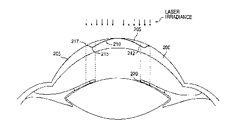
Turning now to the drawings, Fig. I illustrates a schematic side view of a
cornea 200 treated with the invention. The cornea 200 has an anterior surface
that
provides most of the refractive power of the eye. The initial anterior surface
205 of the
cornea 200 has been reshaped to a desired healed profile. The desired healed
profile
includes anterior optical surface 210 and anterior transition surface 215. The
anterior
CA 02306864 2000-04-19
WO 99/44492 PCT/US99/04764
optical surface 210 has a multifocal aspheric shape that corrects for near-
vision centrally
and far-vision peripherally.
While the present invention will often be described with reference to the
mitigation of presbyopia in combination with refractive hyperopia treatment,
it should be
5 understood that the benefits of the present invention are not limited to
these specific
procedures. These presbyopia treatment techniques may be used when no other
refractive
correction (other than the correction, mitigation, and/or inhibition of
presbyopia) is
desired, or the present treatment may be combined with therapies for one or
more of
myopia, astigmatism, irregular refractive aberrations, and the like, as well
as with
10 hyperopia. Still other aspects of the present invention, including methods
and systems
which accommodate and adjust for re-epithelization, may find uses in a broad
variety of
ophthalmic procedures.
The peripheral positioning of the far-vision correction advantageously
permit distance viewing when the pupil is dilated at night. Anterior
transition surface 215
is the anterior surface of the cornea that provides a gradual change in shape
between
anterior optical surface 210 and the portion of the cornea retaining the
initial anterior
surface 205. The outer boundary 212 of the anterior optical surface preferably
extends
entirely across, and is ideally substantially coextensive with, the pupil
which is bounded
by iris 220. The light rays passing through anterior transition surface 215 do
not
contribute to the image formed by anterior optical surface 210. Therefore,
anterior
transition surface 215 is desirably positioned outside the pupil. This
positioning of
anterior transition surface 215 causes the light rays passing through anterior
transition
surface 215 to be substantially occluded by iris 220. This occlusion improves
patient
vision because the light rays are blocked that do not contribute to image
formation, and
which would otherwise reduce the contrast of the image.
The optical correction effected by an ablative surgical procedure to the
cornea is derived from a change in the anterior corneal surface from an
initial anterior
surface 205 to post-operative anterior optical surface 210. The anterior
optical correction
is the post-operative anterior optical surface 210 minus the initial anterior
surface 205.
An ablation profile is a change in an exposed surface profile occurring
immediately after
the tissue removal process. Therefore, the ablation profile is the exposed
surface profile
immediately after the tissue removal process minus the initial exposed surface
profile. As
used herein, "ablated shape" can refer either to an ablation-induced change in
a surface
I II IMIIAI
CA 02306864 2007-05-25
11
topography on a surface of the cornea, or to the surface topography of the
cornea after
ablation. Similarly, "healed shape" can mean either a final corneal topography
once
healing is complete, or a change in the corneal topography from an initial
topography to a
final corneal topography once healing is complete. A healed shape differs
significantly
from an ablated shape when a difference between the two shapes is sufficient
to be
perceptible by a patient. Healing can refer either to an initial covering of
an ablated
surface contour or changes in a tissue structure of the cornea following an
initial covering
of an ablated surface contour.
The relationship of the ablated surface and the anterior corneal surface
overlying the ablated surface is shown in Fig. 2. Initial ablated surface 202
includes
ablated optical zone 211 and ablated transition zone 216. Ablated optical zone
211
includes ablated central optical zone 231 for the correction of near-vision,
ablated
peripheral optical zone 241 for the correction of far-vision, and ablated
intermediate
optical zone 236 for the correction of vision intermediate to near- and far-
vision. Ablated
central optical zone 231 is shaped to appropriately form anterior central
optical
surface 230 when ablated surface 202 is covered and cornea 200 is healed to
form
anterior optical surface 210. Ablated intermediate optical zone 236 is shaped
to form
anterior intermediate optical surface 235 when ablated surface 202 is covered
and
cornea 200 is healed. Ablated peripheral optical zone 241 is shaped to
appropriately form
anterior peripheral optical surface 240 when ablated surface 202 is covered
and
cornea 200 is healed. Ablated transition zone 216 is ablated to minimize the
effect of
corneal healing on anterior optical surface 210.
In one embodiment, covering of the ablated shape will cause the final
shape of anterior optical surface 210 of the anterior surface of cornea 200 to
be different
from ablated optical zone 211. This aspect of the present invention is more
fully
described in the publication entitled "Corneal Ablation Profilometry and Steep
Central
Islands," Journal of Refractive Surgery, Vol. 13, pp. 235-45, 1997.
Initial ablated shape 202 is covered after the ablation. Proximity to ablated
transition zone 216 may cause anterior peripheral optical surface 240 to be a
different
shape than underlying ablated peripheral optical zone 241. However, anterior
central
optical surface 230 of anterior optical surface 210 is distant from ablated
transition
zone 216. Therefore the shape of anterior central optical surface 230 will
more closely
CA 02306864 2000-04-19
WO 99/44492 PCT/US99/04764
12
match the shape of ablated central optical zone 231. In one aspect, the
covering may
include regeneration of the epithelial layer following ablation of Bowman's
membrane
and adjacent stromal layers. In another aspect, covering includes replacing a
resected
portion of the cornea as is described in U.S. Patent No. 4,903,695, issued
February 27,
1990, for "Method and Apparatus for Performing a Keratomileusis or the Like
Operation." In this aspect, the resected portion includes an epithelial layer.
In a yet
further aspect of covering, a tear film forms over the epithelial layer to
form the anterior
surface when cornea 200 is fully healed. The final shape of anterior optical
surface 210
will substantially determine the optical properties of the cornea. Therefore,
it may be
desirable to ablate cornea 200 to form ablated optical zone 211 that is a
different shape
than the shape of anterior optical surface 210.
In another embodiment, ablated optical zone 211 includes ablated central
optical zone 231 and ablated peripheral optical zone 241. Ablated intermediate
optical
zone 241 may be replaced by extending ablated peripheral optical zone 241 and
ablated
central optical zone 231 to border one another. Ablated central optical zone
231 provides
about 2.5 D of near-vision correction with a range from about 0.5 to 4 D,
preferably about
2 to 3 D and a diameter from about 1.0 to 3.5 mm and preferably from about 2
to 3 mm.
Ablated peripheral optical zone 241 is ablated to provide far-vision
correction and is sized
to extend radially outward from the outer boundary of ablated central optical
zone 231 to
a diameter of about 5 mm with a range from about 3 to 7 mm and preferably from
about 4
to 6 mm. Ablated transition zone 216 extends radially outward from the outer
boundary
of ablated optical zone 211 to a diameter of about 9 mm with a range from
about 6 to
11 mm and preferably from about 7 to 10 mm. Covering of ablated optical zone
211 will
cause anterior intermediate optical surface 240 to form over the border
between ablated
central optical zone 231 and ablated peripheral optical zone 241. Anterior
central optical
surface 230 will form over ablated central optical zone 231. Anterior
peripheral optical
surface 240 will form over ablated peripheral optical zone 241. Therefore,
anterior
optical surface 210 may be formed as a multifocal aspheric surface on cornea
200 by
ablating only two optical zones within ablated optical zone 211.
An illustrative plot of the relative refractive power of anterior optical
surface 210 as a function of radial position across the pupil is shown in Fig.
3. The
refractive power decreases from the center toward the periphery. Anterior
central optical
surface 230 of cornea 200 has a relative refractive power from about I to 4 D,
and
CA 02306864 2000-04-19
WO 99/44492 PCT/US99/04764
13
preferably from about 2 and 3 D that corrects for near-vision. This central
surface ranges
from about 1 to 3 mm in diameter and preferably from about 1.5 to 2.5 mm in
diameter.
Anterior peripheral optical surface 240 corrects for far-vision. This
peripheral surface has
an inner boundary from about 2 and 4 mm in diameter and an outer boundary 212
that
may be scaled to match the outer boundary of the pupil as shown in Fig. 1.
Outer
boundary 212 may be scaled to a diameter of between about 3 and 7 mm. Anterior
intermediate optical surface 235 has continuously varying refractive power.
This region
is desirable and provides focus for objects appropriately positioned
intermediate to near
and far positions.
In an exemplary embodiment, ablated central optical zone 231, ablated
intermediate optical zone, 236 and ablated peripheral optical zone 241 are
scaled to match
a dimension of the pupil. The scaling dimensions may be an area of the pupil,
a diameter
of the pupil, a radius, or the like. For example, ablated optical zone 211 may
be
decreased by about 20% from a diameter of about 5 mm to 4 mm for a patient
with a
4 mm diameter pupil. In this case, ablated central optical zone 231, ablated
intermediate
optical zone 236 and ablated peripheral optical zone 241 are each decreased by
about
20%. This scaling is desirable because it keeps the ratios of near,
intermediate and far-
vision nearly constant for varying pupil size. The inner boundary of ablated
transition
zone 216 is scaled to border the outer boundary of ablated optical zone 211.
During the
scaling of ablated optical zone 211, the outer boundary of ablated transition
zone 216 may
be scaled to match the scaling of ablated optical zone 211. Alternatively, the
outer
boundary of ablated transition zone 216 may be fixed to a constant value while
the inner
boundary of ablated transition zone 216 is varied.
Fig. 4 illustrates a block diagram of an ophthalmic surgery system for
incorporating the invention. As seen in this Figure, a personal computer (PC)
work
station 10 is coupled to an embedded computer 21 of a laser surgery unit 20 by
means of
a first bus connection 11. The PC work station 10 comprises a tangible medium
12 and a
treatment table 14. The laser treatment table 14 includes a listing of
coordinate references
of the laser beam during an ablation of the cornea. The sub-components of
laser surgery
unit 20 are known components and preferably comprise the elements of the VISX
STARTM EXCIMER LASER SYSTEM and of the STAR S2 TM System available from
VISX, Incorporated of Santa Clara, California. Thus, the laser surgery system
20
includes a plurality of sensors generally designated with reference numeral 22
which
CA 02306864 2000-04-19
WO 99/44492 PCT/US99/04764
14
produce feedback signals from the movable mechanical and optical components in
the -
laser optical system, such as the elements driven by an iris motor 23, an
image rotator 24,
an astigmatism motor 25 and an astigmatism angle motor 26. The feedback
signals from
sensors 22 are provided via appropriate signal conductors to the embedded
computer 21.
The embedded computer 21 controls the operation of the motor drivers generally
designated with reference numeral 27 for operating the elements 23-26. In
addition,
embedded computer 21 controls the operation of the excimer laser 28, which is
preferably
an argon-fluorine laser with a 193 nanometer wavelength output designed to
provide
feedback stabilized fluence of 160 mJoules per square centimeter at the cornea
of the
patient's eye 30 via the delivery system optics generally designated with
reference
numeral 29 and shown in Fig. 9. Other lasers having a suitable wavelength may
be used
to make an ablative energy for removing a tissue from the eye. For example,
solid state
lasers such as a yittrium aluminum garnet (YAG) laser producing a fifth
harmonic of a
fundamental wavelength may be used to generate an ablative energy. Other
ancillary
components of the laser surgery system 20 which are not necessary to an
understanding of
the invention, such as a high resolution microscope, a video monitor for the
microscope, a
patient eye retention system, and an ablation effluent evacuator/filter, as
well as the gas
delivery system, have been omitted to avoid prolixity. Similarly, the
keyboard, display,
and conventional PC subsystem components (e.g., flexible and hard disk drives,
memory
boards and the like) have been omitted from the depiction of the PC work
station 10. If
desired, embedded computer 21 may be constructed with PC work station
components
and built into laser surgery system 20. In this case embedded computer 21 may
supplant
PC workstation 10.
The iris motor 23 is used to control the diameter of a variable diameter iris
schematically depicted in Fig. 5. The astigmatism motor 25 is used to control
the
separation distance between a pair of cylinder blades 35, 36 which are mounted
on a
platform 38 for bi-directional translational motion in the direction of arrows
40, 41.
Platform 38 is rotatably mounted on a second platform (not illustrated) and is
rotationally
driven by astigmatism angle motor 26 in a conventional way in order to enable
alignment
of the slit axis (illustrated in a vertical orientation in Fig. 5) with the
appropriate
coordinate axes of the patient's eye. Iris 32 is driven by iris motor 23 in a
known way to
change the diameter of the iris opening from a fully opened position (the
position
illustrated in Fig. 5) to a fully closed position in which the aperture is
closed to a
l i I I I II I
CA 02306864 2007-05-25
minimum diameter of 0.8 mm. It is understood that the variable diameter iris
32 and the
cylinder blades 35, 36 are positioned with respect to the output of laser 28
in such a
manner as to intercept the beam prior to irradiation of the corneal surface of
the patient's
eye 30. For the purpose of this application, it may be assumed that iris 32
and cylinder
5 blades 35, 36 are part of the delivery system optics subunit 29 shown in
Fig. 4.
The system of Figs. 4 and 5 is used according to the invention to effect
presbyopic, hyperopic, myopic, astigmatic, and other error corrections to the
anterior
surface of the cornea, to provide a smooth transition zone between the outer
edge of the
optical zone and the untreated surface of the cornea, and to effect surface
smoothing
10 when desired. Other techniques besides the above area profiling of a laser
beam may be
used to profile the laser beam to a desired size and energy distribution on
the surface of
the eye. For example a lens may be used to profile a beam exiting from an
aperture by
focusing the beam to a suitably small area and desired energy profile as
described in U.S.
Patent 4,718,418. Also a diffractive optic may be used to adjust an energy
profile of the
15 laser beam on the surface of the eye as described in co-pending application
entitled
"Laser Delivery System and Method with Diffractive Optic Beam Integration",
PCT
Publication No. WO 99/039410 filed on January 20, 1999.
With reference to Fig. 6, an imaging lens 51 is laterally offset from an
axis 52 by a variable amount in the manner set forth more fully below. Lens 51
preferably comprises the existing imaging lens found in the delivery system
optics 29 of
the Fig. 4 system. Axis 52 is the axis corresponding to the center of rotation
of lens 51.
Displacing lens 51 by translating the lens in a radial direction off the axis
52, which may
or may not correspond to the laser beam axis, displaces the image 54 of
aperture 53 in a
related manner. By also rotating lens 51 about the axis 52 in an eccentric
fashion, as
illustrated in Fig. 7, the displaced image 54 of aperture 53 can be scanned
about axis 52.
This scanning is along a preselected path, which in the hyperopic correction
procedure
described below is an annular path about the axis 52. Depending upon the
manner in
which the lens offset, lens rotation, slit width, slit rotation and iris
diameter are controlled,
various types of ablation corrections can be effected. These corrections
include
presbyopia correction, hyperopic error corrections, hyperopic astigmatism
corrections,
and other vision error corrections, along with simultaneous or successive edge
contouring
to form a smooth transition zone.
INIIF
CA 02306864 2007-05-25
16
Fig. 8 illustrates the aperture positioning relative to the intended ablation
center when employing the variable diameter iris 32 and cylinder blades 35, 36
of Fig. 5
to effect a refractive error correction. In this Figure, R2 represents the
half width of the
slit between blades 35, 36, Ri is the radius of the iris 32, r is the radius
of a circle covered
by the aperture, s is the radial offset of the center of the image of the slit
aperture relative
to the center of rotation 52, and 0 is the half angle for which the circle of
radius r is
covered by the aperture. The intended ablated optical zone is the central
region bounded
by circle 61 and the intended ablated transition zone is the annular region
bounded by
circles 61 and 62.
The manner in which the slit width and diameter are varied by the
computer depends upon the type of vision correction desired. For a hyperopic
correction,
a fixed value of the refractive correction may be used to generate the cut
profile C(r). For
a hyperopic refractive correction of a given fixed value, the sequencing of
the aperture is
done in such a manner as to satisfy the hyperopic lens equations described in
"Photorefractive Keratectomy: A Technique for Laser Refractive Surgery"
authored by
Munnerlyn et al., J. Cataract Refract. Surg. Vol. 18, pages 46-52 (Jan.,
1988). Also,
European Patent Office publication number EP 0 628 298 Al, published December
14,
1994, discloses an aperture sequencing for correcting hyperopia.
For the correction of presbyopia, it may be desirable to vary the refractive
power across the ablated surface. The cut profile C(r) may be calculated by
calculating
the incremental cut profiles along the surface. The incremental cut profiles
are then
summed to calculate the overall cut profile C(r). The incremental cut profiles
may be
calculated using the above hyperopic lens equation, the desired ablated
refractive
correction, and the position from the center of the aspheric lens.
The cut profile is given by the equation:
C(r) _ (d / ir)E; (n;9(r)) (1)
where n; is the number of laser pulses for the ith aperture in a sequence of
aperture
dimensions and radial positions, and d is the amount of material removed with
each laser
pulse or a scaling factor which also takes into account corneal healing. Once
the cut
profile has been calculated, the sequence of aperture dimensions and pulses
may be
,IIIIõ
CA 02306864 2000-04-19
WO 99/44492 PCT/US99/04764
17
calculated. The sequence of aperture dimensions is created by control of the
width of the
slit and the diameter of iris 32 throughout the surgical procedure. The
sequence of
aperture dimensions and positions are preferably incorporated into a laser
treatment table.
The sequence of aperture dimensions may also be tailored to accommodate
variations in the ablation profiles of individual pulses from the laser beam.
For example,
the spatial variation of tissue ablation may cause the geometry of tissue
ablated with a
single laser pulse to be deeper at the edges of an ablation adjacent the image
of iris 32 and
cylinder blades 35 and 36. For an individual laser pulse, this increased
ablation depth
near the edge of an ablation may be 50% greater than the central ablation
depth.
Therefore, a 4D intended hyperopic ablation that assumes a uniform layer of
tissue is
removed with each laser pulse will ablate about 6 D of correction near the
center of
ablated optical zone 211. Clinically, the inventors have observed that
patients treated
with the above ablation algorithm for 3 to 4 D of hyperopia have also been
successfully
treated for presbyopia. However, with a +2D correction, the correction of
presbyopia is
only partial. Therefore to correct presbyopia and hyperopia, it may be
desirable to
combine the +2 D correction with an aspheric ablation. In this case, the
aspheric
correction is about one half of the aspheric correction that would be ablated
on an eye
with no refractive error.
Preferably, the refractive correction of cut profile C(r) is scaled to match a
dimension of the pupil. This scaling may be achieved by appropriately varying
the
refractive correction entered into the hyperopic lens equation. For example,
consider the
scaling of an ablation for a 5 mm pupil compared to a 4 mm pupil. If the
aspheric surface
includes a 1.5D ablated curvature 1.25 mm from the aspheric lens center for
the 5 mm
pupil, this 1.5D curvature will be ablated 1.0 mm from the aspheric lens
center on a 4 mm
pupil. This scaling maintains a balance of near and far-vision correction by
accommodating individual variability in pupil size. By scaling the cut profile
C(r ), the
scaling of the ablated optical zone is incorporated into the laser treatment
table.
For the example shown in Fig. 8, the values of s and R2 are varied to
produce the correct value of radial offset (s) and slit width (2 x R2) so that
the inner edge
of blade 35 is moved in steps from close to the center of the ablation
(starting at
approximately 0.6 mm from the center) to the edge of the corrected optical
zone at
approximately 2.5 mm. R, (the iris radius) is fixed at a predetermined value
(3 m in one
specific procedure), and s and R2 are chosen to anchor the edge of the
ablation at the
I I I M III 1
CA 02306864 2007-05-25
18
outer edge of the intended transition zone of approximately 5 mm radius. The
number of
pulses for each successive position of the inner edge is calculated to give
the desired
depth from the hyperopic lens equation. For a procedure requiring the least
number of
pulses, the treatment is ended as soon as the inner edge of the aperture
reaches the
boundary of the corrected optical zone. Initially, the slit width is set to a
maximum value
and the imaging lens 51 is positioned laterally of the axis of rotation 52
such that the
inner slit edge is positioned at the minimum distance from the center of the
optical zone
and the intersections of the iris diaphragm 32 and the outer slit edge are
positioned over
the outer edge of the intended transition zone.
The image of the aperture is now ready to be scanned over the anterior
surface of the cornea. While several different scanning sequences are
possible, the
following sequence has been actually implemented with effective results. The
radial
position along the optical zone is broken into a series of discrete,
equidistant (typically
0.1 mm apart) nodes. The number of pulses required to ablate tissue to cut
depth C(r) at a
node adjacent to the edge of the inner slit is calculated using
n = (71 * bC(rn)/ ei (rn) * d)
where n is the number of pulses, SC(rõ) is the difference between the actual
ablation depth
from previous pulses and the desired ablation depth at the node, O (r,,) is
the half angle
coverage of the aperture at rõ as previously defined. The radial ablation
profile from
previous pulses is calculated by summing the ablation depth from previous
positions and
pulses at each node as described by equation I. For the initial position,
SC(rn) = C(r). The
number of pulses required for each subsequent node is calculated for each node
adjacent
to the inner cylinder blade as the blade moves toward the edge of the optical
zone.
Having determined the correct number of pulses at each node, the treatment
must be
smoothed rotationally to ensure that it is correct and free from aberrations.
Fig. 9 is a schematic view of the delivery system optics in an embodiment.
As seen in this Fig., the beam from laser 28 is reflected by a first mirror 71
and a second
mirror 72, and enters a spatial integrator 73, where the beam is modified in
cross-section.
A diffractive optic may be used to modify a cross section of the laser beam as
described
in PCT Publication No. WO 99/039410, entitled "Laser Delivery System and
Method
with Diffractive Optic Beam Integration". The modified
I . 1. 1eIII-' -
II I, 1141
CA 02306864 2007-05-25
19
beam exiting from spatial integrator 73 is reflected by minors 74 and 75 and
passed
through a dove prism 76 to the iris/slit mechanism 78 which contains the
variable width
slit and variable diameter iris described above. The profiled beam exiting
from the unit
78 is reflected by a mirror 79 and enters the image offset control unit 80
which contains
imaging lens 51. The offset profiled image exiting from unit 80 is reflected
from a
mirror 82 onto the patient's eye. To smooth out fluctuations in beam energy
across the
beam area, dove prism 76 is rotatably mounted, and is typically rotated during
beam
generation either continuously or between pulses.
The invention affords great flexibility in performing various types of
corrections by virtue of the fact that the system can be programmed to
accommodate
patients having differently sized physical eye parameters and refractive and
presbyopic
correction requirements. The variable slit width/variable diameter iris
arrangement is
particularly adaptable for use in the simultaneous treatment of presbyopia,
hyperopia,
hyperopic astigmatism and irregular refractive aberrations. For simultaneous
treatment of
presbyopia, hyperopia and hyperopic astigmatism, the ablation geometry is
solved as a
function of radial displacement and angular position of the aperture image
about the
rotational center. Further, in all procedures requiring a smoothing of the
transition zone
at the periphery of the ablation zone, the diameter of the iris is varied over
a
predetermined range along with the slit width variation. For presbyopia and
refractive
aberrations, a device such as a spatially resolved refractometer or a
topography machine
or both may be used to map the irregular surface contour of the cornea to
determine the
exact surface corrections required. Thereafter, the slit width and the iris
diameter can be
programmed such that corneal sculpting will achieve the desired aspheric
surface
geometry on a healed cornea. Alternatively, a wavefront sensor may be used to
map the
irregular refractive aberrations of the eye. One suitable embodiment of such a
wavefront
sensor is the Hartmann-Shack sensor described in U.S. Patent No. 5,777,719.
For any of the above specific correction procedures, a treatment table is
normally constructed. The treatment table contains the value of all of the
discrete radial
and angular positions of the optomechanical elements used to scan the image
over the
relevant portion of the anterior corneal surface. This table also contains the
number of
laser pulses per position. A typical treatment table contains on the order of
about 500
different entries.
l l loll,.
CA 02306864 2000-04-19
WO 99/44492 PCTIUS99/04764
The treatment table for a given procedure may incorporate special features
designed to improve the efficiency of the procedure. For example, for some
procedures
(e.g., simultaneous hyperopic and presbyopic correction) it can be beneficial
to leave a
small zone centered on the optical zone untreated. This can be done by
constraining
5 motion of the inner cylinder blade to guarantee occlusion in the small zone
of interest.
The diameter of the untreated zone varies from about 0.1 to 1.5 mm, is
preferably from
about 0.5 to 1.0 mm and is ideally about 0.7 to 0.9 mm. Also, standard tables
can be
constructed for a specific procedure-e.g., hyperopic correction-to different
Dioptric
correction values, and these standard tables can be sorted and combined to
perform
10 multiple repetitions of one or more standard tables to effect a given
Dioptric correction.
For example, standard tables may be created for a myopic correction for values
of 1/4, 1/2
and 1 Diopter. Using these tables, a 3.75 Diopter correction would proceed by
performing the standard 1 Diopter correction three times, followed by the 1/2
Diopter
correction and the 1/4 Diopter correction.
15 While the invention has been described above with specific reference to
ablation of an anterior corneal surface, various portions of the cornea may
also be treated
using the invention. For example, the epithelium may be mechanically removed
by
scraping, as is typically done in photorefractive keratectomy, and the exposed
surface
may be ablated. Further, the invention can also be used for laser
keratomileusis of
20 corneal lamella removed from the cornea. This procedure is described in
U.S. Patent
No. 4,903,695, issued February 27, 1990, for "Method and Apparatus for
Performing a
Keratomileusis or the Like Operation."
In applying the invention to this procedure, a flap of corneal tissue is
physically removed (either fully or partially) from the cornea, the size of
the removed
portion typically lying in the range from about 8 to 10 mm wide and a variable
thickness
up to 400 microns. This flap of tissue is typically removed using a
microkeratome. Next,
the flap is placed in a suitable fixture - typically an element having a
concave surface -
with the anterior surface face down. Thereafter, the required ablation is
performed on the
reverse exposed surface of the flap, after which the ablated flap is
repositioned on the
cornea. Alternatively, after the flap is removed from the cornea, the exposed
stromal
tissue of the eye can be ablated according to the invention, after which the
flap is
reattached over the freshly ablated stromal tissue.
CA 02306864 2000-04-19
WO 99/44492 PCT/US99/04764
21
The technique of shaping a cornea is further illustrated in Figs. 10-15.
These figures illustrate measured ablation profiles, intended optical
corrections and
measured anterior corneal surface optical corrections. The effect of the
spatial variance
of ablation on ablation shape is illustrated in Fig. 10. A measured ablation
shape is
plotted as a function of radial position over the ablated optical zone. This
figure
illustrates an ablated optical zone using an ablation algorithm that assumes a
uniform
layer of tissue is removed with each laser beam pulse. The intended optical
correction is a
+3 D optical correction 410. However, illustrated ablated optical zone 420 is
significantly different. The ablated optical zone 420 is overcorrected by
about 100% in
the central ablation zone 422. The ablated optical zone 420 is over corrected
in the
peripheral ablation zone 424 by about 60%. The initial shape of ablated
optical zone 420
differs significantly from the healed anterior surface shape, and the healed
shape
substantially corrects the initial hyperopic refractive error of the eye.
The covering and healing of the ablated surface decrease the difference
between the intended optical correction and the anterior corneal surface
optical correction
as illustrated in Fig. 11. A measured anterior corneal surface optical
correction is plotted
as a function of radial position over an ablated optical zone. The anterior
optical
correction 430 of the healed cornea more closely matches the intended +3 D
spherical
optical correction 410. However, errors between the intended optical
correction 410 and
the anterior surface optical correction 430 are still present. The central
anterior optical
correction 432 is over corrected compared to the intended +3 D spherical
optical
correction 410. This over correction of the central optical correction 432 is
by about 25%
relative to the intended +3 D optical correction, and corresponds to a 0.75 D
near-vision
correction at 2 mm. However, the peripheral anterior optical correction 434 is
slightly
under corrected relative to the intended +3 D optical correction. This
correction of the
peripheral anterior optical correction 434 appropriately provides distance
vision
correction. Therefore, the anterior optical correction 430 is multifocal and
will provide
some correction of presbyopia. This multifocal effect occurs because the
ablated shape
compensates for changes in corneal shape as the cornea heals. The peripheral
ablated
optical zone is overcorrected to provide distance vision on a healed cornea.
The central
optical zone is overcorrected to provide near-vision on the healed cornea.
Commercially
available corneal topography systems measure healed anterior corneal surfaces.
CA 02306864 2000-04-19
WO 99/44492 PCT/US99/04764
22
Examples of such systems include the Atlas Corneal Topography SystemTM
available
from Humphrey Instruments of San Leandro, California and the PAR CTS SystemTM
available from PAR Vision Systems Corporation of New Hartford, New York.
The effect of the covering and corneal healing of an ablated optical zone is
illustrated in Fig. 12. This figure illustrates the difference in shape
between an ablated
shape and the final anterior optical correction on the anterior surface of the
cornea. This
difference in shape is described as a healing-induced change 440 shown in Fig.
12. The
healing-induced chan ge 440 is illustrated for a patient treated for +3 D of
hyperopia. The
ablated shape is partially filled in by covering and healing to form the
anterior optical
correction. However, this partial filling is not constant over the ablated
optical zone. The
center of the ablated optical zone shows less filling than the peripheral
optical zone. The
peripheral optical zone is filled in by about 50% while the central optical
zone is filled in
by about 30%. A peripheral filling 444 is greater than a central filling 442.
Proximity to
the ablated transition zone causes the peripheral optical surface to be a
different shape
than the underlying ablated peripheral optical zone. However, the anterior
central optical
surface is distant from the ablated transition zone. Therefore, the shape of
the anterior
central optical surface more closely matches the shape of the ablated central
optical zone.
With the above differential healing, an optical zone ablated to a
substantially spherical
shape for correcting hyperopia will heal to an aspheric shape that corrects
for presbyopia.
By estimating a healing-induced change, an initial ablated surface shape
may be derived from a desired anterior corneal surface shape and a healing-
induced
change as illustrated in Fig. 13. For example, consider a desired anterior
surface
correction 450 that corrects for +3 D of hyperopia and corrects for presbyopia
with a
central zone providing +3 D of near-vision correction. The desired anterior
surface
correction 450 is also illustrated in Fig. 13. An initial ablated surface
shape 460 is
calculated from the healing-induced change 440 and the desired anterior
surface
correction 450. The initial ablated shape 460 for the desired anterior surface
correction 450 is illustrated in Fig. 13. The initial ablated shape 460 is
overcorrected
relative to the desired anterior surface correction. The initial ablated shape
460 is
calculated by multiplying the desired anterior surface correction 450 by the
ratio of the
ablated shape 420 to the healed shape 430. A processor may be used to generate
the
ablated shape in response to the desired correction input by the system
operator, typically
CA 02306864 2000-04-19
WO 99/44492 PCT/US99/04764
23
making use of the embedded computer of the laser workstation, the PC
workstation,
and/or the programming and hardware of an external computer.
The ablated shape may be restricted or reduce relative to a desired anterior
surface correction to obtain the desired anterior surface correction. The
diameter of the
relative restriction is between about 0.1 and 2 mm, preferably between about
0.2 and
1 mm and is ideally between about 0.3 and 0.7 mm. In an exemplary embodiment
this
restriction is about 0.5 mm as illustrated in Fig. 14. After covering an
ablated corneal
surface feature (such as a presbyopia correction) and allowing healing of a
cornea, an
anterior surface correction may extend beyond the initial ablated dimensions
of the
ablated surface feature. Ablated central zone 470 on reference 480 includes
dimension 472 across the central ablated zone. Ablated central zone 470 also
includes
elevation 474 relative to the reference 480. Reference 480 may be any suitable
reference
such as a spherical reference surface on an anterior optical surface or an
ablated surface.
Covering of ablated central zone 470 and healing of the cornea will form a
central
anterior optical surface 490. Central anterior optical surface 490 includes
dimension 492
across the central anterior optical surface and elevation 494 relative to
reference 480. A
1.5 mm dimension 472 across the ablated central zone 470 will typically extend
to a
2 mm dimension 492 across the central anterior optical surface 490. Therefore,
to form a
2 mm central anterior optical surface, the ablated central zone is preferably
restricted by a
relative amount of about 0.5 mm. Also, it may be desirable to increase the
elevation 474
of the ablated feature by a relative amount as illustrated above. For example,
an ablation
intended to produce a 4 um surface elevation 494 relative to a reference 480
on the
anterior surface of a healed cornea may be over ablated as an 8 um surface
elevation 474
relative to a reference 480. This overcorrecting of the ablated feature is by
a relative
amount of 4 um. Relative over-correction ranges from about 1 to 25 um. A
desired final
2 mm diameter zone on an anterior surface to correct for near-vision with 3 D
might
typically have an elevation of about 4 um. To correct presbyopia using such a
healed
shape (in other words, to produce a central zone having a diameter of about 2
mm and an
elevation of about 4 um on the anterior surface of a healed cornea), a central
ablation
zone having a restricted diameter of about 1.5 mm and an overcorrected
elevation of
about 8 um is ablated onto an exposed surface of the cornea. Although the term
"diameter" is used to indicate a lateral dimension of these features (and in
general in this
application), it should be understood that the features need not necessarily
be circular.
CA 02306864 2000-04-19
WO 99/44492 PCTIUS99/04764
24
In some instances, it may be desirable to treat presbyopia by leaving a
central region of the optical zone untreated as illustrated in Fig. 15. A
small untreated
zone 500 centered on the optical zone 502 of an ablated cornea has a dimension
504
across the untreated zone. The untreated zone 504 is smoothed by covering and
healing
of the cornea and contributes to the formation of a central anterior optical
surface that
corrects presbyopia.
The above techniques can be used to calculate initial ablation shapes for
treating conditions besides hyperopia and presbyopia. These techniques may be
used to
calculate the initial ablation shapes used to treat astigmatism, myopia and
irregular
refractive aberrations of the eye. For example, the higher order aberration
terms of an
irregular refractive aberration may be over corrected on an ablated corneal
surface to form
an anterior surface on a healed cornea with a desired optical correction of
the higher order
aberrations.
The above technique of making a multifocal optical correction on the
anterior surface of the cornea can be applied to both eyes of a patient to
provide an
improved correction of presbyopia with binocular vision. The correction of
presbyopia
preferably covers about a 3 D range. However, with binocular vision this
approximately
3 D range of presbyopia correction may be treated by treating each eye with a
multifocal
optical correction having less than the full 3 D range of presbyopia
correction. In this
case, the average refraction of each of the two eyes is different to provide
clear vision
over the full 3 D range. A first eye is corrected for near-vision, and a
second eye is
corrected for distance vision. The multifocal anterior optical surface
provides improved
focus for objects intermediate to near and far-vision. For example, one eye is
treated to
have an average refraction of -0.75 D with a 1.5 D multifocal range of focus.
This eye
has an effective focus from 0 to -1.5 D. The other eye is treated to have an
average
refraction of about -2.25 D with a 1.5 D multifocal range of focus. This eye
has an
effective focus from about -1.5 D to -3 D. The effective range of focus of the
two eyes
combined is about 3 D. The multifocal range on each eye varies between about
0.5 and
2.0 D, and is preferably between about 1.0 and 1.5 D. The difference between
the
average refraction of the two eyes varies between about 0.5 and 2.5 D, and is
preferably
between about 1 and 2 D.
While the above provides a full and complete disclosure of the preferred
embodiments of the invention, various modifications, alternate constructions
and
CA 02306864 2000-04-19
WO 99/44492 PCT/US99/04764
equivalents may be employed as desired. For example, while the invention has
been
described with specific reference to the system of Figs. 4 through 9, other
systems may
be employed, as desired. Further, lasers of other appropriate wavelengths than
laser 28
may be used, if desired and effective. Also, laser systems which operate on
the principle
5 of thermal ablations, such as lasers having wavelengths lying in the
infrared portion of the
electromagnetic spectrum, may be used to implement the invention. In addition,
while
the radial and angular positioning of the profiled beam is accomplished with
imaging
lens 51 in the preferred embodiment, other optical scanning elements- such as
rotating
mirrors and prisms - may be employed, if desired. Therefore, the above
description and
10 illustrations should not be construed as limiting the invention, which is
defined by the
appended claims.