Note: Descriptions are shown in the official language in which they were submitted.
CA 02308200 2000-04-20.
WO 99120192 PCT/US98/22463
METHOD AND APPARATUS FOR LOCATING RESECTION AT THE DISTAL CONDYLAR FEMUR,
TIBIA, AND
PATELLA
.. CRO8S-REFERENCE TO RELATED APPLICATIONS
This application is a continuation-in-oars
application to United States latent Application Serial Nc.
09/049,781, (fled Marca 2';, 1998 entitled '~ETHCD r?ND
APPAR~.?'US FOR LOCAT=_dG SCNE ,.;:~_'S .'-_'=' ''_'uE D~S'~A:. . =:4GR.yL
_., CONDYLyS 'T_'O RECEI'?E ~ ~'=:'!ORAr. ?ROT:~ySiS W 1C 'T'J ,.~GRDi~I~:~'
TIBIAL AND PATELLAR RESECTION AND REPr:ACEMENT WITH FEMORAL
RESECTION AND REPLACEMENT, now pending, .which is a
continuation-in-part application to T~nited States Patent
Application Serial No. 081956,015, filed Oc~ober 22, 1°°;
~ entitled METHOD AND APPARATUS FOR LOCATING BONE CTJTS AT THE
DISTAL CONDYLAR FEMUR REGION TO RECEIVE A FEMORAL PROTHESIS
AND PROPERLY ARTTCULATED WITH PATELLAR AND TIBIAL
PROTHESIS, now pending, ;which is a continuation-in-part
application to United States Patent Application serial No.
20 08/455,985, filed May 31, 1995, entitled METHOD AND
APPARATUS FOR LOCATING BONE CUTS AT THE DISTAL CONDYLAR
FEMUR REGION TO RECEIVE a FEMORAL PROSTHESIS, now U.S.
Patent No. 5,776,137, issued July 7, 1998, the disclosures
of which are incorporated by reference herein.
SUBSTITUTE SHEET (RULE 26)
CA 02308200 2000-04-20
WO 99/20192 PCT/US98/22463
- 2 -
FIEhD OF THE INVENTION
The invention relates to methods and apparatus
for locating bone cuts on the medial and lateral femoral
5 condyles to form seating surfaces for a femoral knee
prosthesis, and to coordinate tibial and patellar resection
and replacement with femora resection and replacement.
The invention further relates to a tool for
locating said cuts.
10 BACKGROUND OF THE INVENTION
Over the years, the concepts of designs for the
total knee arthroplasty have evolved to the point where
with few exceptions, most are quite comparable in the
design of femoral, tibial and patellar prostheses.
15 Major discrepancies and problems encountered are
caused by physician error and failure to understand the
principles of more complex alignment or ligament problems
to be corrected at surgery. With the more complex
alignment or "routine" degenerative knee, the major
20 differences are the ease and consistency of instrumentation
for alignment and proper bone cuts allowing proper ligament
balance. This allows satisfactory motion and stability
post operatively.
SUBSTITUTE SHEET (RULE 26)
CA 02308200 2000-04-20
WO 99/20192 PCT/LJS98/22463
_ 3 _
The distal femoral cuts must be placed to provide
the knee prosthesis with a proper flexion and extension
gap, proper varus-valgus alignment, proper patellofemoral
relationship and proper rotation. It is customary to use
5 an intramedullary rod placed in a retrograde fashion
between the medial and lateral femoral condyles just
anterior to the intercondylar notch to establish a single
point of reference for subsequent bone cuts. A major
problem is in the instrumentation to indica~e the location
l0 of the femoral cuts wh?ch relies upon zhe ~'exoerience" or
"eyeballing" of the surgeon. Over the years, two basic
instrument system designs have become popular.
In one design (anterior referencing), the total
knee alignment system takes its point of reference from a
15 centrally placed rod and careful attention is given to the
pateilofemoral joint by using an anteriorl~a placed feeler
gage. The distal femoral cut is consistent with the
thickness of the prosthesis.
This instrument system operates on the principle
20 of anatomic anterior and distal femoral cuts to allow
proper ligament balancing and stability in extension as
well as consistent patellofemoral placement on the anterior
surface. The femur is not notched, and the anterior
surface of the femoral prosthesis not elevated above the
25 anterior surface of the femur. Notching the femur may
SUBSTITUTE SHEET (RULE 26)
CA 02308200 2000-04-20
WO 99/20192 PCT/US98/22463
- 4 -
cause a decrease in strength of the distal femur.
Elevation of the anterior surface of the prosthesis will
cause extremely high patellofemoral pressures in a joint
that seems to be prone to a high rate of post-operative
5 failure.
By establishing the anterior femoral cut as the
benchmark or datum starting point, however, the anterior
referencing instruments result in the installation of a
knee prosthesis which sacr_fices consistent stapilit,r in
flexion due to the formation of a aostericr femoral
condylar cut that may leave the posterior space either too
wide or too narrow. This can cause instability in flexion,
or restrict flexion and cause increased wear.
The second type of instrument design (posterior
15 referencing) is based on the concept that the flexion and
extension stability are more important and the
patellofemoral joint is of secondary importance. This
system also uses an intramedullary rod for referencing.
Although I consider all three joints as "important", when
20 a compromise must be made, the posterior ref erencing
systems compromise the patellofemoral joint while the
anterior reference systems sacrifice stability in flexion
(the posterior tibial femoral joint). Both systems
allegedly equally address the distal tibial-femoral space.
SUBSTITUTE SHEET (RULE 26)
CA 02308200 2000-04-20
WO 99/20192 PCT/US98/22463
- 5 -
Neither consistently addresses the distal rotation of the
femoral component.
Neither system tries to preserve the joint line
at or near an "anatomic" level. By elevating the
5 jointline, the patella is distalized. The femur is also
shortened. Since the arthritic knee often has a loss of
cartilage, there may be a patella infers of 2 - 3 mm
initially. Elevating the distal femoral resection beyond
this will:
l0 1) Further alter the patellofemoral
relationship.
2) Change the isometric and rotational balance
of the MCL and the LCL.
3) Shorten the femur in flexion and may cause
15 increased roll back, anterior lift off, and
increased posteromedial wear.
4) Elevate the level of tibial resection
necessitating a major amount of posterior
femoral resection to achieve a satisfactory
20 flexion space.
When performing a unicompartmental knee
replacement, it is imperative to maintain the jointline.
As a consequence, it is desirable to maintain a full range
of motion.
SUBSTITUTE SHEET (RULE 26)
CA 02308200 2000-04-20
WO 99120192 PCT/US98/22463
- 6 -
SUI~IARY OF THE 7GNVENTION
An object of the invention is to provide methods
and apparatus for locating bone cuts on the medial and
lateral femoral condyles to form seating surfaces for a
5 femoral knee prosthesis, and to coordinate tibial and
patellar resection and replacement with femoral resection
and replacement which reliably and anatomicaily provide:
1. Consistent distal tibio-femoral stability.
2. Consistent distal femoral rotation.
ZO 3. Consistent placement or' the anterior cut
flush with the anterior surface of the femoral cortex,
i.e., without notching or elevation.
4. Consistent placement of the posterior
femoral cut such that the distal and posterior cuts are
15 equal (when indicated) allowing for satisfactory extension
and flexion stability and motion.
The method and apparatus of the invention
contemplate placement of the anatomic joint line which, in
extreme cases, varies up to the difference between the
20 anterior-posterior A-P internal measurements of the size
prostheses. Based on my knowledge of total knee
replacement, personal experience with numerous routine
total knee replacements, numerous more complicated cases
consisting of knees with flexion deformities and revision
25 surgery, a maximum of a few mm proximal or distal
SUBSTITUTE SHEET (RULE 26)
CA 02308200 2000-04-20
WO 99/20192 PCT/US98/22463
7
displacement of the joint line is considerably less harmful
than:
1. A lax flexion gap;
2. Sloping the proximal tibial cut to
5 accommodate for an inconsistent posterior femoral condylar
cut;
3 . Signif icantly notching the femur anteriorly;
4. Raising the anterior flanges of the
prosthesis and thus one patel~ofemora' joint;
=0 5. Not a_'_owing lull extens:.on;
6. Raising the joint line;
7. Tightness in flexion;
8. Malrotation; and
9. Patient pain.
15 With an understanding of the measurements
involved in total knee replacement, a new instrument system
and methodology has been developed to allow flexion 120 -
130 degrees; to perform less soft tissue releasing; and
decrease surgical time. Starting with a "normal" knee, the
20 goal should be to maintain the anatomic landmarks as close
to normal as possible. Then, if deformities are present,
the procedure can be modified to accommodate the situation.
In accordance with the invention, a method is
provided for forming planar cuts on the medial and lateral
SUBSTITUTE SHEET (RULE 26)
CA 02308200 2000-04-20
WO 99/20192 PCT/US98/22463
_ g _
condyles of the femur to form seating surfaces to receive
a femoral knee prosthesis, comprising:
determining a prospective planar cut at the
posterior of the condyles of the femur at which the
5 distance between the anterior surface of the femoral cortex
and the prospective planar cuts is substantially equal to
the interior dimension of a knee prosthesis to be fitted on
said femur at the anterior surface and the cut planar
surf ace ,
10 determining the ~hickness of the poster'_or
lateral or medial condyle which will be resected by said
prospective planar cut,
cutting the distal ends of the condyles along a
plane at which the maximum thickness of resection of the
15 more prominent condyle at said distal end is substantially
equal to the thickness determined to be resected at the
posterior medial or lateral condyle by said prospective
planar cut, and
cutting the condyles along a plane substantially
20 flush with the anterior surface of the femoral cortex, and
along said prospective planar cut.
The method further contemplates loosely placing
a longitudinal intramedullary rod in the femur such that an
end of the rod projects from the femur, mounting a tool on
25 the projecting end of the rod, establishing, by said tool,
SUBSTITUTE SHEET (RULE 26)
CA 02308200 2000-04-20
WO 99/20192 PGTNS98122463
g _
an angular position of said prospective planar cut along a
plane rotated at an angle of between 0 and 15° with respect
to a tangential plane at the posterior of the lateral and
medial condyles about an axis located in said tangential
5 plane.
In further accordance with the method, the tool
is rotated with said rod through said angle and a datum or
benchmark is established by the rotated rod or by pins
installed in the condyles on the basis of the rotated
10 position of the tool. ~ cutting guide can be mounted on
said tool, to enable the distal end of the condyles to be
cut along said plane. Thereafter, the tool is removed and
a second A-P cutting guide is mounted on the selected
benchmark, i.e., the rod or the pins and the posterior and
15 anterior cuts are made. The axis about which the plane of
the prospective cut is rotated is located in said
tangential plane at the posterior surfaces of the medial
and lateral condyles and can be located at either of the
condyles or at any location therebetween. It is a feature
20 of the invention that the tool may remain on the rod both
for the measurements and for the cutting of the distal end
of the femur.
The invention also contemplates that the cutting
guide supports a means which enables the cutting guide to
SUBSTITUTE SHEET (RULE 26)
CA 02308200 2000-04-20
WO 99/20192 PC'f/US98/22463
- 10 -
be secured to the condyles during the cutting of the distal
ends of the condyles.
The invention further contemplates an apparatus
for forming planar resections on the medial and lateral
condyles of a femur to form seating surfaces to receive a
femoral prosthesis and to properly articulate with a tibial
and patellar prosthesis comprising:
a caliper feeler and measurement plate to measure
for the size of the femoral prosthesis to be received, said
caliper feeler and measurement plate adapted to determine
a first distance between an anterior surface of the femoral
cortex and a plane tangent to a posterior surface of the
medial and lateral condyles of a femur, the caliper feeler
referencing the anterior surface of the femoral cortex and
the measurement plate referencing the plane tangent to the
posterior surface of the medial and lateral condyles;
a graduated scale to compare the first distance
to at least two standard femoral prosthesis sizes and to
determine the smaller of the at least two standard femoral
prosthesis sizes;
a graduated scale to measure a second distance
between the first distance and the size of the smaller
standard femoral prosthesis size, so that a thickness or
thicknesses can be measured to be resected at the posterior
surface of the medial and lateral condyles of the femur by
SUBSTITUTE SHEET (RULE 26)
CA 02308200 2000-04-20
WO 99/20192 PCT/US98/22463
- 11 -
adding the average thickness of the posterior condyles of
the smaller standard femoral prosthesis and the second
distance;
a tool to resect the medial and lateral condyles
5 along a plane at the anterior surfaces thereof flush with
the anterior surface of the femoral cortex; and
a tool to resect distal ends of the medial and
lateral condyles at a resected thickness equal to the
average thickness of the distal condoles cf the smaller
l0 standard femoral prosthesis plus the second distance.
The apparatus further contemplates a tool to
resect the measured thickness at the posterior surface of
the medial and lateral condyles of the femur.
The invention also contemralates a method for
15 forming planar resections on the medial and lateral
condyles of a femur to form seating surfaces to receive a
femoral prosthesis and to properly articulate with a tibial
and patellar prosthesis comprises:
measuring for the size of the femoral prosthesis
20 to be received by determining a first distance between an
anterior surface of the femoral cortex and a plane tangent
to a posterior surface of the medial and lateral condyles
of a femur;
using a graduated scale to compare the first
25 distance to at least two standard femoral prosthesis sizes;
SUBSTITUTE SHEET (RULE 26)
CA 02308200 2000-04-20
WO 99/20192 PCT/US98/22463
- 12 -
measuring a second distance between the first
distance and the size of the smaller standard femoral
prosthesis size; and
measuring a thickness or thicknesses to be
5 resected at the posterior surface of the medial and lateral
condyles of the femur, the thickness being equal to the
average thickness of the posterior condyles of the smaller
standard femoral prosthesis plus the second distance.
The methcd further cante:~plates the steps of
10 resecting the medial and lateral condoles along a plane at
the anterior surfaces thereof substantially flush with the
anterior surface of the femoral cortex; and
measuring a thickness or thicknesses to be
resected at the distal ends of the medial and lateral
15 condyles, the thickness being equal to the average
thickness of the distal surface of the smaller standard
femoral prosthesis plus the second distance, and resecting
the distal ends of the medial and lateral condyles at the
measured thickness.
20 Other features and advantages of the present
invention will become apparent from the following
description of the invention which refers to the
accompanying drawings.
SUBSTITUTE SHEET (RULE 26)
CA 02308200 2000-04-20
WO 99/20192 PCT/US98/22463
- 13 -
HRIEF DF~SCRIPTION OF T8E DRAWINGS
Fig. 1 is a diagrammatic, lateral view of the
femur and tibia at a knee joint showing prospective cuts to
be made on the femur for installation of a femoral
5 prosthesis.
Fig. 2 is a diagrammatic illustration of the knee
joint of Fig. l seen anteriorly of the joint.
Fig. 3 is an end view from the distal end of the
femur of the knee joint.
10 Fig. 4 is a sectional wie~a of a femoral !nee
prosthesis adapted for placement on the femur after the
planar cuts have been made on the femur.
Fig. 5 is a side view similar to Fig. 1 in which
the tibia has been turned 90° to expose the distal end of
15 the femur, an intramedullary rod has been inserted into the
femur and a tool placed on the rod, the tool being partly
broken away and shown in section.
Fig. 6 is an end view of the tool taken in the
direction of arrow 6-6 in Fig. 5.
20 Fig. 7 is a sectional view taken on line 7-7 in
Fig. 6.
Fig. 8 is a broken, perspective view of a lower
caliper feeler of the tool.
SUBSTITUTE SHEET (RULE 26)
CA 02308200 2000-04-20
WO 99/20192 PC'T/US98/22463
- 14 -
Fig. 9 is similar to Fig. 6 and illustrates a
first stage in which the rod is angularly rotated by a
specific amount.
Fig. l0 is similar to Fig. 9 in a subsequent
5 stage.
Fig. 11 is an exploded view showing a cutting
guide to be installed on the tool.
Fig. 12 is a top, plan view showing the cutting
guide installed on the tool.
10 Fig. 1~ il'_ustrates the distal end of the femur
after the distal end has been cut and an AP cutting guide
has been placed on the rod.
Fig. 14 is an end view similar to Fig. 6 of a
second embodiment of the tool.
15 Fig. 15 shows the tool of Fig. 14 in a rotated
state.
Fig. 16 is an end view similar to Fig. 6 of a
third embodiment of the tool.
Fig. 17 shows the tool of Fig. 16 in a rotated
20 state.
Fig. 18 is an end view similar to Fig. 6 of a
fourth embodiment of the tool.
Fig. 19 shows the tool of Fig. 18 in a rotated
state.
SUBSTITUTE SHEET (RULE 26)
CA 02308200 2000-04-20
WO 99/20192 PCT/US98/22463
- 15 -
Fig. 20 is similar to Fig. 13 but shows a
modification adapted to the embodiment of Figs. 16 and 17.
Fig. 21 is an end view similar to Fig. 6 of a
fifth embodiment of the tool including an A-P measuring
5 guide.
Fig. 22 is a side view similar to Fig. 5 of the
tool of Fig. 21 mounted on the distal femur.
Fig. 23 is a top view of the tool of Fig. 21
mounted on the distal femur.
10 Fig. 24 is an exploded view of a d=stal cutting
block to be installed on the tool of Fig. 21.
Fig. 25 is a side view of the tool of Fig. 21
mounted on the femur installed with the distal cutting
block.
15 Fig. 26 is a top view of the tool of Fig. 25
mounted on the femur.
Fig. 27 is an enlarged fragmentary view of the
sliding scale of the distal cutting block.
Fig. 28 is a side view of the femur with the
20 distal cutting block mounted thereon.
Fig. 29 is a top view of the A-P cutting block
mounted on the distal femur.
Fig. 30 is a view similar to Fig. 13 illustrating
the distal end of the femur after the distal end has been
25 cut and the A-P cutting block has been mounted thereon.
SUBSTITUTE SHEET (RULE 26)
CA 02308200 2000-04-20
WO 99/20192 PCT/US98/22463
- 16 -
Fig. 31 is a top view of the A-P cutting block
mounted on the distal end of the femur after the distal end
has been cut and the A-P cutting block has been mounted
thereon.
5 Fig. 32 is a side view of the distal end of a
femur after it has been cut and a preferred prosthesis is
ready to be mounted thereon.
Fig. 3:; is a front view of the preferred
prosthesis to be used with the tool of Fig. 21.
10 Fig. 34 is an alternative embodiment of the tool
of Fig. 22, including posterior clips.
Fig. 34a is a rear perspective view of a
posterior clip of Fig. 34.
Fig. 34b is a front perspective view of a
15 posterior clip of Fig. 34.
Fig. 35 is a top view of the tool of Fig. 34.
Fig. 36 is a front view of the tool of Fig. 34.
Fig. 37 is rear view of the tool of Fig. 34.
Fig. 38 is a top view of the tool of Fig. 34
20 mounted on the femur installed with a distal femoral
cutting block and a distal femoral resection caliper.
Fig. 39 is a top view of the femur with the
distal femoral cutting block mounted thereon.
SUBSTITUTE SHEET (RULE 26)
CA 02308200 2000-04-20
WO 99/20192 PCT/US98/Z2463
- 17 -
Figs. 40 and 41 are perspective views of distal
femoral resection calipers for use in right and left
femurs.
Fig. 42 is a front view of a tibial resection
5 guide of the present invention mounted on a tibia.
Fig. 43 is a side view of the tibial resection
guide of Fig. 42.
Fig. 44 is a top view of the tibial resection
guide of Fig. 42.
10 Fig. 45 is a side view of a tibias external
resection guide of the present invention mounted on a
tibia.
Fig. 46 is a top view of a spacer of the present
invention.
15 Fig. 46a is an end view of the top extension
portion of the spacer of Fig. 46.
Fig. 46b is an end view of the bottom flexion top
extension portion of the spacer of Fig. 46.
Fig. 47 is a side view of the spacer of Fig. 46.
20 Fig. 48 is a front view of the knee space
including a spacer in flexion.
Fig. 49 is a front view of the knee space
including a spacer in extension.
SUBSTITUTE SHEET (RULE 26)
CA 02308200 2000-04-20
WO 99IZ019Z PCT/US98/2Z463
- 18 -
Fig. 50 is a side view of a tibial reresection
guide in accordance with the present invention mounted on
a tibia.
Fig. 51 is a front view of the tibial reresection
5 guide of Fig. 50.
Fig. 52 is a top view of a tibial reresection
guide of Fig. 50.
Fig. 53 is a partial side view of a patellar
clamp including a hinge feature in accordance with the
l0 present invention.
Fig. 53a is a partial top view of the hinge
feature of the patellar clamp of Fig. 53.
Fig. 54 is an end view of the patellar clamp of
Fig. 53 showing a scale to measure the patella thickness.
15 Fig. 55 is a side view of a patellar clamp in
accordance with the present invention without the hinge
feature shown reaming a patella.
Fig. 56 is a top view of the patellar clamp of
Fig. 55 reaming a patella.
20 Fig. 57 is a side partially broken away view of
a patella having a patella insert fitted therein.
Fig. 58 is a side view of an improved nail in
accordance with the present invention.
Fig. 59 is a side view of the tool of Fig. 34
25 mounted on a femur installed with a distal femoral cutting
SUBSTITUTE SHEET (RULE 26)
CA 02308200 2000-04-20
WO 99/20192 PCT/US98/22463
- 19 -
block and a distal femoral resection caliper of the present
invention, showing the nail of Fig. 53 being removed from
the femur in accordance with an improved slap hammer of the
present invention.
5 DETAILED DESCRIPTION OF THE II~1VENTION
When performing a unicompartmental knee
replacement, it is imperative to maintain the jointline at
or near anatomic level. ~s a consea_uence, t:~is maintains
a full range of :notion. The instrument system of the
10 present invention has been developed which combines the
advantages of anterior and posterior referencing systems to
maximize motion in a rebroducible fashion and can easilv be
incorporated into an operative protocol. with the
instrument system of the present inventicn, orthopedic
15 surgeons can reconstruct a knee and retain "anatomic"
landmarks. This makes it possible to deal with many of the
deformities confronting the orthopedic surgeon in an
arthritic knee.
There are three ways that joint surgeons can
20 insert a total knee replacement:
1) Resect the distal femur to accommodate the
thickness of the femoral prosthesis. Resect the proximal
tibia to accommodate the thickness of the tibial
prosthesis. This recreates any lost motion and requires
SUBSTITUTE SHEET (RULE 26)
CA 02308200 2000-04-20
WO 99/20192 PCT/US98/22463
- 20 -
major soft tissue releasing. The flexion and extension
resection spaces are not coordinated.
2) Rebuild the "normal" knee by compensating
for loss articular cartilage and bone in the measurements
5 for bony resection; then soft tissue releases can be
performed to accommodate the proper dimensions. This
places even greater demands on contracted soft tissues.
Although this may be most anatomically correct, it requires
such major tissue releases as to make it impractical.
10 3) Accent bony and articular cartilage loss.
Resect the amount of bone in flexion and extension to
accommodate full extension and as much flexion as deemed
necessary. This method relies on a coordinated resection
of the flexion-extension spacing. It relies on accurate
15 measurements to allow for the resection of bone and minor
soft tissue release. Within certain parameters, zn~s
method is preferable and can only be possible with better
instrumentation, such as the instrumentation of the present
invention.
20 Referring now to Fig. 1, the drawing
diagrammatically illustrates the femur 1 and tibia 2 of a
knee joint 3. The invention is concerned with the
placement of planar resections or cuts at the distal
condylar region 4 of the femur 1 to receive a femoral knee
25 prosthesis 5 (Fig. 4) . Typically, a total knee replacement
SUBSTITUTE SHEET (RULE 26)
CA 02308200 2000-04-20
WO 99/20192 PCT/US98I22463
- 21 -
also requires placing a planar cut at the proximal tibia of
the tibia 2 to receive a tibial prosthesis, not shown. The
tibial prosthesis typically consists of a tibial baseplate,
not shown, that is fitted on the proximal tibia after the
5 tibial cut is made, and an articular insert, not shown,
secured to the baseplate to articulate with the femoral
prosthesis 5.
The cut made on the tibia 2 and installation of
the tibial knee prosthesis should be as close to the
10 anatomic level as possible and should be substantial~.v
parallel to the floor in the mediolateral plane. This
maintains the joint line at or close to anatomic level.
Moreover, the angle of the proximal tibias resection should
correspond to the angle of the distal femoral resections
15 12. For example, the proximal tibias is in mild varus and
is resected such that the resection in the mediolateral
plane is parallel to the floor and oriented posteriorly
about 3°. Accordingly, the cuts made on the femoral
prosthesis, discussed below, must also take into account
20 this 3° mediolateral orientation in order to align the
femoral prosthesis with the tibias prosthesis as will be
explained later.
Assuming normal anatomy, it is also important
that the resected space medially in extension between the
25 tibia 2 and the femur 1 of the knee equals the combined
SUBSTITUTE SHEET (RULE 26)
CA 02308200 2000-04-20
WO 99/20192 PCT/US98/22463
- 22 -
thickness of the medial tibial prosthesis and the distal
medial femoral prosthesis; that the resected space
laterally in extension between the tibia 2 and the femur 1
of the knee equals the combined thickness of the lateral
5 tibial prosthesis and the distal lateral femoral
prosthesis; that the resected space medially in flexion
between the tibia 2 and the femur 1 of the knee equals the
combined thickness of the medial tibial prosthesis and the
posterior medial femoral prosthesis; that the resected
10 space laterally in flexion between the tibia 2 and the
femur 1 of the knee equals the combined thickness of the
lateral tibial prosthesis and the posterior lateral femoral
prosthesis; and that the resected space between the tibia
2 and the femur 1 of the knee in flexion must be equal to
15 or greater than the resected space between the tibia 2 and
the femur 1 of the knee laterally in extension, assuming
normal ligament balance.
The condylar region 4 of the femur 1 is formed
with a medial condyle 6 and a lateral condyle 7 separated
20 by an intercondylar notch 8. The femur 1 includes a shaft
9 forming the femoral cortex, the condylar region 4 being
at the distal end of the shaft 9.
In order to install the femoral knee prosthesis
5 on the distal condylar region 4 of the femur 1, three
25 planar cuts are made in the condylar region 4 to form
SUBSTITUTE SHEET (RULE 26)
CA 02308200 2000-04-20
WO 99/20192 PCTNS98/22463
- 23 -
seating surfaces for the prosthesis 5. These cuts consist
of an anterior cut 10, a posterior cut 11 and a distal end
cut 12. The placement of these cuts 10, il, 12 is crucial
to the installation of the prosthesis 5 and its effect on
5 the overall function of the prosthetic knee joint.
The invention is based on complying with the
following conditions.
i. Forming the planar cut l0 at the anterior
surface of the femoral condylar region flush with the
10 anterior surface 13 of the Femoral cor~°_Y SO 3S to form a
continuous surface therewith free of formation of either a
notch or elevation at the juncture of cut 10 and surface
13.
2. Forming the planar cut 11 at the posterior
15 surface of the femoral condylar region at a distance D from
planar cut 10 equal to the interior dimension S between the
anterior and posterior mounting surfaces 14, 15 of the
prosthesis 5. The dimension S is the so-called A-P
distance of the prosthesis and this distance varies for
20 different size prostheses. For example, prosthesis are
categorized as small, small(+), medium, large, large(+) and
extra large and the A-P distance increases in proportion to
the size increase.
With reference to Fig. 3, therein is seen a plane
25 T tangential to the medial and lateral condyles at the
SUBSTITUTE SHEET (RULE 26)
CA 02308200 2000-04-20
WO 99120192 PCT/US98122463
- 24 -
posterior surface 16 of the condylar region. The planar
cut 11 is made at an angle A, with respect to plane T to
angularly align the femoral prosthesis with the tibial
prosthesis. Normally, the angle would be 3° to match the
5 angle of the tibial prosthesis, however, due to anatomical
conditions of the patient such as wear of the medial or
lateral condyles posteriorly the angle ~ can vary
substantially, generally between 0 and 15 ° . The planar cut
11 wily result in resec~ion of bone of a thickness at
10 the medial condyle 5 and a thickness t, at tae ?atsral
condyle. The thickness t3 is usually less than t, and
controls the location of planar cut 11 so that a minimum
thickness of bone is resected at the posterior surfaces of
the condyles. In this regard, the thickness t, is
15 established as the difference between distance D' between
the anterior surface 13 of the femoral cortex and a plane
P tangent to the posterior surface of the lateral condyle
7 and parallel to planar cut 11 and distance D between the
anterior surface of the femoral cortex 13 and planar cut
20 11.
The thickness t3 and the location of the
prospective planar cut 11 therefore can be established
based on measurement of the distance D and the A/P
dimension of the selected size of the prosthesis. The size
25 of the prosthesis is determined .on the basis of the
SUBSTITUTE SHEET (RULE 26)
CA 02308200 2000-04-20
WO 99/20192 PCT/US98/22463
- 25 -
measurement of the distance D' and in general, the
prosthesis size will be selected so that the thickness t3
falls within a relatively narrow range, generally at least
6 mm and between 6 and 11 mm. The resected thickness of
5 bone t, and t3 at the medial and lateral condyles are
generally unequal.
The distal end cut 12 is made so that the maximum
thickness t~ of bone resected at the distal end is
substantially equal to t" i . e. , the maximum thickness t= of
10 bone resected at the more or~minent ccndyl~ a~ the distal
end (the medial condyle 6 in Fig. 2) is equal to the
minimum thickness t~ of bone resected at the posterior
surface.
Referring now to Fig. 5, in order to establish
15 the precise positions of the three planar cuts 10, 11, 12
to be made on the femur 1, a.referencing or datum system is
utilized which in the description herein is in the form of
an intramedullary rod 20 installed in a bore 21 formed in
the femur 1. The use of the intramedullary rod 20 as a
20 benchmark or datum is known in the art and is illustrated
herein by way of example. Other referencing or datum
systems can be employed as well, for example, utilizing two
pins placed in the condyles as set distance below the
anterior femoral cut to position an AP cutting guide
25 thereon. This will be described later.
SUBSTITUTE SHEET (RULE 26)
CA 02308200 2000-04-20
WO 99/20192 PCTNS98/22463
- 26 -
The bore 21, which is approximately 8 mm in
diameter, is formed longitudinally in the shaft 9 and in
the condylar region 4 of the femur 1 at a location which is
slightly anterior and medial of the intercondylar notch 8.
5 The rod 20 has a cylindrical portion 22 which snugly fits
in the bore 21 but is able to be rotated in the bore 2i.
The rod 20 may include radial flutes 23 extending outwardly
a distance slightly greater than the diameter of the bore
21. The Mutes 23 are initiailv outside the bore 21 and
10 are intended t:~ be driven :nzo the bore __ tc f=xedlw
secure the rod 20 in the bore 21. rFOr this purpose, the
flutes 23 are tapered to facilitate driving them into the
bore 21 and grip the bore tightly in the distal femur 1
when driven therein. The outer ends of the flutes 23 can
15 be saw-tooth or jagged as shown in Fig. 7 ~o provide a
resilient gripping action.
The rod 20 includes an adjunct end or stub 24
which is non-circular in cross-section. The stub 24 may
extend at an angle with respect to the longitudinal axis of
20 the rest of the shaft so as to be substantially
perpendicular to the joint and the prospective distal end
cut 12 and parallel to the weight bearing axis of the leg.
Shafts having stubs with different angles varying about 5-
7° may be provided and selection is made on the sex,
SUBSTITUTE SHEET (RULE 26)
CA 02308200 2000-04-20
WO 99/20192 PCT/US98/22463
- 27 -
anatomical condition, and other conditions of the patient.
This is conventional in prior usage.
The angular position of the non-circular stub 24
in bore 21 when the flutes 23 are driven into the bore 21
5 is a measure of the angle A at which the posterior and
anterior cuts 11, 10 are made and, consequently, of the
angular position of the knee prosthesis 5 on the femur 1
relative to the weight bearing axis of the leg.
The anatomical conditions governing the angular
10 position of the rod 20 in the bore 21 is based on anatomy
to maintain a straight line between the hip joint or the
center of the femoral head in neutral rotation, the center
of the knee joint and the midmedial third of the tibial
plafond.
15 If the rod 20 initially assumes an angular
position parallel to plane T, the rod is rotated by angle
A to reach its datum position from which the cuts 10, il,
12 will eventually be made. Nominally, the rotation is at
an angle 3° to match the angle of the tibia prosthesis.
20 However, due to wear of the condyles, and anatomical
conditions of the patient the rotation of the rod must be
varied from 3° to match the tibia prosthesis. The surgeon
is readily able to estimate this angle based on the anatomy
and on X-rays of the patient. Heretofore, however, the
25 surgeon had to estimate the angle at which to set the rod
SUBSTITUTE SHEET (RULE 26)
CA 02308200 2000-04-20
WO 99/20192 PCT/US98/22463
- 28 -
20 when the rod is driven into the bore 21. An imprecise
estimate of the rotational orientation of the stub 24 can
lead to angulation and placement errors of the prosthesis.
Stated succinctly, the estimate of the surgeon of the
5 angulation of cut 11 based on patient anatomy is accurate,
but the "eyeballing" of the rotational position of the stub
is often inaccurate.
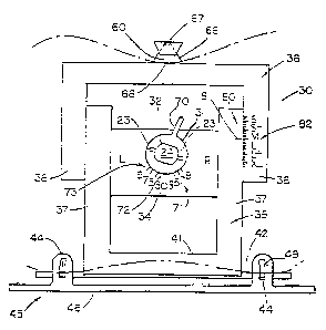
The invention provides a tool or instrument 30
which is fitted on the stub 24 of rod 20 and accurately
10 establishes rotation of the rod 20 when it is driven into
the bore 21 and which measures the distance D' which in
turn will determine the location of the planar cuts 10, 11,
12.
The tool 30 includes a sleeve 31 having a
15 circular-like bore 32 of the same shape as the stub 24 in
order to be fitted on the stub 24 for common rotation
therewith. The bore 32 should include longitudinal slots
or striations, e.g., star-shaped. The sleeve 31 has
grooves 33 aligned with flutes 23 to permit passage of the
20 flutes 23 through the sleeve 31 when the rod 20 is driven
into the bore 21 in the shaft 9 of the femur 1. The sleeve
31 is rotatably supported in a slider 34 which is slidably
supported by a lower half 35 of a caliper means whose upper
half 36 is slidably engaged with lower half 35. The upper
25 and lower halves 3 6 , 3 5 are formed as open U-shaped members
SUBSTITUTE SHEET (RULE 26)
CA 02308200 2000-04-20
WO 99/20192 PCT/US98/22463
- 29 -
forming adjacent legs 37, 38 which are slidably engaged by
tongue and groove engagement means 39. The slider 34 is
slidably engaged in the legs 37 of the lower half 35 of the
caliper means by a tongue and groove engagement means 40.
5 A cross leg 41 at the closed end of the lower
half 35 of the caliper means engages a bar 42 for slidable
movement in a direction substantially perpendicular to the
direction of slidable movement of slider 34. The bar 42 is
formed with opposed flats 43 on which the cross ieg 4? can
10 slide without undergoing rotation. The bar 42 is provided
with forwardly facing pins 44 at end regions thereof.
A posterior caliper 45 is mounted on the pins 44.
The posterior caliper 45 includes a caliper plate 46 with
spaced caliper feelers 47 (Fig. 8) for respectively
15 contacting the posterior surfaces of the medial and lateral
condyles. A pair of upright legs 48 are provided on plate
46 and the legs 48 are provided With respective slots 49 to
receive respective pins 44 of bar 42. The slots 49 are
part-circular in extent and have a common center such that
20 either pin 44 can ride its respective slot 49 and change
the angle of bar 42 relative to the caliper plate 46. The
ends of the pins 44 are threaded and nuts 50 are engaged on
the threaded ends to lock the position of the pins 44 in
the slots 49.
SUBSTITUTE SHEET (RULE 26)
CA 02308200 2000-04-20
WO 99120192 PCT/US98/22463
- 30 -
At the top of upper half 36 of the caliper means
is an integral upstanding projection 60 which is integral
with a guide bar 61. The guide bar 61 extends
substantially perpendicular to the plane of the caliper
5 halves 36, 37. The guide bar 6i has a bore 62 at one end
thereof in which is slidably fitted an end of a rod 63 of
an anterior caliper feeler o4 for extension and retraction
adjustment movement of the anterior caliper feeler 6.
nut 65 secures the nos~tion of the rod 5~. ~.t the end of
10 the red 53 of the anterior ca'_=per feeler 5~ .s a setter
plate 66 which is pivotally supported at 67 by the rod 63.
The sector plate 66 has a part-circular surface 63 adapted
to contact the anterior surface 13 of the femoral cortex.
The surface 68 has its center at the pivotable support
15 point 67.
In operation, the femur 1 is rotated 90 ° from the
position shown in Fig. 2 to the position in Fig. 3 or 5 so
that the distal end of the femur 1 is exposed. The bore 21
is formed in the femur 1 and the rod 20 is inserted into
20 the bore 21. The tool 30 is then installed in the rod 20
by fitting the bore 32 in sleeve 31 on the stub 24 of the
rod 20 projecting from the distal end of the femur 1. The
posterior caliper feelers 47 are respectively brought into
contact with the posterior surfaces of the respective
SUBSTITUTE SHEET (RULE 26)
CA 02308200 2000-04-20
WO 99/Z0192 PC'T/US98/22463
- 31 -
medial and lateral condyles. This effectively establishes
the position of plane T as described in Fig. 3.
A radially projecting tab 70 on the sleeve 31 is
manually engaged to rotate the sleeve 31 through angle A
5 representing the angle determined by the surgeon as
explained previously. A scale 71 is provided to indicate
the angle through which the sleeve 3~, and thereby the rod
20, has been turned. The scale 71 comprises an index
marker 72 on the sleeve and an angle scat a 73 :~n the slider
l0 34. The scats :'3 is :harked for left and r;art ~e:nurs. ~or
left femurs (described and illustrated in the drawing) the
sleeve and rod are rotated to the right (clockwise) whereas
when the tool is mounted on a rod in the right femur, the
sleeve and rod are rotated to the left (counter clockwise) .
15 When the scale 71 indicates the desired angle of rotation,
the sleeve 31 is rotatably locked in the slider 34 by
suitable means (not shown) and the rod 20 is driven into
the bore 21 of the femur 1 to be angularly secured thereon
in the desired rotational position relative to the plane T
20 tangential to the posterior surfaces of the medial and
lateral condyles. This is the position shown in Fig. 9.
In order to set the caliper means in position to
measure the distance D', the nuts 50 on pins 44 are
loosened and the upper and lower caliper halves 36 and 37
25 are rotated as a unit around pin 44 at the lateral femoral
SUBSTITUTE SHEET (RULE 26)
CA 02308200 2000-04-20
WO 99/20192 PC"T/US98/22463
- 32 -
condyle until the index marker 72 returns to its zero
setting on the scale 73 as shown in Fig. 10. The nuts 50
are then tightened and the caliper halves are now in a
position to measure distances perpendicular to the plane P
5 tangent to the posterior surface of the lateral condyle.
The capability of slidable movement of the slider 34 on the
lower caliper half 35 and of the caliper half 35 relative
to bar 42 and posterior caliper feeler permits the
rotation of the caliper halves abcut p_n 44 a~ the lateral
10 condy'e -while the sleeve ~~ and tae slider ~~ a;e sngaced
with the stub 24 of rod 20.
The anterior feeler 64 is then positioned so that
sector plate 66 contacts the anterior surface 13 of the
femoral cortex. A distance scale 80 is provided between
15 the upper and lower caliper halves 36, ::5 and comprises a
marker 81 on leg 37 and a scale 82 on leg 38. The scale 82
indicates the prosthesis size and hence is a measure of the
distance D. The calibration is such that when the marker
81 is in correspondence with a mark on scale 82 for a
20 particular prosthesis, when this prosthesis is utilized,
the difference between D and D' (the thickness t, resected
at the posterior condyle) will be substantially equal to
the thickness of the prosthesis to be inserted. If the
scale falls between prosthesis markings on scale 82,
25 generally the smaller prosthesis is selected and the
SUBSTITUTE SHEET (RULE 26)
CA 02308200 2000-04-20
WO 99/20192 PCT/US98I22463
- 33 -
5
resected thickness of the lateral condyle will be slightly
increased accordingly. The scale markings can also be
calibrated with reference to the resected thickness t, at
the medial condyle to reflect the normally greater
thickness resected thereat.
With the tool still mounted on the rod 20, Fig.
11, the anterior feeler 64 is removed and a guide 90 is
slidably fitted on guide bar 6i. At the top of the guide
bar 61 another scale 91 is provided. The scare .1 is
10 marked in millimeters and =epresents t'~e distance ..rom
plane perpendicular to the rod and tangent to the high
point of the distal end surface of the more prominent of
the medial or lateral condyles. In other words, when the
tool 30 remains on the rod 20 and is brought into abutment
15 with the condyles, this is the zero position of the scale
91. The guide 90 has four upstanding pegs 92 which fit
into four holes 93 of a distal end cutting guide 94.
The cutting guide 94 is provided with slots 95
extending in a plane substantially perpendicular to the
20 axis of stub 24. The slots 95 extend from the medial and
lateral side surfaces of the cutting guide 94 towards the
center thereof. The slots 95 are adapted to guide a narrow
cutting blade (not shown) for respectively cutting the
medial and lateral condyles 6, 7 along planar cut 12. The
25 slots 95 are separated by a solid, intermediate section 96.
SUBSTITUTE SHEET (RULE 26)
CA 02308200 2000-04-20
WO 99/20192 PGTNS98/22463
- 34 -
The position of the slots 95 relative to the
scale indicate the thickness t, to be resected by the
planar cut 12 at the distal end of the femur 1. The
invention contemplates that the thickness t, may be equal
5 to the thickness tj determined by the measurement of
distance D'. Therefore, the guide 90 is moved until the
slots 95 are aligned with the di stance on scale 91 equal to
the determined thickness t,. The guide 90 is then locked
on guide bar 51 by suitable means (not shown).
10 Depending feet 97 are slidably mounted on cutting
guide 94 in respective pairs on opposite sides of each slot
95. After the cutting guide 94 has been moved to its
cutting position as indicated on scale 91, the depending
feet 97 are slidably moved to abut against respective
15 portions of the condyles. The feet 97 are provided with
nail holes 98 and nails (not shown) are driven into the
holes 98 to secure the cutting guide 94 to the femur 1. A
conventional cutting blade is then inserted in guide slots
95 to cut the distal ends of the condyles 6, 7 along the
20 planar cut 12. The feet 97 nailed to the condyles prevent
skewing or sliding of the cutting guide during the cutting
operation.
The tool 30 is then removed from the rod 20 and
a conventional AP cutting guide 100 (Fig. 13) is fitted on
25 the end of the rod 20 and abutted against the planar
SUBSTITUTE SHEET (RULE 26)
CA 02308200 2000-04-20
WO 99/Z0192 PCT/US98/22463
- 35 -
surface 12 now cut at the distal end of the femur 1. The
cutting guide 100 is provided with guide slots 101 and 102
which can be precisely placed for guiding a cutting blade
to produce the anterior and posterior cuts 10, 11
5 respectively. The cut 10 will be flush with anterior
surface 13 of the femoral cortex and the cut 11 will be at
distance D therefrom. The AP cutting guide 100 also
includes angular slots 103, 104 to form chamfer cuts 105,
106 on the =emus 1 which matc:~ corresponding angular
10 surfaces 107, 108 on the knee prost:~esis 5.
Figs. 14 and 15 illustrate a second embodiment of
a tool 30A which is a simpler version of the first
embodiment of Figs. 5-10 and wherein the same reference
characters are used to designate like elements.
15 Essentially, the embodiment of the tool 30A of
Figs. 14 and 15 differs from that of Figs. 5-10 in
eliminating the rotatable sleeve 31 and directly engaging
the stub 24 of rod 20 in bore 32 now provided directly in
the slider 34. The slider 34 thus serves as the engaging
20 means for the stub 24. The legs 48 on the caliper plate 46
are provided with spaced holes 149 instead of the
continuous slot 49 of the embodiment of Figs. 5-10 and
angular markings 173 are provided adjacent to the holes 149
to indicate the magnitude of angle A between the caliper
25 plate 46 and bar 42, serving as a measurement plate, when
SUBSTITUTE SHEET (RULE 26~
CA 02308200 2000-04-20
WO 99/20192 PCT/US98/22463
36
the pin 44 is in the respective hole 149. In the
illustrated embodiment in Figs. 14 and 15, the holes 149
are placed to provide angulations of 0, 3, 5, 7 and 9° left
and right between bar 42 and caliper plate 46.
5 In operation, the stub 24 is engaged in the bore
32 in slider 34 and pins 44 are placed in the 0° holes in
respective legs 48. The caliper feelers 47 are placed into
tangential contact with the postericr surfaces of the
medial and lateral condyles 5, 7 respectively. The pin 4:~
10 in the leg :~8 corresponding to the medial ccndyle is then
removed from the 0° hole and placed in the hole 149
corresponding to the desired angulation of the rod 20.
This is shown in Fig. 15 where pin 44 is set in the hole
149 to ungulate the bar 42, 7° relative to the caliper
15 plate 46 and thereby relative to the plane T tangent to the
medial and lateral condyles. By virtue of the slidable
support of slider 34 in legs 37 and the slidable support of
cross leg 41 on bar 42, the tool 30A is capable of
remaining in position on stub 24 and rotating around pin 44
20 at the posterior surface of the lateral condyle 7.
The measurement by the caliper means to determine
the size of the prosthesis and the resected thickness t, at
the lateral condyle is carried out in the same way as in
the first embodiment and the planar cuts are then made on
25 the condyles as previously described.
SUBSTITUTE SHEET (RULE 26)
CA 02308200 2000-04-20
WO 99/20192 PCTNS98/22463
- 37 -
As was described for the first embodiment of tool
30, it is also possible to effect measurement with the tool
30A to determine thickness t, at the medial condyles and to
utilize this thickness to establish the thickness t,, distal
5 cut 12 .
Both the first and second embodiments have been
described with regard to the intramedullarj rod 20 with
radial Tlutes 23 to embed the rod securely in the bore 21
in the femur ' to establish the datum or benchmark position
10 for attaching the cutting guide 94 ~o efFect the distal end
cut 12 and thereafter the AP cutting guide 100 to effect
the anterior and posterior planar cuts 10, 11. However,
other suitable means can be employed to secure the angular
position of the rod instead of the flutes 23. Moreover,
15 since the rod 20 is ultimately removed from the femur 1
after the planar cuts 10, 11, 12 have been made, the
absence of the flutes 23 makes removal simpler.
Figs. 15 and 17 illustrate a third modified
embodiment of the tool 30B which secures the angular datum
20 position by use of a rod without flutes 23. The same
reference characters as in the first two embodiments
designate like elements.
In the third embodiment, the rod 20 is smooth and
devoid of flutes 23. The rod 2o is rotated to its adjusted
25 angular position, as in the first and second embodiments,
SUBSTITUTE SHEET (RULE 26)
CA 02308200 2000-04-20
WO 99120192 PCTNS98/22463
- 38 -
and in order to secure an angularly adjusted datum
position, lateral plates 110, 111 are secured to the legs
38 of the upper caliper half 36. Each plate 110, 111
contains two vertical rows 112 of overlapped holes 113.
5 The rows 112 are designated for right and left femurs and
the holes 113 are respectively graduated in size order from
the scale 82. When the caliper means cf the tool 3oB has
been rotated to the desired degree of angulation, pins lI5
or similar fasteners are placed in the aotiror~riate holes
10 113 in the lateral plates 110, 111 and secured in the
distal ends of the medial and lateral condyles so that the
pins 115 project from the distal ends of the condyles. The
pins 115 establish an angular datum position. representing
the rotation of the tool. The steps oz measurement of
15 prosthesis size, and oz effecting the planar cut with the
guide 94 are carried out as in the previously described
embodiments. However, after the distal end cut 12 is made,
the tool 30B is removed leaving the pins 115 in place in
the condyles, the rod 20 is removed from the femur 1, and
20 a guide 100', Fig. 20, is mounted on the pins 115 which
serve to accurately position the guide 100' so that the
slots 101-104 will be precisely located for exact placement
of the cuts 10, 11, 105 and 106. The guide 100' has holes
116 to receive the pins 115 which are precisely located
25 with regard to the slots 101-104 to insure accurate
SUBSTITUTE SHEET (RULE 26)
CA 02308200 2000-04-20
WO 99/20192 PCT/US98/22463
- 39 -
location of the cuts when the guide 100' is mounted on the
pins 115. After the cuts have been ma~A_ thr~ nine ~
removed from the condyles. As evident from the above, the
embodiment contemplates the use of the pins 115 as the
5 means to provide the datum position for the cutting guide
100' in lieu of the rod 20. The use of the plates 110, 111
and of the pins 115 is applicable to the other embodiments
as well.
Figs. 13 and 19 _?lustrate a fourth modified
10 embodiment of the tocl 30C which is a simpl~.=~.Qd version of
the second embodiment of Fig. 14 and uses the same
reference characters to designate like elements.
The tool 3oC utilizes slider 34 which engages the
rod end 24 and is slidably engaged in the legs 37 of the
15 lower caliper half 35. The legs 37 of the lower caliper
half 35 are slidably engaged with the legs 38 of the upper
caliper half 36.
At its lower end, the lower caliper half 35
includes a cross bar 141 from which a leg 142 depends. The
20 leg 142 supports a pivot 143 which slidably rides in a slot
144 in a bracket 145 integral with posterior caliper plate
146. The posterior caliper plate 146 is similar to caliper
plate 46 of the second embodiment and includes posterior
caliper feelers for contacting the medial and lateral
25 condyles 6 and 7. The slot 144 extends substantially
SUBSTITUTE SHEET (RULE 26)
CA 02308200 2000-04-20
WO 99/20192 PCT/US98/22463
- 40 -
parallel to the caliper plate 146 in the plane of
tangential contact of the posterior feelers with the
posterior surfaces of the medial and lateral condyles. An
angle scale 147 is provided between the leg 142 and the
5 bracket 145.
In the initial position of the tool, the slider
34 is fitted on the end 24 of t:~e rod and the posterior
feelers are brought into tangential contact with the medial
and lateral condyles. The caliper means 35, 36 ar=_
10 rotated, while the rod 24 is he~d nixed, unti' the angle
scale 147 reads zero. The pivot 143 is disposed in the
slot 144 substantially in the plane T tangent to the
posterior surfaces of the medial and lateral condyles. The
tool 30C is then rotated to cause the end 24 to rotate
15 through an angle A corresponding to the determined angle of
rotation. The angle A is read on the angle scale 147. The
pin 143 undergoes slidable movement in slot 144 while the
slider 34 undergoes slidable movement in lower caliper half
35 to accommodate the rotation of the tool. The pin 144
20 remains in the tangential plane T. The scale 80 is a
measure of the distance from the anterior feeler in contact
with the anterior femoral cortex and the pin 143 along a
perpendicular line from the anterior femoral cortex to a
plane P passing through the pin 144 and inclined relative
25 to posterior caliper plate 146 by the angle of rotation A
SUBSTITUTE SHEET (RULE 26)
CA 02308200 2000-04-20
WO 99/20192 PCT/US98/22463
- 41 -
of the tool. Any difference between the distance from
pivot point 67 to the surface 68 of the sector plate 66 and
the corresponding distance measured along the perpendicular
to the incline plane P is negligible and even for an angle
5 A of 12° the difference will be less than one-third mm.
As an alternative to the slot 144, the bracket
145 can be provided ~rith a series of holes representing
different angles of the caliper. means 3~, 36 relative to
the plate 146, corresponding to different angles ~, as in
l 0 Fig, 1.~ . The holes are provide~ aicng ~:~e a:~ s cf slct w :-'.
in order to be in tangential plane T of the posterior
feelers on the posterior surfaces of the condyles. When
the pin 143 is secured in a respective hole the caliper
means is secured at the angle designated by the associated
15 hole. In the use of this alternative, with the tool not
yet fitted on the end 24, the angle of the caliper means is
set by inserting the pin 143 into the selected hole and the
20
posterior feelers on plate 146 are brought into tangential
contact with the condyles 6, 7. The tool is then fitted on
the end 24 which now assumes the angle of the caliper means
relative to the plate 146. The rod 20 is then driven into
the femur 1 as before, or alternatively, as in the
embodiment of Figs., 16 and 17, pins 115 are installed in
the condyles through holes in plates 110, 111 installed on
SUBSTITUTE SHEET (RULE 26)
CA 02308200 2000-04-20
WO 99120192 PCT/US98/22463
- 42 -
the upper caliper halt of the tool. The subsequent
operations are the same as previously described.
Figs. 21-33 illustrate a fifth modified
embodiment of a tool 30D which is a simplified version of
5 the first four embodiments of Figs. 1-20 and wherein like
reference characters are used to designate like elements.
Preferably, the tool 30D is used in connection with a
GENESIS I_T Total Knee System supplied by Smith & Nephew
Richards, Inc. of Memphis, TN. ;t should be realized,
10 however , t:~a t t:~e tool 3 oD can ae adapted to be used ai t:.
knee systems of other manufacturers.
Referring now to Fig. 33, there is shown a
GENESIS II femoral prosthesis 198. The thickness of the
distal femoral condyles of the prosthesis is about 9.5 mm
15 (about 9-9.5 mm), i.e., the thickness of the distal medial
femoral condyle 204 and the distal lateral femoral condyle
206 are about the same.
The prosthesis 198 has a 3° external rotation or
varus angulation built therein. This is accomplished by
20 altering the thickness of the femoral condyles posteriorly.
For example, the thickness of the posterior lateral femoral
condyle 200 of the prosthesis is about 3 mm thicker (about
2.5-3 mrn) than the posterior medial femoral condyle 202,
assuming that the most prominent portions of the medial and
25 lateral condyles are two inches apart. The difference of
SUBSTITUTE SHEET (RULE 26)
CA 02308200 2000-04-20
WO 99/20192 PCT/US98/22463
- 43 -
thickness of the posterior condyles of the prosthesis will
vary directly with the distance between the condyles. The
GENESIS II tibial prosthesis assembly, not shown, has a
tibial baseplate (metal) thickness of about 2 mm and a
minimal tibial prosthetic (plastic) thickness of about 7.5
mm.
When using the tool 30D and the GENESIS II
prosthesis 198, the joint line will be realigned parallel
to the door. This chances a normally 3° varus angle to
10 0°. A 3° angle amounts to approxi:natelv =.5 :nm per linear
inch. Assuming the tibio femoral weight-bearing area is 2
inches apart on average, then 3 mm more laterally than
medially must be resected from the tibia to achieve the
resection parallel to the floor. To achieve a rectangular
15 extension space and a trapeziodal flexion space, it follows
that 3 mm more from the distal medial femoral condyle than
the distal lateral femoral condyle must also be resected.
The posterior condyles, however, are neutrally resected.
Referring now to Figs. 21-33, once the bore 21 is
20 formed longitudinally in the shaft 9 and in the condylar
region 4 of the femur 1, the tool 30D is fitted over the
rod 20 until it contacts the distal femur, i.e., the distal
end of the femur 1. Before the tool 30D is fitted over the
rod 20, the tool 30D is first fitted with a collet 206
25 which is similar to the stub 24. Like stub 24, collets
SUBSTITUTE SHEET (RULE 26)
CA 02308200 2000-04-20
WO 99/20192 PCT/US98/224b3
- 44 -
having different angles varying about 5-7° may be provided
and selection is made based on the anatomical condition and
other conditions of the patient. The collet 206 is similar
to the valgus angle bushing available from Smith & Nephew
5 Richards, Inc.
The tool 30D is somewhat similar in structure to,
but an improved version of, the valgus alignment guide
and/or valgus alignment assembly available from Smith &
Nephew Richards, Inc. The tool 30D includes a distal
femoral sizes made up of a lower half 208 and an upper bait
210 slidable in the lower half 208. When the distal
femoral sizes is fitted with collet 206, collet 206 fixes
the angle of the distal femoral sizes and may be referred
to as a valgus alignment guide.
I5 The lower half 208 includes a pair of posterior
caliper feelers 47 for respectively contacting the
posterior surfaces of the medial and lateral condyles. The
caliper feelers 47 can be elongated to accommodate smaller
and larger femurs, corresponding to prosthesis sizes 1-5
and 4-8 respectively.
The tool 30D includes a graduated scale 212. The
graduated scale 212 includes markings 213 on the upper half
210 and a marker 214 on the lower half. The markings 213
on the graduated scale 212 indicate prosthetic sizes and
hence is a measure of the distance D or S. For example,
SUBSTITUTE SHEET (RULE 26)
CA 02308200 2000-04-20
WO 99/20192 PCT/US98/22463
- 45 -
the markings 213 in Fig. 21 indicate prosthetic sizes 3-8.
The upper half 210 can be adapted to indicate other
prosthetic sizes as well.
The lower half 208 includes scale 215 to indicate
5 differences in size between respective prosthetic sizes,
i.e., the number of millimeters over or under the
prosthetic size. In a preferred embodiment, the scale 215
is calibrated in one millimeter increments. The
calibration is such that when the marker 214 directly
10 corresponds with a mark 213 on the scale 212 for a
particular prosthesis, e.g., size 4, when this size 4
prosthesis is utilized, the difference between D and D'
(the thickness t3 resected at the posterior condyles and
the distal femoral condyles) will be substantially equal to
15 the thickness S (Figs. 4 and 34) of the size 4 prosthesis
to be inserted.
If the marker 214 falls between prosthesis
markings 213 on scale 215, generally the smaller prosthesis
size is selected and the resected thicknesses of the
20 posterior condyles and the distal femur will be slightly
increased accordingly. For example if the marker 214 falls
one increment, i.e., one millimeter, beyond prosthesis size
4, then the resected thickness at the posterior condyles
will be the average thickness of the posterior condyles of
25 the size 4 prothesis (e. g., 19.5 mm) plus 1 mm. Similarly,
SUBSTITUTE SHEET (RULE 26)
CA 02308200 2000-04-20
WO 99/20192 PGT/US98/22463
- 46 -
the resected thickness of the distal femoral condyles may
be electively increased 1 mm, i.e., 9.5 mm plus 1 mm.
It should be realized that if a surgeon were to
choose the larger prosthesis, then the marker 214 and the
5 scale 215 would indicate how much less thickness from the
prosthetic size would be resected at the posterior and
distal femoral condyles. In this case, appropriate
compensation must be made on the distal femoral resection
and possibly the prcxima~. tibias resection, depending on
10 the deformity of the tree, to achieve sa~isfactor~~ :notion
and ligament balance.
The tool 30D includes an anterior-posterior (A-P)
measuring guide or anterior caliper feeler 64 which along
with the posterior caliper feelers 47 measure distance D'.
15 The A-P measuring guide 64 includes a tab 216 which allows
the A-P measuring guide to be releasably attached to. the
tool 30D.
The A-P measuring guide 64 is somewhat similar in
structure to, but an improved version of, a femoral sizing
20 guide available from Smith & Nephew Richards, Inc. The A-P
measuring guide 64 includes a rod 63 and a sector plate 66
adapted to contact the anterior surface 13 of the femoral
cortex. Unlike the tool 30 of the first preferred
embodiment of Figs . 1-3 , the sector plate 66 need not be
25 pivotally attached to the rod 63.
SUBSTITUTE SHEET (RULE 26)
CA 02308200 2000-04-20
WO 99/20192 PCT/US98/Z2463
- 47 -
The tool 30D is also adapted to be used in
connection with a distal femoral resection caliper 88, a
distal femoral cutting block 89 (Figs. 24-28) and an A-P
cutting block 100 (Figs. 29-31). The distal femoral
5 cutting block 89 is used to resect the distal ends of the
femur 1. The A-P cutting block 100 is used to resect the
posterior medial and lateral condyles, to make the final
anterior resection and to :hake the posterior and anterior
chamfer resections as described above. The distal femoral
10 cutting block 39 and the ~-P cu~~_ng block 100 are semewhac
similar in structure to, but an improved version of, the
distal femoral resection stylus and cutting block, and the
femoral A-P cutting block, respectively, available from
Smith & Nephew Richards, Inc. Like the ~-P measuring guide
15 64, the distal femoral cutting block 89 (arid resection
caliper 88 when joined, as explained below) are releasably
attached to tool 30D by tab 218.
Referring now to Figs. 21-33, in use, once the
tool 30D with the properly angled collet 206 is fitted over
20 the rod 20, which is inserted in the bone 21 of the femur
1, the tool 30D is made to contact the distal femur 1. As
best seen in Figs. 23 and 26, the side of tool 30D that
contacts the distal femur 1 should include a 3 mm lateral
offset 220 to contact the lateral surf ace of the distal
25 femoral condyle. This ensures that the distal resection is
SUBSTITUTE SHEET (RULE 26)
CA 02308200 2000-04-20
WO 99/20192 PCT/IJS98/22463
- 48 -
substantially parallel to the proximal tibial resection in
the medial lateral plane, and that the resultant distance
between the tibial and femoral resections will be
substantially equal to the thickness of the combined tibial
and femoral prosthesis in flexion and extension.
In an alternative embodiment, if collet 206 is
angled 8-10° instead of 5-~°, the 3 mm lateral offset 220
is not necessary. The 8-10° angulation is preferable
because it reflects the true angulation of the distal
l0 femur.
Next, external rotation must be oriented from the
posterior condyles (or any other consistent anatomic
landmark). This requires adjustable posterior feelers or
"feet" 47 to contact the posterior condyles at 3° of
external rotation, or to be able to compensate for
deformities and achieve posterior proper rotation.
To achieve the 3 ° external rotation, the tool 30D
is then rotated so that the posterior caliper feelers 47
contact both corresponding posterior surfaces of the medial
20 and lateral condyles (Figs. 22 and 23) assuming equal or no
bone substance loss. This sets the rotation or angle of
the preliminary anterior resection 10 and the posterior
resection 11 which is made by the A-P cutting block 100
(Figs. 29-31), and equal amounts of substance will be
resected from the medial and lateral posterior condyles.
SUBSTITUTE SHEET (RULE 26)
CA 02308200 2000-04-20
WO 99/20192 PCT/US98/22463
- 49 -
The rotation or angle of the posterior resection 11 is also
set because an anterior portion 248 of the A-P cut~ing
block loo rests on the preliminary anterior resection to
and thus orients the posterior resection 11 from the
5 rotation or angle as the anterior resection 10. Referring
now to Fig. 23, nails 222 should then be inserted in nail
holes 224 to secure the lower half 208 to the distal femur.
If the posterior surfaces have unequal bone loss,
the corresponding ca'_iper feeler 47 should be made to
10 contact the posterior surface ai=h the ieas;. amount of bone
loss. The tool 30D should then be rotated on the rod 20 so
that the other caliper feeler 47 corresponding to the
posterior condyle with the greater amount of bone loss is
a distance away from that posterior condyle about equal to
I5 the amount of bone loss. This sets the rotation, or angle,
of the preliminary anterior resection l0 and the posterior
resection 11 which is made by the A-P cutting block 100
(Figs. 29-31). Unequal amounts of substance may now be
resected from the medial and lateral posterior condyles.
20 Referring now to Figs. 34-38, in an alternative
embodiment, clips 300 sized to make up for bone loss to the
posterior condyles can be added to the posterior feelers
47. With clips 300, tool 30D does not need to be rotated
as explained above to achieve the 3° external rotation.
25 Preferably, clips 300 are made in 2, 4, 6, 10, and 12 mm
SUBSTITUTE SHEET (RULE 26)
CA 02308200 2000-04-20
WO 99/20192 PCT/US98/22463
- 50 -
sizes, although clip 300 can be made of any other
appropriate size. The user of the tool 30D can estimate
the amount of bone loss to the nearest clip size. In Figs.
34-38, clip 300 is attached to the posterior feeler 47
5 corresponding to the medial posterior condyle because the
medial posterior condyle suffered bone loss.
If both the medial and lateral condyles suffer
bone loss, then the user of tool 30D uses the appropriately
sized clip 300 based upon rel ative bone loss between the
10 medial and lateral condyles. nor example, ;° the media'_
posterior condyle suffers 6 mm bone loss and the lateral
posterior condyle suffers 2 mm bone loss, then a 4mm clip
300 will be attached to posterior feeler 47 corresponding
to the medial posterior condyle.
15 The clips 300 can be attached to the posterior
feelers 47 by any of the known methods. Preferably,
posterior feelers 47 include a slot 302 and edges 304 which
receive a tab 306 and grooves 308, respectively, formed in
clip 300. Clips 300 may even include a spring activated
20 post 310 or the like to secure clip 300 to posterior
feelers 47 once slot 302 and edges 304 of the posterior
feelers 47 receive tab 306 and grooves 308 of clip 300.
Posterior feelers 47 may also include posts 310 to even
more securely attach clips 300 to posterior feeler 47.
SUBSTITUTE SHEET (RULE 26)
CA 02308200 2000-04-20
WO 99/20192 PCT/US98/22463
- 51 -
In the first preferred embodiment of the
invention, the scale 71 should be set to rotate the sleeve
31 and thereby the rod 20 through angle A at 1° for every
millimeter of bone loss. For example if the surgeon
5 determines that there is 2 mm bone loss at one of the
posterior condyles, the index marker should be set to
correspond to a 2° angle on the angle scale 73. The sleeve
31 is then rotatably locked in the slider 3.~ and the rod 20
is driven in the bone 21 of the femur l to be angularly
10 secured thereon in the desir?c rota~ional pos:~tion relative
to the plane T targeted to the portion surface of the
medial and lateral condyles. See Fig. 9.
In order to set the caliper means in position to
measure the distance D', the nuts 50 on pins 44 are
15 loosened and the upper and lower caliper halves 36 and 37
are rotated as a unit around pin 44 at the lateral femoral
condyle until the index marker 72 returns to its zero
setting on the scale 73 as shown in Fig. 10. The nuts 50
are then tightened and the caliper halves are now in a
20 position to measure distances perpendicular to the plane P
tangent to the posterior surface of the lateral condyle.
In the fourth preferred embodiment of the
invention, the tool 30C is rotated to cause the end 24 to
rotate through angle A corresponding to the 2 ° angle of
25 rotation. The angle A is read in the angle scale 147.
SUBSTITUTE SHEET (RULE 26)
CA 02308200 2000-04-20
WO 99/20192 PC"T/US98/22463
- 52 -
It should be realized that as explained above, if
a prosthesis other than a GENESIS II prosthesis is used,
i.e., a symmetrical prosthesis, the posterior condyles will
be resected asymmetrically to reflect the 3° external
5 rotation that was otherwise built into the GENESIS II
prosthesis. This angulation may be greater than or less
than 3° to compensate for any bone loss posteriorly.
Referring back now to tool 30D, in order to set
the caliper means in position to measure tze distance D',
10 the upper half 210 of the tool 30D fitted ai t:~ =:~e anterior
caliper feeler 64 is then lowered until the sector plate 66
of the anterior caliper feeler 64 contacts the lateral
portion of the anterior cortex, i.e., the sector plate 66
should contact the lateral side of the anterior cortex
15 (Figs. 22 and 23). The marker 21.1 then indicates a
prostheses size S or distance D. If the marker 214 falls
between two prosthetic sizes, normally the smaller
prosthetic size is chosen. The upper half 210 is then
fixed to the distal femur by inserting a nail 226 in the
20 nail hole 228 that corresponds to the smaller chosen
prosthetic size.
A measurement is now made to determine the
appropriate size A-P cutting block 100 to later be used to
resect the posterior medial and lateral condyles. The
25 approximate size cutting block 100 corresponds to the
SUBSTITUTE SHEET (RULE 26)
CA 02308200 2000-04-20
WO 99/20192 PCT/US98/22463
- 53 -
chosen prosthetic size. If the marker 214 fell between two
prosthetic sizes and the smaller size is chosen, i.e.,
anterior referencing, a measurement must be made to
determine how many millimeters extra would be resected
5 posteriorly (i.e., the thickness of the prosthesis
posteriorly plus the number of mm over resection). This
measurement is then taken from the scale 21~ and is equal
to the number of millimeters the marker 21.k is away from
the smaller prosthetic size. This measurement is then
10 added to the average th'_ckness of the pos~erior condy~es oT
the prosthesis to determine the posterior resections. Each
type of prosthesis has its own average thickness. For
example, if the marker 214 indicates 1 :nm greater than
prosthetic I98 size 3, 1 mm extra will be resected
15 posteriorly. The total posterior resection would then be
the average thickness of the posterior condyles of the
prosthesis 198 (e. g., 8.5 mm) plus 1 mm for a total
thickness of 9.5 mm.
With the tool 30D still mounted on the rod 20,
20 the anterior caliper feeler 64 is removed from the upper
half 210 by depressing the tab 216. A saw blade, not
shown, is then inserted into guides or slot 230 to make a
preliminary cut of the anterior condyles to meet the
surface of the anterior cortex in proper rotational
25 alignment in the mediolateral plane.
SUBSTITUTE SHEET (RULE 26)
CA 02308200 2000-04-20
WO 99120192 PCT/US98/22463
- 54 -
The distal femoral cutting block 39 secured to
distal femoral resection caliper 88 is then attached to the
upper half 210 of tool 30D. Distal femoral resection
caliper 88 is releasably attached to distal femoral cutting
5 block 89 through a channel 234 fixed therein. Distal
femoral resection caliper 88 is secured to distal femoral
cutting block 89 by a cam mechanism 2~6 and to the upper
half 210 by tab 218.
The distal femoral resection caliper 88 includes
10 a sliding scale 233 that is ca?ibrated at one mi'_limeter
increments from the average size or thickness of the distal
femoral condyles of the prosthesis. The average size or
thickness of the distal femoral condyles of a prosthesis
ranges from about 6 mm to 12 mm depending on the particular
15 prosthesis chosen. A typical thickness of the distal
femoral condyles of prosthesis 198 is about 9.5 mm.
The distal femoral cutting block 89 should be
inserted on the upper half 210 until it abuts the resected
surface of the anterior cortex (Figs. 25,26). The cutting
20 block 89 should then be set at "size" plus (or minus) the
previously taken measurement of how many extra (or fewer)
millimeters would be resected posteriorly, i.e., 1 mm, at
the surgeon's discretion. As explained above, "size"
equals the. average of the expected resection of the medial
25 and lateral distal femoral condyles. This equals 9.5 mm
SUBSTITUTE SHEET (RULE 26)
CA 02308200 2000-04-20
WO 99/20192 PCT/US98/22463
- 55 -
assuming normal anatomy. This will resect 11 mm from the
distal medial femoral condyle and 8 mm from the distal
lateral femoral condyle assuming a two inch distance
between the two, i.e., the two most prominent portions of
5 the distal femoral condyles.
The distal femoral cutting block 89 is then
locked into place on sliding scale 23a by the cam mechanism
236. The distal femoral cutting block 39 is then secured
to the anterior cortex by na'_ls 240 through nail holes 242.
10 The rod 2 0 ~.s then r emoved f rom the tocl 3 OD .
The cam mechanism 236 is disengaged and the distal femoral
resection caliper 88 and the tool 30D should be removed
from the distal femoral cutting block 89. Only the distal
femoral cutting block 89 should remain on the femur 1 (Fig.
15 28).
The distal femur should then be resected along
the mediolateral plane 244 of the distal end 246 of the
distal femoral cutting block 89. The preliminary anterior
and final distal cuts 10 and 12, respectively, have now
20 been made.
The plane 244 or cut 12 should be substantially
parallel in the mediolateral plane to the proximal tibial
resection, i.e., parallel to the floor, assuming normal
ligament balance. If the ligaments are not normally
25 balanced, then the ligaments should be released by any of
SUBSTITUTE SHEET (RULE 26)
CA 02308200 2000-04-20
WO 99/20192 PC'T/US98/22463
- 56 -
the known methods until the planes are parallel in the
mediolateral direction.
Referring now to Figs. 38-41 in an alternative
embodiment, if collet 206 is angled 8-10° and the 3 mm
5 lateral offset 220 is not used as explained above, distal
femoral resection caliper 88 must compensate for the 3°
increase in the angle of collet 206. Preferably, distal
femoral resection caliper 88 compensates for the increase
in the angla of col l et 206 by itsel ~ being angl ed 3 ° as
l0 shown in Figs. 38, 40, 41. For the leF= Femur as shown in
Fig. 38, distal femoral resection caliper 88 is angled 3°
laterally as shown in Fig. 40,(i.e., follows the direction
of the intramedullary rod). For the right femur, not
shown, distal femur resection caliper as is angled 3°
15 laterally as shown in Fig. 41 (and would also follow the
direction of 'the intramedullary rod). The angulation of
the distal femoral resection caliper 88 insures that the
angle of the distal femoral resection corresponds to the
medio-lateral plane of the tibial resection in a normal
20 knee. Thus if one of the distal condyles suffers bone
loss, the distal resection will remain at the proper level.
In Fig. 38, there is shown a collet 206 angled 9°. Using
distal femoral resection caliper 88 angled at 3° insures
that the distal femoral resection is made 3° less, or 6°.
SUBSTITUTE SHEET (RULE 26)
CA 02308200 2000-04-20
WO 99/20192 PC'T/US98/22463
- 57 -
Referring now to Figs. 29-32, the distal femoral
cutting block 89 should be removed from the distal femur,
and the appropriately sized A-P cutting block 100 should be
inserted thereon. The A-P cutting block 100 is used to
make the final anterior resection 10 and to resect the
posterior surfaces of the medial and lateral condyles 11.
If the flexion space is of concern, the preliminary
anterior resection, distal femoral resection, and posterior
condylar resection should be performed. Preferably, the
l0 flexion and extension spacing or "balance" ~ait:~ the
appropriately-sized spacers are tested before continuing.
See Figs. 48-49.
The A-P cutting block 100 is placed onto the
distal femur secured by angled nails through the sides of
15 the cutting block 100, not shown. The A-P cutting block
100 includes an anterior portion 248 that sits flush with
the anterior cortex of the femur 1. If desired, the A-P
cutting block 100 can also be secured to the distal femur
by nails (not shown) in nail holes 250. The A-P cutting
20 block 100 should now sit flush with the cut anterior
surface l0 and the distal surface 12.
The A-P cutting block 100 includes slots 102 and
101 which are precisely placed for guiding a resector or
cutting blade to produce the final posterior and anterior
25 cuts 11, 10 respectively. Because of the asymmetric
SUBSTITUTE SHEET (RULE 26)
CA 02308200 2000-04-20
WO 99/20192 PCT/US98/Z2463
- 58 -
buildup of metal on the posterior condyles of the GENESIS
II femoral prosthesis 198, e.g., about 2.5 - 3 mm thicker
on the posterior lateral condyle, the posterior femoral
resection 11 must be altered to accommodate this
5 difference. The resultant posterior femoral condylar joint
line should be parallel to the resultant tibial joint line,
i.e., parallel to the floor.
The posterior femoral resection 11 should be
approximately 3° of varus ~e.a., if using the Genisis II
10 knee) in the mediolatera'_ plane reverenced from the
horizontal assuming no wear or equal wear posteriorly. The
A-P cutting block 100 assures this due to its alignment
with the preliminary anterior femoral condyle resection 10.
The A-P cutting block 100 is so aligned because the
15 anterior portion 248 rests on the preliminary anterior cut
10 which has already been resected at the desired rotation
or angle. The posterior condylar resection will be equal
posterior medially and posterior laterally assuming no wear
or equal wear of the posterior condyles. Moreover, the
20 posterior cut 11 will be made in a constant relationship,
e.g., diverge 3°, from the proximal tibial resection. In
other words there will be an opening wedge laterally.
As explained above, if there is unequal wear,
then tool 30D would have been rotated appropriately to
25 account for the asymmetric loss of substance. The
SUBSTITUTE SHEET (RULE 26)
CA 02308200 2000-04-20
WO 99/20192 PG"T/US98/22463
_ 5g _
posterior resections will not be equal posterior medially
and laterally under this circumstance.
The A-P cutting block 100 also includes angular
slots 103 and 104 to form chamfer cuts 105, 106 which also
5 match corresponding angular surfaces 107, 108 on the
prosthesis 198. Preferably, the femur 1 should be resected
in the following order: the posterior resection 11, the
posterior chamfer 106, the final anterior resection 10 and
the anterior chamfer 105 . ~ The A-P cutting block 100 is
then removed and the prothesis 198 is installed on the
distal femur by any of the known methods.
As shown above, since the prosthetic femoral
condylar dimensions were 9.5 mm distally and posteriorly,
a "standard" resection would be set to resect 11 mm from
15 the distal medial and posteromedial condyles, and 8 mm from
the distal lateral and posterolateral condyles. This would
then produce a 9.5 mm resection at the midline. When
combined with the tibial resection, explained below, this
would give a 19 mm rectangular space to receive the
prosthetic components.
Preferably, the instrument system of the present
invention is for femoral resection and replacement, tibial
resection and replacement and patellar resection and
replacement, i.e., a total knee replacement system.
Preferably, in the total knee replacement system of the
SUBSTITUTE SHEET (RULE 26)
CA 02308200 2000-04-20
WO 99/20192 PCT/US98/22463
60
present invention, the tibia is resected before the femur
and the tibia then reresected, if necessary, to properly
articulate the tibial and femoral prosthesis. It should be
realized, however, by those skilled in the art that the
5 tibia and femur can be resected and replaced in any order.
Referring now to Figs. 42-44, there is shown a
tibial resection guide 400 of the present invention mounted
on tibia 2. Tibial resection guide 400 is similar tc, but
an improved versicn of, the tibia? alignment assembly
10 marketed by Smith & NepheTa Richards, Inc., in '~emphis,
Tennessee under the PROFIX~ total knee system and adapted
to GENESIS II prosthetic dimensions and specifications.
Tibial resection guide 400 includes a bore 402
for receiving a reamer rod 404 therethrough. Bore 402 is
15 drilled in tibial resection guide 40o at a 3 ° posterior
tilt, i.e. tilted down from the anterior to posterior, or
down from the horizontal, 3°. Tibial resection guide 400
is mounted on tibia 2 by any of the known methods.
Initially, the knee should be exposed in the
20 standard fashion everting the patella. The anterior
cruciate ligament and the PCL should be released, and the
medial osteophytes removed, if necessary. Preferably,
using a standard femoral drill, e.g., 3/8", the proximal
tibial medullary canal 406 is opened at, or just posterior
25 to, the tibial attachment of the anterior cruciate
SUBSTITUTE SHEET (RULE 26)
CA 02308200 2000-04-20
WO 99/20192 PCT/US98/22463
- 61 -
ligament. The reamer rod 404 is then placed in the drilled
tibial shaf t just anterior to and between the tibial spines
by any of the known methods.
Tibial resection guide 400 is mounted on rod 404
5 in the manner shown in Figs. 42-44. Tibial resection guide
400 includes a pair of styluses 408 rotatable in tibial
resection guide 400. Preferably, the styluses are spaced
about 2 inches apart. Styluses 408 are independently
rotatable so they can contact the highest, or the most
10 intact side, of the tibias plateau. Styluses 408 include
angled or curved foot extensions 410. Preferably foot
extensions 410 are angled so they extend approximately 3-4
mm further posterially than reamer rod 404.
once a stylus 408 is made to contact the most
15 intact or highest point on the tibias plateau, that
particular stylus 408 remains stationary while tibias
resection guide 400 is moved up or down rod 404 until it
coincides with an "M" marking 412 if the most intact side
is the medial side, or with "L" marking 414 if the most
20 intact side is the lateral side. It is not necessary to
use both styluses. Tibias resection guide 400 is then
secured in that position by for example tightening a
sliding screw. The markings 412, 414 set the proper tibias
resection. Preferably, for a tibias prosthesis 9.5 mm
25 thick, the proper resection corresponding to the "L"
SUBSTITUTE SHEET (RULE 26)
CA 02308200 2000-04-20
WO 99/20192 PCT/US98/22463
- 62 -
marking 414 is 11 mm and 8 mm f or the "M" marking 412 , thus
representing a 3 mm difference between the markings,
assuming the styluses are two inches apart. One millimeter
or more markings can be utilized therebetween. For
5 prostheses with differing thicknesses, the resection
lengths for the markings 412, 414 are adjusted taking into
account that the tibial plateau varies approximately 1.5 mm
per inch . Thus , the thickness of the prosthes is , a . g . , 9 . 5
mm, should represent an average thickness between the
medial lateral resection, namel~~ o mm and ~.'_ mm,
respectively.
Tibial resection guide 400 is now in the proper
position to remove the desired amount of bone described
above, i.e. , resect at 8 mm medially or 11 mm laterally.
15 At this time it is preferable to protect the posterior
cruciate ligament (PCL) if still intact.
To resect the bone, a resector is inserted
through slot 416 by any of the known methods. Because of
the 3° proximal medio-lateral varus tilt of the tibia, the
20 bone is resected on a 3 ° valgus medio-lateral tilt relative
to the tibial plateau. Regardless of the tilt, using
either an intramedullary or extramedullary rod, the
resultant resection of the tibial plateau in the medio-
lateral plane must be parallel to the floor.
SUBSTITUTE SHEET (RULE 26)
CA 02308200 2000-04-20
WO 99/20192 PCT/US98/22463
- 63 -
Referring now to Fig. 43, because bore 402 is
drilled in tibial resection guide 400 at a 3 ° posterior
tilt, i.e. tilted down from the anterior side to the
posterior side 3 ° , tibia 2 is resected 3 ° down from the
5 horizontal, i.e., tilted higher anteriorly than
posteriorly.
Preferably, slot 416 of tibial resection guide
400 extends through most of its width, and does not include
a stop in the center like tibial resection apparatus of the
10 prior art. without such a center stop, better access is
afforded to the tibia.
In a preferred embodiment, tibial resection guide
400 includes stops 418 at the ends thereof. Stops 418
protect the patellar tendon and the medial and lateral
15 ligaments.
Referring now to Fig. 45 there is shown an
alternative embodiment to the present invention. Fig. 45
shows a tibial external alignment guide 420. Preferably,
rod 404 should be angled 3 ° to account for the 3 ° posterior
20 tilt bored into tibial resection guide 400 as explained
above. Tibial external alignment guide 420 is otherwise
constructed, and resects tibia 2, in the same way as tibial
resection guide 400.
Now that tibia 2 and preferably femur 1 have been
25 resected, tibial secondary finishing apparatus is employed
SUBSTITUTE SHEET (RULE 26)
CA 02308200 2000-04-20
WO 99/20192 PCT/US98/22463
- 64 -
to achieve a properly articulated knee in flexion and
extension. For example, tibias resection guide 422, Fig.
50 described below, can be used to downsize, i.e.,
reresect, tibia 2, versus downsizing femur 1, i.e., resect
5 more bone off of, to gain increased flexion if the knee is
tight following the femoral and tibias resections.
Referring now to Figs. 46-4',, there is snown
spacer apparatus which is used to check the space between
the tibias and femoral surfaces in flexion and extension
10 after the appropriate femoral and tibias resec~ions. '~he
spacer apparatus is typically necessary only in cases where
the spacing is put in question, e.g., where the knee has a
complex deformity or a severe loss of motion. The goal is
to cut off the least amount of bone and have proper
15 ligament balance medially and laterally in flexion and
extension. The ligament balance should be solid in
extension and slightly loose in flexion. The spacing
apparatus is advantageous because it determines how much
additional bone to resect before the bone is actually
20 resected.
Referring now to Fig. 46, spacer apparatus
includes paddle 424 having an extension spacer 426 and a
flexion spacer 428 located at opposite ends thereof. As
shown in Fig. 46A, flexion spacer 428 tapers from 10.5 to
SUBSTITUTE SHEET (RULE 26)
CA 02308200 2000-04-20
WO 99/20192 PC'T/US98/22463
- 65 -
7 mm in thickness. As shown in Fig. 46B, extension spacer
426 is uniformly 7 mm thick.
Paddle 424 can be used in connection with snap-on
spacers, not shown, which are available from any of the
know suppliers and can be secured to paddle 424 by any of
the known methods. The snap-on spacers are used to
increase the thickness of extension and flexion spacers
426, 428. Preferably, the snap-on spacers should come in
sizes cf 2-i6 mm or 2-2'~-~m, and be available in 2 mm
increments, e.g., 2, 4, 5 :nm etc.
Heretofore, paddles of the prior art had
extension spacers 19 mm thick and flexion spacers that
tapered from 19 - 20 mm in thickness. These paddles are
disadvantageous because (1) they can not be used in spaces
15 less than 19 mm thick; and (2) they do not accommodate
ligamentous laxity (trapezoidal space).
Paddle 424 and snap-on spacers are used in
accordance with known methods to ensure a properly.
articulated knee in flexion and extension and proper
20 ligament balance.
Referring,now to Fig. 50, there is shown a tibial
reresection guide 430 that is used to take additional bone
off of tibia 2 if, for example, this is found to be
necessary as a result of using the spacer apparatus
25 described above. Tibial reresection guide 430 is similar
SUBSTITUTE SHEET (RULE 26)
CA 02308200 2000-04-20
WO 99120192 PC'T/US98I22463
- 66 -
to, but an improved version of, the tibial secondary prep
guide marketed by Smith & Nephew Richards, Inc., in
Memphis, Tennessee under the PROFIX~ total knee system.
Like tibial resection guide 400, tibial
5 reresection guide 430 includes a bore 432 drilled in the
reresection guide at a 3° posterior tilt, i.e. tilted down
from the anterior to posterior, or down from the
horizontal, 3°. Tibia 2 is reresected an appropriate
amount using any of the known methods.
10 Referring now to Figs. 53-57, t:~ere is shown an
apparatus in accordance with the present invention for
patellar replacement. The apparatus of the present
invention allows for medialization of the patellar
prosthesis and offers the following advantages:
15 - containment with circumferential bone
- load sharing with the "intact" patella
- maintenance of the patellar ridge
- "replaced" surface conforming to trochlear
notch
20 - minimal sacrifice of the patellar bone.
Referring now to Fig. 53 , a patellar clamp 500 is
shown therein. The knee must first be fully extended and
the patella completely exposed on the tendinous surface.
The patella is then inverted and the patellar clamp 500
25 placed on the lateral aspect of the patella. Patellar
SUBSTITUTE SHEET (RULE 26)
CA 02308200 2000-04-20
WO 99/20192 PCT/US98/ZZ463
- 67 -
clamp 500 is similar to, but an improved version of, the
patellar clamp marketed by Smith & Nephew Richards, Inc.,
in Memphis, Tennessee under the PROFIX~ total knee system.
Patellar clamp 500 is first used to measure the
5 thickness of patella 502 so the surgeon knows how much of
patella 502 will be left while accommodating the patellar
insert 504, Fig. 57. It is undesirable to leave too little
bone. It is desirable to have patellar insert 504 well
contained and stable in the patellar bone supporting it
to circumferentially.
To measure the thickness of patella 502, the
patella is placed between an appropriately sized collet 506
and a base plate 511. Collet 506 has four prongs 508
equally spaced around and over the peak 509 of the patella
15 502. Prongs 508 are separated by arcuate sections 512.
Collet 506 is then centered over the patellar ridge and
patellar clamp 500 is tightened by a thumb screw 513 or by
any of the known methods.
Preferably, collet 506 .is pivotably hinged to the
20 arm 507 of patellar clamp 500, Figs. 53, 53a. This hinged
arrangement allows all of the prongs 508 to contact the
patellar peak 509 if it is disposed at an angle.
The thickness of patella 502 for purposes of the
present invention is a distance d between where prongs 508
25 intersect the patella, and base plate 511. As a result,
SUBSTfTUTE SHEET (RULE 26)
CA 02308200 2000-04-20
WO 99/20192 PCT/US98/22463
- 68 -
patella 502 is reamed or drilled from where prongs 508
intersect patella 502.
Patellar clamp 500 includes a scale 514, Fig. 54,
located at an end thereof which measures distance d. The
5 scale determines if patella 502 is thick enough for
reaming. In Fig. 54, for example, distance d is determined
to be 20 mm.
Next, patellar reamer 516 corresponding in size
to coliet 506, is placed in patellar collet 506 to drill or
10 ream away the r ecuir ed amount of pate l l a 5 G2 From where
prongs 508 intersect patella 502 to accommodate patellar
insert 504 by any of the known methods. In a preferred
embodiment, patellar reamer 516 includes a depth scale 518 ,
Fig. 55, located therein to indicate how much patellar
15 reamer 516 reams or drills into patella 502. Preferably,
markings 518 correspond to the thickness of patellar insert
504, e.g., 8 mm or 12 mm.
By way of example, in Figs. 55-56, (hinged
embodiment not shown) scale 514 measured a patellar
20 thickness of 20 mm. As a result, if a surgeon decides to
use an 8 mm patellar insert 504, and accordingly ream
patella 502 8 mm's, he then knows there will be 12 mm of
bone left anteriorly, which is a sufficient amount left for
reaming and to accommodate patellar insert 504. As there
25 is a significant amount of intratendonous patella distally,
SUBSTITUTE SHEET (RULE 26)
CA 02308200 2000-04-20
WO 99/20192 PCT/US98/Z2463
- 69 -
at least a 4-5 mm margin superiorly (if possible) should be
left.
The surgeon would then ream patella 502 8 mm from
where prongs 508 intersect patella 502 or until depth scale
5 518 indicates 8 mm. Preferably, it is recommended to
rongeur the excess substance superiorly and inferiorly to
level off the. patellar ridge. The surgeon would then
insert patellar insert 504 by any of the known methods,
Fig. 57.
10 Referring now to Figs. 58-59, there .s shown an
improved nail and slap hammer apparatus of the present
invention. Fig. 58 shows an improved nail 600 of the type
to secure apparatus, e.g., distal remoral cutting block 89,
Fig. 59, to femur 1, or tibia 2.
15 Nail 600 includes two flat sides 602 adjacent two
round sides 604. This allows nail 600 to be drilled rather
than hammered into the bone. Preferably, nail 600 is
lengthened 1/2 - 3/4" from prior art nails, and includes a
stop 606 to prevent nail 600 from being drilled too far
20 into the apparatus. This leaves sufficient room between
the head 608 of the nail and slap hammer 609 to remove nail
600 as shown in Fig. 59. Nail 600 further includes a
hexagonal head 608 so the nail can be used in connection
with a quick-release chuck.
SUBSTITUTE SHEET (RULE 26)
CA 02308200 2000-04-20
WO 99/20192 PCT/US98/22463
_ 70 _
Referring now to Fig. 59, slap hammer 609
includes a flat shaft 610 which creates a path of travel
for a head 611 to travel up and down. Preferably, shaft
610 should be flat, and a channel in head 611, not shown,
5 similarly shaped, to prevent head 611 from spinning on
shaft 610. Preferably, slap hammer 609 should include a
claw like end 612 to more easily secure nail 600 and remove
it from the bone.
As mentioned above, an objective of the apparatus
of the present inventicn is tc mainta'_r. the prosthet~.c
joint line as near anatomic as possible. For example,
assuming the above prosthetic dimensions for the GENESIS II
femoral prosthesis 198, 8 mm must be resected from the
medial tibial condyle or 11 mm from the lateral tibial
15 condyle. This will give a 9.5 mm resection at the midpoint
at 0°. With the tibial insert of 9.5 mm replaced, the
joint level will be elevated 1.5 mm medially and lowered
1.5 mm laterally, but the patellofemoral joint level will
be near anatomic. It follows then that 11 mm from the
20 medial femoral condyle and 8 mm from the lateral femoral
condyle (both distally and posteriorly) must be resected to
achieve the desired 19 mm bony resection to accommodate the
19 mm dimensions of the prosthetic implants.
The surgeon must decide whether to resect the
25 thickness of the. combined tibial-femoral prosthesis (i.e.,
SUBSTITUTE SHEET (RULE 26)
CA 02308200 2000-04-20
WO 99/20192 PC'T/US98/22463
- 71 -
19 mm) from either the medial or lateral side of the femur
1. The most intact side, least affected or the convex side
should be chosen.
When calculating the amount to be resected for
the convex side, the surgeon should include an estimate of
the "millimeters" of convex ligament laxity. Otherwise, a
too thick tibial prosthesis may be necessary.
Normally the distal femoral .resection guide i.e.,
the lower half 208 of tool SOD f,Fig. 21) will contact the
a
media'_ femora_ condy==_ and be apcroxi:narel'~ 3 mm of= t
lateral femoral condyle. In thi s situation, 11 mm would be
resected from the medial femoral condyle. If the knee is
in varus and the distal femoral resection guide contacts
the lateral femoral condyle, only 3 mm of bone from the
15 lateral femoral condyle must be excised. Therefore, the
medial side must be under-resected by 3 mm. Any
measurements between these extremes can be easily
calculated.
Resecting more than 3 mm medially or 11 mm
20 laterally from the tibial plateau (proximal tibia) may
detach or significantly weaken the posterior cruciate
ligament ("PCL") insertion more than is compatible with
useful function. A tibial plateau that compensates for a
lost PCL may be necessary. Also, lowering the tibial
SUBSTITUTE SHEET (RULE 26)
CA 02308200 2000-04-20
WO 99/20192 PCT/US98/22463
- 72 -
resection may place the tibial prosthesis on to a less
supportive cancellous surface.
It is important to achieve proper ligament
balancing and not equal flexion-extension spacing. The
5 "normal" knee is stable in full extension and has some
laxity in flexion. A surgeon should allow an extra 2-3 mm
of laxity in flexion to accommodate the normal laxity. If
the knee has full unhindered motion, resection in flexion
and extension are equal. _-_ flexion is limited, however,
10 an extra few ~nm in f' exicn gnus t be ; esec ~e~' . ; See "T.oss cf
Flexion" situation described below).
A 1 mm resection is approximately equal to 5°
increased motion in both flexion and extension. Although
this varies slightly from smaller patients to larger
15 patients, the results are relatively consistent. This also
implies that if the femur 1 is under-resected distally by
1 mm or over-resected posteriorly by 1 mm, 5° extra flexion
should be achieved.
When checking range of motion before closing, at
20 least 10° of laxity is required in flexion and extension.
When arthroscoping a knee, initially the joint is quite
snug. After "wrestling" with the knee for 5-10 minutes,
the joint seems to "loosen up". This can be attributed to
"stretching" the ligamentous structures around the knee.
25 There is no normally organized elastin or reticulin in the
SUBSTITUTE SHEET (RULE 26)
CA 02308200 2000-04-20
WO 99/20192 PCTNS98/22463
_.
soft tissues surrounding the knee, but these structures are
capable of approximately lengthening by approximately 10%
before failure (i.e., loss of elastic deformation and
recovery). Generally, this is approximately 2 mm, which
5 translates to approximately l0° of motion lost after
closure.
Any varus or valgus malalignment must be
compensated by removal of osteophytes and by appropriate
medial or lateral ligament and soft ;.issue release, (i.e.,
10 concave balancing). Flexion and extension defor:aities are
managed with a combination of bony resection, soft tissue
release, and possibly posterior cruciate release.
The placement of the tibial plateau is also
important. Aside from establishing proper rotation,
15 posterior placement of the tibial plateau may be useful to:
1) compensate for shortening of the femur by
allowing the tibia to move posteriorly
under the femur;
2) decrease posterior impingement and roll-
20 back;
3) decrease posteromedial tibial wear;
4) decrease posterior soft tissue impingement;
5) allow for maintenance of the
posterior cruciate ligament if
SUBSTITUTE SHEET (RULE 26)
CA 02308200 2000-04-20
WO 99120192 PCT/US98/22463
- 74 -
the femur is shortened only a few
millimeters; and
6) decrease patellof emoral pressure by
anteriorly placing the tibial tubercle
5 relative to the femur.
If the femur 1 is shortened more than
approximately 4 mm from "anatomic," a surgeon should
consider releasing the posterior cruciate ligament to allow
the tibial prosthesis to fall back under the femoral
10 prosthesis in flexion. In addition, releasing the
posterior cruciate ligament allows increased laxity in
flexion. Generally, up to about 3-7 mm of extra space can
be achieved in flexion and 0-2 mm in extension.
If the posterior cruciate ligament is left
15 intact, flexion often causes posterior impingement (i.e.
the "kinetic conflict"). The PCL must be released if:
1) There is significant deformity (varus,
valgus, flexion, extension).
2) It has become significantly contracted.
20 3) There has been a patellectomy.
4) The jointline is elevated and/or the femur
shortened greater than or equal to 4 mm.
5) The patient has an inflammatory arthritis.
6) Flexion under anesthesia is less than 115°.
SUBSTITUTE SHEET (RULE 26)
CA 02308200 2000-04-20
WO 99/20192 PCTNS98/22463
- 75 -
If the knee is unstable in flexion, consider
under-resection of the proximal tibia. A thicker plastic
insert can also be inserted to make up for the laxity.
If the collateral ligaments are compromised, a
5 more constrained knee may be indicated. Care must be taken
not to create a patella infera. If this is of concern,
then one must pre-operatively determine the proper
combination of under-resection of the femur and/or under
resection of the tibia.
Tf the knee is stable i:~ flexion or lacks full
flexion, the distal femur should be under-resected 1 mm for
every 5° of desired motion. If the knee is still too
tight, more can be resected after trialing. If the flexion
space is too loose after resection, then a slightly thicker
15 plastic insert can be inserted with a corresponding loss of
flexion.
Preferably, the instrumentation system of the
present invention should correspond to the protocol
described below.
20 The following standard protocol can be followed
if a simple procedure is desired.
Standard Protocol.
- Resect proximal tibia. Measure from most
intact or convex side (least affected)
SUBSTITUTE SHEET (RULE 26)
CA 02308200 2000-04-20
WO 99/20192 PGT/US98/22463
- 76 -
Evaluate and "replace" for asymmetric loss
of bone from the posterior condyles
- Measure AP size
- Resect distal femur
5 - Downsize femur ,~ resect condyles and
chamf ers
- Maintain PCL
- Resect posterior osteophytes
~f the knee lacks 0°-15' extension and 115° -
10 full flexion, then:
1. Resect proximal tibia at standard
2. Measure AP and downsize to next smaller
size
3. Resect distal femur < 3 mm extra as needed
15 4. Leave PCL and resect posterior osteophytes
5. Downsize to next smaller femur
6. If tight in flexion, release PCL and ream
for PCL substitution
A more advanced protocol for more specific knee
20 conditions is described below.
SUBSTITUTE SHEET (RULE 26)
CA 02308200 2000-04-20
WO 99/20192 PCTNS98/22463
_ 77
Advanced Protocol
"Solvina" the tle$ion space
In order to get "full" (i.e., 130°) flexion, the
surgeon must balance the size of the prosthesis, bony
5 resection, soft tissue releasing, proper rotation and
proper relationship to the extension space.
If the parameter of PCL release is assumed to be
4 mm, wherein 1 mm bone resection results in 5° gain of
motion, then an adequate flexion space with an "anatomic"
2-3 mm laxity is easily produced.
The following situations maintain the jointline
between 3 mm distally and or 4 mm proximally starting with
flexion from 90° - 130° and extension from 55° -
0°. The
above measurements and parameters will allow for a
resultant range of motion of o° - 130° in most patients.
Initial observations re: Measurement variations
As the radius of curvature of the prosthetic
condyles increase, the amount of motion gained from:
a. 1 mm bony resection
b. PCL release
c. Posterior capsular release
decreases as the radius of curvature of the prosthetic
condyles increase, i.e., increased size of the distal
femur.
SUBSTITUTE SHEET (RULE 26)
CA 02308200 2000-04-20
WO 99/20192 PCT/US98/22463
_ 78 _
1~GZ cessation
This procedure generally produces about 3-7 mm of
flexion space laxity, which seems to be in inverse
relationship to the AP size of the distal femur ( i. e. , size
5 8 = 3 mm laxity, size 4-5 = 4 mm laxity, size 2-3 = 5-6 mm.
In the present example, the "gained" space is determined to
be 4 mm.
1 mm resection = 5° gained motion.
Tt has been found that 1 :nm resection a? lows 3 °
10 6° of increased motion which also appears to be in inverse
relationship to the AP size of the distal femur. This is
similar to the motion gained with PCL release.
Therefore, for simplicity, 4 mm of gained space
for PCL resection and 5°/1 mm bone resection has been
15 allowed in the present example. If the motion range is
still tight at the end of the procedure, it would be a
simple matter to resect an extra 1-2 mm from proximal
t' 'a.
varus or valgus Deformity
20 Before measuring flexion or extension loss,
release the contracted medial or lateral figments.
SUBSTITUTE SHEET (RULE 26)
CA 02308200 2000-04-20
WO 99/20192 PCT/US98/22463
- 79
0 15 lack of extension and 95-i05 flexion
1. Resect proximal tibia at standard
2. Measure, AP, femoral size
3. Under-resect distal femur 3 mm
4. Downsize to next smaller femur - Resect
onlv posterior condyles now.
5. Release the PCL
6. Release posterior capsule as needed
7. Re-resect tibia < 3 mm as needed if tight
in flexion
8. Check flexion and extension spaces. Make
sure the flexion space is ample and that
extension is stable at 0-l0
hyperextension.
9. Resect distal femur as needed
10. Resect posterior and anterior condyle
11. Ream for PCL substitution and resect
chamfer
~5-25 lack of
extension and
full flexion
1. Resect tibia as needed
2. Measure AP femoral size
3. Resect distal femur per standard
4. Upsize to next larger femur
5. Release PCL
SUBSTITUTE SHEET (RULE 26~
CA 02308200 2000-04-20
WO 99/20192 PCT/US98/22463
- 80 -
6. Release posterior capsule and osteophytes
7. Measure extension space and re-resect
distal femur as necessary
8. Resect anterior and posterior condyles
5 9. Ream for PCL substitution and resect
chamfers
15°-25° lack of extension and 115°-120° flexion
1. Resect tibia as indicated
2. Measure AP femora'_ size
lp 3. Resect distal femur per standard
4. Downsize to next smaller femur
5. Release PCL
6. Resect anterior and posterior condyles
7. Release posterior capsule and osteophytes
15 8. Measure extension space with blocks
9. Re-resect distal femur as necessary
l0. Ream for PCL substitution and resect
chamf ers
15°-25° lack of extension and 95°-115° flexion
20 1. Resect tibia as indicated
2. Measure AP femoral size
3. Under-resect distal femur 3 mm
4. Downsize to next smaller femur
SUBSTITUTE SHEET (RULE 26)
CA 02308200 2000-04-20
WO 99/20192 PCT/US98/22463
- 81 -
5. Release PCL
6. Resect posterior condyles
7. Release posterior capsule and osteophytes
8. Measure extension space with blocks
5 9. Re-resect proximal tibia as needed
10. Re-resect proximal distal femur as needed
11. Ream for PC~ substitution and resect
chamfers
25°-40° lack of extension > 120° flexion
10 1. Resect tibia as indicated
2. Measure distal femoral AP size
3. Resect distal femur as standard
4. Upsize to next larger femur
5. Resect posterior condyles
15 6. Release posterior capsule PCL and
osteophytes
7. Measure extension space with blocks
8. If full extension not achieved, over-resect
distal femur < 3 mm and/or proximal tibia
20 as needed
9. Complete resection of anterior condyles
10. Ream for PCL substitution and resect
chamf ers
SUBSTITUTE SHEET (RULE 26)
CA 02308200 2000-04-20
WO 99/20192 PCT/US98/22463
- 82 -
25-40 lack of axtsns~on and 105-115 flexion
1. Standard resection of tibia
2. Standard resection of distal femur
3. Measure AP size of, femur
4. QQwnsize to next smaller femur
5. Resect posterior condyles
6. Release posterior capsule PCL
7. Measure extension with spacer block
8. Re-resect distal femur < 3 mm i~ full
l0 extension nct ach~.eveandjor proxima~
tibia as needed
9. Re-resect posterior and anterior condyles,
if needed
10. Ream for PCL substitution and resect
chamfers
25°-~0° lack of extension and 90°-105° flexion
1. Resect tibia < 3 mm as needed (choose
amount as needed to accommodate flexion or
extension)
2. Measure AP size of femur
3. Resect distal femur as standard
4. Downsize to next smaller femur and resect
posterior condyles
5. Release PCL
SUBSTITUTE SHEET (RULE 26)
CA 02308200 2000-04-20
WO 99120192 PCT/US98/22463
- 83 -
6. Release posterior capsule
7. Check flexion - extension space with blocks
8. Re-resect distal femur < 3 mm if full
extension not achieved
5 9. Complete resection of anterior and
posterior condyles
10. Ream for PCL substitution and resect
chamf ers
40°-55° lack of extension and full flexion
1. Resect tibia as needed
2. Measure AP size of femur
3. Resect distal femur as standard
4. Upsize to next larger femur
5. Resect posterior condyles
15 6. Release posterior capsule PCL and
osteophytes
7. Measure extension with spacer block
8. Resect < 4 mm if from distal femur if needed
9. a. If full extension, resect anterior
20 condyles and proceed
b. If still not extending, and soft
tissue has been released, re-resect
proximal tibia 3< mm. Then complete
SUBSTITUTE SHEET (RULE 2fi)
CA 02308200 2000-04-20
WO 99/20192 PCTIUS98/22463
- 84 -
resection of anterior and posterior
condyles
10. Ream for PCL substitution and resect
chamf ers
5 40°-55° lack of extension and 115°-120° flexion
1. Resect tibia as indicated
2. Measure distal femoral AP size
3. Over-resect distal femur ~ mm
4. Downsize to nex~ smal?er femur
5. Resect posterior condyles
6. Release posterior capsule PCL and
osteophytes
7. Measure extension with spacer block
8. If still not extending and soft tissue has
15 been released, re-resect Proximal tibia < 3
mm
9. Ream for PCL substitution and resect
chamfers and anterior condyles
40°-55° lack of extension and 105°-115° flexion
1. Resect proximal tibia
2. Measure AP size of femur
3. Resect distal femur at standard
4. Downsize to next smaller femur
SUBSTITUTE SHEET (RULE 26)
CA 02308200 2000-04-20
WO 99/20192 PCTNS98/22463
- 85 -
5. Resect posterior condyles
6. Resect PCL and release posterior capsule
7. Measure extension and flexion space with
spacer block
8. a. Re-resect proximal tibia < 3 mm if
-
tight in bot flexion and extension.
Then re-resect distal femur <_ 4 mm if
still not extended.
b. I~ flexion ok and lack full extension,
re-resect distal femur < =~ mm
9. Complete resection of anterior and
posterior condyles
10. Ream for PCL substitution and complete
chamf ers
40°-55° of extension and 90°'105° flexion
1. Resect proximal tibia plus < 3 mm extra
resection as needed
2. Measure AP size of femur
3. Resect distal femur at standard minus over-
resection of tibia.
4. Downsize to next smaller femur
5. Resect posterior condyles
6. Resect PCL
SUBSTITUTE SHEET (RULE 26)
CA 02308200 2000-04-20
WO 99/20192 PCT/US98/22463
- 86 -
7. Release posterior capsule and resect
osteophytes
8. Measure flexion-extension space with spacer
block
9. Re-resect distal femur as necessary
l0. Complete resection of anterior and
posterior condyles
12. Ream for PCL substitution and complete
chamf ers
l0 H~,~erextension deformity
1. If the knee is unstable in flexion,
consider under-resecting the proximal
tibia. A thicker plastic insert can also
be inserted to make up for the laxity.
15 If the collateral ligaments are
compromised, a more constrained knee may be
indicated.
Care must be taken not to create a patella
infera. If this is of concern, then one
20 must pre-operatively determine the proper
combination of under-resection of the femur
and/or under-resection of the tibia.
2. If the knee is stable in flexion or lacks
full flexion, under-resect the distal femur
SUBSTITUTE SHEET (RULE 26)
CA 02308200 2000-04-20
WO 99/20192 PCTNS98/22463
_ 87 _
1 mm per 5°. If too tight, more can be
resected after trialing. If too loose,
then a slightly thicker plastic insert can
be inserted with a corresponding loss of
5 flexion.
3. If there is and initial hyperextension
deformity, leave the knee in neutral at the
end and not in 10° hyperextension.
yarus or yalaus laxity
10 Pre-operative assessment of medial or lateral
laxity is important. If on standing films one can
ascertain excess laxity on the convex side of the knee,
then appropriate compensation must be made.
1. If there is full flexion, then under-resect
15 the distal femur 2-3 mm
2. If there is a lack of full flexion, under-
resect the distal femur 2-3 mm and consider
releasing the PCL
If there is significant instability, consider a
20 more constrained knee.
It should be realized that to achieve better
results with total knee replacement, orthopedic surgeons
must:
1) have accurate measurements;
SUBSTITUTE SHEET (RULE 26)
CA 02308200 2000-04-20
WO 99/20192 PCT/US98/22463
_ 88 _
2) have coordinated measurements;
3) maintain an "anatomic" joint line;
4) have access to dimensions of components;
and
5 5) have the ability to compensate for
deformities with a minimum of soft tissue
release and bony resection.
Although the invention has been described with
reference to specific embodiments thereof, it will become
10 apparent to those skilled in the art that numerous
modifications and variations can be made within the scope
and spirit of the invention as defined in the attached
claims.
SUBSTITUTE SHEET (RULE 26)