Note: Descriptions are shown in the official language in which they were submitted.
s CA 02309281 2000-OS-08
1
METHOD FOR COUNTING LEUKOCYTES AND
APPARATUS FOR COUNTING LEUKOCYTES
Technical Field
The present invention relates to a method for
counting leukocytes and an apparatus for counting
leukocytes. In particular, the present invention
relates to a method and an apparatus suitable to
count leukocytes in a platelet preparation or an
erythrocyte preparation.
Background Art
Platelet preparations and erythrocyte
preparations are mainly used for alleviation of
thrombocytopenia and anemia, surgical operations and
so forth. Considering side effects and the like, it
is not desirable from a viewpoint of quality that
leukocytes are present in a platelet preparation or
an erythrocyte preparation. Thus, the number of
leukocytes that can be contained in a small amount
in a platelet preparation or an erythrocyte
preparation is measured for quality control.
Usually, the leukocyte count in a platelet
preparation or an erythrocyte preparation is
measured by baring nuclei of leukocytes and staining
them. That is, leukocytes are accumulated by a
centrifuge or the like, stained and then placed in a
CA 02309281 2000-OS-08
2
Nageotte chamber (hemocytometer) so that observers
visually count the number using a microscope. Since
platelets are rarely dissolved in this method,
however, leukocytes are buried in the platelets,
which results in deteriorated measurement accuracy.
In addition, visual measurement is extremely
inefficient. Furthermore, in this measurement
method, observers often contact blood preparations
with a possibility of biohazard (biological
contamination). Therefore, a safe method that
achieves automatization and facilitation of the
measuring operation as well as improvement of
measurement accuracy is presently desired.
On the other hand, in general, nuclei of
leukocytes must be bared to stain the leukocytes for
measurement. It has been known that a surfactant is
added for this purpose. However, no method for
counting leukocytes has been known, wherein a
cytolytic agent that bares nuclei of leukocytes and
solubilizes platelets or erythrocytes is used to
solubilize platelets or erythrocytes in a platelet
preparation or an erythrocyte preparation.
Disclosure of the Invention
The present invention has been accomplished in
the light of the above circumstances. The object. of
the present invention is to provide a method and an
CA 02309281 2000-OS-08
3
apparatus for readily measuring leukocyte count in a
platelet preparation or an erythrocyte preparation.
As a result of the present inventors' efforts
to achieve the aforementioned object, they have been
found that measurement of the leukocyte count can be
facilitated and a measurement apparatus without
requiring visual measurement can be obtained by
utilizing a cytolytic agent that can bare nuclei of
leukocytes and solubilize platelets or erythrocytes,
because such a cytolytic agent can bare nuclei of
leukocytes and solubilize platelets or erythrocytes
when it is added to a platelet preparation solution
or an erythrocyte preparation solution. Thus, the
present invention has been accomplished.
That is, the present invention provides a
method for counting leukocytes in a platelet
preparation by staining the leukocytes, comprising
adding a cytolytic agent capable of baring nuclei of
leukocytes and solubilizing platelets to a solution
of the platelet preparation to bare nuclei of the
leukocytes and solubilize platelets in the solution
of the platelet preparation.
In the present specification, terms "platelet
preparation" and "solution of the platelet
preparation" are used. As for these terms, if a
platelet preparation is originally in the form of a
solution, "platelet preparation" is equivalent to
"solution of the platelet preparation". It is also
CA 02309281 2000-OS-08
4
contemplated that, even if a platelet preparation is
in the form of a solid or the like, the preparation
can be used as a solution after dissolution.
The present invention also provides a method
for counting leukocytes in a platelet preparation by
staining the leukocytes, comprising:
mixing and shaking a solution of the platelet
preparation solution, a cytolytic agent capable of
baring nuclei of leukocytes and solubilizing
platelets and a dye, in an accumulation container
comprising an opening, a sidewall portion and a
bottom portion, a part or all of the sidewall
portion having a horizontal sectional area gradually
increasing in a direction from the bottom portion
towards the opening, to solubilize platelets, bare
nuclei of the leukocytes and stain the leukocytes,
setting the accumulation container on a
centrifuge to accumulate the stained leukocytes on
the bottom portion of the accumulation container,
and
counting the stained leukocytes.
In measurement by baring nuclei of leukocytes
and staining the leukocytes, what is actually
measured is usually DNA aggregates of stained bared
nuclei of individual leukocytes. In the present
specification, the term "leukocytes" may be used to
refer not only to leukocytes in the normal state,
but also to the DNA aggregates of stained bared
CA 02309281 2000-OS-08
nuclei of leukocytes.
In the above method for counting leukocytes in
the platelet preparation, the cytolytic agent added
to the solution of the platelet preparation is
preferably selected from the group consisting of
anionic surfactants, cationic surfactants,
amphoteric surfactants and nonionic surfactants.
The amount of the cytolytic agent added to the
solution of the platelet preparation solution is
preferably 0.2 to 5$ (w/v).
The present invention also provides a method
for counting leukocytes in a platelet preparation by
staining the leukocytes, comprising:
placing a solution of the platelet preparation
in an accumulation container comprising an opening,
a sidewall portion, and a bottom portion having a
membrane filter through which leukocytes are
impassable, a part or all of the sidewall portion
having a horizontal sectional area gradually
increasing in a direction from the bottom portion
towards the opening,
filtering the solution of the platelet
preparation through the membrane filter provided at
the bottom portion of the accumulation container
containing the solution of the platelet preparation
to accumulate the leukocytes on the bottom portion,
adding a surfactant and a dye to the
leukocytes accumulated on the bottom portion to bare
w CA 02309281 2000-OS-08
6
nuclei of the leukocytes and stain the leukocytes,
and
counting the stained leukocytes.
The present invention also provides a method
for counting leukocytes in an erythrocyte
preparation by staining the leukocytes, comprising
adding a cytolytic agent capable of baring nuclei of
leukocytes and solubilizing erythrocytes to a
solution of the erythrocyte preparation to bare
nuclei of the leukocytes and solubilize erythrocytes
in the solution of the erythrocyte preparation.
In the present specification,
terms "erythrocyte preparation" and "solution of the
erythrocyte preparation" are used. As for these
terms, if an erythrocyte preparation is originally
in the form of a solution, "erythrocyte preparation"
is equivalent to "solution of the erythrocyte
preparation". It is also contemplated that, even if
an erythrocyte preparation is in the form of a solid,
the preparation can be used as a solution after
dissolution.
The present invention also provides a method
for counting leukocytes in an erythrocyte
preparation by staining the leukocytes, comprising:
mixing and shaking a solution of the
erythrocyte preparation, a cytolytic agent capable
of baring nuclei of leukocytes and solubilizing
erythrocytes and a dye, in an accumulation container
CA 02309281 2000-OS-08
7
comprising an opening, a sidewall portion and a
bottom portion, a part or all of the sidewall
portion having a horizontal sectional area gradually
increasing in a direction from the bottom portion
towards the opening, to solubilize erythrocytes,
bare nuclei of the leukocytes and stain the
leukocytes,
setting the accumulation container on a
centrifuge to accumulate the stained leukocytes on
the bottom portion of the accumulation container,
and
counting the stained leukocytes.
In the above method for counting leukocytes in
the erythrocyte preparation, the cytolytic agent is
preferably selected from the group consisting of
anionic surfactants, cationic surfactants,
amphoteric surfactants and nonionic surfactants.
The amount of the cytolytic agent added to the
solution of the erythrocyte preparation is
preferably 0.1 to 10~(w/v).
The present invention also provides a
leukocyte accumulation container comprising an
opening, a bottom portion and a sidewall portion, a
part or all of the sidewall portion having a
horizontal sectional area gradually increasing in a
direction from the bottom portion towards the
opening. The present invention also provides such a
leukocyte accumulation container wherein the bottom
CA 02309281 2000-OS-08
portion has a membrane filter through which
leukocytes are impassable. The maximum diameter of
the bottom portion of the accumulation container
according to the present invention is preferably 0.2
to 5 mm. The maximum diameter of the bottom portion
means the longest diameter irrespective of the shape
of the bottom portion. For example, if the bottom
portion has a circular shape, the diameter of the
circle is the maximum diameter. If it has a
quadrangular shape, the length of the diagonal is
the maximum diameter.
The present invention also provides an
apparatus for counting leukocytes comprising:
any one of the above leukocyte accumulation
containers having an opening, a bottom portion and a
sidewall portion, a part or all of the sidewall
portion having a horizontal sectional area gradually
increasing in a direction from the bottom portion
towards the opening,
a lens portion for projecting the state of the
bottom portion of the leukocyte accumulation
container as an image of which magnification can be
changed by the lens portion,
detection means for detecting the number of
the leukocytes accumulated on the bottom portion of
the leukocyte accumulation container by analyzing
the image of the bottom portion of the leukocyte
accumulation container projected via the lens
CA 02309281 2000-OS-08
9
portion, and
output means for outputting detection results
obtained by the detection means,
wherein the detection means comprises an
image-capturing portion having an image-capturing
surface for capturing an image of the bottom portion
of the leukocyte accumulation container projected
via the lens portion, an image analysis processor
that identifies leukocytes in the image of the
bottom portion of the leukocyte accumulation
container on the image-capturing surface and a
counter for leukocyte count, and
the bottom portion of the leukocyte
accumulation container has a size such that the
image of the entire bottom portion is in the image-
capturing surface of the detection means as one
image. The image-capturing portion preferably
comprises CCD image-processing means.
The present invention will be described in
detail below.
<Method for counting leukocytes in platelet
preparation>
In the first method for counting leukocytes of
the present invention, a cytolytic agent capable of
baring nuclei of leukocytes and solubilizing
platelets is added to a solution of the platelet
preparation to bare nuclei of the leukocytes and
solubilize platelets in the solution of the platelet
CA 02309281 2000-OS-08
preparation; a dye or the like is used to stain the
leukocytes; and then leukocytes in the solution of
the platelet preparation are counted. After adding
the cytolytic agent, it is preferable to
appropriately shake the solution of the platelet
preparation so that the cytolytic agent is
sufficiently diffused in the solution.
The cytolytic agent used in the method of the
present invention is not particularly limited so
long as it can bare nuclei of leukocytes and
solubilize platelets. Specifically, however,
examples thereof include anionic surfactants,
cationic surfactants, amphoteric surfactants,
nonionic surfactants, and so forth.
The above anionic surfactants include,
specifically, sodium dodecylsulfate, sodium
taurodeoxycholate, sodium deoxycholate, sodium
tetradecylsulfate, sodium dodecylsulfonate, sodium
tetradecylsulfonate, sodium cholate, sodium
taurocholate and so forth. The above cationic
surfactants include, specifically,
cetyltrimethylammonium bromide,
tetradecyltrimethylammonium chloride,
dodecylpyridinium bromide, cetylpyrimidinium
chloride and so forth. The above amphoteric
surfactants include, specifically, CHAPS (3-[(3-
cholamidopropyl)dimethylammonio]-propanesulfonate),
CHAPSO (3-[(3-cholamidopropyl)dimethylammonio]-2-
CA 02309281 2000-OS-08
11
hydroxy-1-propanesulfonate), palmitoyl lysolecithin,
dodecyl-N-betaine and so forth.
The above nonionic surfactants include,
specifically, Triton X-100 (trade name), Nonidet P-
40 (trade name), Igepal CA-630 (trade name),
octylglucoside, Tween 20 (trade name), Tween 80
(trade name), Triton X-405 (trade name),
dodecylglucoside, Sterox 67-K (trade name), Triton
X-102 (trade name), heptylthioglucoside,
decylglucoside, nonylthioglucoside, octylmaltoside,
dodecylmaltoside, decanoyl-N-methylglucamide,
polyoxyethylene dodecyl ether (for example, those
commercially available with the trade names of Brij
series, Lubrol W and AL series etc..),
polyoxyethylene heptamethylhexyl ether (for example,
those commercially available with the trade names of
Nikkol BTD series etc.), polyoxyethylene isooctyl
phenyl ether (for example, those commercially
available with the trade names of Triton X series,
Nikkol OP series etc.), polyoxyethylene nonyl phenyl
ether (for example, those commercially available
with the trade names of Triton N series, Nikkol NP
series etc.), polyoxyethylene fatty acid ester (for
example, those commercially available with trade
names of Span series, Sterox CO series etc.),
sucrose fatty acid ester, polyoxyethylene sorbitol
ester (for example, those commercially available
with the trade names of Tween series, Emasol series
~
~ CA 02309281 2000-OS-08
12
etc.) and so forth.
Among these surfactants, preferably used for
the present invention as the cytolytic agent are
sodium dodecylsulfate, sodium taurodeoxycholate,
Triton X-100, Nonidet P-40, Igepal CA-630,
octylglucoside, Tween 20 and so forth. More
preferably, Triton X-100, Nonidet P-40, Igepal CA-
630 and so forth are used. Triton X-100 or the like
is particularly preferred.
In the present invention, one or more
cytolytic agents may be used.
The preferred amount of the cytolytic agent
added to the solution of the platelet preparation
(concentration of the cytolytic agent in the
solution of the platelet preparation) can be
determined by performing a preliminary experiment.
Although it depends on the types of the platelet
preparation and the cytolytic agent, the
centrifugation conditions and so forth, the
concentration of the cytolytic agent in the solution
of the platelet preparation is preferably 0.2 to 5~
(w/v), more preferably 0.5 to 4~ (w/v) and
particularly preferably 0.8 to 2~ (w/v). At a
concentration within this range, almost all the
platelets are solubilized and the added cytolytic
agent is rarely precipitated. Therefore, the
solution of the platelet preparation shows excellent
light transmittance. Furthermore, this range is
CA 02309281 2000-OS-08
13
within a range where nuclei of leukocytes can be
bared to such an extent that sufficient staining and
accurate leukocyte count are enabled.
According to the method of the present
invention, accurate leukocyte count can be readily
obtained even for a sample containing a small amount
of leukocytes because platelets are solubilized so
that leukocytes are unlikely to be covered with
platelets.
When the cytolytic agent used for the present
invention is used at a concentration within the
above range suitable for solubilizing platelets, it
can also bare nuclei of leukocytes. That is, the
cytolytic agent can be used to bare nuclei of
leukocytes and solubilize platelets in the solution
of the platelet preparation.
After adding the cytolytic agent, it is
preferable to stir the solution of the platelet
preparation to bare nuclei of leukocytes and
solubilize platelets. It is preferable to stir the
solution for 5 seconds to 2 minutes, particularly
preferably for 10 seconds to 1 minute, by using a
stirrer generally used for measurement instruments.
If stirring is performed for duration within this
range, nuclei of leukocytes are sufficiently bared
for staining and bared nuclei are rarely destroyed.
In the method of the present invention,
leukocytes can be stained by a usual method. For
CA 02309281 2000-OS-08
14
example, a cytolytic agent capable of baring nuclei
of leukocytes and solubilizing platelets is added to
the solution of the platelet preparation; the
mixture is stirred by using a stirrer to bare nuclei
of leukocytes; and a dye is added thereto to stain
bared nuclei of the leukocytes. Alternatively, both
of the cytolytic agent and the dye can be added
before the solution is stirred, which is encompassed
by the method of the present invention. If the
cytolytic agent and the dye are added at the same
time, they may be separately added to the solution
of the platelet preparation. However, it is
preferable from a viewpoint of operability to add a
mixture obtained by mixing the two reagents
beforehand to the solution of the platelet
preparation.
Preferred dyes for staining bared nuclei of
leukocytes include cyanine, phenanthridine/acridine
and indole/imidazole dyes. Specifically, propidium
iodide, ethidium bromide and ethidium homodimer are
preferred among the phenanthridine/acridine dyes.
Hoechst 33258, Hoechst 33342, DAPI (4',6-diamidino-
2-phenylindole), DIPI (4',6-(diimidazolin-2-yl)-2-
phenylindole) and so forth are preferred among the
indole/imidazole dyes.
Further, detection of leukocytes by "staining"
in the present invention includes detecting
leukocytes by using "luminescence", "fluorescence"
CA 02309281 2000-OS-08
15
or the like, widely used in the immunoanalytical
methods. For example, in order to detect and
differentiate two types of leukocytes having
different antigenic determinants, a first antibody-
fluorochrome conjugate is prepared by binding a
first fluorochrome with an antibody corresponding to
an antigenic determinant specific to one type of
leukocytes and a second antibody-fluorochrome
conjugate is prepared by binding a second
fluorochrome with an antibody corresponding to an
antigenic determinant specific to the other type of
leukocytes, and the conjugates are both added to a
sample containing a plurality of types of leukocytes.
The first antibody-fluorochrome conjugate and the
second antibody-fluorochrome conjugate separately
bind to leukocytes corresponding to each antibody.
Leukocytes having each of two different antigenic
determinants can be individually counted using
fluorescence filters capable of differentially
detecting each of the first fluorochrome and the
second fluorochrome, for example. When the total
leukocyte count in a measurement sample is measured,
the number of leukocytes bound to neither the first
antibody-fluorochrome conjugate nor the second
antibody-fluorochrome conjugate can be also measured.
By utilizing the above first method for
counting leukocytes, leukocytes in the solution of
the platelet preparation can be counted in a simple
CA 02309281 2000-OS-08
16
manner through staining of the leukocytes. That is,
the second method for counting leukocytes of the
present invention is a method for counting
leukocytes according to the above first method using
an accumulation container comprising an opening, a
sidewall portion and a bottom portion, a part or all
of the sidewall portion having a horizontal
sectional area gradually increasing in a direction
from the bottom portion towards the opening.
Specifically, in the second method for
counting leukocytes, the solution of the platelet
preparation solution, the cytolytic agent capable of
baring nuclei of leukocytes and solubilizing
platelets and the dye are mixed in an accumulation
container comprising an opening, a sidewall portion
and a bottom portion, a part or all of the sidewall
portion having a horizontal sectional area gradually
increasing in a direction from the bottom portion
towards the opening; then the resulting solution is
shaken to solubilize platelets, bare nuclei of
leukocytes and stain the leukocytes; the
accumulation container is set on a centrifuge to
accumulate the stained leukocyte nuclei at the
bottom portion of the accumulation container; and
the leukocyte nuclei are counted. As the
accumulation container used here, a leukocyte
accumulation container described below can be
preferably used. Leukocytes may be stained at the
CA 02309281 2000-OS-08
17
same time as when nuclei of the leukocytes are bared
as described above, or stained in a separate process
after bared nuclei are obtained.
The third method for counting leukocytes is a
method for counting leukocytes in a platelet
preparation by staining the leukocytes, wherein
platelets are removed by filtration using an
accumulation container comprising an opening, a
sidewall portion, and a bottom portion having a
membrane filter through which leukocytes are
impassable, a part or all of the sidewall portion
having a horizontal sectional area gradually
increasing in a direction from the bottom portion
towards the opening, without requiring
solubilization of platelets as an essential process.
That is, the third method for counting
leukocytes is characterized by placing a solution of
the platelet preparation in the aforementioned
accumulation container of which bottom portion has
the aforementioned membrane filter, filtrating the
solution of the platelet preparation through the
membrane filter at the bottom portion of the
accumulation container containing the solution of
the platelet preparation so that leukocytes are
accumulated on the bottom portion, adding a
surfactant and a dye to the leukocytes accumulated
at bottom portion to bare nuclei of the leukocytes
and stain them, and counting the leukocyte nuclei.
CA 02309281 2000-OS-08
I$
As the accumulation container, a leukocyte
accumulation container of the present invention
described below can be preferably used. If a
platelet preparation is used as a sample, any
membrane filter through which platelets are passable
and leukocytes are impassable can be used at the
bottom portion of the accumulation container.
Preferably, the pore size is about 3 to 7 um,
particularly preferably about 4 to 6 um.
In the third method for counting leukocytes,
it is sufficient that nuclei of leukocytes can be
bared and stained with the surfactant and the dye,
and it can be attained by a usual method. For
example, as the surfactant, surfactants of Span,
Arlacel, Tween, Triton series and so forth can be
used at a usual concentration for baring nuclei of
leukocytes. In the third method for counting
leukocytes, the aforementioned cytolytic agent
capable of baring nuclei of leukocytes and
solubilizing platelets can be used instead of the
surfactant. It should be understood that such an
embodiment is also encompassed by the third method
for counting leukocytes of the present invention.
If leukocytes are accumulated on the bottom
portion of the above accumulation container without
solubilizing or separating and removing platelets by
filtration, leukocytes are embedded in many
platelets present in the solution of the platelet
CA 02309281 2000-OS-08
19
preparation, which makes it difficult to detect the
leukocytes. However, platelets are solubilized
using the first method for counting leukocytes, or
the platelets can be removed by filtration.
Therefore, even if a leukocyte accumulation
container having the bottom portion of a small area
is used to accumulate leukocytes at the bottom
portion of the container, leukocytes are unlikely to
be embedded in the platelets. Thus, leukocytes can
be detected in a small area, and thereby labor
required for the detection will be reduced.
Detection of the leukocytes that are
accumulated on the bottom portion and stained can be
performed by, for example, a usual method such as
visual measurement by the observer using a
microscope. However, apparatuses for counting
leukocytes and the above leukocyte accumulation
containers suitable for practicing the above methods
will be described in detail below.
<Method for counting leukocytes in erythrocyte
preparation>
Using methods and apparatuses similar to those
for the platelet preparation described above,
leukocytes present in an erythrocyte preparation can
be counted by solubilizing erythrocytes, baring
nuclei of leukocytes and staining them. When the
leukocyte count in the erythrocyte preparation is
CA 02309281 2000-OS-08
measured, a cytolytic agent to be added to a
solution of the erythrocyte preparation is one that
can bare nuclei of leukocytes and solubilize
erythrocytes. Specific examples and preferred
examples thereof are similar to those described for
the above cytolytic agent capable of baring nuclei
of leukocytes and solubilizing platelets. For the
case where leukocytes in the erythrocyte preparation
are counted, a preferred concentration of the
cytolytic agent added to the solution of the
erythrocyte preparation is as follows.
The preferred amount of the cytolytic agent
added to the solution of the erythrocyte preparation
(concentration of the cytolytic agent in the
solution of the erythrocyte preparation) can also be
determined by performing a preliminary experiment.
Although it depends on types of the erythrocyte
preparation and the cytolytic agent, the
centrifugation conditions and so forth, the
concentration of the cytolytic agent in the solution
of the erythrocyte preparation is preferably 0.1 to
10~ (w/v), more preferably 0.2 to 5~ (w/v),
particularly preferably 0.5 to 3~ (w/v). At a
concentration within this range, almost all the
erythrocytes are solubilized and the added cytolytic
agent is rarely precipitated. Therefore, the
solution of the erythrocyte preparation has
excellent light transmittance. Furthermore, within
CA 02309281 2000-OS-08
21
this range, nuclei of leukocytes are sufficiently
bared to such an extent that sufficient staining and
accurate leukocyte count measurement are enabled.
<Leukocyte accumulation container and apparatus for
counting leukocytes of the present invention>
The apparatus for counting leukocytes of the
present invention (also referred to as "measurement
apparatus of the present invention" hereafter)
comprises:
(1) a leukocyte accumulation container comprising an
opening, a bottom portion and a sidewall portion, a
part or all of the sidewall portion having a
horizontal sectional area gradually increasing in a
direction from the bottom portion towards the
opening,
(2) a lens portion for projecting the state of the
bottom portion of the leukocyte accumulation
container as an image of which magnification can be
changed by the lens portion,
(3) detection means for detecting the number of the
leukocytes accumulated at the bottom portion of the
leukocyte accumulation container by analyzing the
image of the bottom portion of the leukocyte
accumulation container projected via the lens
portion, and
(4) output means for outputting detection results
obtained by the detection means,
CA 02309281 2000-OS-08
22
wherein said detection means comprises:
(5) an image-capturing portion having an image-
capturing surface for capturing an image of the
bottom portion of the leukocyte accumulation
container projected via the lens portion,
(6) an image analysis processor for identifying
leukocytes from the image of the bottom portion of
the leukocyte accumulation container on the image-
capturing surface, and
(7) a counter for leukocyte count, and
(8) said bottom portion of the leukocyte
accumulation container has a size such that the
image of the entire bottom portion is in the image-
capturing surface of the detection means as one
image.
The above apparatus for counting leukocytes
uses the leukocyte accumulation container of the
present invention comprising an opening, a bottom
portion and a sidewall portion, a part or all of the
sidewall portion having a horizontal sectional area
gradually increasing in a direction from the bottom
portion towards the opening (hereafter, the
leukocyte accumulation container of the present
invention may be referred to as the "container of
the present invention"). This leukocyte
accumulation container of the present invention will
be described first with reference to Figs. 4 to 12.
CA 02309281 2000-OS-08
23
<1> Leukocyte accumulation container of the present
invention
The container of the present invention is used
to accumulate leukocytes at bottom portion thereof
by centrifugation or the like and comprises a bottom
portion, a sidewall portion and an opening. The
shape of the bottom portion is not particularly
limited. For example, it may be in a circular shape,
quadrangular shape or the like. However, when the
container is attached to an apparatus for counting
leukocytes of the present invention, it is
preferable that the shape of the bottom portion is
similar to that of an image-capturing surface
possessed by the apparatus for counting leukocytes.
Although the maximum diameter of the bottom portion
depends on the size of the image-capturing surface
contained in the apparatus for counting leukocytes
as described below, it is preferably 0.2 to 5 mm,
particularly preferably 1 to 3 mm. The maximum
diameter of the bottom portion is the longest
diameter of the bottom portion irrespective of the
shape. For example, if the bottom portion has a
circular shape like the leukocyte accumulation
container (1) shown in Figs. 4 to 6, the diameter of
the circle is the maximum diameter. If it has a
quadrangular shape like the individual sample
solution reservoir (10) in the leukocyte
accumulation container (lA) shown in Figs. 7 to 9
CA 02309281 2000-OS-08
24
below, the length of the diagonal is the maximum
diameter.
The bottom portion can have a membrane filter
through which leukocytes are impassable so that
leukocytes can be accumulated at the bottom portion
by filtration. The leukocyte accumulation container
using a membrane filter is more favorably used to
count leukocytes in a platelet preparation.
The shape of the opening is not particularly
limited, either. The maximum diameter is preferably
2 to 20 mm, particularly preferably 3 to 15 mm.
A container of the present invention has a
sidewall portion a part or all of which has a
horizontal sectional area gradually increasing in a
direction from the bottom portion towards the
opening (hereafter, this portion may be referred to
as "tapered portion"). Since the tapered portion is
provided, a sample solution of an amount sufficient
for the measurement can be placed in the container
even if a sample solution contains a small amount of
leukocytes like a solution of the platelet
preparation or the like and the bottom portion of
the container has a small preferred diameter as
described above. When leukocytes are accumulated by
centrifugation, the leukocytes can substantially be
accumulated on the bottom portion by one
centrifugation although it depends on centrifugation
conditions, and thereby the measurement can be made
CA 02309281 2000-OS-08
to be easy.
The tapered portion may constitute all or a
part of the sidewall portion. Preferably, the
tapered portion is provided from the portion
adjacent to the bottom portion, or a portion which
has a constant horizontal sectional area is provided
from the portion adjacent to the bottom portion and
the tapered portion is provided thereon, for example.
More specifically, there can be mentioned a tapered
portion that is provided so as to constitute all of
the sidewall portion like the container shown in
Figs. 4 to 6, a tapered portion that is provided
from the bottom portion on which a portion that has
a constant horizontal sectional area is provided
like the container (sample solution reservoir) shown
in Figs. 7 to 9 and so forth. Furthermore, there
can also mentioned a tapered portion provided on a
portion which has a constant horizontal sectional
area and is provided from the portion adjacent to
the bottom portion as in the container shown in Figs.
10 to 12 and so forth.
To form a container of the present invention,
usual materials can be used. When leukocytes are
measured from below, a transparent material is
preferred. Preferably, polystyrene resin, glass and
acrylic resin, particularly preferably, polystyrene
resin and so forth are mentioned as such materials.
The container of the present invention is used
CA 02309281 2000-OS-08
26
with covering the opening with a sheet or the like
having an adhesive portion to place a lid on the
opening, as required.
Figs. 4 to 6 show an example of the leukocyte
accumulation container of the present invention
(also referred to as a "container of Embodiment 1"
hereafter). Fig. 4 shows a front sectional view of
the leukocyte accumulation container of the present
invention. Fig. 5 shows a plane view of the
leukocyte accumulation container of the present
invention. Fig. 6 shows a perspective view of the
leukocyte accumulation container of the present
invention.
The container (1) of Embodiment 1 has a
circular opening (11), a circular bottom portion
(13) and a sidewall portion (12) all of which
constitutes a tapered portion. The bottom portion
has a diameter of 3 mm, and the opening has a
diameter of 10 mm. The height is 20 mm. Since the
container is formed with a polystyrene resin and
transparent, accumulated leukocytes can be seen
through the bottom portion.
Figs. 7 to 9 show another example of the
leukocyte accumulation container of the present
invention (also referred to as a "container of
Embodiment lA" hereafter). Fig. 7 is a plane view
of the example of the collective type leukocyte
accumulation container of the present invention.
CA 02309281 2000-OS-08
27
Fig. 8 is a front sectional view of the example of
the collective type leukocyte accumulation container
of the present invention. Fig. 9 is a sectional
side view of the example of the collective type
container of the collective type leukocyte
accumulation container of the present invention.
The container (lA) of Embodiment lA is a
collective type leukocyte accumulation container
having a plurality of sample solution reservoirs
(10). Each sample solution reservoir (10) is a
container for storing each sample solution. The
sample solution reservoir (10) has a square shaped
bottom portion (13A) and a square shaped opening
(11A). A tapered portion is provided from a portion
of the sidewall portion (12A) adjacent to the bottom
portion. Furthermore, a portion that has a constant
horizontal sectional area is provided on the tapered
portion.
The container of Embodiment lA has a length of
48 mm, width of 88 mm and height of 22 mm. The
maximum diameter of the bottom portion (diagonal of
the square bottom portion) of the sample solution
reservoir is about 2.8 mm. The same material as
used for the container of Embodiment 1 is used.
A bucket that can accommodate the container of
Embodiment lA can be used to set the container on a
centrifuge. The bucket may be one generally used
for setting a collective type container similar to a
CA 02309281 2000-OS-08
28
container of Embodiment lA on a centrifuge.
Figs. 10 to 12 show a further example of the
leukocyte accumulation container of the present
invention (also referred to as "container of
Embodiment 1C" hereafter). Fig. 10 is a front
sectional view of the further example of the
leukocyte accumulation container of the present
invention. Fig. 11 is a plane view of the further
example of the leukocyte accumulation container of
the present invention.,. Fig. 12 is a perspective
view of the further example of the leukocyte
accumulation container of the present invention.
The container (1C) of Embodiment 1C has a
sidewall portion consisting of a tapered portion
(l2Ba) and a cylindrical portion (l2Bb). The
cylindrical portion of Embodiment 1C is connected to
the periphery of the bottom portion (13B) and have a
constant horizontal sectional area. The tapered
portion (l2Ba) is provided on the cylindrical
portion up to the opening (11B).
<2> Apparatus for counting leukocytes of present
invention
The apparatus for counting leukocytes of the
present invention is an apparatus for detecting
leukocytes accumulated at the bottom portion of the
above leukocyte accumulation container of the
present invention and counting the leukocytes.
CA 02309281 2000-OS-08
29
In an apparatus for counting leukocytes of the
present invention, the state of the bottom portion
of the leukocyte accumulation container is projected
as an image via a lens portion by which the
magnification of the obtained image can be changed
and the obtained image is captured by detection
means having an image-capturing surface. That is,
any lens portion can be used that can project the
state of the bottom portion of the leukocyte
accumulation container as an image and has the
magnification that can change the size of the image
of the bottom portion on the image-capturing surface
of the detection means to a size in which leukocytes
can be identified by the detection means and the
number can be counted. Preferably, the
magnification of 1 to 10 is used. Any lens portion
usually used to change the magnification of an image
in measurement instruments can be used so long as
that can change the magnification of the image as
described above. A plurality of lenses may be used
although only one lens is illustrated in the
examples shown in Figs. 15 and 16 for simply
representing the systems and the principles of the
measurement instruments.
The detection means comprises an image-
capturing portion having an image-capturing surface,
an image analysis processor for identifying
leukocytes in the image of the bottom portion of the
~
CA 02309281 2000-OS-08
leukocyte accumulation container on the image-
capturing surface and a counter for leukocyte count.
The image-capturing portion captures an image
of the bottom portion. The image-capturing surface
provided on the image-capturing portion has a size
such that the image of the entire bottom portion
projected via the lens is within one field. For
this purpose, it is contemplated that the size of
the bottom portion, the magnification of the lens
and the size of the image-capturing surface, or a
combination thereof are adjusted. In the present
invention, the size of the image-capturing surface
can be set within the range generally used for
measurement instruments by using the above leukocyte
accumulation container of the present invention.
It is preferable that the image-capturing
portion comprises CCD image-processing means, in
view of connection with an image analysis processor
or the like described below.
The leukocytes projected on the image-
capturing surface of the imaging section are
identified by an image analysis processor. Any
image analysis processor can be used so long as it
can identify stained leukocytes, that is, there can
be used an image analysis processor that can
identify fluorochrome, fluorescence substance,
luminescence substance and so forth, which are used
for staining leukocytes. The leukocytes identified
CA 02309281 2000-OS-08
31
by the image analysis processor is counted by the
leukocyte counter.
The measured leukocyte count is output from
the output means. Any output means can be used that
allows a measurer to recognize the leukocyte count.
Usual means such as a printer or an image display by
a monitor can be used.
An instrument or apparatus generally used as
an optical measurement instrument may be connected
to the measurement apparatus of the present
invention. For example, it can have a light source
for lighting the observed surface to project an
image on the image-capturing surface such as a xenon
lamp, a YAG laser (532 nm), a halogen lamp, a metal
halide lamp or an ultra high-pressure mercury lamp,
a filter that transmit only a specific wavelength
such as excitation light filter and fluorescence
filter, a dichroic mirror and so forth.
In the apparatus for counting leukocytes of
the present invention, leukocytes are accumulated on
the bottom portion of the leukocyte accumulation
container and can be detected within a small area.
If the bottom portion of the container to be
observed is not in the image-capturing surface as
one image, the entire bottom portion must be scanned
by moving the lens portion or the like. However,
this operation is not necessary for the measurement
apparatus of the present invention. Therefore,
CA 02309281 2000-OS-08
32
there can be provided a simple measurement apparatus
that does not require a system or a program for
integrating a plurality of images scanned by the
lens or the like.
The apparatus for counting leukocytes of the
present invention is suitable for practicing the
above methods for counting leukocytes of the present
invention, and enables mechanization of the
leukocyte count in the solution of the platelet
preparation or the solution of the erythrocyte
preparation. Therefore, it enables to perform the
measurement in a simple manner. The leukocyte count
can also be automatized using the mechanized
measurement apparatus. Samples that can be measured
using the apparatus for counting leukocytes of the
present invention are not limited to a platelet
preparation and an erythrocyte preparation, but
leukocyte counts in other blood preparations can
also be measured using it.
The apparatus for counting leukocytes of the
present invention will be described below with
reference to Figs. 15 to 17. Fig. 15 shows the
principle of an example of the apparatus for
counting leukocytes of the present invention. Fig.
15 (i) shows the entire measurement apparatus (5),
and (ii) shows an image on the image-capturing
surface.
Accumulated leukocytes (3) are present at the
CA 02309281 2000-OS-08
33
bottom portion (13) of the leukocyte accumulation
container (1). The entire bottom portion is
enlarged via a lens portion (51) and projected on
the image-capturing surface (521a) as an image. On
an image-capturing surface (521a), the entire bottom
portion of the leukocyte accumulation container (1),
which is a field to be observed (521c), is captured
as one image (521b). A CCD image processor (521)
converts the image (521b) captured on the image-
capturing surface (521,a) to electrical signals and
transmits them to an image analysis processor (522A).
In the image analysis processor (522A), stained
leukocyte (3) in the transmitted image are
identified. The number of the identified leukocytes
(3) is counted by a leukocyte counter (523) and the
total count is obtained. The total count of the
leukocytes (3) on the image is output by an output
printer (53).
Fig. 16 (i) shows the principle of a
comparative example of the apparatus for counting
leukocytes. Fig. 16 (ii) shows the imaging area in
the bottom portion of the leukocyte accumulation
container that can be projected on the image-
capturing surface. Differences compared with the
apparatus for counting leukocytes shown in Fig. 15
will be mainly described below. In the apparatus
for counting leukocytes shown in Fig. 16, the entire
image of the bottom portion cannot be captured by
CA 02309281 2000-OS-08
34
the lens portion (51). Only the hatched part (521d)
in (ii) can be captured as one image. Therefore, in
order to detect the leukocytes from the entire
bottom portion of the leukocyte accumulation
container (1B), which is a field to be observed
(521e), the bottom portion must be scanned by the
lens portion to obtain a plurality of images and
integrate them to measure the total count of the
leukocytes. Therefore, the apparatus for counting
leukocytes of the comparative example comprises a
built-in image-integrating program in the image
analysis processor (522B). Such a program is not
essential for the apparatus for counting leukocytes
of the present invention.
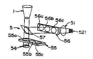
Fig. 17 shows the principle of another
embodiment of the apparatus for counting leukocytes
of the present invention. For the apparatus for
counting leukocytes shown in Fig. 17, the same
numbers are given to the same items in the apparatus
for counting leukocytes of the present invention
shown in Fig. 15, and only differences will be
described.
In the apparatus for counting leukocytes (5)
shown in Fig. 17, a light source (54) is provided
below the leukocyte accumulation container (5). An
excitation light filter slider (55) having three
kinds of excitation light filters (55a, 55b and 55c)
is provided between the light source (54) and the
CA 02309281 2000-OS-08
bottom portion of the leukocyte accumulation
container (5). Since the excitation light filter
slider (55) is disposed between the light source
(54) and the bottom portion, any one of the
excitation filters can be selected by sliding the
excitation light filter slider.
A dichroic mirror (57) is further provided
above the excitation light filter slider (55). The
light selected by the excitation light filter is
transmitted through the dichroic mirror (57) and
irradiated on the observed surface. The generated
fluorescence is reflected by the dichroic mirror
(57), transmitted through the fluorescence filter
(56a, 56b or 56c) and projected on the imaging
section of the CCD image processing means (521) via
the lens portion (51). Any one of the three kinds
of fluorescence filters can be selected by sliding
the fluorescence filter slider (56).
The apparatus for counting leukocytes shown in
Fig. 17 can readily measure the number of the
leukocytes selectively stained with different
fluorochromes by selecting appropriate ones from
three kinds for each of excitation light filters
(55a, 55b and 55c) and fluorescence filters (56a,
56b and 56c).
Brief Description of the Drawings
Fig. 1 shows the measurement results for
CA 02309281 2000-OS-08
36
platelet count in a platelet preparation at each
concentration of added Triton X-100.
Fig. 2 shows light transmittance of a solution
of a platelet preparation at each concentration of
added Triton X-100.
Fig. 3 shows the measurement results for
erythrocyte count in an erythrocyte preparation and
the measurement results for the bared leukocyte
nucleus count by a flow cytometer at each
concentration of added Triton X-100.
Fig. 4 shows a front sectional view of an
example of the leukocyte accumulation container of
the present invention.
Fig. 5 shows a plane view of an example of the
leukocyte accumulation container of the present
invention.
Fig. 6 is a perspective view of an example of
the leukocyte accumulation container of the present
invention.
Fig. 7 is a plane view of an example of the
collective type leukocyte accumulation container of
the present invention.
Fig. 8 is a front sectional view of an example
of the collective type leukocyte accumulation
container of the present invention.
Fig. 9 is a sectional side view of an example
of the collective type leukocyte accumulation
container of the present invention.
CA 02309281 2000-OS-08
37
Fig. 10 is a front sectional view of another
example of the leukocyte accumulation container of
the present invention.
Fig. 11 is a plane view of another example of
the leukocyte accumulation container of the present
invention.
Fig. 12 is a perspective view of another
example of the leukocyte accumulation container of
the present invention.
Fig. 13 shows the process of accumulation of
leukocytes in a solution of a platelet preparation
using the leukocyte accumulation container of the
present invention from a point before the
accumulation to a point after the accumulation.
There are provided front sectional views of the
leukocyte accumulation container. (i) shows the
state before the accumulation, (ii) shows the state
after nuclei of the leukocytes are bared and stained
and platelets are solubilized, and (iii) shows the
state after the accumulation.
Fig. 14 shows the process of accumulation of
leukocytes in a solution of a platelet preparation
by using the leukocyte accumulation container of the
present invention provided with a membrane filter at
the bottom portion from a point before the
accumulation to a point after the accumulation.
There are provided front sectional views of the
leukocyte accumulation container. (i) shows the
r
CA 02309281 2000-OS-08
38
state before the accumulation, (ii) shows the state
after the accumulation by filtration, and (iii)
shows the state after nuclei of the leukocytes are
bared and stained.
Fig. 15 shows principle of an example of the
apparatus for counting leukocytes of the present
invention. (i) shows the entire measurement
apparatus, and (ii) shows an image on the image-
capturing surface.
Fig. 16 shows principle of a comparative
example of an apparatus for counting leukocytes.
(i) shows the entire measurement apparatus, and (ii)
shows an image on the image-capturing surface.
Fig. 17 shows the principle of another example
of the apparatus for counting leukocytes of the
present invention.
Best Mode for Carrying out the Invention
Examples of the present invention will be
described below.
Example 1: Leukocyte count in platelet preparation
<1> Solubilization of platelets in solution of
platelet preparation
15 uL of Triton X-100 surfactant was added to
a solution of a platelet preparation at various
concentrations so that the final concentration was
CA 02309281 2000-OS-08
39
0.01 to 10~. Each solution of the platelet
preparation to which Triton X-100 was added was
stirred by a vortex mixer (Scientific Industry) for
20 seconds to accelerate baring nuclei of leukocytes,
and the platelet count in the solution of the
platelet preparation was measured by an automatic
hemacytometer (Sysmex (trade name), Toa Medical
Electronics Co., Ltd.) to evaluate the
solubilization of platelets by Triton X-100. At the
same time, the light transmittance of the solution
of the platelet preparation was measured by a
spectrophotometer (Beckman).
The measurement results for the platelet count
in the solution of he platelet preparation and the
light transmittance are shown in Figs. 1 and 2. In
Figs. 1 and 2, PRP and PPP represent platelet rich
plasma and platelet poor plasma, respectively.
As shown in Fig. 1, it was revealed that the
platelet count in the solution of the platelet
preparation decreased depending on the concentration
of Triton X-100 and that the platelets were
solubilized depending on the concentration of Triton
X-100. It was revealed, in particular, that most of
platelets were solubilized when the concentration of
Triton X-100 was higher than 0.2~.
As shown in Fig. 2, it was revealed that the
light transmittance of the sample increased with the
increase in the concentration of Triton X-100 up to
' CA 02309281 2000-OS-08
the concentration of Triton X-100 of 1$, but the
solution of the platelet preparation began to show
turbidity due to deposition of Triton X-100, and the
light transmittance was lowered when the
concentration exceeded 1$.
<2> Leukocyte count in solution of platelet
preparation
(1) Method of accumulating stained leukocytes by
centrifugation
As shown in Fig. 13, 0.45 ml of platelet
preparation (2), 0.05 ml of Triton X-100 at a
concentration of 10~ and 0.015 ml of propidium
iodide at a concentration of 1 mM were added to the
above container of Embodiment 1 (Figs. 4 to 6) and
stirred by a vortex mixer for 20 seconds so that
platelets were dissolved and nuclei of leukocytes
were bared and stained. In Fig. 13, 3a and 3b show
leukocytes before nuclei thereof were bared and
stained and leukocytes after nuclei thereof were
bared and stained, respectively. The accumulation
container was set on a centrifuge (Tomy Seiko Co.,
Ltd., Model LC06-SP) for 5 minutes to accumulate
stained leukocytes.
The container where leukocytes were
accumulated on the bottom portion was set on the
apparatus for counting leukocytes provided with a
CCD image processor to detect leukocytes accumulated
CA 02309281 2000-OS-08
41
on the bottom portion by the CCD image processor and
measure the leukocyte count. The image of the
bottom portion of the container of Embodiment 1
could be within the image-capturing surface of the
CCD image processor as one image. Therefore, the
image-capturing surface or the like did not need to
be scanned.
(2) Method for accumulating leukocytes by filtration,
baring nuclei and staining them
As shown in Fig. 14, 0.5 ml of platelet
preparation (2) was placed in the container similar
to Embodiment 1 except that the bottom portion
consisted of a membrane filter (131), and leukocytes
were accumulated on the bottom portion by suction
filtration. 0.1 ml Triton X-100 at a concentration
of 1.0~ and 0.003 ml propidium iodide at a
concentration of 1 mM were added so that nuclei of
the leukocytes were bared and stained. The numerals
3a and 3b represent the same items as in the
aforementioned Fig. 13.
The container where leukocytes were
accumulated on the filter was set on an apparatus
for counting leukocytes provided with a CCD image
processor to detect the leukocytes accumulated on
the bottom portion by the CCD image processor and
measure the leukocyte count. The image of the
bottom portion of the container of Embodiment 1
CA 02309281 2000-OS-08
42
could be within the image-capturing surface of the
CCD image processor as one image. Therefore, the
image-capturing surface or the like did not need to
be scanned.
Example 2: Leukocyte count in erythrocyte
preparation
<1> Solubilization of erythrocytes in erythrocyte
preparation
15 uL of a surfactant, Triton X-100, was added
to a solution of an erythrocyte preparation at
various concentrations so that the final
concentration was 0.01 to 10~. Each solution of the
erythrocyte preparation to which Triton X-100 was
added was stirred by a vortex mixer (Scientific
Industry) for 20 seconds to accelerate baring nuclei
of the leukocytes, and the erythrocyte count in the
solution of the erythrocyte preparation was measured
by an automatic hemacytometer (Sysmex (trade name),
Toa Medical Electronics Co., Ltd.) to evaluate the
solubilization of erythrocytes by Triton X-100. At
the same time, bared nuclei of the leukocytes
(leukocyte nuclei) were counted by a flow cytometer
(Coulter, Model EICS XL).
The measurement results for the erythrocyte
count and the bared leukocyte nucleus count in the
solution of the erythrocyte preparation are shown in
Fig. 3.
CA 02309281 2000-OS-08
43
<2> Leukocyte count in solution of erythrocyte
preparation
87 ~L of an erythrocyte preparation, 10 uL of
Triton X-100 at a concentration of 10~ and 3 uL of
propidium iodide at a concentration of 1 mM were
added to a container of the above Embodiment 1 (Figs.
4 to 6) and stirred by a vortex mixer for 20 seconds
so that erythrocytes were dissolved and nuclei of
leukocytes were bared and stained. The accumulation
container was set on a centrifuge (Tomy Seiko Co.,
Ltd., Model LC06-SP) for 5 minutes to accumulate the
stained leukocytes (corresponding to a case where
the numeral 2 in Fig. 13 indicates an erythrocyte
preparation).
The container where leukocytes were
accumulated on the bottom portion was set on an
apparatus for counting leukocytes provided with a
CCD image processor to detect the leukocytes
accumulated on the bottom portion by the CCD image
processor and measure the leukocyte count. The
image of the bottom portion of the container of
Embodiment 1 can be within the image-capturing
surface of the CCD image processor as one image.
Therefore, the image-capturing surface or the like
does not need to be scanned.
The results of the measurement revealed that
the leukocyte count in a bag of erythrocyte
CA 02309281 2000-OS-08
44
preparation (200 ml) used as a sample was 2 x 10'.
Industrial Applicability
According to the method for counting
leukocytes of the present invention, even if a
sample contains a small amount of leukocytes, the
leukocytes are unlikely to be covered with platelets
or erythrocytes because platelets or erythrocytes
are solubilized, and thus accurate and easy
measurement of the leukocyte count can be enabled.
In particular, since leukocytes can be detected
within a small area by using the leukocyte
accumulation container of the present invention,
labor required for the measurement can be reduced.
In addition, the apparatus for counting leukocytes
of the present invention can be constituted by a
simple system. The apparatus for counting
leukocytes of the present invention allows
mechanized measurement of the leukocyte count and
also enables automatic measurement.