Note: Descriptions are shown in the official language in which they were submitted.
CA 02312931 2000-06-02
WO 99/27841 PCT/US98/25336
TITLE OF THE INVENTION
Automated Visual Function Testing Via Telemedicine
BACKGROUND OF THE INVENTION
Field of the Invention - This invention is in the field of performance of
visual field
tests and other tests of visual function, for the diagnosis of eye conditions.
Background Information - On a world-wide basis, glaucoma is one of the leading
causes of blindness. Unlike cataract blindness, which is correctable with
modern surgical
techniques. blindness from glaucoma is permanent. The target organ of glaucoma
is the
to optic nerve, which transmit signals of light from the retina to the brain.
No known method
is available for repairing, or transplanting, an injured optic nerve.
A major diagnostic problem is that visual loss from glaucoma is almost without
exception painless. The patient is not aware of the ravages of glaucoma until
it is too late.
To compound the problem, the intraocular pressure in glaucoma is often not
elevated
~ 5 (termed "low-tension" glaucoma), and therefore reliance upon tonometry to
measure the
patient's intraocular pressure frequently leads to a blatantly false sense of
security. The
patient is told that glaucoma is not present, when, in reality, the disease is
insidiously
attacking the patient's optic nerve, causing irreversible neurological damage
to the visual
system.
2o Visual field testing is mandatory for glaucoma diagnosis and treatment. The
current
gold standard of measurement of optic nerve function is visual field testing,
called
"perimetry.'' A problem with this technology, however, is that far too many of
the
examiners performing visual field testing are inadequately trained to
recognize subtle
patterns in the patient's visual field indicative of glaucoma (or other
neurological disease).
25 Such misdiagnosis, which is unfortunately frequent, again gives the patient
a false sense of
security.
Millions upon millions of patients throughout the world have glaucoma and are
completely unaware of this. The particularly sad aspect of glaucoma blindness
is that it is
generally preventable with proper diagnosis and treatment. The proposed
invention, which
3o incorporates the use of telemedicine for real-time feedback and for
autointerpretation of
visual field performance, will play a major role in eliminating the all-too-
common errors in
visual field interpretation and the unnecessary blindness which accompanies
such
CA 02312931 2000-06-02
WO 99/27841 PCT/US98/25336
ignorance. By making the proper diagnosis virtually instantaneously over the
Internet or
other telemetric vehicle, glaucoma treatment can be instituted. Millions of
patients will be
spared the ravages of glaucoma.
In addition to testing for glaucomatous. damage to the optic nerve, visual
field
s testing is also used to test for a variety of neurological disorders,
including cerebrovascular
accidents ("strokes"), trauma, brain tumors, and other diseases. The proposed
invention,
which incorporates real-time feedback to monitor the patient's performance,
and accurate,
instantaneous diagnosis available through autointerpretation on a world-wide
telemetric
basis, addresses a major medical need.
With the huge data base developed by a large-scale, world-wide telemedicine
system, leading international experts on glaucoma and other neurological
diseases can be
employed to improve the accuracy of the entire system.
Investigational work has been done on the use of neural nets "trained to
interpret
the visual fields from an automated perimeter," as described in
"Interpretation of
15 Automated Perimetry for Glaucoma by Neural Net," by Goldbaum, et al.
Spenceley, et al.
have also published work in the field in an article entitled, "Visual Field
Analysis Using
Artificial Neural Networks." Brigatti, Hoffman, and Caprioli have worked with
neural
networks for glaucoma identification, as described in their article entitled,
"Neural
Networks to Identify Glaucoma With Structural and Functional Measurements."
These
2o works are limited to conventional globe-like perimeter systems.
BRIEF SUMMARY OF THE INVENTION
The presently-described invention uses a data processing system to provide
automatic interpretation of visual field and other test data received from
testing apparatus
25 in a system which can feature a virtual reality head-mounted display
system. Using virtual
reality and associated head-gear configuration in an interactive computerized
system
allows unprecedented freedom of movement of the head and body, thus minimizing
or
even eliminating the stress and fatigue common with conventional non-virtual
reality
visual field testing systems.
3o The combination of automatic visual field interpretation with a head-
mounted
display system is unique and novel. The use of telemedicine for centralized
interpretation
2
CA 02312931 2000-06-02
WO 99/27841 PCT/US98/25336
of visual field testing at remote locations, and interactively modulating the
performance of
the patient is likewise unique and novel.
The present invention also contemplates the use of a standard visual field
testing
machine, utilizing telemedicine between a central station and several remote
test sites, for
supplying, testing, measuring, quantifying, and autointerpreting visual
information to and
from the visual pathways of the eye and the retina, the optic nerve, the optic
chiasm, the
visual pathways to the brain, and the brain itself. A machine such as, for
example, those
manufactured by Humphrey Instruments, Dicon, or Octopus, can be used to
present visual
stimuli to a patient. Audio feedback stimuli, such as voice, or a tone or
series of tones, or
tactile feedback stimuli, such as a vibration, monitor the test performance in
real-time.
These stimuli are generated and controlled by software in an associated
computer, which
receives interactive feedback stimuli from the patient. The content of the
software is
dictated by the need to provide technically acceptable protocols, such as for
examining
wide and narrow fields of view, selected areas, such as the blind spot or the
fovea, and
~ 5 measurements of thresholds for sensitivity to light intensity, or, if
desired, color. Active
feedback sensing alerts the system to patient loss of attention in general, or
loss of fixation
in particular, for notation and reiteration of test stimuli. The system is
configured to allow
test stimuli to be presented to one eye at a time, or to both eyes
simultaneously. Individual
test points are reiterated when a result is found to be inconsistent with a
predetermined
20 norm.
The novel features of this invention, as well as the invention itself, will be
best
understood from the attached drawings, taken along with the following
description, in
25 which similar reference characters refer to similar parts, and in which:
BRIEF DESCRIPTION OF THE SEVERAL VIEWS OF THE DRAWINGS
Figure 1 is a schematic view of the local test site apparatus used in the
present
invention;
3o Figure 2 is a side view of the apparatus of Figure 1 measuring a vertical
angular
field of view;
3
CA 02312931 2000-06-02
WO 99/Z7841 PCTNS98/Z5336
Figure 3 is a top view of the apparatus of Figure 1 measuring a horizontal
angular
field of view;
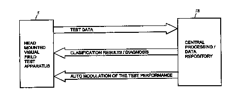
Figure 4 is a schematic diagram of the information flow in the system of the
present invention; and
Figure 5 is a schematic diagram of the automatic interpretation portion of the
system of the present invention.
DETAILED DESCRIPTION OF THE INVENTION
Fig. 1 shows a schematic of one embodiment of the local virnial reality visual
field
1 o testing apparatus 5 which can be incorporated in the present invention, in
which a head-
gear assembly 12 is connected to a local or even imbedd~ processing system 13,
which
delivers a visual signal to a head-gear display screen 12(a), and an audio
signal to a head-
gear earphone 14.
The head-mounted visual display apparatus, head-gear 12, which remains in a
fixed
~ 5 spatial relationship to the patient's head during testing of the visual
field, is adjustable to
suit the individual patient, and is mounted on the patient's head by
conventional means.
The screen display 12(a) is part of the head-gear 12 and encompasses the
maximum field
of view required. The head-gear 12 is provided with an integral microphone 15
and a
speaker or earphone 14, for audio communication and feedback, and a mufti-
element gaze-
zo aim sensor array 17. The microphone 15 provides feedback audio response to
the
processing system 13. The head-gear assembly 12 is connected, by appropriate
means, to
the processing system 13 which provides the necessary visual and audio stimuli
for the
patient, and which receives the feedback responses to enable interactive
functioning of the
system. A hand-operated switch 16 is incorporated to provide the patient's
response
25 feedback to the processing system 13, and the gaze sensor 17, mounted in
the direction of
gaze, provides optical gaze direction feedback to the processing system 13.
Fig. 2 shows, by dashed line 6, a vertical image surface covering an angular
field of
view 7 on the screen display 12(a).
Fig. 3 shows, by dashed line 10, a horizontal image surface covering an
angular
30 field of view 11 on the screen display 12(a).
An element of the virtual reality visual field testing apparatus 5 is that it
allows the
patient the freedom to shift his/her gaze, while in the test mode, without
disruption of the
4
CA 02312931 2000-06-02
WO 99/27841 PCT/US98/25336
process, thus relieving one of the causes of patient stress. Another feature
provided is the
ability to modulate the background scene brightness, contrast, color, optical
stimulus size
and detail, and duration of the test stimuli, all of which serve to relieve
fatigue of the
patient. Of paramount significance is that the patient may move around bodily,
since the
head gear 12 is portable and, in addition, electrical interfaces to the
processing system 13
may be wireless.
In addition to a vastly more patient-friendly and portable test setting, a
further
significant advantage of the presently-described method and apparatus is that
background
light intensity and other parameters can be easily calibrated to predetermined
settings, thus
t o eliminating the requirement mandated by conventional visual field testers
to calibrate these
parameters for the entire room. For instance, the fact that room brightness
can vary almost
imperceptibly, but yet significantly, from day to day in conventional visual
field testing
situations creates built-in unreliability of the test data received from the
patient.
Furthermore, feelings of anxiety frequently displayed by patients undergoing
conventional visual field testing in which first one eye and then the fellow
eye is covered
with an occluder patch can be eliminated in the preferred embodiment, since
both eyes can
be tested simultaneously, or separately and independently, through the use of
individual
eye goggles, or an appropriate face mask, to provide gaze separation.
In other embodiments of the present invention, a standard visual field testing
2o machine can be used in lieu of the head-mounted display, where preferred. A
local
processing system 13 would still be employed, however.
The system of the present invention, as illustrated in Figure 4, includes a
local
visual field test apparatus 5, which can include a head mounted visual field
test apparatus
12 or a standard visual field testing machine, and a local processing system
13 which can
form an integral part of the head-mounted diagnostic apparatus 12. The expert
supervision
of the testing process and interpretation of the results can be performed via
long-distance
transmission vehicles, such as, but not limited to, optical fiber or Internet,
thus providing,
telemetrically, not only essentially instantaneous autointerpretation, but
also telemetric
monitoring of the patient's performance of the test in real time. A central
world-wide
3o processing/data collection system 18 (consisting of a single station or a
series of stations,
such as one for the United States, one for Japan, one for France, etc.) can be
linked via the
Internet to a multitude of local test stations 5 and provide multiweb-like
integration.
5
CA 02312931 2000-06-02
WO 99/27841 PCT/US98/25336
Alternatively, as international long-distance communication becomes more and
more
affordable, one central station could have global capability via direct
connection over
telephone lines. The data processing portion of the system incorporates the
local
processing system 13 and the central processing system and data repository 18,
to provide
the classification of the visual field test data in tenors of presence or
absence of all
diseases, or any particular disease (e.g., glaucoma). The data processing
portion of the
system also may assign a probability of detection and/or a numerical value
indicating the
severity of the disease. This provides a tool for monitoring disease
progression.
Functions of the local processing system include the following:
(a) provision of visual stimuli,
(b) automatic customization of the stimuli sequence based on the patient
response,
including repetition of the stimuli for which no adequate response was
registered (due
either to the patient's loss of attention or to disease-induced damage to the
visual field),
and adjustment of the amplitude of stimuli, and
t 5 (c) pre-processing of the patient response data, such as elimination of
those
measurement points (patient's responses) that are deemed inadequate,
normalization to a
pre-defined standard, and formatting for transmission to the remote processing
system.
Functions of the remote processing system include the following:
(a) automatic interpretation of the visual field test data, and
(b) formulation of corrections to the data collection protocol, based on the
results
of auto-interpretation and comparative analysis employing the database of
interpreted and
medically verified visual field tests.
The central processing/data collection system 18 includes an automated
interpretation system, incorporating a neural network, which functions as
shown in Figure
5. Integration of a multitude of local testing stations S into a world-wide
system results in
a telemedicine system which is "intelligent" in that ongoing data accumulation
and
analyses thereof improve the computational model and provide, over time,
increasingly
more accurate identification of very subtle disease processes.
A database of empirical, semi-empirical, or simulated visual field test data
is used
3o to build a neural network model of the visual field test data. This model,
when applied to
previously unseen test results, is capable of automatically interpreting and
classifying the
test data in terms of the presence and/or severity of abnormal (diseased)
regions and states.
6
CA 02312931 2000-06-02
WO 99/27841 PCT/US98/Z5336
The auto-interpretation system utilizes the results of visual stimuli
(consisting of
dots, symbols, shapes, or patterns, with or without color, etc.) presented to
the patient,
which are converted into numerical representation for data processing, such as
in the
standard automated perimetry schemes (cf. Humphrey Field Analyzer). Other
inputs,
resulting from standard pre-processing of the test data, such as visual field
indices, can
also be employed by the neural network. Inclusion of all available individual
components
of perimetric examination is useful for proper clinical interpretation of the
visual test
examination. Thus, the information provided to the neural network may include:
(a) ancillary data, such as pupil size during testing, the patient's age, and
visual
1 o acuity;
(b) reliability indices, such as fixation behavior and accuracy, and response
fluctuation;
(c) visual field indices, such as average deviation of sensitivity at each
test location
from age-adjusted normal population values, the index of the degree of
irregularity of
~ 5 visual field sensitivity about the normal slope, and sensitivity analysis
of clusters of points;
(d) results of point-by-point comparison of test results with age-matched
normal
population values;
(e) results of high-pass resolution perimetry, if available from the given
implementation of the test apparatus; and,
20 (f) results of pattern discrimination perimetry and other available tests.
The use of the entire gamut of available information for automatic
interpretation by
the neural network is also novel. Previously known neural network systems
included only
the straight visual field data.
The preferred embodiment of the neural network based auto-interpretation
system
25 is shown in Figure 5. The system consists of some or all of the modules
described below.
The data reduction module 22 is employed to reduce the size of the data vector
presented to the neural network classifier. This module employs singular value
decomposition, principal component analysis (PCA), learning vector
quantization, or other
clustering and data size reduction methods. Typically, application of any of
these methods
3o results in at least a two-fold decrease in the size of the data vector.
Such a reduction
increases the ability of the neural network to generalize the data contained
in the training
set. The clustering and linear decomposition methods (such as PCA) are also
useful for
7
CA 02312931 2000-06-02
WO 99/Z7841 PCT/US98/25336
'novelty detection', i.e., for establishing if the current data vector is
outside the region
encompassed by the training data set. The neural network model is likely to
fail for such
data and thus, the ability to detect novelty is crucial for minimizing the
number of
erroneous interpretations.
The data normalization module 24 performs amplitude normalization of the data
presented to the neural network.
The neural network classifier module 26 performs pattern recognition and
classification of the visual field test data. The probability of
classification (or, degree of
membership) is quantified for each of the classes considered in the model. In
the preferred
t 0 embodiment, a non-linear classification scheme exemplified by the
multilayer perceptron
or the radial/ellipsoidal basis function neural network is used. However,
other
classification schemes such as multivariate analysis. linear regression,
statistical classifiers
or discriminators (such as Bayesian classifiers) may also be employed. The
neural
networks are especially useful for the automatic application scheme because
they provide a
non-parametric, empirical model of the visual field test data and are
computationally non-
intensive. i.e., the classification computations can be performed quickly on
inexpensive
computers.
The neural network may be a binary classification system, which will indicate
the
presence or absence of a particular disease, such as glaucoma, or a multi-
class system,
2o which provides recognition and classification of a large variety of
possible visual field
disorders. including, but not limited to, neurological tumors, cerebrovascular
accidents and
strokes, optic nerve disorders. compression syndromes of the optic nerve or
optic chiasm,
demyelinating diseases, and diseases of the retina.
The implementation may be in the form of a single-level neural network system
or
a hierarchical system. In the single-level system, all the input data, which
are deemed
relevant for the interpretation task, are inputted and processed
simultaneously. In the
hierarchical system, different input data types are modeled by dedicated
separate sub-
systems, and these outputs are subsequently fused thmugh a suitable
computational
architecture, to produce the final classification result.
3o The output module 28 creates a graphical representation of the visual field
test
data, such as isopter/scotoma plots, or gray scale or color-coded plots, with
superimposed
identification of the regions that the system classified as abnormal.
8
CA 02312931 2000-06-02
wo ~msa~ Pc rius9ans336
The automatic interpretation system is an expert system trained on a set of
empirical, semi-empirical, and/or simulated data. The construction of a proper
training
database is essential for achieving good performance of the interpretation
system (good
sensitivity and specificity). The training database may contain all, or any,
of the following
s types of visual field data:
(a) empirical data, i.e., data obtained for patients with normal and abnormal
visual
fields;
(b) semi-empirical data, i.e., data obtained by modification of the empirical
data, as
described above, by:
( 1 ) emphasizing or de-emphasizing certain aspects of the visual field test
to
bring out the characteristic features of certain diseased states;
(2) adding noise or measurement uncertainty components which may be
associated with a real visual field examination; and,
(3) any other modification of the visual field test data and their associated
t 5 classification; and,
(c) simulated data, i.e., data that are constructed to simulate the real-world
results
of a visual field test for both normal and abnormal visual fields.
Training of the classification system is performed off line with active
participation
of a human expert. That is, all visual field test data in the training
database are examined
2o by an expert and the medical diagnosis is verified and validated before the
data is used to
build the neural network model. The centralized processing enables collection
of a large
number of diverse examples of normal and abnormal visual field test data. The
novelty
detection capability of the system alerts the system custodian to the
necessity for expert
examination of the novel data. After completion of such examination, the data
may be
25 included in the model by including the new data in the training database
and re-training the
system.
While the particular invention as herein shown and disclosed in detail is
fully
capable of obtaining the objects and providing the advantages hereinbefore
stated, it is to
be understood that this disclosure is merely illustrative of the presently
preferred
3o embodiments of the invention and that no limitations are intended other
than as described
in the appended claims.
9