Note: Descriptions are shown in the official language in which they were submitted.
CA 02314100 2000-06-14
WO 99/34864 PCT/IL99/00008
An Integrated sleep apnea screening system.
FIELD AND BACKGROUND OF THE INVENTION
S The present invention relates to medical monitoring devices and, in
particular, it relates to a monitor for the detection of sleep apnea.
It is known that sleep related breathing disorders are a common medical
problem. Two common sleep pathology syndromes are Obstructive Sleep
Apnea (OSA) and Central Sleep Apnea (CSA).
Obstructive Sleep Apnea (OSA) occurs when the upper airway (the nose,
mouth or throat) become obstructed in some way during sleep, and is usually
accompanied by a decrease in the oxygen saturation of the blood (Sp02).
Snoring indicates an intermittent obstruction, which at times may become
complete, stopping air flow. Apnea (the cessation of breathing) may occur
hundreds of times during one night of sleep, leading to severe sleep
disruption
and excessive daytime somnolence. As such, the patient may easily fall asleep
during working hours, such as when the patient is driving a car or a truck.
Many
commercial trucking firms thus require that their drivers undergo sleep
studies
CA 02314100 2000-06-14
WO 99/34864 PCT/IL99/00008
2
to determine if they suffer from OSA. Furthermore, OSA may cause heart
problems such as cardiac arrhythmias and Cor Pulmonale.
Central Sleep Apnea Syndrome (CSA), in contrast, occurs due to a
defect in central nervous system control of the respiratory drive, and is most
commonly seen in patients with neurological disorders affecting respiratory
control and in the elderly. CSA may also result in frequent awakenings and
their associated impact on daytime performance.
Definitive diagnosis of these respiratory-sleep pathologies is currently
achieved by means of an in-lab, full night, formal sleep study. In such a
study,
the patient is required to sleep for a whole night in a controlled environment
(a
"sleep laboratory") while connected to multiple monitoring devices, which
continuously measure such physiological parameters as respiratory effort,
nasal
and oral airflow, brain electrical activity (EEG), muscle electrical activity
(EMG), heart rate and rhythm (ECG), and blood oxygen saturation. These
parameters are recorded on paper or stored in a memory bank for later
analysis.
A trained sleep technician is required to oversee the study so as to ensure
that
all parameters are recorded properly. The data is then analyzed, either
manually
or by specialized software, to produces a "hypnogram" which describes the
nature of the patients sleep. Indices in the hypnogram, such as an "apnea
index"
and a "leg movement index", are then used, by a sleep specialist, to diagnose
the patients pathology.
CA 02314100 2000-06-14
WO 99/34864 PCT/IL99/00008
3
The formal sleep study as a means of diagnosing and following-up
patients with respiratory related sleep problems, however, suffers from
several
deficiencies and limitations:
1. The study requires the use of multiple medical monitoring
devices and the continuous presence of a trained technician. It is
thus labor intensive to perform, and requires the use of multiple,
expensive, resources.
2. The patient is asked to sleep in a non natural sleep environment,
which may itself affect his sleep patterns.
3. The patient is inconvenienced by having to be in a hospital
setting for a night.
4. There is no patient privacy.
As such, sleep laboratories are a limited resource, each containing only a
limited number of beds. This is particularly problematic as studies are often
conducted on "suspicious" patients, in whom the outcome is frequently
negative. In such patients, for whom there was no need for the study at all, a
limited screening study may have been sufficient to exclude sleep pathology.
The study price often prohibits repeating studies on a regular basis for
purposes
of patient follow-up.
In order to overcome some of these drawbacks, the performance of home
studies by means of ambulatory systems has become popular. These studies
utilize miniature ambulatory recorders, and are limited to a relatively small
CA 02314100 2000-06-14
WO 99/34864 PCT/IL99/00008
4
number of information recording channels. The patient is prepared for the
study
at the sleep lab, and returns home with all sensors appropriately attached.
Alternatively, a technician may come to the patients home, or the patient may
attach the sensors by himself after receiving appropriate instruction from a
technician. The study is then conducted in the patient's home, as he sleeps in
his own bed, and the recorded data stored in a memory device. In the morning
the recorder and memory device are returned to the sleep lab for data
downloading to an analysis station. Some of these ambulatory systems can
correct for some data recording problems, by adjusting the gain or filtering
during data recording or when post-processing the data. Alternatively, the
study
can be monitored from the sleep lab via a modem.
Although ambulatory sleep-apnea monitoring systems are much more
convenient to the patient, and considerably less expensive than formal, in-
lab,
sleep studies, all current ambulatory sleep monitoring systems suffer from
several deficiencies:
1. Performance of the study still requires the participation of a
trained technician (for the purposes of either attaching the
monitoring device or instructing the patient how to do so) and
the participation of a formal sleep laboratory (for the purposes of
downloading and analyzing the test results, and maintaining the
equipment necessary for the performance of the test). Such tests
are thus still labor and resource intensive.
CA 02314100 2000-06-14
WO 99/34864 PCT/IL99/00008
2. As analysis of the recorded data is performed off-line in the
sleep laboratory, the ambulatory monitoring device must be able
to store all registered data in a suitable memory storage device,
until such data can be downloaded. Alternatively, if the data is
5 relayed to the sleep laboratory in real time, a modem and
telephone line are necessary. Current ambulatory devices are
therefore relatively complex and expensive to manufacture. As
such, ambulatory studies are still too expensive to perform on a
regular basis (currently approximately $500 per study), thus
precluding their widespread use as a screening tool or for
purposes of frequent patient follow-up. In addition, the cost of
such studies does not justify their use on "difficult" patients,
such as mental health patients or small children, in whom the
likelihood of technical failure of the study is high.
There is therefore a need for a sleep-apnea screening system which is
suitable for widespread use for patient screening and follow-up. Such a system
should be sufficiently simple to implement as to allow patients to perform the
study at home, without the need for assistance from a trained technician. In
addition, such a system should provide the patient with an easily
understandable
result at the end of the study, without the need for data processing at a
sleep
laboratory, and without the need for interpretation of the result by a
physician
CA 02314100 2000-06-14
WO 99134864 PCT/IL99/00008
6
or technician. Finally, such a system should be sufficiently inexpensive as to
make multiple and frequent studies practical to finance.
SUMMARY OF THE INVENTION
The present invention is an ambulatory sleep-apnea screening system.
The invention integrates a minimal data collection and analysis system into a
disposable, single use device that achieves data collection and analysis in
real
time, and outputs the study result in an easily understood format immediately
following the study.
The entire system is incorporated into a single small, flexible, plastic unit
which
can be easily positioned, or placed, under the patients nose, that is, upon
the
patients philtrum. The system is powered by a lithium battery, which is
irreversibly activated by means of the patient pulling on a tab. Once
activated, a
respiration detector (such as that which measures temperature differences in
an
airflow, by which is meant a flow of inhaled and exhaled nasal or oral air)
inputs data describing the pattern of respiration into a micro-processor, via
an
analog to digital converter. A flashing LED display indicates to the user that
the
device is correctly positioned. A software module detects the absence of hot
airflow for a predetermined period - indicating apnea. Apnea duration is
measured, normal breaths between apneas are counted, and, together with real-
time clock information, the presence, and severity of, episodes of apnea is
documented. Data can be sampled continuously, or in segments each a few
CA 02314100 2000-06-14
WO 99/34864 PCT/IL99/00008
7
minutes long, so as to conserve battery power. After a predefined period of
time, non volatile output flags (in the form of heat sensitive colored dots)
are
set by the software. Once activated, the output flags undergo a permanent
color
change. As such, they produce an easily read hard copy of the study results,
informing the user whether significant apnea was detected and whether a
physician need be consulted. Hereinafter, output flags which undergo a
permanent change in color when activated by heat are referred to as "heat
sensitive permanent color display elements".
The integration, onto a respiratory sensor, of a sleep apnea screening
system which is capable of analyzing respiratory data in real time and
generating an immediate report thereof, is unique to the current invention. By
"real time" is meant that the processing of the respiratory data and the
sensing
of the respiratory pattern occur during the same time interval, rather than
the
processing occurring after all respiratory sensing has been completed.
As data is analyzed in real time, the need for a large memory storage unit
to store data for later analysis, and the need for complex downloading
hardware, are obviated. This feature allows the entire system to be
manufactured in a small and inexpensive format, and provides the user with the
result of the study immediately upon conclusion of the study, without the need
for data processing and analysis by medical professionals at a sleep
laboratory.
Furthermore, as the power source, processor, and display mechanism of the
device are all integrated with the respiratory sensor into a small, single,
unit,
CA 02314100 2000-06-14
WO 99/34864 PCT/IL99/00008
without the need for cables or wires connecting these components to each
other,
and as an easily seen flashing light confirms to the user that placement and
operation of the device are correct, the device is simple and straightforward
to
use. The device can thus be operated without supervision by trained medical
professionals. Accordingly, the cost per study is sufficiently low as to
justify
performing studies frequently for screening purposes (whenever there is even a
slight chance of true pathology being present) or for regular patient follow-
up.
As their are no cables or wires connecting the respiratory sensor with the
rest of
the device, the possibility that the sensor might be pulled of off the users
face,
due to the cable becoming entangled while the user is asleep, is obviated.
It is an object of the current invention to provide a sleep apnea screening
system which can be easily and reliably used by a patient without the need for
professional supervision.
It is a further object of the current invention to provide a sleep apnea
screening system which does not require the use of complex data storage and
analysis hardware.
It is an additional object of the current invention to provide a sleep apnea
screening system which is sufficiently simple and inexpensive as to facilitate
performance of multiple sleep apnea screening studies on the same patient, on
unreliable patents, or on patients with a low likelihood of having real
pathology.
CA 02314100 2000-06-14
WO 99/34864 PCT/IL99/00008
9
It is a yet further object of the current invention to provide a sleep apnea
screening system which allows the study to be performed in the patients
natural
sleep environment
It is a yet further object of the current invention to provide a sleep apnea
screening system which does not infringe patient privacy.
According to the teachings of the present invention there is provided a
sleep apnea screening system, including a respiration sensor, for sensing a
respiratory pattern, at a location on a respiratory tract; a processor, for
analyzing the respiratory pattern to determine the presence of a pattern of
apnea, and for correlating the pattern of apnea with a diagnosis; a display,
for
displaying the diagnosis; a power source, for powering the respiration sensor,
the processor, and the display; and a housing, for housing the processor, the
display, and the power source, on the respiration sensor, the housing being
placeable at the location on the respiratory tract. There is also provided a
sleep
apnea screening method, including the steps of placing a housing at a location
on a respiratory tract; sensing a respiratory pattern at the housing during a
time
interval; processing the sensed respiratory pattern to detect the presence of
a
pattern of apnea, the processing occurring during the time interval;
correlating
the pattern of apnea with a diagnosis, the correlating occurring during the
time
interval; and displaying the diagnosis on the housing.
CA 02314100 2000-06-14
WO 99/34864 PCT/IL99/00008
BRIEF DESCRIPTION OF THE DRAWINGS
The invention is herein described, by way of example only, with
reference to the accompanying drawings, wherein:
FIG. 1 is a line drawing of the physical structure of an apnea screening
5 system;
FIG. 2 is a schematic depiction of the structure of an apnea screening
system;
FIG. 3 is a block diagram of the data flow within the processor of an
apnea screening system; and
10 FIG. 4 is a diagram of the positioning of an apnea screening system on
the face of a user.
DESCRIPTION OF THE PREFERRED EMBODIMENTS
The present invention is a sleep apnea screening system, integrated on an
I S airflow sensor.
The principles and operation of a sleep apnea screening system,
according to the present invention, may be better understood with reference to
the drawings and the accompanying description.
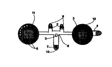
Referring now to the drawings, Figure 1 is a line drawing of the overall
structure of the current invention. As can be seen, a thin, flexible housing
$,
shaped like a thin strip, serves as a base for the system. In the preferred
embodiment, housing 8 is made of a flexible plastic film. Housing 8 is shaped
CA 02314100 2000-06-14
WO 99/34864 PCT/IL99/00008
11
such that it can be attached between the nose and the upper lip of the user,
such
that protrusions 9 and 10 will overly the two nostrils and the mouth,
respectively. Housing 8 includes two larger circles, 11 and 12, each
approximately 1.5" in diameter, which house electronic components of the
system. A double sided adhesive foam backing (not shown), covering the entire
area of the back of housing 8, allows for the comfortable attachment of the
device to the face of the user.
A power source 3, in the form of a flat lithium battery, is housed in circle
12. Power source 3 powers the functioning of all elements of the sleep apnea
monitoring system. The negative contact of power source 3 is insulated from a
conductive electrode (not shown) on housing 8 by a pull-tab 4. When tab 4 is
pulled out by the user, contact is made between the negative contact of power
source 3 and the electrode completing the electrical circuit, and operation of
the
system commences.
Two nasal NTC (Negative Temperature Coefficient) thermistors 2 and
an oral NTC thermistor 7 are located on protrusions 9 and 10 respectively,
such
that they are located inside the air streams emanating from nose and mouth
when housing 8 is properly positioned on the face of the user. Examples of
thermistors suitable for use as nasal and oral thermistors 2 and 7 are SMT
components (Thermometrics Inc., Tounton, UK). Alternatively, thermistors 2
and 7 can be replaced with other respiration sensors, such as humidity
sensors,
pressure sensors, or respiration sounds detectors. Thermistors 2 and 7 are
CA 02314100 2000-06-14
WO 99/34864 PCT/IL99100008
12
connected in series to the input of a processor (CPU) 1, which is housed in
circle 11. The flow of cyclically hot and cold air streaming over thermistors
2
and 7 (during expiration and inspiration respectively) causes a cyclical
change
in resistance within thermistors 2 and 7. This changing resistance is
registered
by CPU 1 as breathing data. In the preferred embodiment, CPU 1 is a RISC
processor running a continuos monitoring and scoring program, which will be
detailed below. CPU 1 analyzes the received respiratory data in real time, and
reaches one of several possible predefined study conclusions.
A LED display 5 is located on housing 8 such that it can be easily seen
by the user, when looking in a mirror, once the system has been attached to
the
face of the user and operation commenced. LED 5 is operative to flash with
each breath taken by the user, so as to indicate that proper placement of
thermistors 2 and 7 has been achieved ands that the system is functioning
properly.
When the sleep apnea study is complete, CPU 1 issues a command to
flow an electric current through one (or more) non-volatile markers 6. In the
preferred embodiment, each one of markers 6 comprises a miniature heating
element, and a coating of a heat sensitive material. When current is passed
though one of the elements it heats up, inducing a change in the color of the
coating material (such as rendering the coating material permanently black).
This color change is permanent, even after cooling down of the element.
Hereinafter, such markers are also referred to as "display elements". The
choice
CA 02314100 2000-06-14
WO 99/34864 PCT/IL99/00008
13
of which of markers 6 to activate depends on the study conclusion, as
determined by CPU 1. Each non-volatile marker 6 corresponds to one of several
possible diagnoses. By "diagnoses" is meant possible study outcomes
describing the degree of severity of apnea and recommended courses of action
to be taken by the user in response thereto, for example:
1 ) Severe Apnea detected - must refer to a sleep lab
2) Medium Apnea detected - must .refer to a sleep lab
3) Mild Apnea detected - Advised to consult your GP.
4) Possible problem - consult a physician,
5) No problem detected
6) Bad data - Perform a new study (if, for example, apneas
lasting longer than 2 minutes were detected).
In FIG. 2, a simplified block diagram of the device is shown.
Thermistors 2 and 7 input flow data to a signal conditioner and A/D (analog to
digital) converter 13, which may be part of CPU 1. The resultant digital data
stream is input to CPU l, which runs specialized data acquisition and analysis
software. Each time a breath is sensed by thermistors 2 or 7, a command is
output to LED 5, which flashes once. When a conclusion is reached at the end
of the study, CPU 1 outputs a command to one of non-volatile markers 6. The
entire system is powered by power source 3.
CA 02314100 2000-06-14
WO 99/34864 PCT/IL99/00008
14
In normal operation, after switching the device on by pulling out tab 4,
the user stands in front of a mirror, attaches the sensor under his nose and
over
his cheeks, and breathes through his nose and then through his mouth. If LED
flashes with each breath, the user knows that proper placement and operation
5 of the device has been achieved. The user then waits approximately thirty
minutes prior to going to sleep, during which time the device collects normal
data, that is, respiratory data without episodes of apnea, over the course of
several minutes. The user then goes to sleep. CPU 1 resumes collecting and
processing data automatically after 1 hour, analyzes breathing patterns in
real
time for several minutes, and then enters a sleep mode for approximately 30
minutes.This cycle is then repeated several times, until CPU 1 reaches a
conclusion as to whether sleep apnea was detected and estimates its severity,
or
until more than 5 hours have passed since the time that power source 3 was
activated. CPU 1 then outputs the analysis result to non-volatile indicators
6.
Upon awakening in the morning, the user checks to see which of indicators 6
have been activated, and is thus informed of the result of the study. In the
event
that significant apnea was detected during the study, the user is advised (by
indicator 6) to consult with a physician or sleep clinic for further
investigation.
The device, with it's permanent color-coded study outcome, can be kept for
later reference.
FIG. 3 describes the data flow within CPU 1 of the sleep apnea
screening system.
CA 02314100 2000-06-14
WO 99/34864 PCT/IL99/00008
Data in from thermistors 2 and 7 is smoothed and converted to digital
format by A/D converter l3.The resultant digital data is input to a
breath/apnea
detector 14. Apnea detector 14 is a software module that monitors the data
generated by thermistors 2 and 7, reflecting temperature differences caused by
5 cold air being inhaled and hot air being exhaled. Apnea detector 14 locates
the
maximum and minimum registered temperatures, and calculates the difference
between those values, which approximates the volume of inspired air. Apnea
detector 14 also calculates the time from one maximum temperature value to
the next. After processing several cycles during the thirty minute period
prior to
10 the user falling asleep (i.e. the period of no apnea), a maximum time
between
maximum temperature values is determined, and defined as the maximum
normal time interval between breaths. In addition, a minimum percentage
difference between maximum and minimum temperature values for one
respiratory cycle is determined, and defined as the minimum normal peak-to-
1 S peak value of a breath. If the registered difference between the maximum
and
minimum temperatures of one cycle is less than the minimum normal peak-to-
peak value, a low flow state can be defined as being present, while if the
registered difference is zero, a zero flow state can be defined as being
present.
The condition of zero flow and undetectable rhythmic temperature pattern
indicates the state of apnea, and this state is counted as a real apneac
episode by
an apnea counter 17 if it lasts for more than 10 seconds.
CA 02314100 2000-06-14
WO 99/34864 PCT/IL99/00008
16
Apnea detector 14 thus locates local minima and maxima for each
respiratory cycle, calculates the time from the last maxima to the current
maxima, and calculates the peak to peak value of the current breath cycle. If
the
time from last breath is more than a prescribed value (typically 10 sec), or
the
peak-to-peak value is less than a prescribed value (typically 30%), an apnea
mark is issued by apnea detector 14 and input to apnea counter 17. The number
of normal breaths during the study period are counted by a normal breath
counter module 15. The duration of each apneac episode is measured by an
apnea duration timer 18. Apnea duration timer 18 commences timing once
cessation of airflow is detected, and stops timing as soon as airflow resumes.
This module also calculates the mean and standard deviation for all recorded
apneac episodes during the study. The "accumulated apnea time", meaning the
total number of rriinutes in apnea state during the course of the study, is
calculated by an accumulated apnea timer 16. LED 5 is activated by apnea
detector 14 via a LED driver 20 whenever a normal respiratory cycle is
detected, and flashes.
The above described process is repeated several times, under the
command of a cycle timer 18. Cycle timer 18 runs the data collection and
analysis software in epochs of several minutes each every half hour, and then
may switch CPU 1 to a sleep mode, in order to conserve battery power.
Decision integrator 19 compares the data for each epoch with all prior epoch
data, and when "convergence" of data (by which is meant approximately
CA 02314100 2000-06-14
WO 99/34864 PCT/IL99/00008
17
equivalent apnea behavior in several epochs) is detected, the data acquired
during the study is assumed to be a reliable depiction of reality. When
decision
integrator 19 detects convergence of values, or when cycle timer 18 issues a
command to decision integrator 19 after a predefined maximum period of time
has elapsed (such as five hours), decision integrator 19 accesses all data
stored
in apnea counter 17, apnea duration timer 18, and accumulated apnea timer 16.
Decision integrator 19 then compares the number and nature of apneac episodes
detected to a predefined "diagnostic table" which categorizes all apnea
patterns
as falling into one of several diagnostic categories. Each diagnostic category
corresponds to a particular non-volatile marker 6, which is activated by
decision
integrator 19 if the study is defined as falling into its corresponding
diagnostic
category. Based on the accumulated apnea time (as determined by accumulated
apnea timer 16), the total number of apneac episodes per hour (as determined
by apnea counter 17), and the breathing rate (as derived from the total breath
count divided by the length of the study), decision integrator 19 activates
one of
the following markers 6:
"No problem" marker - no apnea detected.
"Minor problem" marker - average 1-5 apneas per hour.
"Moderate problem" marker - average 6-10 apneas per hour.
"Severe problem" marker - average over 10 apneas per hour.
"Bad study" marker- apneas lasting longer than 120 seconds
detected, or a change of normal respiration amplitude over time
CA 02314100 2000-06-14
WO 99/34864 PCT/IL99/00008
18
of over 50% (poor steady state values), or a lack of normal
respiration pattern during the first ten minutes after turn-on.
As the markers retain their appearance indefinitely, the device can be
kept indefinitely as a medical record, and test results can be compared from
study to study.
It will be appreciated that the invention as described herein may be
supplemented in several ways, without departing from the spirit of the
invention. For example, a heat sensitive element, to sense skin temperature
during the study, may be incorporated into the device. This element would
indicate if the device was removed during the night, prior to the end of the
study. In addition, a light sensor may be incorporated into the device so as
to
determine that the lights were switched off during the study, as a fraud
detection mechanism.
FIG. 4 illustrates the preferred positioning of the device of the present
invention on the face of the user. The device is positioned between the nose
and
the upper lip, covering the philtrum. In the preferred embodiment the device
is
held in place by double sided adhesive tape, although in alternative
embodiments any mechanism suitable for securely holding an object against the
face may be used, such as adjustable straps 21. As illustrated, protrusions 9
are
positioned in proximity to the nares, protrusion 10 over the mouth, and
circles
11 and 12 over the cheeks of the user.
CA 02314100 2000-06-14
WO 99/34864 PCT/1L99/00008
19
As a very low cost screening method, the device of the current invention
may have several applications:
1. Follow-up of sleep apnea patients after dietary treatment,
surgery, CPAP treatment, fitting of an anti-snoring oral
appliance, or a change is sleeping posture.
2. Screening infants for higher risk of Sudden Infant Death
Syndrome. (SIDS), by detecting non-regular breathing pattern.
3. Screening for candidates for a full feature sleep study.
4. Screening of applicants for high risk jobs like truck-driving or
shift-working.
There has thus been described a sleep apnea screening system which can
be easily and reliably used without the need for professional supervision or
the
use of complex data storage and analysis hardware. The system is sufficiently
simple and inexpensive as to facilitate performance of multiple sleep apnea
screening studies on the same patient, on unreliable patents, or on patients
with
a low likelihood of having real pathology. The system allows the study to be
performed in the patients natural sleep environment, and does not infringe
patient privacy.