Note: Descriptions are shown in the official language in which they were submitted.
CA 02316361 2007-02-19
30317-21
-1-
A METHOD FOR PREDICTTNG AN ABNORMAL LEVEL OF CLOTI'ING PROTEINS
BACKGROUND OF THE INVENTION
This application is a cont.inuation-in-part of
U.S. Patent 6,101,449 to Givens et al. filed
May 21, 1997, which is a continuation of U.S.
Patent 5,708,591 to Givens et al. filed June 7, 1995.
This application also relates to U.S. Patent 5,646,046
to Fischer et al. This application is further related
to the following publications:
1. B. Pohl, C. Beringer, M. Bomhard, F. Keller,
The quick machine - a mathematical model for the
extrinsic activation of coagulation, Haemostasis, 24,
325-337 (1994).
2. J. Brandt, D. Triplett, W. Rock, E. Bovill,
C. Arkin, Effect of lupus anticoagulants on the
activated partial thromboplastin time, Arch Pathol Lab
Med, 115, 109-14 (1991).
3. I. Talstad, Which coagulation factors
interfere with the one-stage prothrombin time?,
Haemostasis, 23, 19-25 (1993).
4. P. Baumann, T. Jurgensen, C. Heuck,
Computerized analysis of the in vitro activation of
the plasmatic clotting system, Haemostasis, 19,-309-
321 (1989).
5. C. Heuck, P. Baumann, Kinetic analysis of
the clotting system in the presence of heparin and
depo.lymerized heparin, Haemostasis, 21, 10-18 (1991).
3 5 6. M. Astion and P. Wilding, The application of
backpropagation neural networks to problems in
CA 02316361 2000-06-22
WO 99/34208 PCT/US98/27865
-2-
pathology and laboratory medicine, Arch Pathol Lab
Med, 116, 995-1001 (1992).
7. M. Astion, M. Wener, R. Thomas, G. Hunder,
and D. Bloch, Overtraining in neural networks that
interpret clinical data, Clinical Chemistry, 39,
1998-2004 (1993).
8. J. Furlong, M. Dupuy, and J. Heinsimer,
Neural network analysis of serial cardiac enzyme data,
A.J.C.P., 96, 134-141 (1991).
9. W. Dassen, R. Mulleneers, J. Smeets, K. den
Dulk, F. Cruz, P. Brugada, and H. Wellens, Self-
learning neural networks in electrocardiography, J.
Electrocardiol, 23, 200-202 (1990).
10. E. Baum and D. Haussler, What size net gives
valid generalization? Advances in Neural Information
Processing Systems, Morgan Kauffman Publishers, San
Mateo, CA, 81-90 (1989).
11. A. Blum, Neural Networks in C++, John Wiley
& Sons, New York, (1992).
12. S. Haykin, Neural Networks A Comprehensive
Foundation, Macmillan College Publishing Company, New
York, (1994).
13. J. Swets, Measuring the accuracy of
diagnostic systems, Science, 240, 1285-1293 (1988).
14. M. Zweig and G. Campbell, Receiver-operating
characteristic (ROC) plots: a fundamental evaluation
tool in clinical medicine, ClinicaZ Chemistry, 39,
561-577 (1993).
CA 02316361 2000-06-22
WO 99/34208 PCT/US98/27865
-3-
15. D. Bluestein, L. Archer, The sensitivity,
specificity and predictive value of diagnostic
information: a guide for clinicians, Nurse
Practitioner, 16, 39-45 (1991).
16. C. Schweiger, G. Soeregi, S. Spitzauer, G.
Maenner, and A. Pohl, Evaluation of laboratory data
by conventional statistics and by three types of
neural networks, Clinical Chemistry, 39, 1966-1971
(1993).
Blood clots are the end product of a complex
chain reaction where proteins form an enzyme cascade
acting as a biologic amplification system. This
system enables relatively few molecules of initiator
products to induce sequential activation of a series
of inactive proteins, known as factors, culminating in
the production of the fibrin clot. Mathematical
models of the kinetics of the cascade's pathways have
been previously proposed.
In [1], a dynamic model of the extrinsic
coagulation cascade was described where data were
collected for 20 samples using quick percent,
activated partial thromboplastin time (APTT), thrombin
time (TT), fibrinogen, factor(F) II, FV, FVII, FX,
anti-thrombin III (ATIII), and factor degradation
product (FDP) assays. These data were used as input
to the model and the predictive output compared to
actual recovered prothrombin time (PT) screening assay
results. The model accurately predicted the PT result
in only 11 of 20 cases. These coagulation cascade
models demonstrate: (1) the complexity of the clot
formation process, and (2) the difficulty in
associating PT clot times alone with specific
conditions.
CA 02316361 2000-06-22
WO 99/34208 PCT1US98/27865
-4-
Thrombosis and hemostasis testing is the in vitro
study of the ability of blood to form clots and to
break clots in vivo. Coagulation (hemostasis) assays
began as manual methods where clot formation was
observed in a test tube either by tilting the tube or
removing fibrin strands by a wire loop. The goal was
to determine if a patients blood sample would clot
after certain materials were added. It was later
determined that the amount of time from initiation of
the reaction to the point of clot formation in vitro
is related to congenital disorders, acquired
disorders, and therapeutic monitoring. In order to
remove the inherent variability associated with the
subjective endpoint determinations of manual
techniques, instrumentation has been developed to
measure clot time, based on (1) electromechanical
properties, (2) clot elasticity, (3) light scattering,
(4) fibrin adhesion, and (5) impedance. For light
scattering methods, data is gathered that represents
the transmission of light through the specimen as a
function of time (an optical time-dependent
measurement profile).
Two assays, the PT and APTT, are widely used to
screen for abnormalities in the coagulation system,
although several other screening assays can be used,
e.g. protein C, fibrinogen, protein S and/or thrombin
time. If screening assays show an abnormal result,
one or several additional tests are needed to isolate
the exact source of the abnormality. The PT and APTT
assays rely primarily upon measurement of time
required for clot time, although some variations of
the PT also use the amplitude of the change in optical
signal in estimating fibrinogen concentration.
Blood coagulation is affected by administration
of drugs, in addition to the vast array of internal
factors and proteins that normally influence clot
CA 02316361 2000-06-22
WO 99/34208 PCT/US98/27865
-5-
formation. For example, heparin is a widely-used
therapeutic drug that is used to prevent thrombosis
following surgery or under other conditions, or is
used to combat existing thrombosis. The
administration of heparin is typically monitored using
the APTT assay, which gives a prolonged clot time in
the presence of heparin. Clot times for PT assays are
affected to a much smaller degree. Since a number of
other plasma abnormalities may also cause prolonged
APTT results, the ability to discriminate between
these effectors from screening assay results may be
clinically significant.
Using a sigmoidal curve fit to a profile,
Baumann, et al [4] showed that a ratio of two
coefficients was unique for a select group of blood
factor deficiencies when fibrinogen was artificially
maintained by addition of exogenous fibrinogen to a
fixed concentration, and that same ratio also
correlates heparin to FII deficiency and FXa
deficiencies. However, the requirement for
artificially fixed fibrinogen makes this approach
inappropriate for analysis of clinical specimens. The
present invention makes it possible to predict a
congenital or acquired imbalance or therapeutic
condition for clinical samples from a time-dependent
measurement profile without artificial manipulation of
samples.
The present invention was conceived of and
developed for predicting the presence of congenital or
acquired imbalances or therapeutic conditions of an
unknown sample based on one or more time-dependent
measurement profiles, such as optical time-dependent
measurement profiles, where a set of predictor
variables are provided which define characteristics of
profile, and where in turn a model is derived that
represents the relationship between a congenital or
_~--- _ _
CA 02316361 2000-06-22
WO 99/34208 PCTIUS98/27865
-6-
acquired imbalance or therapeutic condition and the
set of predictor variables (so as to, in turn, utilize
this model to predict the existence of the congenital
or acquired imbalance or therapeutic condition in the
unknown sample).
SUMMARY OF THE INVENTION
The present invention is directed to a method and
apparatus for predicting the presence of at least one
congenital or acquired imbalance or therapeutic
condition from at least one time-dependent measurement
profile. The method and apparatus include a)
performing at least one assay on an unknown sample and
measuring a respective property over time so as to
derive a time-dependent measurement profile, b)
defining a set of predictor variables which
sufficiently define the data of the time-dependent
profile, c) deriving a model that represents the
relationship between a diagnostic output and the set
of predictor variables, and d) utilizing the model to
predict the existence of a congenital or acquired
imbalance or therapeutic condition in the unknown
sample relative to the diagnostic output. In one
embodiment, training data is provided by performing a
plurality of assays on known samples, the model is a
multilayer perceptron, the relationship between the
diagnostic output and the set of predictor variables
is determined by at least one algorithm, and the at
least one algorithm is a back propagation learning
algorithm. In a second embodiment of the present
invention, the relationship between the diagnostic
output and the set of predictor variables is derived
by a set of statistical equations. Also in the
present invention, a plurality of time-dependent
measurement profiles are derived, which time-dependent
CA 02316361 2007-02-19
30317-21
-7-
measurement profiles can be optical time-dependent
measurement profiles such as ones provided by an automated
analyzer for thrombosis and hemostasis, where a plurality of
optical measurements are taken over time, and where the
plurality of optical measurements are normalized. The
optical profiles can include one or more of a PT profile, a
fibrinogen profile, an APTT profile, a TT profile, a
protein C profile, a protein S profile and a plurality of
other assays associated with congenital or acquired
imbalances or therapeutic conditions.
According to one aspect of the present invention,
there is provided a method for predicting the presence of an
abnormal level of one or more proteins of a coagulation
cascade from at least one time-dependent measurement
profile, comprising: a) performing at least one time-
dependent measurement on an unknown sample of a property
over time, which property changes when said sample undergoes
coagulation, so as to derive at least one time-dependent
measurement profile; b) defining a set of a plurality of
predictor variables which sufficiently define at least one
time-dependent measurement profile; c) deriving a model that
represents the relationship between the abnormal level of
said one or more proteins in the coagulation cascade and the
set of a plurality of predictor variables; and d) utilizing
the model of step c) to predict the existence of the
abnormal level of said one or more proteins in the
coagulation cascade and to predict which protein or proteins
in the coagulation cascade are said one or more proteins
which are at an abnormal level as compared to a known
sample.
According to another aspect of the present
invention, there is provided the method as defined above,
wherein the concentration of the one or more proteins in the
CA 02316361 2007-02-19
30317-21
-7a-
coagulation cascade is further estimated by utilizing the
model of step c).
BRIEF DESCRIPTION OF THE DRAWINGS
Figure 1 is a general neuron diagram relating to
the embodiment of the present invention utilizing a neural
network;
Figure 2 is a diagram of a multilayer perceptron
for predicting congenital or acquired imbalances or
therapeutic conditions, relating to the neural network
embodiment of the present invention;
Figure 3 is an optical profile with first and
second derivatives of a normal clotting sample;
Figure 4 is an illustration of two learning
curves;
Figure 5 is an illustration of an unstable
learning curve;
Figure 6 is a graph showing a comparison of
training and cross-validation learning curves;
Figure 7 is a graph showing a comparison of
training error for training tolerances of 0.0 and 0.1;
CA 02316361 2000-06-22
WO 99/34208 PCT/US98/27865
-8-
Figure 8 is a ROC illustrating the effect of
decision boundary on classification;
Figure 9 is a Table comparing hidden layer size
with prediction error;
Figure 10 is a receiver operator characteristic
plot related to predicting an abnormality in relation
to Factor VIII;
Figure 11 is a graph demonstrating the ability to
predict actual Factor VIII activity;
Figure 12 is a receiver operator characteristic
plot related to predicting an abnormality in relation
to Factor X;
Figure 13 is a chart listing examples of
predictor variables for use in the present invention;
Figures 14 - 21 show ROC curves for neural
networks trained to predict FIi, FV, FVII, FVIII, FIX,
FX, FXI, and FXII deficiencies from PT parameters
alone, from APTT parameters alone, or from combined
APTT and PT parameters;
Figure 22 shows the constituency of the training
and cross-validation sets with regard to each factor
deficiency;
Figure 23 shows results of classification of
coagulation factor deficiencies as determined from
area under ROC curves;
Figure 24 shows areas under ROC curves for three
networks trained to classify factor deficiencies based
on three different diagnostic cutoffs;
Figure 25 shows results from linear regressions
comparing factor concentrations estimated using neural
network with measured factor concentrations;
CA 02316361 2000-06-22
WO 99/34208 PCT/US98/27865
-9-
Figure 26 shows the correlation between neural
network output and measured fibrinogen concentration
for cross-validation data set from neural networks
trained to estimate fibrinogen concentration;
Figure 27 shows the correlation between neural
network output and measured FX concentration for
cross-validation data set from neural networks trained
to estimate FX concentration;
Figure 28 shows SOM contour plots derived from
APTT optical data for the six specimen categories;
Figure 29 shows contour plots for self-organizing
feature maps trained with PT data;
Figure 30 shows the sensitivity, specificity,
efficiency and predictive value of positive test (PPV)
and the predictive value of negative test (NPV), based
on either APTT or PT parameters;
Figure 31 is a chart illustrating key aspects of
the present invention;
Figure 32 is a graph of True Positive Proportion
vs. False Positive Proportion for a PT assay; and
Figure 33 is a graph of True Positive Proportion
vs. False Positive Proportion for an APTT assay.
DESCRIPTION OF THE PREFERRED EMBODIMENTS
In the present invention, both a method and
apparatus are provided for predicting the presence of
at least one congenital or acquired imbalance or
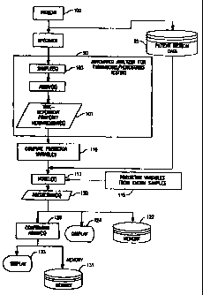
therapeutic condition. As can be seen in Figure 31,
one or more time-dependent measurements (101) are
performed on an unknown sample (103). The term "time-
CA 02316361 2000-06-22
WO 99/34208 PCT/US98/27865
-10-
dependent measurement" is referred to herein to
include measurements derived from assays (e.g. PT,
APTT, fibrinogen, protein C, protein S, TT, ATIII,
plasminogen and factor assays). The terms "unknown
sample" and "clinical sample" refer to a sample, such
as one from a medical patient (100), where a
congenital or acquired imbalance or therapeutic
condition associated with thrombosis/hemostasis is not
known (or, if suspected, has not been confirmed). In
the present invention, a coagulation property is
measured over time so as to derive a time-dependent
measurement profile. In a preferred embodiment, the
time-dependent measurement is an optical measurement
for deriving an optical profile. For example, a PT
profile, a fibrinogen profile, a TT profile, an APTT
profile and/or variations thereof can be provided
where, an unknown sample is analyzed for clot
formation based on light transmittance over time
through the unknown sample. In another preferred
embodiment, two (or more) optical profiles are
provided, such as both a PT profile and an APTT
profile.
After the time-dependent measurement profiles are
provided, a set of predictor variables are defined
(110) which sufficiently define the data of the time-
dependent profile. One or more predictor variables
comprise the set. And, in one embodiment, three or
more, and in a preferred embodiment, four or more
predictor variables were found to desirably make up
the set. It was found that the characteristics of the
time-dependent measurement profile could best be
defined by one or more predictor variables, including
the minimum of the first derivative of the optical
profile, the time index of this minimum, the minimum
of the second derivative of the optical profile, the
time index of this minimum, the maximum of the second
CA 02316361 2000-06-22
WO 99/34208 PCT/US98/27865
-11-
derivative, the time index of this maximum, the
overall change in transmittance during the time-
dependent measurement, clotting time, slope of the
optical profile prior to clot formation, and slope of
the optical profile after clot formation.
After defining the set of predictor variables, a
model (113) is derived which represents the
relationship between a congenital or acquired
imbalance or therapeutic condition and the set of
predictor variables. This model can be derived from
a neural network in one embodiment of the present
invention. In another embodiment, the model is
derived via a set of statistical equations.
Neural networks represent a branch of artificial
intelligence that can be used to learn and model
complex, unknown systems given some known data (115)
from which it can train. Among the features of neural
networks that make them an attractive alternative for
modeling complex systems are :
1. They can handle noisy data well and recognize
patterns even when some of the input data are
obscured or missing.
2. It is unnecessary to determine what factors are
relevant a priori since the network will
determine during the training phase what data are
relevant, assuming there are at least some
meaningful parameters in the set.
Neural networks are formed from multiple layers
of interconnected neurons like that shown in Figure 1.
Each neuron has one output and receives input.il...iõ
from multiple other neurons over connecting links, or
synapses. Each synapse is associated with a synaptic
weight, w,. An adder E or linear combiner sums the
products of the input signals and synaptic weights
CA 02316361 2000-06-22
WO 99/34208 PCT/US98/27865
-12-
ij*wj. The linear combiner output sum, and 0, (a
threshold which lowers or a bias which raises the
output) are the input to the activation function f()
The synaptic weights are learned by adjusting their
values through a learning algorithm.
After deriving the model (113), whether based on
neural networks or statistical equations, the model is
utilized to predict (120) the existence of a
congenital or acquired imbalance or therapeutic
condition in the unknown sample relative to the time-
dependent measurement profile(s). As such, a
congenital or acquired imbalance or therapeutic
condition can be predicted. Conditions which can be
predicted as being abnormal in the present invention
can include, among others, a) factor deficiencies,
e.g. fibrinogen, Factors II, V, VII, VIII, IX, X, XI
and XII, as well as ATIII, plasminogen, protein C,
protein S, etc., b) therapeutic conditions, e.g.
heparin, coumadin, etc., and c) conditions such as
lupus anticoagulant. In one embodiment of the present
invention, the method is performed on an automated
analyzer (90). The time-dependent measurement
profile, such as an optical data profile, can be
provided automatically by the automated analyzer,
where the unknown sample is automatically removed by
an automated probe from a sample container to a test
well, one or more reagents are automatically added to
the test well so as to initiate the reaction within
the sample. A property over time is automatically
optically monitored so as to derive the optical
profile. The predicted congenital or therapeutic
condition (120) can be automatically stored in a
memory (122) of an automated analyzer and/or displayed
(124) on the automated analyzer, such as on a computer
monitor, or printed out on paper. As a further
feature of the invention, if the predicted congenital
CA 02316361 2000-06-22
WO 99/34208 PCT/US98/27865
-13-
or acquired imbalance or therapeutic condition is an
abnormal condition (126), then one or more assays for
confirming the existence of the abnormal condition are
performed on the automated analyzer. In fact, in a
preferred embodiment, the one or more confirming
assays are automatically ordered and performed on the
analyzer once the predicted condition is determined,
with the results of the one or more confirming assays
being stored in a memory (131) of the automated
analyzer and/or displayed (133) on the analyzer.
Also, where the unknown sample is from a medical
patient, both the derived model and other patient
medical data (95) can be used for predicting the
imbalance/condition. If a monitoring system is used,
a plurality of optical measurements at one or more
wavelengths can be taken over time so as to derive the
optical profile, with the optical measurements
corresponding to changes in light scattering and/or
light absorption in the sample. Also, the plurality
of optical measurements can each be normalized to a
first optical measurement. If the time-dependent
measurement is an optical profile, this can be
provided automatically by an analyzer, where a sample
is automatically removed by an automated probe from a
sample container to a test well, one or more reagents
are automatically added to the test well so as to
initiate the property changes within the sample, and
the development of the property over time is
automatically optically monitored so as to derive the
optical data profile. And, the predictor variables
can be a plurality of variables, three or more
predictor variables, or more than three predictor
variables.
EXAMPLE 1: Prediction of Heparin in Sample
CA 02316361 2000-06-22
WO 99/34208 PCT/US98/27865
-14-
This example shows a set of predictor variables
that adequately describe screening assay optical
profiles, develops an optimal neural network design,
and determines the predictive capabilities of an
abnormal condition associated with
thrombosis/hemostasis (in this case for the detection
of heparin) with a substantial and well-quantified
test data set.
SimplastinTM L, PlatelinT"' L, calcium chloride
solution (0.025 M), imidazole buffer were obtained
from Organon Teknika Corporation, Durham, NC, 27712,
USA. All plasma specimens were collected in 3.2% or
3.8% sodium citrate in the ratio of one part
anticoagulant to nine parts whole blood. The tubes
were centrifuged at 2000 g for 30 minutes and then
decanted into polypropylene tubes and stored at -80QC
until evaluated. 757 specimens were prepared from 200
samples. These specimens were tested by the following
specific assays: FII, FV, FVII, FVIII, FIX, FX, FXI,
FXII, heparin, fibrinogen, plasminogen, protein C,
and AT-III. Samples represented normal patients, a
variety of deficiencies, and therapeutic conditions.
Of the specimen population 216 were positive for
heparin determined by a heparin concentration greater
than 0.05 units/ml measured with a chromogenic assay
specific for heparin. The remaining specimens,
classified as heparin-negative, included normal
specimens, a variety of single or multiple factor
deficiencies, and patients receiving other therapeutic
drugs. Positive heparin samples ranged to 0.54
units/ml.
PT and APTT screening assays were performed on
each specimen utilizing two automated analyzers (MDATM
180s) and multiple reagent and plasma vials (Organon
Teknika Corporation, Durham NC 27712, USA ) over a
CA 02316361 2000-06-22
WO 99/34208 PCT/US98/27865
-15-
period of five days. When clot-based coagulation
assays are performed by an automated optically-based
analyzer such as the MDA 180, data are collected over
time that represents the normalized level of light
transmission through a sample as a clot forms (the
optical profile). As the fibrin clot forms, the
transmission of light is decreased. The optical
profile was stored from each test.
The network configuration chosen, a multilayer
perceptron (MLP) maps input predictor variables from
the PT and APTT screening assays to one output
variable (see Figure 2) which represents a single
specified condition. A similar network was also
employed for PT-only variables and APTT-only
variables. This specific MLP consists of three
layers: the input layer, one hidden layer, and the
output layer.
A normal optical profile is shown in Figure 3.
The set of predictor variables were chosen with the
intent of describing optical profiles as completely as
possible with a minimum number of variables. They are
summarized in Table 1 where t is time from initiation
of reaction, T is normalized light transmission
through the reaction mixture, and pvik is the kth
predictor variable of assay j.
The predictor variables were scaled to values
between 0 and 1, based on the range of values observed
for each variable for assay type k
CA 02316361 2000-06-22
WO 99/34208 PCT/US98/27865
-16-
j~ ' " \PVJk ' (PVf -"''t (PVJ-vk )m~ =
~
The input variable set includes il 7 for both a PT
assay and APTT assay for each specimen. For known
output variable values, heparin samples with results
of greater than 0.05 units/ml were considered
positive and assigned a value of 1 while negative
samples were assigned a value of 0.
As the ratio of training set sample to
the number of weights in a network decreases, the
probability of generalizing decreases, reducing the
confidence that the network will lead to correct
classification of future samples taken from the same
distribution as the training set. Thus, small samples
sizes, then can lead to artificially high
classification rates. This phenomenon is known as
overtraining. In order to achieve a true accuracy
rate of 80%, a guideline for the number of samples in
the training set is approximately five times the
number of weights in the network. For most of this
work, a 14-6-1 network was used, leading to an upward
bound on the sample size of 0(450). To monitor and
evaluate the performance of the network and its
ability to generalize, a cross-validation set is
CA 02316361 2000-06-22
WO 99/34208 PCTIUS98/27865
-17-
processed at the end of each training epoch. This
cross-validation set is a randomly determined subset
of the known test set that is excluded from the
training set.
Once the input predictor variables and output
values were determined for all specimen optical
profiles, the 757 sets of data were randomly
distributed into two groups: 387 were used in the
training set and 370 were used in the cross-validation
set. These same two randomly determined sets were
used throughout all the experiments.
All synaptic weights and threshold values were
initialized at the beginning of each training session
to small random numbers.
The error-correction learning rule is an
iterative process used to update the synaptic weights
by a method of gradient descent in which the network
minimizes the error as pattern associations (known
input-output pairs) in the training set are presented
to the network. Each cycle through the training set
is known as an epoch. The order or presentation of
the pattern associations was the same for all epochs.
The learning algorithm consists of six steps which
make up the forward pass and the backward pass. In
the forward pass, the hidden layer neuron activations
are first determined
CA 02316361 2000-06-22
WO 99/34208 PCT/US9S/27865
-18-
h = F(iW 1 + Oh)
where h is the vector of hidden-layer neurons, i the
vector of input-layer neurons, W1 the weight matrix
between the input and hidden layers, and F() the
activation function. A logistic function is used as
the activation function
F(x) 1+e
Then the output-layer neurons are computed
o = F(hW2 + 90)
where o represents the output layer, b the hidden
layer and W2 the matrix of synapses connecting the
hidden layer and output layers. The backward pass
begins with the computation of the output-layer error
ea=(o-d)where d is the desired output. If each element of eo
is less than some predefined training error tolerance
CA 02316361 2000-06-22
WO 99/34208 PCTIUS98/27865
-19-
vector TEwõ than the weights are not updated during
that pass and the process continues with the next
pattern association. A training error tolerance of
0.1 was used in all experiments unless otherwise
specified. Otherwise, the local gradient at the
output layer is then computed:
90 =0(1- o)eo.
Next, the hidden-layer local gradient is computed:
9,,' h(1- h)W2ga.
once the hidden layer error is calculated, the second
layer of weights is adjusted
W2m = W2m-1 + OW2
where
OW2 = t7ng0 + 'yAW2m-1
___--
_....._._____.___
CA 02316361 2000-06-22
WO 99/34208 PCT/US98/27865
-20-
is the learning rate, y is the momentum factor, and m
is the learning iteration. The first layer of weights
is adjusted in a similar manner
W1mW1m-1+AW1
where
AW 1 = -qie + -yaW lm-1.
The forward pass and backward pass are repeated for
all of the pattern associations in the training set,
referred to as an epoch, 1000 times . At the end of
each epoch, the trained network is applied to the
cross-validation set.
Several methods were employed to measure the
performance of the network's training. Error, E, for
each input set was defined as
ql
-~_.___.r....~........-..-__ __._..
CA 02316361 2000-06-22
WO 99/34208 PCTIUS98/27865
-21-
The learning curve is defined as the plot of E versus
epoch. The percent classification, V, describes the
percent of the total test set (training and cross-
validation) that is correctly classified based on some
defined decision boundary, ft. Receiver-Operating
Characteristic (ROC) plots have also been utilized to
describe trained networks' ability to discriminate
between the alternative possible outcome states. In
these plots, measures of sensitivity and specificity
are shown for a complete range of decision boundaries.
The sensitivity, or true-positive fraction is defined
as
sensitivity = tme positive
true positive + false negative
and the false-positive fraction , or (1-specificity)
is defined as
~1-- specifcity) = .f~e,positive
false positive + true negative
These ROC plots represent a common tool for evaluating
clinical laboratory test performance.
CA 02316361 2000-06-22
WO 99/34208 PCT/US98/27865
-22-
Using the test set described, experiments were
performed to determine if the presence of heparin
could be predicted with this method. First,
experiments were conducted to determine optimal error-
correction backpropagation learning parameters: (1)
hidden layer size, (2) learning rate, and
(3) momentum. Additional experiments were also
conducted to compare the performance of networks based
on PT and APTT assays alone with that of one combining
the results of both, the effect of the training error
tolerance, and the decision boundary selection.
Figure 9 shows the effect of the hidden layer
size on the training and cross validation error and
the percent correct classification for the optimal
decision boundary, defined as the decision boundary
which yielded the lowest total number of false
positives and false negatives from the total test set.
As the hidden layer size is increased, the error is
decreased. However, the ability to generalize does
not increase after a hidden layer size of 6. The most
significant benefit in terms of both error and
percentage correct classification is between 4 and 6.
A hidden layer size of 6 was used for the remainder of
the experiments.
A series of experiments were conducted with
71 = {0.01,0.1,0.5,0.9} and ~y = {0.0,0.1,0.5,0.9} . Figure 4
shows the learning curves for two of the best
CA 02316361 2000-06-22
WO 99/34208 PCT/US98/27865
-23-
combinations of parameters. Figure 5 shows an example
learning curve when the learning rate is so high it
leads to oscillations and convergence to a higher E.
In general, as n -0 0 the network converged to a lower E
and as ly -30 1, the rate of convergence improved. As ~J
-lo1, the value of E converged too increased and
oscillations increased. In addition, as -y -~ 1
exacerbated the oscillations.
Figure 6 shows a comparison of the learning curve
for the training set and cross-validation set for
n=0.5 and 7=0.1. It is a primary concern when
developing neural networks, and it has been previously
shown that it is important to look not only at the
error in the training set for each cycle, but also the
cross-validation error.
Figure 7 shows the learning curve n=0.5 and y=0.1
and a learning tolerance of 0.0 and 0.1. These
results suggest that a small learning tends to
smoothen the convergence of the learning process.
Figure 8 shows the ROC plot for networks trained
with the predictor variables from each of the two
screening assays with that of them combined. In the
single assay cases, the hidden layer size was'3.
While using the data from one assay does lead to some
success, using the information from both assays makes
CA 02316361 2000-06-22
WO 99/34208 PCT/US98/27865
-24-
a significant improvement in the ability of the
network to correctly predict the presence of heparin.
This graph indicates that a 90% true positive
proportion can be achieved with a false positive
proportion of 15%. Using a single assay, a 60-70%
true positive proportion can be achieved with a false
positive proportion of approximately 15%.
EXAMPLE 2: Factor VIII
Similar tests were run as in Example 1. As can
be seen in Figures 10 and 11, two training sessions
were conducted for predicting a Factor VIII condition
in an unknown sample. Figure 10 is a receiver
operator characteristic plot related to predicting an
abnormality in relation to Factor VIII. In Figure 10,
everything below 30% activity was indicated as
positive, and everything above 30% was indicated as
negative. Cutoff values other than 30% could also be
used. In this Example, the activity percentage has a
known accuracy of approximately + or - 10t. In Figure
11, the actual percent activity was utilized as the
output.
EXAMPLE 3: Factor X
As can be seen in Figure 12, the method of the
present invention was run similar to that as in
Example 2, where here an abnormality in Factor X
CA 02316361 2000-06-22
WO 99/34208 PCT/US98/27865
-25-
concentration was predicted from unknown samples.
Everything below 30% activity was indicated as
positive, and everything above 30% was indicated as
negative. Cutoff values other than 30% could also be
used.
The results of the cross-validation sample sets
throughout the experiments indicate that the sample
size was sufficient for the network to generalize.
While the random distribution of the training and
cross-validation sets were held constant throughout
the experiments presented, other distributions have
been used. These distributions, while all yielding
different results, still lead to the same general
conclusion.
Many alternatives for or additions to the set of
predictor variables were explored. This included
coefficients of a curve fitted to the data profile,
pattern recognition, and clot time-based parameters.
Low order functions tend to lose information due to
their poor fit, and high order functions tend to lose
information in their multiple close solutions. Clot-
based parameters, such as clot time, slope in the
section prior to the initiation of clot formation, and
afterwards, are often available, but not always
(because in some samples, the clot time is not
detectable). The successful results observed
indicate that the set of predictor variables used are
CA 02316361 2000-06-22
WO 99/34208 PCTIUS98/27865
-26-
effective for predicting congenital or acquired
imbalances or therapeutic conditions.
The optimization of the network learning
algorithm's parameters made significant differences in
its performance. In general, performance was best
with low learning rates, high momentum rates, some
small training error tolerance, and a hidden layer
size approximately half of the size of the input
layer.
ADDITIONAL EXAMPLES:
Optical measurements for APTT and PT assays were
performed on MDA 180 instruments at a wavelength of
580 nm. Plasma specimens (n= 200) included normal
patients, patients with a variety of coagulation
factor deficiencies and patients undergoing heparin or
other anticoagulant therapy. Duplicate APTT and PT
screening assays were performed on each specimen with
two MDA 180s using single lots of APTT and PT
reagents. These specimens were also analyzed using
specific assays for FII, FV, FVII, FVIII, FIX, FX,
FXI, FXII, heparin, fibrinogen, plasminogen, protein C
and antithrombin-III.
Data Processing and Neural Networks
CA 02316361 2000-06-22
WO 99/34208 PCT/US98/27865
-27-
Optical profile data files were exported from the
MDA 180s and processed off-line. A set of nine
parameters was derived to describe the timing, rate
and magnitude of coagulation events. These parameters
were calculated for all APTT and PT tests. The
parameter set is modified slightly from that for
Example 1. In this approach, the optical data for a
PT or APTT assay was divided into three segments (a
pre-coagulation segment, a coagulation segment and a
post-coagulation segment) using divisions based on the
minimum and maximum value of the second derivative for
changes in optical signal with respect to time. The
parameters that were analyzed included: (1) the times
at which the onset, midpoint and end of the
coagulation phase occur (tmin2, tminl and tmax2;
respectively); (2) mean slopes for the pre-coagulation
phase and the post-coagulation phase (slopei and
slope3, respectively) and the slope at the mid-point
of coagulation (mini, the coagulation "velocity" at
reaction midpoint, which is analogous to slope2); (3)
terms for coagulation "acceleration" and
"deceleration" (min2 and max2, respectively); and (4)
the magnitude of signal change during coagulation
( de1 ta ) .
Three different sets of data parameters were
used as input to the neural network: (1) the nine
parameters from PT assays, (2) the nine parameters
CA 02316361 2000-06-22
WO 99/34208 PCT/US98/27865
-28-
from APTT assays, and (3) the combined parameters
from the APTT and PT assays. Each specimen was run
in duplicate on two instruments, to give a total of
approximately 800 parameter sets from the 200
specimens. The total number varied slightly because of
missing data due to insufficient sample, mechanical
failure or unspecified failures. The data parameter
sets were divided into training and cross-validation
sets randomly by specimen where all replicates for a
given specimen were grouped either in the cross-
validation set or training set. The same training and
cross-validation sets were used throughout this study.
The method for training and cross-validation of the
back-propagation neural networks has been described in
relation to Example 1. Each neural network was trained
for 1000 epochs. Training parameters were learning
rate, 0.01; momentum, 0.5; learning tolerance, 0.10;
decay, 0.05; input layer size, 18 (or 9 for single
assays); hidden layer size, 9 (or 5 for single
assays); and output layer size, 1. Three types of
networks were trained. These included networks that
classified specimens as deficient or non-deficient
based on a single diagnostic cut-off, sets of networks
that used diagnostic cut-offs at different levels of
the same factor, and networks trained to estimate the
actual concentration of a specific factor.
CA 02316361 2000-06-22
WO 99/34208 PCT/US98/27865
-29-
Classification of Factor Deficiencies Based on a
Single Diagnostic Cut-off Level
In the first set of tests, neural networks were
trained to classify plasma samples into two groups,
positive (factor-deficient) and negative (non-
deficient), and results were compared to classification
based on the measured factor concentration for the
specimens. In most testing, the diagnostic cut-off for
defining factor deficiencies was set as 30%; that is,
specimens with a measured concentration of less that 30%
of normal for a specific factor were defined as
deficient and those with greater than 30% activity were
defined as non-deficient. These diagnostic cut-off
levels were arbitrarily defined, but are based on
clinical requirements and reagent sensitivity. The
desired output from positive samples and negative
samples were defined as '1' and '0', respectively; the
actual output for each specimen was a floating point
value, a, where 0< a< 1. Figure 22 shows the
constituency of the training and cross-validation sets
with regard to each factor deficiency. Classification
of specimens was evaluated at varying "decision
boundaries" that divided the neural network outputs into
positive and negative groups. This positive or negative
classification was then compared to the desired output
(the known classification) for each input data set.
Results were plotted as nonparametric receiver-operating
CA 02316361 2000-06-22
WO 99/34208 PCT/US98/27865
-30-
characteristic (ROC) curves and the areas under the
curves were computed along with their associated
standard errors. ROC curves were also derived for APTT
and PT clot time values for comparison. Data points on
the ROC curves represent the proportion of true-positive
and false-positive classifications at various decision
boundaries. Optimum results are obtained as the true-
positive proportion approaches 1.0 and the false-
positive proportion approaches 0.0 (upper-left corner of
graph). The optimum global measure of the ROC curve is
an area of 1Ø
Classification of Factor Deficiencies at Multiple
Diagnostic Cut-off Levels
A second set of networks was trained for FX
classification in a similar manner to the first set
except that the diagnostic cut-off level was varied
(10t, 30%, and 50%). FX was chosen for this experiment
because the data set contained a greater number of
positive samples at all cut-off levels than other
factors.
Estimation of Factor Concentration Using Neural
Networks
A third set of networks were trained to approximate
actual specific factor activities (FII, FV, FVII, FVIII,
FIX, FX, FXI and FXII) and fibrinogen levels from
CA 02316361 2000-06-22
WO 99/34208 PCT/US98/27865
-31-
combined PT and APTT parameters from unknown samples.
In these cases, the desired output of the training and
cross-validation sets was the measured activity for a
specific factor for each specimen and the actual output
of the neural network was a predicted concentration for
this, specific factor activity. The coefficients of
linear regressions using the desired outputs versus the
actual neural network outputs for the cross-validation
set were used to describe the performance of these
networks. The Pearson product moment correlation
coefficient, r, was used to estimate the correlation
between the two data sets.
Classification of Factor Deficiencies Based on a
Single Diagnostic Cut-off Level
Neural networks were trained to classify samples as
deficient (positive result) or non-deficient (negative
result) for individual plasma factors, using a value of
30% activity as the diagnostic cut-off to define
deficiencies. Results were examined graphically using
receiver-operating curves (ROC). These graphs plot the
true-positive proportion (number of positives detected
divided by the total number of positives) versus the
false-positive proportion (number of negative specimens
incorrectly diagnosed as positive divided by the total
number of negatives). An ROC curve is generated by
determining true-positive and false-positive proportions
CA 02316361 2000-06-22
WO 99/34208 PCT/US98/27865
-32-
at different "decision boundaries" for the diagnostic
test. For example, an ROC plot for diagnosis of FII
deficiencies using PT clot time was generated by varying
the decision boundary (value of PT clot time) used to
differentiate between deficient and non-deficient
specimens. When a short clot time is used as the
decision boundary, most deficient specimens can be
identified but a significant proportion of non-deficient
specimens may also be flagged (false-positives). When
a long clot time is used as the decision boundary, the
proportion of false-positives decreases, but the number
of true-positive specimens that are not diagnosed may
also increase. Under ideal conditions, a decision
boundary can be identified from an ROC curve that
produces a very high proportion of true-positives and a
very low proportion of false-positives. This condition
corresponds to the upper left region of the ROC plot.
Two related terms that are often applied to clinical
diagnostic tests are "sensitivity" and "specificity".
Sensitivity refers to the ability to detect positive
specimens and corresponds to the y-axis of the ROC
plots. Specificity refers to the proportion of
specimens diagnosed as negative which are correctly
identified. The ROC x-axis equals (1-specificity).
Visual assessment of the ROC curves is one method used
to evaluate the performance of the neural networks and
compare them to the diagnostic power of PT and APTT clot
CA 02316361 2000-06-22
WO 99/34208 PCT/US98/27865
-33-
times. Another method is to measure the diagnostic
performance by using the area under the ROC curves. The
area under the ROC curve is equivalent to an estimate of
the probability that a randomly chosen positive specimen
will have a more positive result than a randomly chosen
negative specimen. In the event that ROC curves
overlap, the shape of the curves as well as the areas
beneath them becomes important. An ROC curve
encompassing a smaller area may be preferable to an
overlapping curve with greater area depending on the
desired performance for a given diagnostic system.
Figures 14 - 21 show ROC curves for neural networks
trained to predict FII, FV, FVII, FVIII, FIX, FX, FXI,
and FXII deficiencies from PT parameters alone, from
APTT parameters alone, or from combined APTT and PT
parameters. ROC plots based on classification using
APTT and PT clot times are included for comparison.
Figure 23 shows the area under these curves and their
associated standard errors.
Results for classification of FII deficiencies are
shown in Figure 14. Best results were observed for
neural networks using APTT parameters alone or combined
with PT parameters, with area under ROC curves greater
than 0.99 in both cases (Figure 23). Classification
based on PT or APTT clot times, or from neural networks
using PT data alone resulted in less successful
classification and reduced area under curves.
---
CA 02316361 2000-06-22
WO 99/34208 PCT/US98/27865
-34-
Results from classification of FV deficiencies
showed somewhat different characteristics (Figures 15
and 23). Best results were observed for classification
from a neural network using APTT data parameters, based
on visual inspection and area under the ROC curve. Less
successful classification were obtained from neural
networks using PT data parameters alone or combined with
APTT data, and from PT clot time, as judged from areas
under ROC curves. Classification based on PT clot time
was qualitatively different from neural networks using
PT data, however, and tended toward higher sensitivity
rather than specificity. This type of pattern was
observed for classification of several coagulation
factors, especially factors VIII, X and XI. In
situations where overlapping ROC curves were obtained,
consideration of the relative value of specificity and
sensitivity, as well as the area under ROC curves,
becomes important in comparing diagnostic results.
For several of these plasma factors, including FV,
FVI II , FIX, FX, FXI and FXII (Figures 15, 17, 18, 19,
20 and 21), it appeared that it would be possible to
achieve a moderately high true-positive proportion (>
0.6) while maintaining a low false-positive proportion
(< 0.1) from neural networks using PT, APTT or combined
parameters. This corresponds to a situation where a
significant proportion of deficient specimens are not
CA 02316361 2000-06-22
WO 99/34208 PCT/US98/27865
-35-
detected (moderate sensitivity), but those that are
detected are correctly classified as deficient for that
specific factor (high specificity). In contrast, using
PT or APTT clot times it was possible for most factors
to adjust decision boundaries to identify most
deficiencies (true-positive proportion approaching 1.0,
high sensitivity), but with a relatively high rate of
false-positives (low specificity). This corresponds to
a situation where most or all deficient specimens are
detected, but where the specific factor deficiency is
frequently not correctly identified. The first scenario
involving moderate or high true-positive rates with very
low false positive rates may be preferable in the
diagnostic scheme shown in Figure 13.
For factors II, V, IX and XII, it appeared that an
appropriate choice of neural network gave best
diagnostic performance, as judged from the area under
curves. For factors VIII, X and XI, neural networks were
not visibly superior to diagnosis based on clot times
when areas under ROC curves were the only consideration;
however, neural networks for these factors did provide
better specificity. For one factor (FVII, Figure 16),
neural network classification was less effective than
for other factors, at least in this test system.
The performance of networks using data parameters
from PT or APTT assays alone or in combination varied
for different factors. For factors VIII and XII, best
CA 02316361 2000-06-22
WO 99/34208 PCT/US98/27865
-36-
performance (significantly greater area with no overlap)
was observed when the combined sets of AP1F:~-PT data
parameters were used. For several other factors, use of
a single parameter set provided results that were
comparable to or better than the combined APTT and PT
parameters. A network using only APTT data parameters
(APTT NN) was equivalent (similar area) to a network
using combined APTT-PT data (APTT-PT NN) for FII and FX;
and superior for FV (greater area and no overlap).
Networks using only PT parameters provided results that
were comparable (similar area) to the combined
parameters for FV classification and better (greater
area and insignificant overlap) for FIX classification.
The data for misclassified positive specimens were
examined more closely. Misclassified positive specimens
were clustered in several categories: 1) Specimens with
"no clot" APTT or PT results (specimens with very
prolonged or very weak coagulation reaction for which no
clot time can be reliably calculated); 2) specimens with
multiple deficiencies or abnormalities; 3) specimens
with borderline deficiencies (factor activity marginally
lower than the diagnostic cut-off of 30%); and 4)
specimens with atypically steep slope during the pre-
coagulation phase for APTT assays that were not
characteristic of other specimens in the same
classification (FX deficiencies were not detected for
CA 02316361 2000-06-22
WO 99/34208 PCT/US98/27865
-37-
two specimens exhibiting this characteristic with FX
activities of 26.8% and 16.8%, respectively).
Classification of Factor Deficiencies at Multiple
Diagnostic Cut-off Levels
The ability of neural networks to classify FX-
deficient specimens was tested at varying diagnostic
cut-offs. Areas under the ROC curves for cut-off
levels of 10%, 30% and 50% FX activity are shown in
Figure 24. Results indicate that progressively poorer
classification (as expressed in smaller areas under ROC
curves) was observed as higher cut-off levels were used.
This was true for classification based on neural
networks or PT clot times.
Neural Network Estimation of Factor Concentration
Neural networks were also trained to estimate
actual protein concentrations (as opposed to a
positive/negative classification at a defined cut-off)
for FIi, FV, FVII, FVIII, FIX, FX, FXI, FXII and
fibrinogen. Linear correlation coefficients for the
estimated and measured concentrations are shown in
Figure 25 for all experiments, and plots of the
correlation data are shown in Figure 26 for fibrinogen
and Figure 27 for FX. Correlation data between PT and
APTT clot time and measured concentrations are also
shown in Figure 25 for comparison.
CA 02316361 2000-06-22
WO 99/34208 PCT/US98/27865
-38-
Example: Se1f-organizing Feature Maps
Neural networks using self-organizing feature maps
and learning vector quantization were used to analyze
optical data from clinical coagulation tests. Self-
organizing feature maps using an unsupervised learning
algorithm were trained with data from normal donors,
patients with abnormal levels of coagulation proteins
and patients undergoing anticoagulant therapy. Specimen
categories were distinguishable in these maps with
varying levels of resolution. A supervised neural
network method, learning vector quantization, was used
to train maps to classify coagulation data. These
networks showed sensitivity greater than 0.6 and
specificity greater than 0.85 for detection of several
factor deficiencies and heparin.
An alternative approach to analyzing PT and APTT
data with artificial neural networks (as set forth in
Example 1) is by using self-organizing feature maps.
Self-organizing feature maps contain layers of input and
output neurons only and contain no hidden layers.
Training is based on competitive learning where the
output neurons compete with one another to be activated
and only one output neuron is activated for any given
set of inputs. Output neurons become selectively tuned
to certain input patterns, and data with similar
features tend to be grouped together spatially. This
type of neural network may use either an unsupervised or
CA 02316361 2000-06-22
WO 99/34208 PCT/US98/27865
-39-
supervised learning algorithm. When an unsupervised
method is used, such as the self-organizing map (SOM)
algorithm, unidentified input patterns are presented to
the network during training and the output for each
input pattern is the coordinates of the winning neuron
in the output layer, or map. When a supervised method
is used, such as learning vector quantization (LVQ),
input patterns are presented along with a known sample
classification to the network during training and the
output is a unique predicted classification. The LVQ
method is similar to SOM, except that the map is divided
into classes, and the algorithm attempts to move outputs
away from the boundaries between these classes.
MDA Simplastin L (PT reagent), MDA Platelin L (APTT
reagent) and other reagents were obtained from Organon
Teknika Corporation, Durham, NC 27712, USA, unless
otherwise indicated. Factor-deficient plasmas for
factor assays were obtained from Organon Teknika and
George King Bio-Medical Corporation, Overland Park,
Kansas 66210, USA. Additional factor-deficient plasmas
were obtained from HRF, Raleigh, NC 27612, USA.
Random samples, specimens from patients receiving
heparin or oral anticoagulant therapy, and other
specimens were obtained from Duke University Medical
Center Coagulation Laboratory.
All testing was performed on MDA 180 coagulation
analyzers (Organon Teknika). Optical measurements for PT
CA 02316361 2000-06-22
WO 99/34208 PCTIUS98/27865
-40-
and APTT assays were performed at a wavelength of 580
nm. Plasma specimens (n= 200) included normal patients,
patients with a variety of deficiencies, and patients
undergoing heparin or other anticoagulant therapy.
Duplicate PT and APTT assays were performed on each
specimen using two MDA 180s to give a total of
approximately 800 parameter sets from the 200 specimens.
The total number varied slightly because of missing data
due to insufficient sample, mechanical failure or
unspecified failures. These specimens were also tested
to determine the concentration of coagulation factors
(FII, FV, FVII, FVIII, FIX, FX, FXI, FXII) heparin, and
fibrinogen. The diagnostic cut-off for defining factor
deficiencies was set at 30%; that is, specimens with a
measured concentration of less that 30% of normal for a
specific factor were defined as deficient and those with
greater than 30% activity were defined as non-deficient.
Samples were defined as positive for heparin if the
measured heparin concentration was greater than 0.05
IU/ml.
Optical Data Processing
Optical profile data files were exported from MDA
180s and processed off-line. A set of nine parameters
was derived to describe the timing, rate and magnitude
of coagulation events for PT and APTT tests, as
described previously. In this approach, the optical
CA 02316361 2000-06-22
WO 99/34208 PCT/US98/27865
-41-
data for a PT or APTT assay was divided into three
segments (a pre-coagulation segment, a coagulation
segment and a post-coagulation segment) using divisions
based on the minimum and maximum value of the second
derivative for changes in optical signal with respect to
time. Parameters included: 1) the times at which the
onset, midpoint and end of the coagulation phase occur;
2) mean slopes for the pre-coagulation phase and the
post-coagulation phase and the slope at the mid-point of
coagulation; 3) terms for coagulation "acceleration" and
"deceleration"; and 4) the magnitude of signal change
during coagulation.
Self-Organizing Map Algorithm
A self-organizing feature map neural network
consists of input and output layers of neurons. The
self-organizing map (SOM) algorithm transforms an input
vector (a set of data parameters from PT or APTT optical
data for a single test) to an individual output neuron
whose location in the output layer, or map, corresponds
to features of the input data. These features tend to
be spatially correlated in the map. There are five
steps in the SOM learning process:
1. Unique weight vectors wi(0), are randomly,chosen.
2. A sample from the training set is selected.
3. The best-matching winning neuron i(x) at time n,
using the minimum-distance Euclidean criterion
CA 02316361 2000-06-22
WO 99/34208 PCT/US98/27865
-42-
i(x) = arg min{Ilx(n) - W., (n)II}
is identified.
4. The weight vectors of all neurons are updated with
the formula
{w1(n) + a(n)[x(n) - w, (n),, j E Njn)
w (n+l)= wi(n), j o N, (n)
where a(n) is the learning rate parameter, and N, (n)
is the neighborhood function centered around the
winning neuron i(x) ; both a(n) and N,(n) vary
dynamically during training.
5. Steps 2 through 4 are repeated until the map
reaches equilibrium.
The SOM tests were performed using the Self-
Organizing Map Program Package (SOM PAK) available from
the Helsinki University of Technology, Laboratory of
Computer Sciences. Two different sets of parameters
were used as input to the SOMs: (1) the nine parameters
from a PT assay, and (2) the nine parameters from the
APTT assay. All data sets (786) were used to train the
SOMs. A 10x10 map was trained using a hexagonal
neighborhood in two stages. In the first stage, the map
was trained for 1000 epochs (an epoch is one cycle
through all data sets) with an initial learning rate
CA 02316361 2000-06-22
WO 99/34208 PCT/US98/27865
-43-
parameter of 0.5 (decreasing linearly to zero during
training) and a neighborhood radius of 10 (decreasing
linearly to 1 during training). In the second stage,
the map was trained for 10000 epochs using a learning
rate parameter of 0.1 and a radius of 3.
Learning Vector Quantization
Learning vector quantization (LVQ) is a supervised
learning algorithm often used to fine-tune self-
organizing feature maps in order to use them in the role
of a pattern classifier. The classification accuracy of
the map is improved by pulling the weight vectors away
from the decision surfaces that demarcate the class
borders in the topological map. There are several
variations of the LVQ algorithm; the one used here is
referred to as LVQ1. The learning process is similar to
the SOM algorithm described above, except that known
sample classifications are included when weight vectors
are updated (step 4):
1. Initial weight vectors wj(0), are randomly chosen.
2. A sample from the training set with a known
classification is selected.
3. The best-matching winning neuron i(x) at time n,
using the minimum-distance Euclidean criterion
i(x) = arg minlIlx(n) - w (n)II}
CA 02316361 2000-06-22
WO 99/34208 PCT/US98/27865
-44-
is identified.
4. The weight vectors of all neurons are updated with
the formula
w, (n) + a(n)[x(n) - wj (n),, j = Cõ
wj (n + 1~ = w j(n) - a(n)[x(n) - w, (n)], j = Cx
w j (n), j ~ i
where is the class associated with the vector Wt
and Cx is the class associated with the input vector
X.
5. Steps 2 through 4 are repeated until the map
reaches equilibrium.
The LVQ tests were performed using the Learning
Vector Quantization Program Package (LVQ_PAK), also
available from the Helsinki University of Technology,
Laboratory of Computer Sciences. The sets of parameters
from the APTT assay or PT assays were used for the LVQ
networks. The data parameter sets were divided evenly
into training and cross-validation sets randomly by
specimen, where all replicates for a given specimen were
grouped either in the cross-validation set or training
set. The same training and cross-validation sets were
used throughout this study. The LVQ networks were
trained to classify plasma samples into two categories,
positive (factor-deficient specimens or specimens from
patients undergoing anticoagulant therapy) and negative
(non-deficient or no anticoagulant therapy), and results
CA 02316361 2000-06-22
WO 99/34208 PCTIUS98/27865
-45-
were compared to classification based on the measured
factor concentration or therapeutic condition for the
specimens. LVQ training was performed using 200 weight
vectors, 10000 epochs, initial learning rate parameter
of 0.5 (decreasing linearly to 0), and 7 neighbors used
in knn-classification.
LVQ networks were evaluated using sensitivity (the
proportion of known positive specimens that were
correctly classified as positive by the network),
specificity (the proportion of known negative specimens
that were correctly classified as negative by the
network), positive predictive value (PPV), negative
predictive value (NPV) and efficiency. These terms are
defined below, where TP, TN, FP and FN correspond to
true positive, true negative, false positive and false
negative classifications, respectively.
TP
sensitivity = TP + FN
TN
specificity = FP + TN
CA 02316361 2000-06-22
WO 99/34208 PCT/US98/27865
-46-
TP
PPV=TP+F
TN
NPV = TN + FN
TN+TP
efficiency= TP+FP+FN+TN
Self-Organizing Map Algorithm
Self-organizing feature maps were trained using
optical data parameters from either PT or APTT data for
200 specimens as input. Network output consisted of map
coordinates for each specimen. Contour plots were
constructed for six categories of known specimen
classifications: normal donors, specimens with heparin
> 0.05 IU/ml, fibrinogen >600mg/dl, fibrinogen <200
mg/dl, patients receiving oral anticoagulants, and
factor-deficient specimens (specimens with <30% of
CA 02316361 2000-06-22
WO 99/34208 PCT/US98/27865
-47-
normal activity for FII, FV, FVII, FVIII, FIX, FX, FXI,
or FXII). These contour plots depict the distribution
of specimens within a category according to their map
coordinates.
Figure 28: Contour plots for populations of samples
used in training a self-organizing feature map using the
unsupervised training method SOM based on data from APTT
assays. Optical data parameters from 765 APTT assays
were used to train this self-organizing feature map.
The shaded areas represent the distribution of output
neurons for specific specimen populations within the
feature map. Each contour line represents an
incremental step of one test result located at a given
set of map coordinates.
Figure 28 shows SOM contour plots derived from APTT
optical data for the six specimen categories. Specimens
containing low fibrinogen and high fibrinogen were
classified at opposite borders of the SOM with no
overlap. Normal populations showed some overlapping
with low fibrinogen, factor deficient and oral
anticoagulated categories. Overlap between normal
specimens and edges of the high and low fibrinogen
populations is expected, since some proportion of
healthy donors have fibrinogen levels that are lower or
higher than normal. Overlap between mapping of normal
specimens and factor-deficient plasmas is also not
surprising, since APTT tests are sensitive to some
CA 02316361 2000-06-22
WO 99/34208 PCT/US98/27865
-48-
factor-deficiencies (but not others), whereas PT assays
are sensitive to a separate subset of factor
deficiencies. The low fibrinogen category tended to
overlap the factor-deficient category, consistent with
our observation that many factor-deficient specimens
also had reduced fibrinogen levels. The heparin
category tended to overlap the high fibrinogen category,
again consistent with measured levels of fibrinogen for
these specimens. Little or no overlap was observed
between normal specimens and specimens containing
heparin. Specimens from patients receiving oral
anticoagulant therapy show significant overlap with both
normal and heparin populations. This is consistent with
known properties of APTT assays, which are sensitive to
heparin therapy but relatively insensitive to oral
anticoagulant therapy.
Figure 29: Contour plots for populations of samples
used in training a self-organizing feature map using the
unsupervised training method SOM based on optical data
from 765 PT assays. Experimental details are as
described in the Materials and Methods section and in
Figure 28.
Contour plots for self-organizing feature maps
trained with PT data are shown in Figure 29. Results
are similar to maps from APTT data in several respects:
(1) high and low fibrinogen were well resolved at
opposite sides of the map; (2) normal specimens were
CA 02316361 2000-06-22
WO 99/34208 PCT/US98/27865
-49-
localized in a region that overlapped low fibrinogen
specimens slightly; (3) factor-deficient specimens were
distributed between non-overlapping regions and regions
that overlapped low fibrinogen and normal populations.
Overlap was consistent with measured fibrinogen for some
specimens, and with poor sensitivity of PT reagents to
some factor deficiencies in other cases; (4) oral
anticoagulated specimens showed some overlap with both
normal and heparin populations; and (5) the heparinized
population was distributed over a large portion of the
map. Overlap between heparinized specimens and high
fibrinogen populations was consistent with measured
fibrinogen levels. The resolution of the heparin
population is somewhat surprising, considering that PT
reagents are relatively insensitive to heparin.
These results indicate that self-organizing feature
maps are capable of distinguishing differences in
optical data parameters from APTT and PT assays even
when no information regarding specimen diagnosis is
presented to the neural network. Resolution of specimen
populations was variable, depending on reagent
properties and sensitivities, and on whether specimens
belonged to a given category uniquely or to multiple
overlapping categories.
Example
CA 02316361 2000-06-22
WO 99/34208 PCT/US98/27865
-50-
The following procedure was performed for PT assays
(see Figure 32) and then separately for APTT assays (see
Figure 33).
For Clot Time:
1. Calculate mean and standard deviation (SD) for
clot time from assays (PT or APTT) run on aliquots from
normal specimens (n = 79).
2. Calculate z-scores for clot times from the
normal group in step from the normal group in step 1(n
= 79) and a group from assays performed on aliquots from
abnormal specimens (n=410). The group of abnormals
included various factor deficiencies, oral-
anticoagulated specimens, suspected DIC specimens, and
heparinized specimens. Z-scores are calculated by
subtracting the mean of normals from the clot time and
then dividing the result by the SD.
3. Determine the number of true positives, true
negatives, false positives and false negatives if
specimens with an absolute value of the z-score greater
than x SD (where x=1,2,3,4,5,) are called positive.
For parameters:
1. Calculate Mean and SD for each of the nine
parameters (slope_1, slope_3, delta, index min 1, min_1,
index max 2, max_2, index min_2, min_2) from assays (PT
or APTT) run on aliquots from normal specimens (n=79).
CA 02316361 2000-06-22
WO 99/34208 PCT/US98/27865
-51-
2. Calculate z-scores for each parameter of each
specimen from the normal group (n=79) and abnormal group
(n=410).
3. Determine the number of true positives, true
negatives, false positives and false negatives if
specimens with an absolute value of the z-score greater
than x SD (where x=1,2,3,4,5) for one or more of the
parameters are called positive.
Results:
Sensitivity and specificity for non-specific
abnormals as a group is higher when using all parameters
rather than the traditional clot time used alone. This
method requires only (1) a group of known normal
specimens, (2) calculation of mean and SD for each of
the nine parameters of the normal group and an unknown
specimen, (3) computation of z-scores for the nine
parameters of the unknown specimen.
Learning Vector Quantization
Eighteen LVQ networks were trained to predict the
presence or absence of a specific factor deficiency or
therapeutic condition from APTT or PT optical data.
Results for the cross-validation data are summarized in
Figure 30. Previous studies concluded that back-
propagation neural networks were capable of sensitivity
> 0.6 while maintaining specificity >0.9 for all factors
except FVII using an appropriate choice of PT and APTT
CA 02316361 2000-06-22
WO 99/34208 PCT/US98/27865
-52-
data separately or in combination. In this study, LVQ
networks using APTT data gave sensitivity > 0.6 with
specificity > 0.85 for factors II, X, XI, and XII, and
heparin. LVQ networks using PT data were able to
achieve > 0.6 sensitivity while maintaining > 0.85
specificity for Factors II, X, and XI, and heparin
(Figure 30). Results from LVQ networks showed less
sensitivity for prediction of FVII deficiencies,
consistent with results from back-propagation networks.
For FV, FVIII and FIX, sensitivity for predicting
deficiencies from LVQ cross-validation sets was
generally less (<0.35) than for factors II, X, XI and
XII.
It is to be understood that the invention described
and illustrated herein is to be taken as a preferred
example of the same, and that various changes in the
method and apparatus of the invention may be resorted
to, without departing from the spirit of the invention
or scope of the claims.