Note: Descriptions are shown in the official language in which they were submitted.
CA 02317505 2008-10-16
ENZYME CATALYZED THERAPEUTIC AGENTS
TECHNICAL FIELD
The present invention relates to the field of drug discovery and specifically,
the
design of prodrugs which are substrates for an intracellular enzyme critical
to resistance to
therapeutics in pathological cells and converted to a cell toxin by the
intracellular enzyme.
BACKGROUND
Throughout and within this disclosure, various publications are referenced by
first
author and date, patent number or publication number. The full bibliographic
citation for
each reference can be found within the specification or at the end of this
application,
immediately preceding the claims. The disclosures of these publications are
hereby
incorporated by reference into this disclosure to more fully describe the
state of the art to
which this invention pertains.
Cancer cells are characterized by uncontrolled growth, de-differentiation and
genetic instability. The instability expresses itself as aberrant chromosome
number,
chromosome deletions, rearrangements, loss or duplication beyond the normal
diploid
CA 02317505 2000-07-06
WO 99/37753 PCTIUS99/01332
2
number. Wilson, J.D. et al. (1991). This genomic instability may be caused by
several
factors. One of the best characterized is the enhanced genomic plasticity
which occurs
upon loss of tumor suppression gene function (e.g., Almasan, A. et al.
(1995)). The
genomic plasticity lends itself to adaptability of tumor cells to their
changing environment,
and may allow for the more frequent mutation, amplification of genes, and the
formation
of extrachromosomal elements (Smith, K.A. et al. (1995) and Wilson, J.D. et
al. (1991)).
These characteristics provide for mechanisms resulting in more aggressive
malignancy
because it allows the tumors to rapidly develop resistance to natural host
defense
mechanisms, biologic therapies (Wilson, J.D. et al. (1991) and Shepard, H.M.
et al.
(1988)), as well as to chemotherapeutics. (Almasan, A. et al. (1995) and
Wilson, J.D. et al.
(1991)).
Cancer is one of the most commonly fatal human diseases worldwide. Treatment
with anticancer drugs is an option of steadily increasing importance,
especially for
systemic malignancies or for metastatic cancers which have passed the state of
surgical
curability. Unfortunately, the subset of human cancer types that are amenable
to curative
treatment today is still rather small (Haskell, C.M. eds. (1995), p. 32).
Progress in the
development of drugs that can cure human cancer is slow. The heterogeneity of
malignant
tumors with respect to their genetics, biology and biochemistry as well as
primary or
treatment-induced resistance to therapy mitigate against curative treatment.
Moreover,
many anticancer drugs display only a low degree of selectivity, causing often
severe or
even life threatening toxic side effects, thus preventing the application of
doses high
enough to kill all cancer cells. Searching for anti-neoplastic agents with
improved
selectivity to treatment-resistant pathological, malignant cells remains
therefore a central
task for drug development. In addition, widespread resistance to antibiotics
is becoming
an important, world-wide, health issue. (Segovia, M. (1994) and Snydman, D.R.
et al.
(1996)).
Classes of Chemotherapeutic Agents
The major classes of agents include the alkylating agents, antitumor
antibiotics,
plant alkaloids, antimetabolites, hormonal agonists and antagonists, and a
variety of
miscellaneous agents. See Haskell, C.M., ed., (1995) and Dorf, R.T. and Von
Hoff, D.D.,
eds. (1994).
CA 02317505 2000-07-06
WO 99/37753 PCT/US99/01332
3
The classic alkylating agents are highly reactive compounds that have the
ability to
substitute alkyl groups for the hydrogen atoms of certain organic compounds.
Alkylation
of nucleic acids, primarily DNA, is the critical cytotoxic action for most of
these
compounds. The damage they cause interferes with DNA replication and RNA
transcription. The classic alkylating agents include mechlorethamine,
chlorambucil,
melphalan, cyclophosphamide, ifosfamide, thiotepa and busulfan. A number of
nonclassic
alkylating agents also damage DNA and proteins, but through diverse and
complex
mechanisms, such as methylation or chloroethylation, that differ from the
classic
alkylators. The nonclassic alkylating agents include dacarbazine, carmustine,
lomustine,
cisplatin, carboplatin, procarbazine and altretamine.
Many clinically useful antitumor drugs are natural products of various strains
of the
soil fungus Streptomyces. They produce their tumoricidal effects by one or
more
mechanisms. All of the antibiotics are capable of binding DNA, usually by
intercalation,
with subsequent unwinding of the helix. This distortion impairs the ability of
the DNA to
serve as a template for DNA synthesis, RNA synthesis, or both. These drugs may
also
damage DNA by the formation of free radicals and the chelation of important
metal ions.
They may also act as inhibitors of topoisomerase II, an enzyme critical to
cell division.
Drugs of this class include doxorubicin (Adriamycin), daunorubicin,
idarubicin,
mitoxantrone, bleomycin, dactinomycin, mitomycin C, plicamycin and
streptozocin.
Plants have provided some of the most useful antineoplastic agents. Three
groups
of agents from this class are the Vinca alkaloids (vincristine and
vinblastine), the
epipodophyllotoxins (etoposide and teniposide) and paclitaxel (Taxol). The
Vinca
alkaloids bind to microtubular proteins found in dividing cells and the
nervous system.
This binding alters the dynamics of tubulin addition and loss at the ends of
mitotic
spindles, resulting ultimately in mitotic arrest. Similar proteins make up an
important part
of nervous tissue; therefore, these agents are neurotoxic. The
epipodophyllotoxins inhibit
topoisomerase II and therefore have profound effects on cell function.
Paclitaxel has
complex effects on microtubules.
The antimetabolites are structural analogs of normal metabolites that are
required
for cell function and replication. They typically work by interacting with
cellular enzymes.
Among the many antimetabolites that have been developed and clinically tested
are
methotrexate, 5-fluorouracil (5-FU), floxuridine (FUDR), cytarabine, 6-
mercaptopurine (6-
CA 02317505 2000-07-06
WO 99/37753 PCT/US99/01332
4
MP), 6-thioguanine, deoxycoformycin, fludarabine, 2-chlorodeoxyadenosine, and
hydroxyurea.
Endocrine manipulation is an effective therapy for several forms of neoplastic
disease. A wide variety of hormones and hormone antagonists have been
developed for
potential use in oncology. Examples of available hormonal agents are
diethylstilbestrol,
tamoxifen, megestrol acetate, dexamethasone, prednisone, aminoglutethimide,
leuprolide,
goserelin, flutamide, and octreotide acetate.
Drawbacks of Current Chemotherapeutic Agents
Among the problems currently associated with the use of chemotherapeutic
agents
to treat cancers are the high doses of agent required; toxicity toward normal
cells, i.e., lack
of selectivity; immunosuppression; second malignancies; and drug resistance.
The majority of the agents that are now used in cancer chemotherapy act by an
anti-
proliferative mechanism. Toxicity results because many normal cell types
(e.g., colon
epithelium, hematopoietic cells) have a high proliferative rate. Because of
host toxicity,
treatment has to be discontinued at dose levels that are well below the dose
that would be
required to kill all viable tumor cells.
Another side effect associated with present day therapies is the toxic effect
of the
chemotherapeutic on the normal host tissues that are the most rapidly
dividing, such as the
bone marrow, gut mucosa and cells of the lymphoid system. The agents also
exert a
variety of other adverse effects, including neurotoxicity; negative effects on
sexuality and
gonadal function; and cardiac, pulmonary, pancreatic and hepatic toxicities;
vascular and
hypersensitivity reactions, and dermatological reactions.
CA 02317505 2000-07-06
WO 99/37753 PCT/US99/01332
Table 1
Normal and Tumor Breast Epithelial Cells Are
Equally Sensitive to Doxorubicin Chemotherapy
Cell or Tissue Number of Samples Average IC50
Normal Breast 13 14.8 t 8.7ng/ml
Primary Carcinoma (UT) 19 11.4 f 6.8 ng/ml
Metastatic Carcinoma (UT) 4 36 26.3 ng/ml
Metastatic Carcinoma (Rx) 10 19.8 12.7 ng/ml
From Smith et al. JNC174:341-347 (1985).
Hematologic toxicity is the most dangerous form of toxicity for many of the
5 antineoplastic drugs used in clinical practice. Its most common form is
neutropenia, with
an attendant high risk of infection, although thrombocytopenia and bleeding
may also
occur and be life threatening. Chemotherapy may also induce qualitative
defects in the
function of both polymorphonuclear leukocytes and platelets. The hematopoietic
growth
factors have been developed to address these important side effects. Wilson,
J.D. et al.
(1991) and Dorr, R.T. and Von Hoff, D.D., eds. (1994).
Most of the commonly used antineoplastic agents are capable of suppressing
both
cellular and humoral immunity. Infections commonly lead to the death of
patients with
advanced cancer, and impaired immunity may contribute to such deaths. Chronic,
delayed
immunosuppression may also result from cancer chemotherapy.
The major forms of neurotoxicity are arachnoiditis; myelopathy or
encephalomyelopathy; chronic encephalopathies and the somnolence syndrome;
acute
encephalopathies; peripheral neuropathies; and acute cerebellar syndromes or
ataxia.
Many of the commonly employed antineoplastic agents are mutagenic as well as
teratogenic. Some, including procarbazine and the alkylating agents, are
clearly
carcinogenic. This carcinogenic potential is primarily seen as delayed acute
leukemia in
patients treated with polyfunctional alkylating agents and inhibitors of
topoisomerase II,
such as etoposide and the anthracycline antibiotics. Chemotherapy has also
been
associated with cases of delayed non-Hodgkin's lymphoma and solid tumors. The
present
invention will minimize these effects since the prodrug will only be activated
within tumor
cells.
CA 02317505 2000-07-06
WO 99/37753 PCT/US99/01332
6
The clinical usefulness of a chemotherapeutic agent may be severely limited by
the
emergence of malignant cells resistant to that drug. A number of cellular
mechanisms are
probably involved in drug resistance, e.g., altered metabolism of the drugs,
impermeability
of the cell to the active compound or accelerated drug elimination from the
cell, altered
specificity of an inhibited enzyme, increased production of a target molecule,
increased
repair of cytotoxic lesions, or the bypassing of an inhibited reaction by
alternative
biochemical pathways. In some cases, resistance to one drug may confer
resistance to
other, biochemically distinct drugs. An alternative mechanism of resistance to
cancer,
chemotherapeutics occurs via the functional loss of tumor suppressor genes,
especially
p53, RB and p 16. Loss of function of these gene products leads to derepressed
expression
of enzymes commonly targeted by anti-cancer drugs (e.g., 5FU/thymidylate
synthase and
methotrexate/dihydrofolate reductase). (Lee, V. et al. (1997), Exp. Cell Res.
234:270-6;
Lenz, H.J. et al. (1998), Clinical Cancer Res. 4:1227-34 (1998), Fan, J. and
Bertino, J.
(1987), Oncogene 14:1191-200). Amplification of certain genes is involved in
resistance
to biologic and chemotherapy. Amplification of the gene encoding dihydrofolate
reductase
is related to resistance to methotrexate, while overexpression/amplification
of the gene
encoding thymidylate synthase is related to resistance to treatment with 5-
fluoropyrimidines. Smith (1995). Table 2 summarizes some prominent enzymes in
resistance to biologic and chemotherapy.
CA 02317505 2000-07-06
WO 99/37753 7 PCTIUS99/01332
Table 2
Enzymes Overexpressed in Resistance to Cancer Chemotherapy
Enzyme Biologic or Referenced (Examples)
Chemotherapy
Thymidylate synthase Uracil-based LOnn, U. et al. Cancer 77:107,1996
Folate-based Kobayashi, H. et al. Jpn. J. Cancer Res. 86:1014,
Quinazoline- 1995
based Jackman, AL et al. Anticancer Drug Des. 10:573,
1995
Dihydrofolate reductase Folate-based Banerjee, D. et al. Acta Biochem Pol.
42:457, 1995
Bertino, J.R. et a!. Stem Cells 14:5, 1996
Tyrosine kinases TNF-alpha Hudziak, R.M. et al. PNAS 85:5102, 1988
Multidrug Stilhlinger, M. et al. J. Steroid Biochem 49:39,
resistance 1994
MDR-associated Multidrug Simon, S.M. and Schindler, M. PNAS 91:3497,
proteins resistance 1994
(ABC P-gp proteins) Gottesman, M.M. et al. Annu. Rev. Genet. 29:607,
1995
CAD* PALLA** Smith, K.A. et. al. Philos. Trans. K Soc. Lon. B.
Biol. Sci. 347:49, 1995
Dorr, R.T. and Von Hoff, D.D., eds. "Cancer
Chemotherapy Handbook" 2nd ed. (Appleton and
Lange 1994), pp. 768-773
Topoisomerase I Camptothecin Husain et al. Cancer Res. 54:539, 1994
(Colon & Prostate
Cancers)
Ribonucleotide Hydroxyurea Wettergren, Y. et al. Mol. Genet. 20:267-85, 1994
reductase Yen, Y. et al. Cancer Res. 54:3686-91,1994
* CAD = carbamyl-P synthase, aspartate transcarbamylase, dihydroorotase
** PALA = N-(phosphonacetyl)-L-aspartate
Use of Prodrugs as a Solution to Enhance Selectivity of a Chemotherapeutic
Agent
The poor selectivity of anticancer agents has been recognized for a long time
and
attempts to improve selectivity and allow greater doses to be administered
have been
numerous. One approach has been the development of prodrugs. Prodrugs are
compounds
that are toxicologically benign but which may be converted in vivo to
therapeutically active
products. In some cases, the activation occurs through the action of a non-
endogenous
enzyme delivered to the target cell by antibody ("ADEPT" or antibody-dependent
enzyme
prodrug therapy (U.S. Patent No. 4,975,278)) or gene targeting ("GDEPT" or
gene
SUBSTITUTE SHEET (RULE 26)
CA 02317505 2000-07-06
WO 99/37753 PCT/US99/01332
8
dependent enzyme-prodrug therapy (Melton, R.G. and Sherwood, R.F. (1996)).
These
technologies have severe limitations with respect to their ability to exit the
blood and
penetrate tumors. Connors, T.A. and Knox, R.J. (1995).
Accordingly, there is a need for more selective agents which can penetrate the
tumor and inhibit the proliferation and/or kill cancer cells that have
developed resistance to
therapy. The present invention satisfies this need and provides related
advantages as well.
SUMMARY OF THE INVENTION
This invention provides a method for identifying potential therapeutic agents
by
contacting a target or test cell with a candidate therapeutic agent or prodrug
which is a
selective substrate for a target enzyme in the cell. This new approach is
named "ECTA", for
Enzyme Catalyzed Therapeutic Agents. In one embodiment, the target enzyme is
an
endogenous, intracellular enzyme which is overexpressed and confers resistance
to biologic
and chemotherapeutic agents. In a separate embodiment, the activity of the
enzyme has been
greatly enhanced in a tumor cell as a result of loss of tumor suppressor
function (Smith,
K.A. et al. (1995) and Li, W. et al. (1995)) and/or selection resulting from
previous exposure
to chemotherapy, (Melton, R.G. and Sherwood, R.F. (1996) and Lonn, U. et al.
(1996)). In
a separate embodiment, the target enzyme is an expression product of an
infectious agent
in the cell.
After the cell is contacted in vitro and/or in vivo with the candidate
prodrug, the
cell is assayed for efficacy of the agent by noting if the agent caused a
reduction in cellular
proliferation or if the agent kills the cell. In one aspect of this invention,
the prodrug kills the
cell or inhibits the cellular proliferation by the release of a toxic
byproduct from the prodrug
by the target enzyme. In a further aspect of this invention, one or more
"target enzymes" can
be used to activate the prodrug so that it releases the toxic byproduct.
Another aspect of this invention includes kits for use in assaying for new
prodrugs
having the characteristics described herein against target enzymes. The kits
comprise the
reagents and instructions necessary to complete the assay and analyze the
results.
This invention also provides methods and examples of molecules for selectively
killing a pathological cell by contacting the cell with a prodrug that is a
selective substrate for
a target enzyme, e.g., an endogenous, intracellular enzyme as defined above.
The substrate is
CA 02317505 2008-10-16
9
specifically converted to a cellular toxin by the intracellular target enzyme.
In another aspect
of this invention, the product of an initial reaction is subsequently fully
activated by a
common cellular enzyme such as an acylase, phosphatase or other "housekeeping"
enzymes
(Voet, et al. (1995)) or common cellular constituent (e.g., water) to release
the toxic
byproduct from the prodrug.
Further provided by this invention is a method for treating a pathology
characterized
by pathological, hyperproliferative cells in a subject by administering to the
subject a prodrug
that is a selective substrate for a target enzyme, and selectively converted
by the enzyme to a
cellular toxin in the hyperproliferative cell. The prodrugs of this invention
may be used alone
or in combination with other chemotherapeutics or alternative anti-cancer
therapies such as
radiation.
A further aspect of this invention is the preparation of a medicament for use
in
treating a pathology characterized by pathological, hyperproliferative cells
in a subject by
administering to the subject a prodrug that is a selective substrate for a
target enzyme, and
selectively converted by the enzyme to a cellular toxin in the
hyperproliferative cell.
A still further aspect of this invention is a method for identifying the
optimal
therapeutic for a subject, by isolating cells overexpressing an endogeneous,
intracellular
enzyme and contacting the cells with at least one of the prodrugs of this
invention, and
then identifying which of the one or more prodrugs inhibits the proliferation
or kills the
cells, thereby identifying the optimal therapeutic for the subject.
CA 02317505 2008-10-16
9a
Various embodiments of this invention provide a method for identifying
potential
therapeutic agents, comprising: (a) contacting a target cell with 'a candidate
therapeutic
phosphoryl or phosphoramidate prodrug that is a selective substrate for a
target enzyme,
under conditions that favor the incorporation of the agent into the
intracellular compartment
of the target cell; (b) assaying the target cell for inhibition of cellular
proliferation or cell
killing.
Other embodiments of this invention provide a method for identifying potential
therapeutic agents, comprising: (a) contacting a target cell with a candidate
therapeutic
phosphoryl or phosphoramidate prodrug having a detectably labeled toxic
leaving group
and that is a selective substrate for a target enzyme, under conditions that
favor the
incorporation of the agent into the intracellular compartment of the target
cell; and (b)
assaying the culture media for the amount of label released and comparing it
to the amount
of label released.
Other embodiments of this invention provide use of a phosphoryl or
phosphoramidate prodrug that is selectively converted to a toxin in a
hyperproliferative cell
by an endogenous, intracellular enzyme, for inhibiting proliferation of the
cell. The use
may be for preparation of a medicament for such inhibiting.
Other embodiments of this invention provide use of a phosphoryl or
phosphoramidate prodrug that is converted to a toxin in a hyperproliferative
cell by an
intracellular enzyme that is endogenously overexpressed or over-accumulated in
the cell, for
treating a pathology characterized by the hyperproliferative cell in a
subject. The use may
be for preparation of a medicament for such treating.
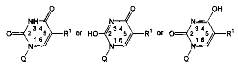
Other embodiments of this invention provide a compound of the formula:
0 O OH
/ / N H N N-
p~(2 6 R~ or HO-12 1 34 34
65/ R1 or 0~2 1 65 Rt
\\
N \
N
Q N Q Q
wherein:
Rl is a moiety of the formula:
Rs (R3)m R4
CA 02317505 2008-10-16
9b
R2 is a divalent electron conduit moiety selected from the group consisting
of:
an unsaturated hydrocarbyl group;
an aromatic hydrocarbyl group comprising one or more unsaturated
hydrocarbyl groups; and,
a heteroaromatic group comprising one or more unsaturated
hydrocarbyl groups;
R3 is a divalent spacer moiety selected from the group consisting of.
-CH2- ~ ~ -CHRS- i ~ -C(R5)2-
--0- ~ ~ -S- ~ t-NH- ~ and t -NRS-
R5 maybe the same or different and is independently a linear or branched
alkyl group having from 1 to 10 carbon atoms, or a cycloalkyl group having
from 3
to 10 carbon atoms;
nis an integer from 0 to 10;
mis0or1;
R4 is a toxophore moiety selected from the group consisting of.
X
Z
Z
i -Z-P-NJ -Z- II -N
N NH2
U X
O
II
-O-NH-C-NH2
CH2
C=0
I
NH OH
-Z-CH2-CH-CH-CH=CH-(CH2)12CH3
O
II
-Z-CFZ CH2-CHF-C-OH
0
11
-Z-CFZ CHF-CH2 C-OH
CA 02317505 2008-10-16
9c
0
11
-Z-CF2=CH2-C-OH
CH3 0
-Z-CF2-CH-C-OH
Y
I
-Z-CF- L; -Y
Y
-Z-CF2 CH2-CH2-N02
and
-Z \ / NO2
X is-C1, -Br or -I;
Y is independently -H or -F;
Z is independently -0- or -S-;
Q is a moiety selected from the group consisting of:
R~--O 0\ R? O S R~-- O,
6 6 6 6 6 6
R- CH2 0 R7 O 0` IJ
and v
6 6 6
R6 is independently -OH, -OC(=O)CH3, or other protected hydroxyl group;
and,
R7 is hydrogen, a phosphate group, or a phosphoramidate group;
and wherein said compound may be in any enantiomeric, diasteriomeric, or
stereoisomeric form, including, D-form, L-form, a-anomeric form, and P-
anomeric
form.
Other embodiments of this invention provide compounds as disclosed herein,
including any enantiomeric, diasteriomeric, or stereoisomeric form, including,
D-form, L-
form, a-anomeric form, and 1i-anomeric form.
CA 02317505 2009-10-19
9d
Other embodiments of this invention provide a method for screening for a
therapeutic agent in vitro, comprising: (a) contacting a first target cell
with a compound of
this invention, under conditions that favor the incorporation of the compound
into the
intracellular compartment of the target cell and a second target cell with a
potential
therapeutic agent, under conditions that favor the incorporation of the
compound into the
intracellular compartment of the target cell; and (b) assaying the second
target cell for
inhibition of cellular proliferation or cell killing.
Other embodiments of this invention provide use of a compound of this
invention,
for inhibiting proliferation of a hyperproliferative cell. The use may be for
preparation of a
medicament for such inhibiting.
Other embodiments of this invention provide use of a compound of this
invention
for inhibiting proliferation of a hyperproliferative cell comprising
contacting the cell in vitro
with the compound.
Other embodiments of this invention provide the use of a compound of this
invention, for preparation of a medicament for treating a pathology
characterized by
hyperproliferative cells. The use may be for preparation of a medicament for
such treating.
Other embodiments of this invention provide a method for screening for a
therapeutic agent, comprising contacting a target cell in vitro with a
compound of this
invention, wherein R4 is:
-Z \ / N02
which target cell favors incorporation of the compound into the target cell,
for the
diagnostic purpose of detecting intracellular levels of thymidylate synthase.
Other embodiments of this invention provide a composition comprising a
pharmaceutically acceptable carrier and a compound of this invention.
CA 02317505 2008-10-16
9e
BRIEF DESCRIPTION OF THE FIGURES
Figure 1 shows the development of resistance to anti-cancer modalities in
cells, and
the consequences.
Figure 2 schematically shows activation pathways of the prodrugs of this
invention.
Figure 3 schematically shows the High Throughput Screen for prodrugs activated
by intracellular enzymes important in drug resistance.
Figure 4 schematically shows how to find a lead human thymidylate synthase
(TS)
pro drug using TS-negative E. coli as the cell target.
Figure 5 shows an example of how to use this screen to simultaneously optimize
the prodrug for reactivity to two target enzymes.
CA 02317505 2008-10-16
Figure 6 shows one embodiment of a multi partitioned substrate for the
generation
of a thymidylate synthase (TS)-activatable prodrug.
Box I represents a masked or absent phosphate group R7. When masked, it is a
5 phosphoramidate or similar derivative that facilitates cell entry and is
processed
intracellularly to a monophosphate which can bind to TS. See Fries, K.M. et
al. (1995).
When absent, R' is a hydrogen atom and the prodrug is a substrate for cellular
thymidine
kinase (TK), which generates the requisite monophosphate in vivo.
Box 2 is a 2'-deoxyribofuranose group or other similar sugar, thio-sugar,
10 carbocyclic, or acyclic group which connects the monophosphate to the
pyrimidine ring in
a manner that supports functional binding of the prodrug to TS and, when R' is
a hydrogen
atom, to TK. This group need not utilize an oxygen atom for attachment to the
R' group of
box 1. Thus, phosphonate analogs of sugar phosphates are acceptable.
Box 3 represents a tether group, wherein n is an integer from 0 to 10, that is
a
mono- or polyunsaturated electron conduit acting to conduct electrons away
from the
pyrimidine ring and toward the leaving group R4 when the prodrug is acted upon
by IS.
The tether group is comprised of 0 to 10 unsaturated moieties like acyclic
vinyl, ethynyl,
imine, or azo units or cyclic unsaturated, aromatic, or heteroaromatic ones
that can be
mixed and matched at will as long as their connectivity provides the requisite
electron-
conducting conduit.
Box 4 represents a spacer unit X, wherein m is an integer from 0 to 1, that
connects
the tether to the leaving group R4. If Box 3, n equals 0, then Box 4, m equals
1. In the
preferred form, X is a methylene (CH2) unit, either bearing substituents or
not.
Additionally, though, X can be an oxygen, sulfur, nitrogen, or other atoms
capable of
forming at least two covalent bonds. When X is absent (Box 4, m equals 0), the
departure
of the leaving group R4 during the processing of the prodrug by TS leaves
behind a
pyrimidine nucleotide-based alkylating entity. See Barr, et al. (1983). When X
is present
(Box 4, m equals 1), the departure of the leaving group R4 occurs early during
the
processing of the prodrug by TS.
Box 5 represents a leaving group R4 that is released by the action of TS on
the
prodrug. It is itself a toxic antimetabolite or alkylating agent or is an
entity that readily
produces a toxic antimetabolite or alkylating agent in vivo. For example, the
leaving group
CA 02317505 2000-07-06
WO 99/37753 PCT/US99/01332
11
R4 can depart as an active metabolite of the anticancer agents Tepa or
Thiotepa, a
phosphoramide mustard, N-acetylsphingosine (C2 ceramide, a tumor suppressor
lipid) or
hydroxyurea (an inhibitor of ribonucleotide reductase) upon release by TS. The
leaving
group R, can also be an a,a-dihalogenated ether, in which case it affords a
carboxylic acid
group when the a,a-dihalogenated alcohol released by TS undergoes hydrolysis.
Thus, the
leaving group R4 can depart as a progenitor to fluoroacetate, fluorocitrate,
malonic acid,
methylmalonic avid, or 3-nitroproprionic acid, all potent inhibitors of
oxidative
phosphorylation.
Figure 7 shows TS Western blot of cell lines transfected by plasmid encoding
neomycin resistance, with or without the HER-2 protooncogene. Lane (1)
MCF7/HER2;
(2) MCF7/neo; (3) MDA-MB-435/HER2; (4) MDA-NM-435/neo; (5) BT-20/HER2; (6)
BT-20/neo.
Figure 8 shows reaction products of prodrug compounds with the enzyme
thymidylate synthase.
DETAILED DESCRIPTION OF THE INVENTION
The invention is achieved by exploiting some of the key genomic and phenotypic
changes intimately linked to resistance to biologic and chemotherapy of cancer
cells. The
invention provides a means for in vivo selectively inhibiting the growth
and/or killing of
cells which have undergone selection by exposure to cancer therapy (including
biologic
therapy such as tumor necrosis factor (TNF) or chemotherapy). (Refer to Table
2). As a
result, certain enzymes which have been activated by mutation or gene
amplification are
resistant to initial or further therapy by the agent. Unlike prior art
therapies directed to
creating more potent inhibitors of endogenous, intracellular enzymes, this
invention
exploits the higher enzyme activity associated with therapy-resistant diseased
cells and
tissues versus normal cells and tissues and does not rely on inhibiting the
enzyme. In one
aspect, the tumor cells successfully treated by the prodrugs of this invention
are
characterized by enhanced target enzyme activity and therefore have a much
higher
potential to convert the prodrug to its toxic form than do normal cells which
do not
overexpress the target enzyme. The term "target enzyme" is used herein to
define enzymes
having one or more of the above noted characteristics.
CA 02317505 2000-07-06
WO 99/37753 PCT/US99/01332
12
The practice of the present invention will employ, unless otherwise indicated,
conventional techniques of molecular biology, microbiology, cell biology and
recombinant
DNA, which are within the skill of the art. See, e.g., Sambrook, Fritsch and
Maniatis,
MOLECULAR CLONING: A LABORATORY MANUAL, 2" d edition (1989); CURRENT PROTOCOLS
IN MOLECULAR BIOLOGY (F. M. Ausubel et al. eds., (1987)); the series METHODS
IN
ENZYMOLOGY (Academic Press, Inc.): PCR 2: A PRACTICAL APPROACH (M.J.
MacPherson,
B.D. Haines and G.R. Taylor eds. (1995)) and ANIMAL CELL CULTURE (RI.
Freshney, ed.
(1987)).
As used in the specification and claims, the singular form "a", "an" and "the"
include
plural references unless the context clearly dictates otherwise. For example,
the term "a cell"
includes a plurality of cells, including mixtures thereof.
An "effective amount" is an amount sufficient to effect beneficial or desired
results.
An effective amount can be administered in one or more administrations,
applications or
dosages.
As used herein, the terms "host cells, "target cells" and "hyperproliferative
cells"
encompass cells characterized by the activation by genetic mutation or the
endogenous
overexpression of an intracellular enzyme. In some embodiments, the
overexpression of
the enzyme is related to loss of tumor suppressor gene product function drug
resistance or
the genetic instability associated with a pathological phenotype. A number of
cellular
mechanisms are involved in drug resistance, e.g., altered metabolism of the
drug,
impermeabilty of the cell with regard to the active compound or accelerated
drug
elimination from the cell, altered specificity of an inhibited enzyme,
increased production
of a target molecule, increased repair of cytotoxic lesions, or the bypassing
of an inhibited
reaction by alternative biochemical pathways. Enzymes activated or
overexpressed and
related to drug resistance include, but are not limited to thymidylate
synthase (TS) (Loan,
U. et al. (1996); Kobayashi, H. et al. (1995); Jackman, A.L. et al. (1995)),
dihydrofolate
reductase (Banerjee, D. et al. (1995) and Bertino, J.R. et al. (1996)),
tyrosine kinases
(TNF-a, Hudziak, R.M. et al. (1988)) and multidrug resistance (Stuhlinger, M.
et al.
(1994)); Akdas, A. et al. (1996); and (Tannock, I.F. (1996)); and ATP-
dependent
multidrug resistance associated proteins (Simon, S.M. and Schindler, M.
(1994)) and, in
some diseases including colon and prostate cancer, topoisomerase I (Husain et
al. (1994)).
Alternatively, resistance to one drug may confer resistance to other,
biochemically distinct
CA 02317505 2000-07-06
WO 99/37753 PCT/US99/01332
13
drugs. While this application is specifically directed to cancer, a similar
approach can be
applied to enzymes encoded by human and animal pathogens, and in which the
inhibitors
have failed due to development of resistance.
Amplification of certain genes is involved in resistance to chemotherapy.
Amplification of dihydrofolate reductase (DHFR) is related to resistance to
methotrexate
while amplification of the gene encoding thymidylate synthase is related to
resistance to
tumor treatment with 5-fluoropyrimidines. Amplification of genes associated
with drug
resistance can be detected and monitored by a modified polymerase chain
reaction (PCR)
as described in Kashini-Sabet, et al. (1988), U.S. Patent No. 5,085,983, or
the method
described herein. Acquired drug resistance can be monitored by the detection
of
cytogenetic abnormalities, such as homogeneous chromosome staining regions and
double
minute chromosomes both of which are associated with gene amplification.
Alternative
assays include direct or indirect enzyme activity assays and both of which are
associated
with gene amplification (e.g., Carreras & Santi (1995)); other methodologies
(e.g.
polymerase chain reaction, Houze, T.A. et al. (1997) or immunohistochemistry
(Johnson,
P.G. et al. (1997)).
Alternatively, the target cell is characterized as having inactivated tumor
suppressor
function, e.g. loss or inactivation of retinoblastoma (RB) or p53, known to
enhance
expression of TS (Li, W. et al. (1995)) or DHFR (Bertino, et al. (1996) and
Li, W. et al.
(1995)).
The prodrugs of this invention are useful to treat or ameliorate any disease
wherein
the disease-associated enzyme is associated with drug resistance to a
chemotherapeutic
whether due to loss of tumor suppressor functionality, in vivo selection by
chemotherapy
or a combination. This includes embodiments, where the enzyme is
overexpressed, over-
accumulated or activated in pathological cells versus normal cells, for
example, the TS
enzyme. Particularly excluded is the enzyme glutathione-S-transferase which
has been
shown to be occasionally elevated in some human tumors. Morgan, A.S. et al.
(1998).
The prodrugs of the subject invention are distinguishable on the basis that
the target
enzymes of this invention are commonly overexpressed, overaccumulated or
activated in
pathological cells versus normal cells. The most important principle which
distinguishes
the current invention from other approaches are:
CA 02317505 2000-07-06
WO 99/37753 PCT/US99/01332
14
(1) This invention describes the synthesis of substrates for enzymes like
thymidylate synthase. The overexpressed enzyme will generate toxin,
preferentially in
diseased cells. Previous approaches have relied on an inhibitor. The
inhibitors lead to
amplified expression of the enzyme, and subsequent resistance to treatment
(see, e.g.,
Lonn, U. et al. (1996).
(2) The current approach is also distinguishable from other "substrate-
prodrug"
approaches, e.g., the glutathione-S-transferase enzymes (see, e.g., Morgan,
A.S. et al.
(1998). The enzymes of the GST family are expressed at increased levels in
response to
toxic insult to the cell. The GST family of enzymes have overlapping substrate
specificities, which makes it difficult to design a substrate reactive with
only a single
species of enzyme with elevated expression in a cancer cell (Morgan, A.S. et
al. (1998)).
Because each of the enzymes of the current invention (e.g., thymidylate
synthase,
dihydrofolate reductase and thymidine kinase) is unique with respect to its
structure and
substrate specificity, it is facile to design unique substrates. Several
examples of substrates
for thymidylate synthase are provided in the specifications of this
application.
(3) In some cases the gene encoding the target enzyme (e.g., thymidylate
synthase)
may have undergone mutation to give resistance to inhibitors, (Barbour, K.W.
et al. (1992)
and Dicken, A.P. et al. (1993)) but will still be capable of carrying out
reaction with non-
inhibitor substrate prodrugs.
(4) A further advantage of this approach is that loss of tumor suppressor
function is
critical to development of malignancy. The majority of tumor cells have lost
one of the
p53, RB or p16 tumor suppressor functions. Such a loss results in increased
expression of
resistance enzymes (e.g., Thymidylate synthase), independent of previous
exposure to
chemotherapy. The prodrugs described herein will be useful in treating early
stages of
malignancy, as well as disease previously treated with chemotherapy.
Substrates for
enzymes like GST require previous exposure of the tumor to chemotherapy in
order to
achieve sufficient overexpression to offer even the possibility of a modest
therapeutic
index.
Drug Assay
This invention provides a method for identifying agents which have therapeutic
potential for the treatment of hyperproliferative or neoplastic disorders.
e.g., cancer. The
CA 02317505 2008-10-16
method also identifies agents that inhibit the growth of cells or cell cycling
of
hyperproliferative cells, such as cancer cells. Other cells that are included
are bacterial, yeast
and parasitic cells which cause disease as a result of inappropriate
proliferation in the patient.
The agent is considered a potential therapeutic agent if cell proliferation,
replication or cell
5 cycling is reduced relative to the cells in a control sample. Most
preferably, the cells are
killed by the agent. The cells can be procaryotic (bacterial such as E. coli)
or eucaryotic.
The cells can be mammalian or non-mammalian cells, e.g., mouse cells, rat
cells, human
cells, fungi (e.g., yeast) or parasites (e.g., Pneumocystis or Leishmania)
which cause disease.
As used herein, a "hyperproliferative cell" is intended to include cells that
are de-
10 differentiated, immortalized, neoplastic, malignant, metastatic or
transformed. Examples of
such cells include, but are not limited to a sarcoma cell, a leukemia cell, a
carcinoma cell, or
an adenocarcinoma cell. More specifically, the cell can be a breast cancer
cell, a hepatoma
cell, a detectable cancer cell, pancreatic carcinoma cell, an oesophageal
carcinoma cell, a
bladder cancer cell, an ovarian cancer cell, a skin cancer cell, a liver
carcinoma cell, or a
15 gastric cancer cell. In an alternative embodiment, the target cell can be
resistant to a drug or
compound used to prevent or kill a cell infected with an infectious agent
which is resistant to
conventional antibiotics. Infectious agents include bacteria, yeast and
parasites, such as
trypanosomes.
Specific examples of target enzymes that are the subject matter of this
invention are
listed in Table 2 (above) or Table 3 (below). These enzymes are involved in
resistance to
chemotherapy, are endogeneously activated, overexpressed or over-accumulated
in a cell
characterized by resistance to cancer therapy and associated with a
pathological or disease
include, but are not limited to enzymes such as a member of the tyrosine
kinase superfamily
or an ATP-dependent MDR-associated protein, CAD, thymidylate synthase,
dihydrofolate
reductase, and ribonucleotide reductase. Table 3 provides a list of enzymes
which may be
targeted by this approach in infectious disease.
CA 02317505 2000-07-06
WO 99/37753 16 PCT/US99/01332
Table 3
Enzymes Overexpressed in Infectious Disease, and which Contribute to Drug
Resistance
Enzyme Provides increased Resistance to:
Beta-lactamases Penicillin and other beta-lactam containing antibiotics
Aminoglycosidase, or Aminoglycoside antibiotics (e.g., streptomycin,
aminoglycoside midifying gentamycin)
enzymes
Chloramphenicol transacetylase Chloramphenicol
Dihydrofolate reductase Trimethoprim
Reference: Mechanisms of Microbial Disease, 2' Ed., M. Schaechter, G. Medloff,
B.I.
Eisenstein, Editor TS Satterfield. Publ. Williams and Wilkins, pp. 973 (1993).
The potentially therapeutic agent identified by the method of this invention
is a
prodrug that is a substrate for the enzyme and is converted intracellularly to
an intracellular
toxin. As used herein, a "prodrug" is a precursor or derivative form of a
pharmaceutically
active agent or substance that is less cytotoxic to target or
hyperproliferative cells as
compared to the drug metabolite and is capable of being enzymatically
activated or converted
into the more active form (see Connors, T.A. (1986) and Connors, T.A. (1996)).
The toxicity
of the agent is directed to cells that are producing the converting enzyme in
an amount
effective to produce a therapeutic concentration of the cellular toxin in the
diseased cell.
This invention also provides a quick and simple screening assay that will
enable
initial identification of compounds with at least some of the desired
characteristics. For
purposes of this current invention, the general scheme of one embodiment is
shown in Figure
3. This drawing describes how the assay is arranged and the materials
necessary for its
process. As shown in Figure 3, the assay requires two cell types, the first
being a control cell
in which the target enzyme is not expressed, or is expressed at a low level.
The second cell
type is the test cell, in which the target enzyme is expressed at a detectable
level, e.g., a high
level For example, a procaryotic E. coli which does not endogenously express
the target
enzyme TS is a suitable host cell or target cell. The cell can have a control
counterpart
(lacking the target enzyme), or in a separate embodiment, a counterpart
genetically modified
to differentially express the target enzyme, or enzymes (containing the
appropriate species of
target enzyme). More than one species of enzyme can be used to separately
transduce
separate host cells, so that the effect of the candidate drug on a target
enzyme can be
SUBSTITUTE SHEET (RULE 26)
CA 02317505 2000-07-06
WO 99/37753 PCT/US99/01332
17
simultaneously compared to its effect on another enzyme or a corresponding
enzyme from
another species.
In another embodiment, a third target cell is used as a control because it
receives an
effective amount of a compound, such as, for example, the compounds shown
below, which
have been shown to be potent prodrugs. This embodiment is particularly useful
to screen for
new agents that are activated by thymidylate synthase.
In another embodiment, transformed cell lines, such as ras-transformed NIH 3T3
cells (ATCC, 10801 University Blvd., Manassas, VA 20110-2209, U.S.A.) are
engineered to
express variable and increasing quantities of the target enzyme of interest
from cloned cDNA
coding for the enzyme. Transfection is either transient or permanent using
procedures well
known in the art and described in Chen, L. et al. (1996), Hudziak, R.M. et al.
(1988), or
Carter, P. et al. (1992), and in the experimental section below. Suitable
vectors for insertion
of the cDNA are commercially available from Stratagene, La Jolla, CA and other
vendors.
The level of expression of enzyme in each transfected cell line can be
monitored by
immunoblot and enzyme assay in cell lysates, using monoclonal or polyclonal
antibody
previously raised against the enzyme for immuno-detection. See, e.g., as
described by Chen,
L. et al. (1996). The amount of expression can be regulated by the number of
copies of the
expression cassette introduced into the cell or by varying promoter usage.
Enzymatic assays
to detect the amount of expressed enzyme also can be performed as reviewed by
Carreras,
C.W. and Santi, D.V. (1995), or the method described in the experimental
section below.
Tumor cell lines can be selected to express enhanced levels of thymidylate
synthase (e.g.,
colon tumor cells, as described by Copur et al. (1995).
As noted above, cells containing the desired genetic deficiencies may be
obtained
from Cold Spring Harbor, the Agricultural Research Service Culture Collection,
or the
American Type Culture Collection. The appropriate strains can also be prepared
by inserting
into the cell a gene coding for the target enzyme using standard techniques as
described in
Miller (1992), Sambrook, et al. (1989), and Spector et al. (1998). Growth
assays can be
performed by standard methods as described by Miller (1992), Sugarman et al.
(1985) and
Spector et al. (1998).
It should be understood by those skilled in the art that the screen shown in
Figure 3
can be applied broadly for the discovery of antibiotics. For example,
thymidylate synthase
from yeast could be substituted for that of E. coli in Figure 4. This would
allow the
CA 02317505 2000-07-06
WO 99/37753 PCT/US99/01332
18
discovery of specific antifungal antibiotics targeting yeast related
pathogens. In addition,
other enzymes can be subjected to this treatment. For example, prodrugs which
target
specifically the dihydrofolate reductase activity of infectious agents, like
Pneumocystis
carnii, could be selected. These agents will be selected for specificity for
the target enzyme,
and can be shown not to activate the enzyme of the natural host by employing
the screening
assay described in Figure 3. The control cellular constructs would contain the
corresponding
normal human enzyme, in order to show lack of toxicity when only the normal
human
enzyme is present.
For example and as shown in Figure 4, a foreign gene, e.g., a human gene
encoding
TS, can be inserted into the host cell such that human TS is expressed. This
genetically
engineered cell is shown as the "test cell" in Figure 3. The "control cell"
does not express the
target enzyme. In some embodiments it may be necessary to supplement the
culture media
with the protein product of the target enzyme.
In a separate embodiment, the wild type host cell is deficient or does not
express
more than one enzyme of interest. As shown in Figure 4, the host cell does not
endogenously
produce thymidine kinase (TK-) or thymidylate synthase (TS-). Genes coding for
the human
counterpart of these enzymes are introduced into the host cell to obtain the
desired level of
expression. The level of expression of enzyme in each transfected cell line
can be
monitored by methods described herein, e.g., by immunoblot and enzyme assay in
cell
lysates, using monoclonal or polyclonal antibody previously raised against the
enzyme for
immunodetection. See, e.g., as described by Chen, L. et al. (1996). Enzymatic
assays also
can be performed as reviewed by Carreras, C.W. and Santi, D.N. (1995) using
detectable
labeled substituents, e.g. tritium labeled substituents. A possible advantage
of the "two
enzyme" system is that the requirement for activation by two enzymes
preferentially
overexpressed in tumor cells will provide increased safety for normal cells.
The test cell is grown in small multi-well plates and is used to detect the
biologic
activity of test prodrugs. For the purposes of this invention, the successful
candidate drug
will block the growth or kill the test cell type, but leave the control cell
type unharmed.
The candidate prodrug can be directly added to the cell culture media or
previously
conjugated to a ligand specific to a cell surface receptor and then added to
the media.
Methods of conjugation for cell specific delivery are well known in the art,
see e.g., U.S.
Patent Nos. 5,459,127; 5,264,618; and published patent specification WO
91/17424
CA 02317505 2000-07-06
WO 99/37753 PCT/US99/01332
19
(published November 14, 1991). The leaving group of the candidate prodrug can
be
detectably labeled, e.g., with tritium. The target cell or the culture media
is then assayed for
the amount of label released from the candidate prodrug. Alternatively,
cellular uptake may
be enhanced by packaging the prodrug into liposomes using the method described
in Lasic,
D.D. (1996) or combined with cytofectins as described in Lewis, J.G. et al.
(1996).
In a separate embodiment, cultured human tumor cells overexpressing the enzyme
of
interest i.e., target enzyme, are identified as described above. The cells are
contacted with the
potential therapeutic agent under conditions which favor the incorporation of
the agent into
the intracellular compartment of the cell. The cells are then assayed for
inhibition of cellular
proliferation or cell killing.
It should be understood, although not always explicitly stated, each
embodiment can
be further modified by providing a separate target cell to act as a control by
receiving an
effective amount of a compound, such as, for example, the compounds shown
below, which
have been shown to be potent prodrugs.
A high throughput screen to identify biologically active compounds is outlined
in
Figures 3, 4 and 5. The basis of the test is the ease of genetic manipulation
and growth of
E. coli, and similar single cell organisms (e.g. yeast), see Miller (1992) and
Spector et al.
(1998). The key step is removing the endogenous enzyme activity corresponding
to an
enzyme target for prodrug design. This can be done by any of the methods
described by
Miller (1992), Sambrook, et al. (1989) or Spector et al. (1998). These methods
include
chemical and biologic (e.g. viral or transponson insertional) mutagenesis,
followed by an
appropriate selection procedure. The TS negative (TS-) cell then becomes a
negative
control for the identification of prodrugs that, when acted upon by
thymidylate synthase,
become cell toxins. A similar approach can be made with other cell types, e.g.
other
bacteria, yeast, or other selectable single cell organisms. In the assay, both
control and
recombinant organisms are compared for sensitivity to the test compounds. As
will be
understood by those skilled in the art, prodrugs which distinguish between
species of
enzyme can also be derived from this procedure. For example, otherwise
identical cells
expressing human and yeast enzymes can be used to detect antibiotic prodrugs
which are
preferentially toxic only to the cells expressing the yeast enzyme. In this
way, novel and
specific antibiotics can be discovered.
CA 02317505 2000-07-06
WO 99/37753 PCT/US99/01332
Example cell lines are ras-transformed NIH 3T3 cells (obtained from the ATCC)
and are engineered to express increasing quantities of human thymidylate
synthase
(Hu TS) from the cloned cDNA. Transfection is done in a transient or permanent
basis
(see Chen, L. et al. (1996), Hudziak, R.M. et al. (1988), and Carter, P. et
al. (1992).
5 NIH-000 (ras-transformed parent cell line); NIH-001 (low expressor of HuTS);
NIH-002
(intermediate expressor of Hu TS); NIH-003 (high expressor of HuTS). The level
of
expression of TS in each cell line is monitored by immunoblot and enzyme assay
in cell
lysates, using antibody directed versus HuTS protein for immunodetection
(e.g., as
described in Chen, L. et al. (1996)). Enzymatic assays are performed as
reviewed by
10 Carreras and Santi (1995).
Human colorectal and breast tumor cell lines are screened for expression of
HuTS
enzyme. Cell lines expressing low, moderate and high levels of HuTS will be
exposed to
drug candidates as described above for the NIH 3T3 cell lines. Growth
inhibition and
cytotoxicity are monitored as described above. Similar tests can be carried
out for each of
15 the enzymes listed in Table 1.
An alternative embodiment for a prodrug taking advantage of TS overexpression
in
tumor cells is a deoxyuridine phosphoramidate, or other modifications (cited
herein)
conjugated with a therapeutic radionuclide. An example of a therapeutic
radionuclide is
rhenium 188. The isotope can be synthesized essentially as described by
Callahan, et al.
20 (1989). Alternatively, it can be obtained commercially, for example from
Mallicrodt
Medical By, The Netherlands. The therapeutic radionuclide can be conjugated
with
deoxyuridine, or deoxyuridine 5'-phosphoramidate, or other derivative, by
standard
methods (for example, as described by Lin, W-Y., et al. (1997)). The
radionuclide-
containing deoxyuridine phosphoramidate will be preferentially taken up into
the DNA of
tumor cells overexpressing thymidylate synthase, and cause their death via
concentrated
emission of beta and gamma radiation. Alternative radionuclides include
rhenium- 186,
and others (Troutner, D.A. (1987)).
In Vivo Administration
The in vitro assays are confirmed in animal models bearing human tumors or
infected with an antibiotic resistant microorganism to determine in vivo
efficacy.
CA 02317505 2008-10-16
21
Another aspect of this invention is a method for treating a pathology
characterized
by hyperproliferative cells in a subject comprising administering to the
subject a therapeutic
amount of a prodrug that is converted to a toxin in a hyperproliferative cell
by an endogenous
intracellular enzyme as defined herein. In a preferred embodiment, the
compound is selected
from the compounds defined in the section "Prodrugs," Infra.
When the prodrug is administered to a subject such as a mouse, a rat or a
human
patient, the agent can be added to a pharmaceutically acceptable carrier and
systemically or
topically administered to the subject.
To determine patients that can be beneficially treated, a tumor sample is
removed
from the patient and the cells are assayed for the level of expression of the
enzyme of
interest. If the expression is above that expressed in normal cells so that a
toxic amount of
the prodrug would cause administered without undesirable side effects, then
the tumor or
cells are determined to be beneficially treated and thus, the patient is
suitable for the
therapy of this invention. For example, if the target enzyme is expressed at
least about 2
times and preferably about 3 times higher than normal cells, the patient is a
suitable subject
for the therapy method of this invention. Therapeutic amounts can be
empirically
determined and will vary with the pathology being treated, the subject being
treated and
the toxicity of the converted prodrug or cellular toxin.
When delivered to an animal, the method is useful to further confirm efficacy
of the
prodrug. As an example of an animal model, groups of nude mice (Balb/c NCR
nu/nu
female, Simonsen, Gilroy, CA) are each subcutaneously inoculated with about
105 to about
109 hyperproliferative, cancer or target cells as defined herein. When the
tumor is
established, the prodrug is administered, for example, by subcutaneous
injection around
the tumor. Tumor measurements to determine reduction of tumor size are made in
two
dimensions using venier calipers twice a week. Other animal models may also be
employed as appropriate. Lovejoy et al. (1997) and Clarke, R. (1996).
Administration in vivo can be effected in one dose, continuously or
intermittently
throughout the course of treatment. Methods of determining the most effective
means and
dosage of administration are well known to those of skill in the art and will
vary with the
composition used for therapy, the purpose of the therapy, the target cell
being treated, and
the subject being treated. Single or multiple administrations can be carried
out with the
CA 02317505 2000-07-06
WO 99/37753 PCT/US99/01332
22
dose level and pattern being selected by the treating physician. Suitable
dosage
formulations and methods of administering the agents can be found below.
The agents and compositions of the present invention can be used in the
manufacture of medicaments and for the treatment of humans and other animals
by
administration in accordance with conventional procedures, such as an active
ingredient in
pharmaceutical compositions.
The pharmaceutical compositions can be administered orally, intranasally,
parenterally or by inhalation therapy, and may take the form of tablets,
lozenges, granules,
capsules, pills, ampoules, suppositories or aerosol form. They may also take
the form of
suspensions, solutions and emulsions of the active ingredient in aqueous or
nonaqueous
diluents, syrups, granulates or powders. In addition to a compound of the
present
invention, the pharmaceutical compositions can also contain other
pharmaceutically active
compounds or a plurality of compounds of the invention.
More particularly, a compound of the formula of the present invention also
referred
to herein as the active ingredient, may be administered for therapy by any
suitable route
including oral, rectal, nasal, topical (including transdermal, aerosol, buccal
and -
sublingual), vaginal, parental (including subcutaneous, intramuscular,
intravenous and
intradermal) and pulmonary. It will also be appreciated that the preferred
route will vary
with the condition and age of the recipient, and the disease being treated.
In general, a suitable dose for each of the above-named compounds, is in the
range
of about 1 to about 100 mg per kilogram body weight of the recipient per day,
preferably
in the range of about I to about 50 mg per kilogram body weight per day and
most
preferably in the range of about 1 to about 25 mg per kilogram body weight per
day.
Unless otherwise indicated, all weights of active ingredient are calculated as
the parent
compound of the formula of the present invention for salts or esters thereof,
the weights
would be increased proportionately. The desired dose is preferably presented
as two, three,
four, five, six or more sub-doses administered at appropriate intervals
throughout the day.
These sub-doses may be administered in unit dosage forms, for example,
containing about
1 to about 100 mg, preferably about 1 to above about 25 mg, and most
preferably about 5
to above about 25 mg of active ingredient per unit dosage form. It will be
appreciated that
appropriate dosages of the compounds and compositions of the invention may
depend on
the type and severity and stage of the disease and can vary from patient to
patient.
CA 02317505 2000-07-06
WO 99/37753 PCTNS99/01332
23
Determining the optimal dosage will generally involve the balancing of the
level of
therapeutic benefit against any risk or deleterious side effects of the
treatments of the
present invention.
Ideally, the prodrug should be administered to achieve peak concentrations of
the
active compound at sites of disease. This may be achieved, for example, by the
intravenous injection of the prodrug, optionally in saline, or orally
administered, for
example, as a tablet, capsule or syrup containing the active ingredient.
Desirable blood
levels of the prodrug may be maintained by a continuous infusion to provide a
therapeutic
amount of the active ingredient within disease tissue. The use of operative
combinations is
contemplated to provide therapeutic combinations requiring a lower total
dosage of each
component antiviral agent than may be required when each individual
therapeutic
compound or drug is used alone, thereby reducing adverse effects.
While it is possible for the prodrug ingredient to be administered alone, it
is
preferable to present it as a pharmaceutical formulation comprising at least
one active
ingredient, as defined above, together with one or more pharmaceutically
acceptable
carriers therefor and optionally other therapeutic agents. Each carrier must
be "acceptable"
in the sense of being compatible with the other ingredients of the formulation
and not
injurious to the patient.
Formulations include those suitable for oral, recta, nasal, topical (including
transdermal, buccal and sublingual), vaginal, parenteral (including
subcutaneous,
intramuscular, intravenous and intradermal) and pulmonary administration. The
formulations may conveniently be presented in unit dosage form and may be
prepared by
any methods well known in the art of pharmacy. Such methods include the step
of
bringing into association the active ingredient with the carrier which
constitutes one or
more accessory ingredients. In general, the formulations are prepared by
uniformly and
intimately bringing into association the active ingredient with liquid
carriers or finely
divided solid carriers or both, and then if necessary shaping the product.
Formulations of the present invention suitable for oral administration may be
presented as discrete units such as capsules, cachets or tablets; each
containing a
predetermined amount of the active ingredient; as a powder or granules; as a
solution or
suspension in an aqueous or non-aqueous liquid; or as an oil-in-water liquid
emulsion or a
CA 02317505 2000-07-06
WO 99/37753 PCT/US99/01332
24
water-in-oil liquid emulsion. The active ingredient may also be presented a
bolus,
electuary or paste.
A tablet may be made by compression or molding, optionally with one or more
accessory ingredients. Compressed tablets may be prepared by compressing in a
suitable
machine the active ingredient in a free-flowing form such as a powder or
granules,
optionally mixed with a binder (e.g., povidone, gelatin, hydroxypropylmethyl
cellulose),
lubricant, inert diluent, preservative, disintegrant (e.g., sodium starch
glycolate, cross-
linked povidone, cross-linked sodium carboxymethyl cellulose) surface-active
or
dispersing agent. Molded tablets may be made by molding in a suitable machine
a mixture
of the powdered compound moistened with an inert liquid diluent. The tablets
may
optionally be coated or scored and may be formulated so as to provide slow or
controlled
release of the active ingredient therein using, for example,
hydroxypropylmethyl cellulose
in varying proportions to provide the desired release profile. Tablets may
optionally be
provided with an enteric coating, to provide release in parts of the gut other
than the
stomach.
Formulations suitable for topical administration in the mouth include lozenges
comprising the active ingredient in a flavored basis, usually sucrose and
acacia or
tragacanth; pastilles comprising the active ingredient in an inert basis such
as gelatin and
glycerin, or sucrose and acacia; and mouthwashes comprising the active
ingredient in a
suitable liquid carrier.
Pharmaceutical compositions for topical administration according to the
present
invention may be formulated as an ointment, cream, suspension, lotion, powder,
solution,
past, gel, spray, aerosol or oil. Alternatively, a formulation may comprise a
patch or a
dressing such as a bandage or adhesive plaster impregnated with active
ingredients and
optionally one or more excipients or diluents.
For diseases of the eye or other external tissues, e.g., mouth and skin, the
formulations are preferably applied as a topical ointment or cream containing
the active
ingredient in an amount of, for example, about 0.075 to about 20% w/w,
preferably about
0.2 to about 25% w/w and most preferably about 0.5 to about 10% w/w. When
formulated
in an ointment, the prodrug may be employed with either a paraffinic or a
water-miscible
ointment base. Alternatively, the prodrug ingredients may be formulated in a
cream with
an oil-in-water cream base.
CA 02317505 2000-07-06
WO 99/37753 PCT/US99/01332
If desired, the aqueous phase of the cream base may include, for example, at
least
about 30% w/w of a polyhydric alcohol, i.e., an alcohol having two or more
hydroxyl
groups such as propylene glycol, butane- l,3-diol, mannitol, sorbitol,
glycerol and
polyethylene glycol and mixtures thereof. The topical formulations may
desirably include
5 a compound which enhances absorption or penetration of the prodrug
ingredient through
the skin or other affected areas. Examples of such dermal penetration
enhancers include
dimethylsulfoxide and related analogues.
The oily phase of the emulsions of this invention may be constituted from
known
ingredients in an known manner. While this phase may comprise merely an
emulsifier
10 (otherwise known as an emulgent), it desirably comprises a mixture of at
lease one
emulsifier with a fat or an oil or with both a fat and an oil. Preferably, a
hydrophilic
emulsifier is included together with a lipophilic emulsifier which acts as a
stabilizer. It is
also preferred to include both an oil and a fat. Together, the emulsifier(s)
with or without
stabilizer(s) make up the so-called emulsifying wax, and the wax together with
the oil
15 and/or fat make up the so-called emulsifying ointment base which forms the
oily dispersed
phase of the cream formulations.
Emulgents and emulsion stabilizers suitable for use in the formulation of the
present invention include Tween 60, Span 80, cetostearyl alcohol, myristyl
alcohol,
glyceryl monostearate and sodium lauryl sulphate.
20 The choice of suitable oils or fats for the formulation is based on
achieving the
desired cosmetic properties, since the solubility of the active compound in
most oils likely
to be used in pharmaceutical emulsion formulations is very low. Thus the cream
should
preferably be a non-greasy, non-staining and washable product with suitable
consistency to
avoid leakage from tubes or other containers. Straight or branched chain, mono-
or dibasic
25 alkyl esters such as di-isoadipate, isocetyl stearate, propylene glycol
diester of coconut
fatty acids, isopropyl myristate, decyl oleate, isopropyl palmitate, butyl
stearate, 2-
ethylhexyl palmitate or a blend of branched chain esters known as Crodamol CAP
may be
used, the last three being preferred esters. These may be used alone or in
combination
depending on the properties required. Alternatively, high melting point lipids
such as
white soft paraffin and/or liquid paraffin or other mineral oils can be used.
Formulations suitable for topical administration to the eye also include eye
drops
wherein the active ingredient is dissolved or suspended in a suitable carrier,
especially an
CA 02317505 2000-07-06
WO 99/37753 PCT/US99/01332
26
aqueous solvent for the prodrug ingredient. The prodrug ingredient is
preferably present in
such formulation in a concentration of about 0.5 to about 20%, advantageously
about 0.5 to
about 10% particularly about 1.5% w/w.
Formulations for ectal administration may be presented as a suppository with a
suitable base comprising, for example, cocoa butter or a salicylate.
Formulations suitable for vaginal administration may be presented as
suppositories,
tampons, creams, gels, pastes, foams or spray formulations containing in
addition to the
prodrug ingredient, such carriers as are known in the art to be appropriate.
Formulations suitable for nasal administration, wherein the carrier is a
solid,
include a coarse powder having a particle size, for example, in the range of
about 20 to
about 500 microns which is administered in the manner in which snuff is taken,
i.e., by
rapid inhalation through the nasal passage from a container of the powder held
close up to
the nose. Suitable formulations wherein the carrier is a liquid for
administration as, for
example, nasal spray, nasal drops, or by aerosol administration by nebulizer,
include
aqueous or oily solutions of the prodrug ingredient.
Formulations suitable for parenteral administration include aqueous and non-
aqueous isotonic sterile injection solutions which may contain anti-oxidants,
buffers,
bacteriostats and solutes which render the formulation isotonic with the blood
of the
intended recipient; and aqueous and non-aqueous sterile suspensions which may
include
suspending agents and thickening agents, and liposomes or other
microparticulate systems
which are designed to target the compound to blood components or one or more
organs.
The formulations may be presented in unit-dose or multi-dose sealed
containers, for
example, ampoules and vials, and may be stored in a freeze-dried (lyophilized)
condition
requiring only the addition of the sterile liquid carrier, for example water
for injections,
immediately prior to use. Extemporaneous injection solutions and suspensions
may be
prepared from sterile powders, granules and tablets of the kind previously
described.
Preferred unit dosage formulations are those containing a daily dose or unit,
daily
subdose, as herein above-recited, or an appropriate fraction thereof, of a
prodrug
ingredient.
It should be understood that in addition to the ingredients particularly
mentioned
above, the formulations of this invention may include other agents
conventional in the art
having regard to the type of formulation in question, for example, those
suitable of oral
CA 02317505 2000-07-06
WO 99/37753 PCT/US99/01332
27
administration may include such further agents as sweeteners, thickeners and
flavoring
agents.
Prodrugs and compositions of the formula of the present invention may also be
presented for the use in the form of veterinary formulations, which may be
prepared, for
example, by methods that are conventional in the art.
Provided below is a brief summary of cells and target enzymes that are useful
to
activate the prodrugs of this invention.
Tyrosine Kinases
The tyrosine kinase superfamily comprises the EGF receptor (EGFR), the
macrophage colony-stimulating factor (CSF-1) receptor (v-fins), and the
insulin receptor,
which shows 30 to 40% identity with the product of the ros oncogene. More
specifically,
the members of this superfamily include v-src, c-src, EGFR, HER2, CSF-1
receptor, c-fins,
v-ros, insulin receptor, and c-mos. See Figure 8.5 of Burck, K.B. et al., eds.
(1988).
Overexpression of members of the type 1 receptor tyrosine kinase superfamily
has been
documented in many types of cancer (Eccles, S.A. et al. (1994-95)).
Overexpression of
tyrosine kinases is linked to exposure to the a-cancer biologic agent TNF-a
(Hudziak,
R.M. et al. (1988) and Hudziak, R.M. et al. (1990)) and to chemotherapy
(St0hlinger et al.
(1994)).
The transforming gene of the Rous sarcoma virus, v-src, encodes an enzyme that
phosphorylates tyrosine residues on proteins. The c-src proto-oncogene is
found on
chromosome 20. Tissues and cell lines derived from tumors of neuroectodermal
origin
having a neural phenotype express high levels of c-src accompanied by high
specific
kinase activity.
Several groups of investigators have reported overexpression of c-erbB-2/neu
("HER2") oncogene in cancer cells. Brison (1993) noted that erbB proto-
oncogene is
amplified in human tumors with resultant overexpression in most cases.
Amplification of
the c-erbB-2/neu oncogene has been reported in human mammary tumors (Slamon,
et al.
(1987), van de Vijver et al. (1987), Pupa et al. (1993), and Andersen et al.
(1995)) and in
bladder tumors (Sauter et al. (1993)), and in every case amplification was
accompanied by
overexpression. c-erbB-2/neu overexpression also has been reported in ovarian
cancer
CA 02317505 2000-07-06
WO 99/37753 PCT/US99/01332
28
tissue samples (Slamon, et al. (1989), Meden et al. (1994), and Felip et al.
(1995)), and
tumors derived from the peripheral nervous system. Sukumar and Barbacid,
(1990).
To perform the drug screening assay, tumor cell lines will be assayed for
expression of the oncogene or will be engineered to express varying levels of
tyrosine
kinase. Selected cell lines are cultured and candidate drugs are added in
varying
concentrations. The cells are assayed for cell killing or inhibition of
cellular proliferation,
as described in Hudziak, R.M. et al. (1988) and Hudziak, R.M. et al. (1990).
Dihydrofolate Reductase
Methotrexate is a potent inhibitor of dihydrofolate reductase, an enzyme
necessary
for intracellular folate metabolism. Dihydrofolate reductase functions to
regenerate
tetrahydrofolate from dihydrofolate, a product of the thymidylate synthase
reaction
(Voet, et al. eds. (1995), p. 813). It is well established that an important
mechanism of
resistance of cells to methotrexate is an increase in DHFR activity due to
amplification of
the DHFR gene. Banerjee, D. et al. (1995), Schimke, R.T. et al. (1988). Lonn,
U. et al.
(1996) reported that amplification of the DHFR gene occurred in breast cancer
patients
who previously received adjuvant chemotherapy (cyclophosphamide, methotrexate,
5-
fluorouracil [CMF]) after surgery. Lack of the retinoblastoma (Rb) may also
lead to
enhanced MTX resistance as a consequence of an increase in DHFR mRNA
expression
activity without gene amplification. Li, W.W. et al. (1995). Cell lines with
mutated p53
have been shown to undergo gene amplification, and the resistant cells are
selected by
chemotherapy. Banerjee, D. et al. (1995), Yin, Y. et al. (1992) and
Livingston, L.R. et al.
(1992). For the purposes of performing the assay of this invention, Schimke,
R.T. et al.
(1988) describes several mouse, hamster and human cell lines. Alternatively,
the PCR
method of Lonn U. et al. (1996) is used to assay DHFR gene amplification and
identify
cells that are useful in the method of identifying therapeutic agents as
described herein.
The nucleotide sequence of the cDNA coding for the human dihydrofolate
reductase is
provided in Masters, J.N. and Attardi, G. (1983) and cells can be engineered
to express
varying levels of the enzyme as noted herein. Dicken, A.P. et-al. (1993)
describes a mutant
DHFR gene selected by chemotherapy. Purification of DHFR and assays related to
enzyme function are described in Nakano, T. et al. (1994). Alternatively, cDNA
encoding
CA 02317505 2000-07-06
WO 99/37753 PCT/US99/01332
29
DHFR is transfected into NIH 3T3 cells. Candidate drugs are added in varying
concentrations and cell killing and inhibition of proliferation are assayed.
Antimetabolites dependent on dihydrofolate reductase activity can be
synthesized
by the attachment of, for example, an alkylating group to either the N5 or the
C6 position
of dihydrofolate. Reduction of the NS-C6 bond by DHFR will result in the
release of the
alkylating agent. In addition to the alkylating groups, any moiety whose
release by DHFR
results in the production of a toxin or an antimetabolite will be useful in
the practice of the
invention. These compounds can be further modified by the addition of a
phosphatese or
phosphoramidate moiety.
Multidrug Resistant Tumors
Multidrug resistance (MDR) is a generic term for the variety of strategies
tumor
cells use to evade the cytotoxic effects of anticancer drugs. MDR is
characterized by a
decreased sensitivity of tumor cells not only to the drug employed for
chemotherapy but
also to a broad spectrum of drugs with neither obvious structural homology nor
common
targets. This pleiotropic resistance is one of the major obstacles to the
successful treatment
of tumors. MDR may result from structural or functional changes at the plasma
membrane
or within the cytoplasm, cellular compartments, or nucleus. Molecular
mechanisms of
MDR are discussed in terms of modifications in detoxification and DNA repair
pathways,
changes in cellular sites of drug sequestration, decreases in drug-target
affinity, synthesis
of specific drug inhibitors within cells, altered or inappropriate targeting
of proteins, and
accelerated removal or secretion of drugs.
One of the mechanisms implicated in MDR results from amplification and over-
expression of a gene known as the ATP-dependent multidrug resistant associated
protein
(MRP) in drug selected cell lines. For a review of the mechanisms of MDR, see
Gottesman, M.M. et al. (1995) and Noder et al. (1996).
To establish MDR cell lines, drug selections are conducted in either a single
step or
in multiple steps as described in Gottesman, M.M. et al. (1995) and Simon,
S.M. and
Schindler, M. (1994), and references cited therein. The isolation of DNA
sequences
coding for MDR from various mammalian species is described in Gros, P. et al.
(1986),
Gudkov, A.V. et al. (1987), and Roninson, I.B. et al. (1984), and reviewed in
Gottesman,
M.M. et al. (1995), and cells can be engineered to express varying levels of
this enzyme as
CA 02317505 2000-07-06
WO 99/37753 PCT/US99/01332
described above. The prodrug targeting MDR will be based upon the ATPase
activity of
this transporter.
Ribonucleotide Reductase
5 The enzyme ribonucleotide reductase reduces ribonucleoside diphosphates to
the
corresponding deoxyribonucleoside diphosphates. The enzyme is a tetramer made
up of
two a-subunits and two n-subunits. Hydroxyurea specifically blocks this
reaction by
interacting with the tyrosyl free radical (Tyr-122) of the (32-substrate
complex. Voet et al.
(1995). The goal in targeting this reaction is to allow the accumulation of
the free radical
10 product 0,-, which is highly cytotoxic.
Application of Technology to Other Diseases
While the primary focus of this application is directed to cancer, it should
be
recognized that the technology is broadly applicable to other diseases,
especially antibiotic
15 resistant bacterial infections. The (3-lactam antibiotics encounter
resistance in bacteria as
the result of overexpression of (3-lactamases. Hamilton-Miller, J.M.T. and
Smith,-J.T. eds.
(1979) p. 443. Other enzymes, such as the aminoglycoside phosphotransferese
Type III,
are induced and selected for following treatment with aminoglycoside
antibiotics, such as
kanamycin. McKay, G.A. et al. (1994). For the purpose of this application,
prodrug
20 substrates derived from known substrates will be prepared that will not
block enzyme
activity, but will instead take advantage of the high enzyme activity to
generate
intracellular toxins to the infectious agents.
Thymidylate Synthase
25 The overexpression of thymidylate synthase is associated with colon cancer,
breast
cancer, gastric cancer, head and neck cancer, liver cancer and pancreatic
cancer. These
diseases are currently treated by antimetabolite drugs (uracil-based, folate-
based, or
quinazoline-based, (see Table 1)). In each of these cases it is likely that
tumor suppressor
loss and/or 5-fluorouracil therapy can lead to amplified activity of TS, or
select for drug
30 resistant forms of the enzyme, and thereby lead to drug-resistance of the
disease relapse.
Lonn, U. et al. (1996) reported that amplification of the TS gene occurred in
breast cancer
patients who previously received adjuvant chemotherapy (cyclophosphamide,
CA 02317505 2000-07-06
WO 99/37753 PCT/US99/01332
31
methotrexate, 5-fluorouracil [CMF]) after surgery. This enhanced TS expression
is in
addition to the basic increase of TS which results from loss of tumor
suppressor function.
The principal reaction normally performed by TS is the synthesis of
deoxythymidine
monophosphate (dTMP) and dihydrofolate (DHF) from deoxyuridine monophosphate
(dUMP) and N(5),N(10)-methylene-tetrahydrofolate (THF). In one embodiment, a
derivative of uracil or THE is provided to cells expressing IS. For purposes
of this
invention, "uracil" (base only) and "uridine" (base and sugar) are used
interchangeably and
synonomously. Table 4 (below) summarized the many cancer types impacted by
elevated
TS expression.
CA 02317505 2000-07-06
WO 99/37753 PCT/US99/01332
32
Table 4
Thymidylate Synthase Overexpression Impacts Survival of Cancer Patients
Median Survival Time (Months)
Cancer TS (Low) TS (High) References
Breast 84 54 Pestalozzi et al., 1997
NSCLC 46 10 Volm and Mattern, 1992
Colon 13.6 8.2 Leichman et al., 1997
Rectal > 60 24 Johnston et al., 1994
Head and Neck > 72 24 Johnston et al., 1997
Stomach 43 6 Lenz et al., 1995
Average > 53.1 Average 21
Fold difference between averages > 2.5
The derivative or "prodrug" is converted by the enzyme into highly cytotoxic
metabolites. The low level of TS expressed in normal cells will not produce a
toxic
amount of the converted toxin. High levels of TS expressed in disease tissues
generate
more toxin and thereby lead to an inhibition of cell proliferation and/or cell
death. For
example, current therapy utilizes 5-fluorodeoxyuridylate to inhibit TS
activity. During the
reaction with substrate, the fluorine atom irreversibly becomes attached to
the TS enzyme
and inhibits it. In one embodiment, the proposed new therapeutic allows TS to
complete
the reaction but generates a modified product that, when incorporated into
DNA, causes a
toxic effect. The enzyme product may also block other critical cellular
functions (e.g.
protein synthesis or energy metabolism). Conversion of the prodrug also can
release a
metabolite, such as CN- which is toxic to the cell. Derivatives of uracil/dUMP
and
N(5)(10)-THF can be synthesized, all of which have the potential of generating
toxic
product after metabolic transformation by TS.
Primary sequences show that TS is one of the most highly conserved enzymes.
Perry, K. et al. (1990). Crystal structures of TS from several procaryotic
species,
Lactobacillus casei (Hardy, L.W. et al. (1987); Finer-Moore, J. et al. (1993))
and
Escherichia coli (Perry, K. et al. (1990)); an eukaryote Leishmania major
(Knighton,
E.R. et al. (1994)); and T4 phage (Finer-Moore, J.S. et al., (1994)) have been
determined
CA 02317505 2000-07-06
WO 99/37753 PCT/US99/01332
33
and indicate that tertiary structure also is very well conserved. The sequence
alignment of
the species of TS whose three dimensional structures have been determined and
is shown
in Schiffer, C.A. et al. (1995). From these amino acid sequences, the DNA
sequences can
be deduced or isolated using methods well known to those of skill in the art.
Sambrook, et
al. (1989). Alternatively, some 29 TS sequences from different organisms have
been
cloned and deposited into the DNA databases as described in Carreras, C.W. and
Santi,
D.V. (1995). The sequence of human thymidylate synthase gene, its cloning,
expression
and purification is provided in Takeishi, K. et al. (1985), Davisson, V.J. et
al. (1989) and
Davisson, V.J. et al. (1994). Genes encoding the TS protein and containing the
necessary
regulatory sequences, are constructed using methods well known to those of
skill in the art.
The gene encoding TS is introduced to target cells by electroporation,
transformation or
transfection procedures. Sambrook, et al. (1989). Alternatively, the gene is
inserted into
an appropriate expression vector by methods well known in the art, e.g., as
described in
Carreras, C.W. and Santi, D.V. (1995), Miller (1992) and Spector et al.
(1998). The
expression vector inserts the TS gene into the cells. The cells are then grown
under
conditions which favor the expression and production of TS protein.
Human gastric cancer cell lines, MKN-74, MKN-45, MKN-28 and KATO-III can
be used in the assay described above to identify potential therapeutic agents
which are
selective substrates for TS. MKN-74 and MKN-45 are established from well and
poorly
differentiated adenocarcinomas, respectively. These cell lines and culture
conditions are
described in Osaki, M. et al. (1997) and references cited therein.
Alternatively, tumor cell
lines such as those described by Copur, S. et al. (1995), which have been
selected by 5-FU
to overexpress thymidylate synthase may be used.
Quantitation of TS can be performed using enzymatic biochemical assays that
are
well known to those with skill in the art. To quantify the level of TS protein
and TS gene
expression from human tumor tissue samples, the methods as reported by
Johnston, P.G. et
al. (1991) and Horikoshi, T. et al. (1992) provide sensitive assays.
Alternatively, the PCR
method of Lonn, U. et al. (1996) is used to assay TS gene amplification and
identify cells
that are useful in the method of identifying therapeutic agents as described
herein.
As is apparent to one skilled in the art, control- cell culture systems
without drug and
separately with a reference drug such as the compounds exemplified below, also
are assayed.
A lead compound is one which preferentially kills target cells with about 2-
fold and
CA 02317505 2000-07-06
WO 99/37753 PCT/US99/01332
34
preferably about 3-fold or greater activity than normal cells. This invention
also provides
the agents identified by the methods described herein.
In another aspect, this invention provides a method for inhibiting the
proliferation of
a hyperproliferative cell, by first conducting the above assay. A prodrug
identified by this
assay is contacted with the cell and converted to a toxic metabolite in the
cell by an
endogenous intracellular enzyme as described above. Growth or cytotoxicity of
bacteria,
yeast and other cell types can be monitored as described by Miller et al.
(1992), Sugarman et
al. (1985) and Spector et al. (1998).
TS Prodrugs
In a preferred embodiment, the present invention involves two classes of
compounds activated by TS, each a derivative of 5-substituted deoxyuridine
monophosphate or a protected 5-substituted uracil or deoxyuridine
monophosphate.
Protected 5-substituted deoxyuridine monophosphate derivatives are those in
which the
phosphate moiety has been blocked through the attachment of suitable chemical
protecting
groups. Protection of 5-substituted deoxyuridine monophosphate derivatives can
improve
solubility, facilitate cellular penetration, facilitate passage across the
blood-brain barrier,
and prevent action of cellular or extracellular phosphatases, which might
otherwise result
in loss of the phosphate group. In another embodiment, 5-substituted uracil or
uridine
derivatives are administered to cells containing nucleoside kinase activity,
wherein the 5-
substituted uracil/uridine derivative is converted to a 5-substituted uridine
monophosphate
derivative. Uridine derivatives may also be modified to increase their
solubility, cell
penetration, and/or ability to cross the blood-brain barrier.
Action of thymidylate synthase upon 5-substituted uridine monophosphate
derivatives can release the substituent attached to the 5-position ("leaving
group") of the
pyrimidine ring. The released substituent is then capable, either inherently
or following
reaction with another cellular component, of acting as a toxin or an inhibitor
of cellular
proliferation.
General Synthesis of Compounds of Class I
The L and D isomers of the compounds of Class I have the structure:
CA 02317505 2008-10-16
0 OH 0
R, Rj Rj
5 a 3NH 5 N 5 ~ 3N
~ or 3 or ~ ~1
6 1 6 ~ 6 I
N O N N OH
Q Q Q
In the above formulae. R, (at the 5-position) is or contains a leaving group
which is a
5 chemical entity that has a molecular dimension and electrophilicity
compatible with
extraction from the pyrimidine ring by thymidylate synthase, and which upon
release from
the pyrimidine ring by thymidylate synthase, has the ability to inhibit the
proliferation of the
cell or kill the cell.
In the above formulae, Q is a phosphate or phosphoramidate derivative
containing a
10 chemical entity selected from the group consisting of sugar groups, thio-
sugar groups,
carbocyclic groups, and derivatives thereof. Examples of sugar groups include,
but are not
limited to, monosaccharide cyclic sugar groups such as those derived from
oxetanes (4-
membered ring sugars), furanoses (5-membered ring sugars), and pyranoses (6-
membered
ring sugars). Examples of furanoses include threo-furanosyl (from threose, a
four-carbon
15 sugar); erythro-furanosyl (from erythrose, a four-carbon sugar); ribo-
furanosyl (from
ribose, a five-carbon sugar); ara-furanosyl (also often referred to as arabino-
furanosyl;
from arabinose, a five-carbon sugar); xylo-furanosyl (from xylose. a five-
carbon sugar);
and lyxo-furanosyl (from lyxose, a five-carbon sugar). Examples of sugar group
derivatives include "deoxy", "keto", and "dehydro" derivatives as well as
substituted
20 derivatives. Examples of thio sugar groups include the sulfur analogs of
the above sugar
groups, in which the ring oxygen has been replaced with a sulfur atom.
Examples of
carbocyclic groups include C4 carbocyclic groups, C5 carbocyclic groups, and
C6
carbocvclic groups which may further have one or more subsituents, such as -OH
groups.
In one embodiment, Q is a f3-D-ribofuranosyl group of the formula:
CA 02317505 2000-07-06
WO 99/37753 PCT/US99/01332
36
R7 O CH2
O
R3 R2
wherein R7 is selected from the group consisting of phosphoryl,
phosphoramidate and
derivatives thereof, and wherein R2 and R3 are the same or different and are
independently -H
or -OH.
In some embodiments, R, is an alkenyl group, i.e., (-CH=CH)n R4, wherein n is
an
integer from 0 to 10, and R4 is a halogen such as is I- or Br, CN- or mercury;
wherein R2 is H
and R3 is -OH; wherein R2 is OH and R3 is H; wherein R2 and R3 are H; or
wherein R2 and R3
are OR
In another aspect, R, is an alkenyl group, i.e., (-CH=CH)n-R4, wherein n is an
integer
from 0 to 10, and R4 is or contains a group selected from the group consisting
of H, a
halogen, alkyl, alkene, alkyne, hydroxy, -O-alkyl, -0-aryl, 0-heteroaryl, -S-
alkyl, -S-aryl, a
cyanide, cyanate and thiocyanate halovinyl group, a halomercuric group, -S-
heteroaryl, -NH2,
-NH-alkyl, -N(alkyl)2, -NHCHO, -NHOH, -NHO-alkyl, NH2CONHO-, and NHNH2. In
these embodiments, further aspects include: wherein R2 and R3 are H; wherein
R2 is OH and
R3 is H; herein R2 is H and R3 is OH; or wherein R2 and R3 are OR
A preferred embodiment for the substituent in the R, position is one that
could
undergo an allylic interchange as shown in Figure 8.
CA 02317505 2000-07-06
WO 99/37753 PCT/US99/01332
37
In a still further aspect, the candidate therapeutic agent is a compound of
the
formula:
0
H (CH=CH)õ-CH2 O A
O N
Q
wherein n is an integer from 0 to 10; wherein A is a phosphoramide derivative,
or a
compound of the formula:
0
11
P -N(CH2CH2CI)2
NH2
and wherein Q is selected from the group consisting of H, an unsubstituted or
substituted sugar as defined above and a substituted or unsubstituted
carbocyclic as defined
above.
In a further embodiment, the compounds described above, are modified by Q
having
the structure:
R7 O CH2
O
R3 R2
wherein R7 is selected from the group consisting of H, phosphoryl,
phosphoramidate
and derivatives thereof, and wherein R2 and R3 are the same or different and
are
independently -H or -OH. In one embodiment, R' is not H. For these
embodiments, R, also
can have the structure -(CH=CH)n R4, wherein n is an integer from 0 to 10, and
R, is selected
CA 02317505 2000-07-06
WO 99/37753 PCT/US99/01332
38
from the group consisting of H, a halogen, alkyl, alkene, alkyne, hydroxy, -0-
alkyl, -0-aryl,
O-heteroaryl, -S-alkyl, -S-aryl, -S-heteroaryl, -NH2, -NH-alkyl, -N(alkyl)2, -
NHCHO, a
cyanide, cyanate and thiocyanate halovinyl compound, a halomercuric compound, -
NHOH,
-NHO-alkyl, NHNH21 and NH2CONHO-.
Additionally, in a further aspect, the candidate therapeutic agent is a
compound of the
formula:
NH2
C=o
R-(CH=CH)n (CH2)m 0 NN. H
where R = 2'-deoxy-5-uridyl, in is 0 or 1, and n is an integer from 0 to 10.
Where appropriate, the compounds can be in any of their enantiomeric,
diasteriomeric, or stereoisomeric forms, including, for example, D- or L-
forms, and can be
in any stereochemical configuration, including, for example, a- or 1i-anomeric
form.
Synthesis of the above noted 5-substituted pyrimidine nucleosides and 5-
substituted
pyrimidine nucleoside monophosphates can be accomplished by methods that are
well-
known in the art. For example, treatment of 5-chloromercuri-2'-deoxyuridine
with
haloalkyl compounds, haloacetates or haloalkenes in the presence of Li2PdC14
results in the
formation, through an organopalladium intermediate, of the 5-alkyl, 5-acetyl
or 5-alkene
derivative, respectively. Wataya, et al. (1979) and Bergstrom, et al. (1981).
Another
example of C5-modification of pyrimidine nucleosides and nucleotides is the
formation of
C5-trans-styryl derivatives by treatment of unprotected nucleotide with
mercuric acetate
followed by addition of styrene or ring-substituted styrenes in the presence
of Li2PdCl4.
Bigge, et al. (1980). Pyrimidine deoxyribonucleoside triphosphates were
derivatized with
mercury at the 5 position of the pyrimidine ring by treatment with mercuric
acetate in
acetate buffer at 50 for 3 hours. Dale, et al. (1973). Such treatment would
also be
expected to be effective for modification of monophosphates; alternatively, a
modified
triphosphate could be converted enzymatically to a modified monophosphate, for
example,
by controlled treatment with alkaline phosphatase followed by purification of
monophosphate. Other moieties, organic or nonorganic, with molecular
properties similar
CA 02317505 2000-07-06
WO 99/37753 PCT/US"/01332
39
to mercury but with preferred pharmacological properties could be substituted.
For general
methods for synthesis of substituted pyrimidines, for example, U.S. Patent
Nos. 4,247,544;
4,267,171; and 4,948,882; and Bergstrom et al. (1981). The above methods would
also be
applicable to the synthesis of derivatives of 5-substituted pyrimidine
nucleosides and
nucleotides containing sugars other than ribose or 2'-deoxyribose, for example
2'-3'-dideoxyribose, arabinose, furanose, lyxose, pentose, hexose, heptose,
and pyranose.
An example of a 5-position substituent is the halovinyl group, e.g. E-5-(2-
bromovinyl)-2'-
deoxyuridylate. Barr, P.J. et al. (1983). This compound is synthesized as
follows as
described in the experimental section.
Alternatively, 5-bromodeoxyuridine, 5-iododeoxyuridine, and their
monophosphate
derivatives are available commercially from Glen Research, Sterling, VA (USA),
Sigma-
Aldrich Corporation, St. Louis, MO (USA), Moravek Biochemicals, Inc., Brea, CA
(USA),
ICN, Costa Mesa, CA (USA) and New England Nuclear, Boston, MA (USA).
Commercially-available 5-bromodeoxyuridine and 5-iododeoxyuridine can be
converted to
their monophosphates either chemically or enzymatically, though the action of
a kinase
enzyme using commercial available reagents from Glen Research, Sterling, VA
(USA) and
ICN, Costa Mesa, CA (USA). These halogen derivatives could be combined with
other
substituents to create novel and more potent antimetabolites.
General Synthesis of Compounds of Class II
In a further aspect, the prodrug contacted with the cell overexpressing
thymidylate
synthase is an L or D isomer of a compound of the formula:
O O OH
NH N N
=< -- 2 2 3 45 R1 or HO 3 45 R1 or O 2 3 45 R1
O
N16 N16 N16
Q Q Q
In the above formulae, R' is a moiety of the formula:
R2 R3 R4
m
CA 02317505 2000-07-06
WO 99/37753 PCTIUS99/01332
In the above formulae, R2 is or contains a divalent electron conduit moiety.
In one
embodiment, R2 is or contains a mono- or polyunsaturated electron conduit
acting to
conduct electrons away from the pyrimidine ring and toward the leaving group
R'. In one
embodiment, R2 is selected from the group consisting of: an unsaturated
hydrocarbyl group;
5 an aromatic hydrocarbyl group comprising one or more unsaturated hydrocarbyl
groups; and,
a heteroaromatic group comprising one or more unsaturated hydrocarbyl groups.
In one embodiment, R2 is an unsaturated hydrocarbyl group having a structure
selected from the group consisting of:
and
In one embodiment, R2 and R3, taken together form a structure selected from
the
group consisting of:
'LLL
and
In one embodiment, R2 is an aromatic hydrocarbyl group having a structure
selected
from the group consisting of
and
CA 02317505 2000-07-06
WO 99/37753 PCT/US99/01332
41
In one embodiment, R2 is a heteroaromatic group having a structure selected
from the
group consisting of
J I and
wherein J is a heteroatom, such as -0-, -S-, or -Se-, or a heteroatom group,
such as -
NH- or -NR-, where RALK is a linear or branched alkyl having 1 to 10 carbon
atoms or a
cycloalkyl group having 3 to 10 carbon atoms.
In the above formulae, R3 is a divalent spacer moiety, also referred to as a
spacer unit.
In one embodiment, R3 is a divalent spacer moiety having a structure selected
from the group
consisting of
-CH2 -CHR5- -C(R5)2
-0- ~ -S- ~ ~ -NH- ~ and ~ -NR5-
wherein R5 is the same or different and is independently a linear or branched
alkyl
group having from 1 to 10 carbon atoms, or a cycloalkyl group having from 3 to
10 carbon
atoms.
In one embodiment, R3 is a divalent spacer moiety having a structure selected
from
the group consisting of
-O S NH ~ and NR5-
In the above formula, n is an integer from 0 to 10 and, m is 0 or 1. In one
embodiment, n is an integer from 0 to 10 and, m is 1. In one embodiment, n is
0 and m is
0. In one embodiment, when R' is -H, then n is not zero. In one embodiment,
when R' is -
H, then m is not zero. In one embodiment, when R' is -H, then n is not zero
and m is not
zero. In one embodiment, when R' is -H, then R4 is not a halogen (i.e., -F, -
Cl, -Br, -I). In
one embodiment, when R7 is -H, and m is zero, then R4 is not a halogen (i.e., -
F, -Cl, -Br, -
I). In one embodiment, when R' is -H, and m is zero and n is zero, then R4 is
not a halogen
(i.e., -F, -Cl, -Br, -I).
CA 02317505 2008-10-16
42
In the above formula, Ra is a toxophore moiety. As used herein, the term
"toxophore" shall mean a moiety which is or contains a leaving group which is
a chemical
entity that has a molecular dimension and electrophilicity compatible with
extraction from
the pyrimidine ring by thymidylate synthase, and which upon release from the
pyrimidine
ring by thymidylate synthase. has the ability to inhibit the proliferation of
the cell or kill the
cell.
In one embodiment, the toxophore is or contains a leaving group that is
activated or
released by an intracellular enzyme overexpressed in the cell. In one
embodiment. R4 is or
contains a group having a structure selected from the group consisting of.
X
II II /
Z-P-NJ Z-P-N
N U NH2 X
O
11
-O--NH-C-NH2
CH3
C I =0
NH OH
I I
Z-CH2-CH-CH-CH=CH-(CH2)12CH3
0
11
Z-CF2-CH2-CHF-C-OH
0
11
Z-CF2--CHF-CH2--C-OH
O
11
Z--'CF2--CH2-C-OH
CH3 0
1 11
Z---CF2-CHC-OH
CA 02317505 2000-07-06
WO 99/37753 PCT/US99/01332
43
Y
Z-CF2 i -Y
I
Y
Z-CF2-CH2-CH2-NO2
and
Z NO2
wherein X is -Cl, -Br, -I, or other potent leaving group (including, but not
limited
to, -CN. -OCN, and -SCN); Y is the same or different, and is independently -H
or -F; and
Z is the same or different and is independently -0- or -S-.
In one embodiment, R4 is or contains a group having a structure selected from
the
group consisting of
x
Z-P-N( and Z-P-N
N NH2
U x .
In one embodiment, R4 is or contains a group having a structure selected from
the
group consisting of:
0 II S
-O-P-N( and -O-P-NJ
U U
In one embodiment, R4 is or contains a group having the structure:
0
(1
-O-NH-C-NH2
CA 02317505 2008-10-16
44
In one embodiment, R4 is or contains a group having the structure:
CH3
I
C=0
I
NH OH
Z-CH2-CH-CH-CH=CH-(CH2)12CH3
In one embodiment, R4 is or contains a group having a structure selected from
the
group consisting of.
0
11
Z-CF2-CH2-CHF-C-OH
0
11
Z-CF2-CHF-CH2-C-OH
0
11
Z-CF2-CH2--C-OH
CH3 0
Z-CF2-CH C-OH and
Z NO2
In one embodiment, R4 is or contains a group having the structure:
Y
Z-CF2-C-Y
I
I
Y
CA 02317505 2000-07-06
WO 99/37753 PCT/US99/01332
In one embodiment, R4 is or contains a group having the structure:
Z-CF2-CH2 CH2-NO2
In one embodiment, R4 is or contains a group having the structure:
5 Z \ /NO2
In one embodiment, R4 is or contains a chemical entity selected from the group
consisting of. -Br, -I, -0-alkyl, -O-aryl, 0-heteroaryl, -S-alkyl, -S-aryl, -S-
heteroaryl, -CN,
-OCN, -SCN1 -NH21 -NH-alkyl, -N(alkyl)2, -NHCHO, -NHOH, -NHO-alkyl, NH,CONHO-,
NHNH,, -N3, and a derivative of cis-platin, such as:
O=C C=0
Pt 0
H3N i NH
In the above formulae, Q is or contains a sugar moiety or a similar moiety
which
supports functional binding of the prodrug to the enzyme, e.g., TS or TK. In
one
embodiment, Q is selected from the group consisting of:
R'~-O 0\ R? O S\ R- O-
R 6 R6 R6 R6 6
R- CH2 0 R7-O O
J
and
R6 R6 R6
CA 02317505 2000-07-06
WO 99/37753 PCT/US99/01332
46
wherein R6 is the same or different and is independently -H, -OH, -OC(=O)CH3,
or
other protected hydroxyl group (including, but not limited to, benzoyl, -
COC6H5, and toluoyl,
-COC6H4CH3); and, R' is hydrogen , a phosphate group, a phosphoramidate group,
or other
phosphorus containing group.
In one embodiment, Q is:
R- O 5
O
4'
3' 2'
OH
In one embodiment, Q is:
R-- O 5
O
4' 1'
3' 2'
OH
In one embodiment, R' is hydrogen. In one embodiment, R' is not hydrogen. In
one
embodiment, R' is a phosphate group or a phosphoramidate group. In one
embodiment, R7 is
a phosphate group or a phosphoramidate group. In one embodiment, R' is a
phosphoramidate group.
In one embodiment, R7 is a phosphoramidate group derived from an amino acid,
including, for example, the twenty naturally occuring amino acids. In one
embodiment, R'
is a phosphoramidate group derived from alanine. In one embodiment, R' is or
contains a
group having the structure:
O
O-P
NH
~,.CH
CH~3 \COOCH3
The above group, and methods for its preparation, are described in McGuigan et
al.
(1993), and McGuigan et al. (1996).
CA 02317505 2000-07-06
WO 99/37753 PCTIUS99/01332
47
In one embodiment, R7 is a phosphoramidate group derived from tryptophan. In
one embodiment, R' is or contains a group having the structure:
O
HO-
NH NH
.CH
"COOCH3
The above group, and methods for its preparation, are described in Abraham et
al.,
(1996).
In one embodiment, R7 is a phosphate group. In one embodiment, R7 is or
contains a
group having a structure selected from the group consisting of:
O O
\%--O-P- O
O O and O
O
The first of the two above groups, and methods for its preparation, are
described in
Freed et al. (1989); Sastry et al., (1992); Farquhar et al. (1994), and
Farquhar et al. (1995).
The second of the two above groups, and methods for its preparation, are
described in
Valette et al. (1996); and Benzaria et al. (1996).
In one embodiment, R' is or contains a group having a structure selected from
the
group consisting of (where R is an. aromatic substituent):
OHO OH
P O
and O O-PI
O (CH2)17CH3 OH
R
The first of the two above groups, and methods for its preparation, are
described in
Meier et al. (1997); Meier et al., (1997); and Meier et al., (1997). The
second of the two
CA 02317505 2000-07-06
WO 99/37753 PCT/US99/01332
48
above groups, and methods for its preparation, are described in Hostetler et
al. (1997); and
Hostetler et al., published International Patent Application No. WO 96/40088
(1996).
In one embodiment, the R7 forms a cyclic group within Q. One such embodiment,
and a method for its preparation, is shown below (where DMTr is 4,4'-
dimethoxytrityl, Boc
is t-butyloxycarbonyl, DCC is 1,3-dicyclohexylcarbodiimide, and 4-DMAP is 4-
dimethylaminopyridine):
DMTr-O
O O
HO DMTr-O
DMTr-CI O Boc-L-alanine Bock NH
DCC, cat. 4-DMAP 0
OH OH s
O
O
HO O U-1 11 O
1. HCI PhOP(O)CI2
NH2 NH
2. Base Imidazole
Y0 O
CH3 CH3
0 0
In one embodiment, the compound may be in any enantiomeric, diasteriomeric, or
stereoisomeric form, including, D-form, L-form, a-anomeric form, and 0-
anomeric form.
In one embodiment, the compound may be in a salt form, or in a protected or
prodrug
form, or a combination thereof, for example, as a salt, an ether, or an ester.
In one embodiment, the prodrug is a compound of the formula:
O
NH Br
0=< 0 N
O-PI -O
CH3, NH
COOCH3 OH
CA 02317505 2000-07-06
WO 99/37753 PCT/US99/01332
49
In one embodiment, the prodrug is a compound of the formula:
O
NH
O=< F
O N
II
O-P-O
CH3 NH
COOCH3 OH
In one embodiment, the prodrug is a compound of the formula:
O
NH
O = < N /
O
II 0=P-N(
O-P-O
CH3 NH
COOCH3 OH
In one embodiment, the prodrug is a compound of the formula:
O
NH
O==<
O N
II
0-0-P-0-
H3 NH 'l`~~~4''. O
C
0=P-N~
0
y
COOCH3 OH
N
U
CA 02317505 2000-07-06
WO 99/37753 PCT/US99/01332
In one embodiment, the prodrug is a compound of the formula:
NH
0==<
O N
II
O-P-O
CH3NH O
(
0
COOCH3 OH N
U
In a separate embodiment, the above structures are further modified to possess
5 thiophosphodiaziridine instead of phosphodiaziridine groups, using the
methods described
below.
General Synthesis Strategy for Compounds of Class II
The structures at the 5-position of uracil are referred to as the tethers
because they
10 connect the proposed leaving group (toxophore) to the heterocycle. Upon
activation of the
heterocycle by reaction with Cys-195 in human TS, a negative charge is
conducted from
the 6-position of uracil into the tether. This mechanism has been described
for the 5'-
monophosphorylated versions of (E)-5-(bromovinyl)-2'-deoxyuridine (BVDU) by
Barr et
al. (1983) and of (E)-5-(3,3,3-trifluoro-l-propenyl)-2'-deoxyuridine (TFPe-
dUrd) by
15 Wataya et al. (1979), Santi (1980), and Bergstrom et al. (1984).
The tether "spacer" between the toxin and dNMP must be unsaturated so that it
can
conduct the toxin-labilizing negative charge supplied by the TS-Cys-sulfhydryl
attack. Of
the many unsaturated organic functionalities available for this purpose, the
vinyl, allyl, and
propargyl units are simple, small, and readily accessible synthetically. The
vinyl and allyl
20 units have the advantage that they can be prepared in either of two non-
interconvertible
geometric isomeric forms. Thus, they can be used as "probes" of prodrug
accommodation
by the TS active site. On the other hand, the propargyl unit has the advantage
of being
cylindrically symmetrical, so that TS-catalyzed toxin release from this type
of tether does
CA 02317505 2000-07-06
WO 99/37753 PCT/US99/01332
51
not depend upon its orientation with respect to dUMP's uracil ring, as is the
case with the
vinyl and allyl molecules.
Two distinct approaches have been taken to design the nucleotide-based
prodrugs
of this invention. One is based on the structure of BVDU monophosphate and
features a
leaving group/toxin directly attached to the terminus of a (poly)vinyl
substituent at C5 of
dUMP. This is the vinyl tether approach. These compounds are defined in part
as "Class
I" compounds. The other is based on the structure of TFPe-dUMP and is similar
to the
first but has a methylene unit separating the leaving group/toxin and the
unsaturated unit
and thus contains an allyl or propargyl unit, described below as "Class II".
This is the allyl
tether approach.
Structures for 5'-phosphoramidate versions of each type are shown below:
The Electron Conduit H The Base O r The Leaving
The Group/Toxophore
NJ-
Q~2 Phosphate N
The Spacer (e.g., -CH2-)
R2 O s ~~`~O''''.
4 ~..
3 2
The Sugar
HO
I II Activation by Thymidylate Synthase
The activated nucleoside Released toxophore attacks
rearranges to give toxic intracellular target
product (e.g., modified dNMP)
Nucleic Acid Other Intracellular Target Mitochondria Other Pleiotropic Cell
Target
The mechanism of activation of a propargyl version of the allyl tether
approach has
a precedent in the interaction of both 5-ethynyl-2'-deoxyuridine 5'-
monophosphate
(EdUMP) and 5-(3-hydroxy-l-propynyl)-2'deoxyuridine 5'-monophosphate (HOPdUMP)
CA 02317505 2000-07-06
WO 99/37753 PCT/US99/01332
52
with TS (Barr et al. 1981, Barr and Robins 1983). EdUMP is a potent inhibitor
of TS (Ki
= 0.1 M), and likely forms an allene-based species at the active site.
HOPdUMP (Ki =
3.0 M) shows unusual inhibition kinetics, which might be due to formation of
a
cumulene-based species at the active site.
5-Alkylidenated 5,6-dihydrouracils similar in structure to the intermediate
common
to both the vinyl and allyl tether approach mechanisms have been synthesized
recently
(Anglada et al. 1996). These were shown to be highly electrophilic. Their
ready reaction
with ethanol to generate 5-(ethoxymethyl)uracils is a precedent for the water
addition that
regenerates catalytically competent TS in the mechanisms shown in Figure 8.
Even more
recently, the existence of the long-elusive C5 methylene intermediate produced
by TS was
demonstrated by trapping studies (Barrett et al. (1998)).
The synthesis of C5 propargylic and allylic alcohol-equipped 2'-deoxyuridines
is
straightforward. Many of these and their close derivatives are reported in the
literature,
and some have even been studied in connection with TS. For example, 5-alkynyl-
dUMPs
including the 5-(3-methoxy-l-propynyl) and 5-(3-hydroxy-l-propynyl) ones have
been
examined as TS inhibitors (Barr et al. (1981)) and some of these have been
shown to
become incorporated into the DNA of TS-deficient cancer cells (Balzarini et
al. (1985)).
Both 5-mercuri- (Ruth et al. (1978)) and 5-iodouridines (Robins et al. (1981))
readily condense with alkenes and alkynes in the presence of a palladium
catalyst to afford
C5 tether-equipped uridines. The latter route is the more often employed
(Robins et al.
(1982)), Asakura and Robins (1988) and (1990)). High-yielding condensations of
protected 5-iodo-2'-deoxyuridines with t-butyidimethylsilyl propargyl ether
(Graham et al.
(1998) De Clercq et al. (1983), methyl propargyl ether (Tolstikov et al.
(1997)) and even
propargyl alcohol itself (Chaudhuri et al. (1995) Goodwin et al. (1993)) have
been
achieved. The 3-hydroxy-l-propynyl substituent introduced by the latter
reaction can also
be accessed by DIBAL-H reduction of a methacrylate group (Cho et al. (1994)),
itself
arising from the same Heck reaction used in the synthesis of BVDU. These
palladium-
catalyzed reactions are so versatile that they can used to condense very long
and
elaborately-functionalized propargyl-based tethers to 5-iodo-2'-deoxyuridines
(Livak et al.
(1992) Hobbs (1989)). (Z)-Allyl-based tethers are generated by the partial
hydrogenation
of a propargylic precursor over Undiar catalyst (Robins and Barr (1983))
whereas the (E)-
CA 02317505 2000-07-06
WO 99/37753 PCT/US99/01332
53
allyl-based ones are best prepared by Heck coupling of an (E)-
tributylstannylated ethylene
(Crisp (1989)).
Closely following the literature procedures, a t-butyldimethylsilyl propargyl
ether-
equipped 3', 5'-di-O-protected 2'-deoxyuridine (Graham et al. (1998), De
Clercq et al.
(1983)) is prepared and a portion of it converted to the corresponding (Z)-
allyl ether
(Robins and Barr (1983)) is reduced. Because the TBAF-mediated removal of a
TBDMS
group generates an oxyanion that ran be functionalized in situ, these TBDMS-
protected
propargyl- and (Z)-allytic-tethered nucleosides will serve as convenient
precursors to some
of the toxophore-equipped targets. For the (E)-allyl alcohol equipped
nucleoside. the
known 0-tetrahydropyranyl ether derivative is prepared by the literature Heck
coupling of
an (E)-tributylstannylated ethylene (Crisp (1989)).
H\ 0 H\ 0
N N
O=< I O=< j-c= =C-CH,-OH
PG-O N PG-O N
propargylic,
(E)-allylic, or
(Z)-allylic
PG-O PG-O
Using a two step literature protocol (Phelps et al. (1980) Hsiao and Bardos
(1981)),
the propargylic and (E) and (Z)-allylic alcohols are converted to their
corresponding bis-
aziridinyl phosphoramidates or thiophosphoramidates so that TS processing of
the 5'-
mononucleotide versions will release an active metabolite of the cytostatic
drugs TEPA or
ThioTEPA (Dirven et al. (1995)), respectively.
CA 02317505 2000-07-06
WO 99/37753 PCT/US99/01332
54
N
O=~ C= =C-CH2-OH
H 0
/
PG-O N ro ar ltc,
p
(E)-allylic, or
(Z)-allylic
PG-O H 0
N O(S)
O j-_c=_=C-CH2--0--N1
PG-O N ,opropargylic,
(E)-allylic, or
(Z)-allylic
PG-O
To avoid the potential hydrolytic lability of esters derived from the
propargylic or
allylic alcohols, the carboxylic acid moiety of toxophores like 3-
nitroproprionic acid is
"masked" by attaching them to the alcohol as a,a-difluoro ethers. To do this,
a propargyl
or allyl thioester tether is first prepared by treating the normal ester with
Lawesson's
reagent (Cava and Levinson (1985)), and then converting the thioester to the
a,a-difluoro
ether in an established fashion with tetrabutylammonium dihydrogentrifluoride
(Kuroboshi
and Hiyama (1991) and (1994)). The a,a-difluoro ether-based tether is then
condensed
with a protected 5-iodo-2'-deoxyuridine as usual. As an a,a-difluoro ether,
the masked
toxophore will be stable until TS releases it as a latent reactive acyl
fluoride. The ensuing
rapid hydrolysis will generate the carboxylic acid-based toxophore in situ.
CA 02317505 2000-07-06
WO 99/37753 PCT/US99/01332
02N 02N
C= =C-CH2-O -~-C= =C-CH2-O-CF2
propargylic, propargylic,
(E)-allylic, or (E)-allylic, or
(Z)-allylic (Z)-allylic
02N 02N 02N
2 - F H2O
-O-CF2 O HO2C
F
p-Nitrophenyl ether derivatives of the CS propargylic and (E) and (Z)-allylic
alcohol-equipped 2'-deoxyuridines provide good reagents for in vitro TS enzyme
assays,
5 because the spectrophotometric, kinetic determination of released p-
nitrophenol will
identify those tethered dUMP platforms that bind to TS in a manner that
permits facile
catalytic release of a leaving group from the end of the tether. The needed p-
nitrophenyl
ethers will be obtained either directly from the alcohols by a base-catalyzed
condensation
with 4-fluoronitrobenzene or de novo by a Heck coupling of an appropriate p-
nitrophenyl
10 propargyl or allyl ether. The 5'-monophosphates needed for the TS assay are
generated
from the nucleosides either enzymatically or chemically according to a well-
established
regioselective 5'-monophosphorylation protocol (Imai et al. (1969)).
CA 02317505 2000-07-06
WO 99/37753 PCTIUS99/01332
56
H O
N
O = < N
PG-O gyp' propargylic, -~ -~
(E)-allylic, or
(Z)-allylic
PG-O
:5= O =C-CH2-O NO2
H 0
PG-O ,,0~0~=,,,, propargylic,
(E)-allylic, or
(Z)-allylic
PG-O
Many 5-substituted 2'-deoxyuridines are not substrates for human TK, but
interestingly 5-(4-hydroxy-l-butynyl)-2'-deoxyuridine was found to be an
exception (Barr
et al. (1981)). Thus, it is expected that some of the toxophore equipped
nucleosides will
also possess propitious TK substrate activity, and so they will be examined
for activity
against tumor cell growth. Still, the preparation of the 5'-phosphoramidates
is now a
simple matter according to this newly developed regioselective procedure, and
so these
pro-prodrug versions are prepared and tested as well.
Cominatorial Synthesis
A rapid way to achieve a lead compound is through the use of combinatorial
chemistry methodologies. Because a great deal is known about the mechanism of
action of
the thymidylate synthase enzyme (Schiffer, et al. (1995)), the following
issues are
anticipated: cellular entry by the prodrug, phosphorylation of the prodrug by
thymidine
kinase. and conversion from prodrug (uridine derivative) to active drug by
thymidylate
synthase. With respect to cellular entry, modifications of a phosphorylated
nucleotide may
be employed (see, for example, U.S. Patent Nos. 5,233,031 and 5,627,165). In
addition,
preferential phosphorylation of the prodrug in tumor cells is facilitated as a
result of
CA 02317505 2000-07-06
WO 99/37753 PCT/US99/01332
57
overexpression of thymidine kinase in most tumor cells as a result of tumor
suppressor
gene loss (Hengstschlager et al. (1996)). Similarly, preferential activation
of the
phosphorylated prodrug or phosphoramidate derivative will occur in tumor cells
because of
the overexpression of thymidylate synthase accompanying tumor suppressor loss
(Li, W. et
al. (1995)) and chemotherapy (Peters, G.J. et al. (1995)). Combinatorial
chemistry
targeting the 5-position of the uridine ring (from which the cell toxin may be
generated), or
the 5'-position of the pentose sugar (to facilitate binding to thymidylate
synthase), will
greatly expedite the discovery of lead compounds by optimizing the structure
of the
leaving group (cell toxin) from the uridine ring, as well as facilitating
optimization of a
phosphorylation competent group at the 5'-position of the pentose sugar. For
purposes of
the screen shown in Figures 3, 4 and 5, the chemical entity being tested will
be generated
by either a single site directed chemistry (Figure 4), or simultaneously at
two sites on the
molecule (Figure 5). Together with the screen shown in Figure 3, a powerful
system for
discovery of prodrugs targeting thymidylate synthase has been developed. The
combinatorial chemistry approach used will be similar to that described in
Lam, K.S.
(1997). In one embodiment, prodrugs that provide toxic leaving groups are
shown in
Figure 6. The uridine substrates shown in Figure 6 take advantage of
phosphoramidase
(present in all cells) and elevated thymidylate synthase (TS) in tumor cells.
As is
understood by those of skill in the art, this rationalized drug design can be
broadly applied
to the synthesis of other prodrugs as defined herein.
It should be understood by those skilled in the art that the screen shown in
Figure 3
can be applied broadly for the discovery of antibiotics. For example,
thymidylate synthase
from yeast could be substituted for that of E. coli in Figure 4. This would
allow the
discovery of specific antifungal antibiotics targeting yeast related
pathogens. In addition,
other enzymes can be subjected to this treatment. For example, prodrugs which
target
specifically the dihydrofolate reductase activity of infectious agents, like
Pneumocystis
carnii, could be selected. These agents will be selected for specificity for
the target
enzyme, and can be shown not to activate the enzyme of the natural host by
employing the
screening assay described in Figure 3. The control cellular constructs would
contain the
corresponding normal human enzyme, in order to show lack of toxicity when only
the
normal human enzyme is present.
CA 02317505 2000-07-06
WO 99/37753 PCT/US99/01332
58
The drugs or agents of the invention or as described herein can be made more
permeable, for example across cell membranes and across the blood-brain
barrier, by
means of various chemical modifications. These would include attachment, to
their
phosphate moiety, of various functional groups which improve membrane
permeability.
Such functional groups include, but are not limited to, those described in
U.S. Patent Nos.
5,233,031 and 5,627,165; Fries, K.M., et al. (1995) and McGuigan, C., et al.
(1984).
Certain phosphoramidate derivatives of dideoxyuridine (ddU) are active against
HIV and
successfully bypass thymidine kinase. McGuigan, C., et al. (1984). Such
chemical
modifications can also serve to protect substituted pyrimidine monophosphate
derivatives
from the action of cellular and extracellular phosphatases. While the mercury
or halogen
derivatives may be effective, it is expected that modifications that release
alkylating or
antimetabolic compounds will be preferred. A preferred embodiment is shown in
Figure 6.
Derivatives of the Compounds of Class 1 and II
Salts, esters, and ethers of the above compounds disclosed herein are also
within
the scope of this invention. Salts of the prodrugs of the present invention
may be -derived
from inorganic or organic acids and bases. Examples of acids include
hydrochloric,
hydrobromic, sulfuric, nitric, perchloric, fumaric, maleic, phosphoric,
glycollic, lactic,
salicyclic, succinic, toluene-p-sulfonic, tartaric, acetic, citric,
methanesulfonic,
ethanesulfonic, formic, benzoic, malonic, naphthalene-2-sulfonic and
benzenesulfonic
acids. Other acids, such as oxalic, while not in themselves pharmaceutically
acceptable,
can be employed in the preparation of salts useful as intermediates in
obtaining the
compounds of the invention and their pharmaceutically acceptable acid addition
salts.
Examples of bases include alkali metal (e.g., sodium) hydroxides, alkaline
earth metal
(e.g., magnesium) hydroxides, ammonia, and compounds of formula NW4+, wherein
W is
C,.4 alkyl.
Examples of salts include: acetate, adipate, alginate, aspartate, benzoate,
benzenesulfonate, bisulfate, butyrate, citrate, camphorate, camphorsulfonate,
cyclopentanepropionate, digluconate, dodecylsulfate, ethanesulfonate,
fumarate,
flucoheptanoate, glycerophosphate, hemisulfate, heptanoate, hexanoate,
hydrochloride,
hydrobromide, hydroiodide, 2-hydroxyethanesulfonate, lactate, maleate,
methanesulfonate,
2-naphthalenesulfonate, nicotinate, oxalate, palmoate, pectinate, persulfate,
CA 02317505 2000-07-06
WO 99/37753 PCT/US99/01332
59
phenylproprionate, picrate, pivalate, propionate, succinate, tartrate,
thiocyanate, tosylate
and undecanoate. Other examples of salts include anions of the compounds of
the present
invention compounded with a suitable cation such as Na+, NH4', and NW4+
(wherein W is a
C14 alkyl group).
For therapeutic use, salts of the compounds of the present invention will be
pharmaceutically acceptable. However, salts of acids and bases which are non-
pharmaceutically acceptable may also find use, for example, in the preparation
or
purification of a pharmaceutically acceptable compound.
Esters of the prodrugs or compounds identified by the method of this invention
include carboxylic acid esters (i.e., -O-C(=O)R) obtained by esterification of
the 2'-, 3'-
and/or 5'-hydroxy groups, in which R is selected from (1) straight or branched
chain alkyl
(for example, n-propyl, t-butyl, or n-butyl), alkoxyalkyl (for example,
methoxymethyl),
aralkyl (for example, benzyl), aryloxyalkyl (for example, phenoxymethyl), aryl
(for
example, phenyl optionally substituted by, for example, halogen, C14alkyl, or
C,4alkoxy or
amino); (2) sulfonate esters, such as alkylsulfonyl (for example,
methanesulfonyl) or
aralkylsulfonyl; (3) amino acid esters (for example, L-valyl or L-isoleucyl);
(4)
phosphonate esters and (5) mono-, di- or triphosphate esters. The phosphate
esters may be
further esterified by, for example, a C,-20 alcohol or reactive derivative
thereof, or by a 2,3-
di-(C6_24)acy1 glycerol. In such esters, unless otherwise specified, any alkyl
moiety present
advantageously-contains from 1 to 18 carbon atoms, particularly from 1 to 6
carbon atoms,
more particularly from 1 to 4 carbon atoms. Any cycloalkyl moiety present in
such esters
advantageously contains from 3 to 6 carbon atoms. Any aryl moiety present in
such esters
advantageously comprises a phenyl group. Examples of lyxo-furanosyl prodrug
derivatives of the present invention include, for example, those with
chemically protected
hydroxyl groups (e.g., with O-acetyl groups), such as 2'-O-acetyl-lyxo-
furanosyl; 3'-O-
acetyl-lyxo-fiuanosyl; 5'-O-acetyl-lyxo-furanosyl; 2',3'-di-O-acetyl-lyxo-
furanosyl and
2',3',5'-tri-O-acetyl-lyxo-furanosyl.
Ethers of the compounds of the present invention include methyl, ethyl,
propyl,
butyl, isobutyl, and sec-butyl ethers.
In a further embodiment, the substrate may not be chemically related to
pyrimidines
or folates, but rather synthesized based upon known parameters of rational
drug design. See
Dunn, W.J. et al. (1996).
CA 02317505 2008-10-16
Chemical assays for products, for example, where a reaction product is an anti-
metabolite of the bromovinyl-derivatives of dUMP, are described in the
Examples
provided below or by Barr, P.J. et al. (1983).
5 Diagnostic Application
Another aspect of the present invention pertains to the methods for screening
for a
therapeutic agent, comprising contacting a target cell with a prodrug compound
this
invention.
In the embodiment, the prodrug is a compound to the group of Class II, as
10 described below, wherein R4 is:
-Z- NO2
which target cell favor the incorporation of the compound into the target
cell, for the
diagnostic purpose of detecting intracellular levels of thymidylate synthase.
15 EXAMPLES
The following examples are specifically directed to the target enzyme TS. It
is
apparent to those skilled in the art that the following methods can be
modified for the
discovery of other prodrugs to target enzymes as defined herein, or used as a
control in the
20 discovery of other prodrugs for TS and another intracellular enzymes.
Synthesis of Prodrugs
A phosphoramidate derivative of 5-fluoro-2'-deoxyuridine (5-FUdR), and (E)-5-
(2-
bromovinyl)-2'-deoxyuridine (BVDU) and a phosphoramidate derivative of BVDU
were
25 synthesized.
BVDU was prepared from 2'-deoxyuridine according to a literature procedure
(Dyer, et al. (1991)). According to standard methods, it was first protected
at the 05'
position with a dimethoxytrityl (DMTr) group and then at the 03' position with
a
tertbutvldimethylsilyl (TBDMS) group, and then the 5'-O-DMTr group was removed
by
30 treatment with acid (see below).
CA 02317505 2000-07-06
WO 99/37753 PCT/US99/01332
61
H+
H2O
Tox O O
t
TS-S-N--, / N-H TS-S" N-H
-Tox
H2O3PO N H2O3PO O
O O
HO HO HO
O
N-H
- TS N_
H203PO-\~
HO
~
To~ H20/-4v
H+
O CC O
TS S N---, N-H - Tox TS-S N
H2O3PO N -H
- H2O3PO ~
, O N O
HO` HO HO`
O
N-H
- TS N_<
H2O3P0
HO
CA 02317505 2000-07-06
WO 99/37753 PCT/US99/01332
62
The 3'-O-TBDMS-protected nucleoside thus obtained reacted with phenyl L-
methoxyalaninyl phosphorochloridate (PMPC) McGuigan, et al. (1992)) to give
the 3'-O-
TBDMS-protected BVDU-PA. However, unexpectedly, the phosphoramidate
functionality was found to be labile to the very mild desilylation conditions
of
tetrabutylammonium fluoride (TBAF) on silica gel, by 1 H nuclear magnetic
resonance
(NMR) spectroscopy. This observation of a sensitivity to mildly basic
conditions provides
one possible explanation why, save for one arabinonucleoside (McGuigan, et al.
(1998)),
all of the nucleoside 5'-phosphoramidate derivatives reported to date have
lacked a 3'-
hydroxy group functionality (see e.g., McGuigan, et al. (1996); McGuigan, et
al. (1993);
McGuigan, et al. (1994), Balzarini, et al. (1997), Balzarini, et al. (1996)).
A novel
synthetic method was developed to solve this problem.
To synthesize (E)-5-(Bromovinyl)-2'-deoxy-5'-uridyl phenyl L-
alaninylphosphoramidate (BVDU-PA), a solution of BVDU (420 mg, 1.26 mmol) in 2
mL
of anhydrous DMF under argon was treated with imidazole (103 mg, 1.51 mmol)
and then
dropwise with phenyl L-methoxyalaninyl phosphorochloridate (350 mg, 1.26 mmol
(McGuigan, et al. (1992)). The reaction mixture was stirred at 23 C under
argon for 24
hours. By TLC on silica gel using 10% methanol in dichloromethane as eluent,
the
generation of BVDU-PA (Rf 0.70) from BVDU (Rf 0.53) had occurred, but only to
a partial
extent (ca. 15%), and so additional imidazole (52 mg, 0.75 mmol) and PMPC
reagent (175
mg, 0.63 mmol) was added and the mixture stirred again at 23 C under argon
for 12
hours. By TLC, the progress of the reaction had increased somewhat (ca. 30%
complete).
The solution was reduced in volume to 0.75 mL by rotary evaporation and then
was diluted
with an equal volume of dichloromethane and was applied directly to a dry 4 mm
silica gel
Chromatotron plate. Radial chromatography using 250 mL of dichloromethane to
elute
residual DMF followed by 10% methanol in dichloromethane to elute product and
starting
material gave 144 mg (20%) of BVDU-PA and 294 mg of unreacted BVDU, for a 67%
yield of BVDU-PA based on unrecovered starting material. If the 'H NMR
spectrum of
product revealed the presence of imidazole (8 7.65 and 7.01) or DMF (8 7.95,
2.89, and
2.73), an additional radial chromatographic purification was performed. In
this way,
BVDU-PA with a purity of at least 98% by TLC and 'H NMR was obtained as an oil
or a
foam-powder. 'H NMR ((CD3)2SO) 8 11.4 (bs, exchanges with D20, 1, 3'OH), 8.28
(t, 1,
H6), 7.35 (m, 2, Ph), 7.31 (d, 1, vinyl 1H), 7.20 (m, 3, Ph), 6.89 (d, 1,
vinyl 2H), 6.19
CA 02317505 2000-07-06
WO 99/37753 PCT/US99/01332
63
(pseudo-t, 1, H1'), 6.08 (t, exchanges with D20, 1, alaninyl NH), 5.45 (bs,
exchanges with
D20, 1, 3'OH), 4.32 (m, 1, H4'), 4.22 (m, 2, 5'CH2), 3.97 (m, 1, H3'), 3.86
(t, 1, alaninyl
CH), 3.58 (s, 3, CO,Me), 2.15 (m, 2, 2'CH2), 1.23 (t, 3, alaninyl CH3). Jvinyl
cH-vinyl cH = 13.5,
JHl'-1.12. - 6.8, JH2'_H3' - 5, Jl ._H4. - 0, alaCH-Ala-NH3 NH - 6 Hz. 'H/'H
COSY 2D NMR
spectroscopy provided confirmation of spectral assignments.
In a similar fashion, 5-Fluoro-2'-deoxy-5'-uridyl phenyl L-
alaninylphosphoramidate
(5FUdR-PA), was obtained in a purity of at least 98%, by TLC and 'H NMR. 'H
NMR
((CD3)2SO) S 11.9 (bs, exchanges with D20, 1, N3H), 7.88 (t, 1, H6), 7.36 (m,
2, Ph), 7.19
(m, 3, Ph), 6.15 (pseudo-t, 1, H1'), 6.07 (t, exchanges with D20, 1, alaninyl
NH), 5.42 (bs,
exchanges with D20, 1, 3'OH), 4.21 (m, 3, H4' and 5'CH2), 3.98 (m, 1, H3'),
3.84 (t, 1,
alaninyl CH), 3.58 (s, 3, C02Me), 2.08 (m, 2, 2'CH2), 1.22 (t, 3, alaninyl
CH3). JH6_5F =
7.1, JHl'-li2' ^- 5.2, JH2'-H3' - 2, JH3'-H4' "' 0, Jalaninyl CH-alaninyl NH 6
Hz. 'H/'H COSY 2D NMR
spectroscopy provided confirmation of spectral assignments. Low-resolution
mass
spectrum (DCI-NH3), m/z 505 (MNH4+), 488 (MH+).
It was reasoned that the direct condensation of an unprotected 2'-
deoxyribonucleoside with PMPC might very well proceed to give the desired
phosphoramidate and in a 5'-O-regioselective manner if it were conducted in
the absence
of base but the presence of a scavenger for the HCl produced. Both BVDU and
5FUdR
condensed with the McGuigan reagent in the presence of imidazole in anhydrous
DMF
solution to give the desired BVDU-PA and 5FUdR-PA, respectively (see below).
The
reaction conditions have not been optimized, and although the reactions do not
proceed to
completion, the readily separable product mixture in both cases consists
largely of only
desired product and starting material, by thin layer chromatography (TLC). The
synthetic
scheme is summarized below.
H O H O
N / Br 101 N / Br
0= PhO- P -CI O O=
HO Me,, ~NH Ph0-I _0-\)~ N
Me,, NH
C02Me
HO imidazole C02Me HO
DMF, 23 C
BVDU BVDU-PA
CA 02317505 2000-07-06
WO 99/37753 PCTIUS99/01332
64
O
H N O H
~
N
O==< / F PhO-P-CI O 0=< / F
HO M66 NH PhO-P-O N
Me6 NH
C02Me
HO imidazole CO2Me HO
DMF, 23 C
5FUdR 5FUdR-PA
(E)-5-(Bromovinyl)-2'-deoxy-5'-uridyl phenyl L-alaninylphosphoramidate (BVDU-
TLC monitoring (10% methanol in dichloromethane as eluent) revealed the
production of BVDU-PA (R f 0.70) from BVDU (R f 0.53). In this way, BVDU-PA
was
obtained in a purity of at least 98%, by TLC and IH NMR. 1H NMR ((CD3)2SO). A
1H/IH COSY 2D NMR spectrum confirmed the spectral assignments. Low-resolution
mass spectrum by direct chemical ionization (DCI) using NH3, m/z 593/591
(M=NH4+),
576/574 (M=H+).
5-Fluoro-2'-deoxy-5'-uridyl phenyl L-alaninylphosphoramidate (5FUdR-PA). In a
similar fashion, 5FUdR-PA was obtained in a purity of at least 98%, by TLC and
I H
NMR. 1H NMR ((CD3)2SO). A 1H/1H COSY 2D NMR spectrum confirmed the spectral
assignments. Low-resolution mass spectrum (DCI-NH3), m/z 505 (M=NH4+), 488
(M=H+).
Thus, one aspect of the invention pertains to methods of preparing a 2'-deoxy,
3'-hydroxy, 5'-phosphoramidate of a furanosyl nucleoside, which methods
involve
reacting an unprotected furanosyl nucleoside which is 2'-deoxy, 3'-hydroxy,
and 5'-
hydroxy, witha phosphochloridate in the presence of an HC1 scavenger. In one
embodiment, the phosphochloridate comprises a phosphorus substituent which is
derived
from an amino acid, such as alanine. In one embodiment, the phosphochloridate
is phenyl-
CA 02317505 2000-07-06
WO 99/37753 PCT/US99/01332
L-methoxyalanine phosphorochloridate. Thus, in a further embodiment, the
present
invention pertains to a method of forming a compound of the formula:
0
/ \ 0-PI 0 Base
0 1 0
NH
,.CH
CH~3 "COOCH3
OH
wherein "Base" denotes a nucleic acid base (such as uracil, thymine, cytosine,
adenine, or
5 guanine, but preferably uracil, or a derivative thereof); which method
comprises the step of
reacting a compound of the formula:
Base
HO --
0
OH
with a compound of the formula:
oO1EI
CH~3 '"COOCH3
10 in the presence of an HCl scavenger.
In one embodiment, the furanosyl nucleoside is a uridine nucleoside. In
another
embodiment, the furanosyl nucleoside is a uridine ribofuranosyl nucleoside. In
another
embodiment, the furanosyl nucleoside is a uridine (3-D-ribofuranosyl
nucleoside. Thus, in
one embodiment, the present invention pertains to a method of forming a
compound of the
15 formula:
CA 02317505 2000-07-06
WO 99/37753 PCT/US99/01332
66
0
NH
0 0==< Rt
11 N
0
-0-P-0-
1 N1_1
NH
I
,CH
CHk3 'COOCH3
OH
wherein R' is a substituent (including, for example, those described above for
R'); which
method comprises the step of reacting a compound of the formula:
0
NH
0~( R~
N
HO
O
OH
with a compound of the formula:
O
Q_o_ci
NH
I
CH
CH3 COOCH3
in the presence of an HCl scavenger.
In one embodiment, the reaction is performed in the absence of base, but in
the
presence of an HCl scavenger, such as imidazole. In an alternate embodiment,
the reaction
is carried out in a non-aqueous medium. In another embodiment, the reaction is
carried out
in a non-aqueous medium comprising anhydrous dimethylformamide (DMF).
CA 02317505 2000-07-06
WO 99/37753 PCT/US99/01332
67
Chemical and Cell-Based Assays
Two cell lines, H630R10 (Copur, et al. (1995)) and normal colon epithelial
cells,
CCD 18co (ATCC) were used in these assays. The H630P (Copur et al. (1995))
cell line
was selected for resistance to 10 M 5-FU to give rise to H630R10. The
characterization
of these cell lines which expresses an elevated level of the TS enzyme. A
normal colon
epithelial cell type CCD18co (available from the ATCC) was used for comparison
with
H630R10 for sensitivity to test compounds. Cell lines expressing p185 - HER2
or
neomycin marker alone were prepared as described by Pegram et al. (1997) shows
that
H630R and H630R10 expresses 10 fold increased thymidylate synthase enzyme as
compared with CCD 18co as determined by Western blot analysis.
The ability of the test compounds to block proliferation of cells was
determined by
the crystal violet procedure (Sugarman et al. (1985) and Antelman et al.
(1995)).
Compounds were dissolved in dimethyl sulfoxide to a concentration of 1 M. They
were further diluted as necessary into DMEM cell culture medium, and
subsequently into
the first wells of the 96 Well microtiter plate. Each concentration was tested
in triplicate
on the target cell line. Compound concentrations from 1 M to 3000 M were
tested.
Cells were incubated with compound for 72 hours, the plates were washed, and
the cells
fixed with methanol and stained with crystal violet as described in Sugarman
et al. (1995)
and Antelman, et al. (1995).
Western Blot Analysis of TS Levels in Cell Lines
Western blot experiments were performed with the human normal colon epithelium
cell type CCD I 8co (obtained from ATCC, Manassas, VA), colon adenocarcinoma
cell line
H630R10 (obtained from Dr. S. Copur, Yale University), and HER2-transfected
breast
cancer cell lines (Pegram et al. (1997)). Cells were lysed in RIPA buffer (50
mM Tris-
HCI, pH 7.5,150 mM NaCl, 0.5% Triton X-100, 0.1% SDS and 0.5% Deoxycholic
acid,
sodium salt and protease inhibitors). Protein concentrations were determined
by using
BCA-200 protein assay kit (obtained from Pierce, Rockford, IL). 10 jig of
proteins from
each cell line were resolved by 12% SDS-PAGE. The separated proteins were
transferred
onto PVDF membrane (obtained from Amersham, England), followed by immunoblot
with
human thymidylate synthase monoclonal primary antibody and anti-tubulin
monoclonal
antibody (manufactured by NeoMarkers, Fremont, CA). Horseradish peroxidase
linked
CA 02317505 2000-07-06
WO 99/37753 PCT/US99/01332
68
sheep anti-mouse Ig was used as secondary antibody (Amersham). The ECL plus
kit
(Amcrsham) was used for detection of immunoreactivity. The bands corresponding
to
thymidylate synthase were quantified and normalized to that of tubulin by
image analysis
(Molecular Dynamics Storm).
RT-PCR Analysis of TS mRNA in Cell Lines
Expression level of human thymidylate synthase transcripts in different cell
lines
were quantified by using RT-PCR. Oligonucleotide primers for amplification of
the
human thymidylate synthase and B-actin were designed as follows: Thymidylate
synthase
sense primer 5'-GGGCAGATCCAACACATCC-3' (corresponding to bases 208-226 of
thymidylate synthase cDNA sequence, Genbank Accession No. X02308), antisense
primer
5'-GGTCAACTCCCTGTCCTGAA-3' (corresponding to bases 564-583), P-actin sense
primer 5'-GCCAACACAGTGCTGTCTG-3' (corresponding to bases 2643-2661 of (3-actin
gene sequence, Genbank accession no. M10277) and antisense primer 5'-
CTCCTGCTTGCTGATCCAC-3' (corresponding to bases 2937-2955).
Total RNAs were isolated from CCD18co and H630R10 cells, using Rneasy mini
kit (obtained from Qiagen, Valencia, CA). To monitor for possible DNA
contamination,
the primers for amplification of (3-actin were designed to span the
exon4/intron5/exon5
junction. Genomic DNA template leads to a 313 bp R-actin fragment, and cDNA
template
generates a 210 bp product.
Reverse transcription reactions were performed, using SuperScript
preamplification
system (Gibco/BRL, Gaithersburg, MD). 3 g total RNA was applied in a volume
of 20 Al
buffer to conduct reverse transcription reaction, followed manufacturer's
protocol.
PCR reactions were performed in a volume of 48 l, containing 3 1 of cDNA
mixture
from reverse transcription reaction, 3 mM MgCl2, 50 mM KCI, 20 mM Tris-Cl, pH
8.4, 0.2
mM of each dNTP, 0.4 gM of thymidylate synthase sense and antisense primers
and 3
units of Tag DNA polymerase (obtained from Promega, Madison, WI). The reaction
mixtures were incubated at 94 C for 3 min, followed by 10 cycles of I min
incubation at
94 C, 1 min incubation at 58 C , and then 1 min incubation at 72 C. After 10
cycles,
human R-actin primers in 2 l were added to achieve a final concentration of
0.2 M,
bringing the final reaction volume to 50 l. PCR reaction was continued to a
total of 28
cycles, followed by a 7 min incubation at 72 C.
CA 02317505 2000-07-06
WO 99/37753 PCT/US99/01332
69
L of PCR products were resolved by electrophoresis in 2% agarose gel,
followed by staining with SYBR Gold nucleic acid gel stain (obtained from
Molecular
Probes, Eugene, OR). The DNA bands corresponding to thymidylate synthase were
quantified and normalized to that of R-actin by Molecular Dynamics Storm.
5
Northern Blot Analysis
Northern blots were obtained from Invitrogen (Carlsbad, CA) and hybridized to
a
cloned TS cDNA probe. Hybridization signals were normalized vs. ribosomal
protein S9
as a housekeeping transcript. A tumor was considered to overexpress TS mRNA if
the
normalized signal was enhanced at least 2-fold as compared to the normal
tissue control.
Cloning, Expression and Purification of Human Thymidylate Synthase
An E. coli plasmid expression vector for human thymidylate synthase (TS) was
constructed using a modified human TS cDNA in pGCHTS. This clone, obtained
from Dr.
Dan Santi, contains the complete open reading frame for human TS.
For expression in E. coli the complete human TS ORF was subcloned into the T7
promoter expression vector pET28a (Novagen Inc.). The resulting plasmid vector
encodes
the synthesis of human TS as a recombinant fusion protein with a six histidine
followed by
a thrombin cleavage site. This fusion protein adds a total of 20 extra amino
acids to the
amino terminus of human IS. To produce recombinant protein, the human TS
expression
vector was introduced into the E. coli strain BL21(DE3), a strain with the T7
RNA
polymerase gene inserted into the chromosome under the control of the lac
operator.
The human TS-poly-His fusion protein was purified using a metal chelating
affinity
resin (Novagen Inc.). Protein purification was followed by electrophoresis on
10% SDS
polyacrylamide gels. Protein concentrations were determined using the Pierce
BCA
protein assay. Approximately 10 mg of human TS fusion protein was recovered
from a
500 ml culture of E. coli. When visualized with silver stain only the band
corresponding to
human TS was apparent. The identity of the human TS band was confirmed by
performing
a Western blot with the anti-TS monoclonal antibody TS 106 (NeoMarkers).
CA 02317505 2000-07-06
WO 99/37753 PCT/US99/01332
Thymidylate Synthase Enzyme Activity Assay
TS activity was measured using the spectrophotometric assay of Wahba and
Friedkin (1961). In this assay, TS activity is monitored by measuring the
increase in
absorbency at 340 nm that occurs when the co-factor 5,10 methylene
tetrahydrofolte is
5 oxidized to dihydrofolate as dUTP is converted to dTTP. Enzyme prepared by
this method
has a specific activity of 0.5 - 0.65 units/mg protein. One unit is defined as
the production
of one micromole of dTNP per minute. This value is similar to that reported by
Pedersen-
Lane et al. (1997).
10 Experimental Results
These results establish the feasibility of utilizing thymidylate synthase as a
cytotoxin generator in tumor cells. In this assay, CCD18co cells (normal colon
epithelium), and H630R10, a derivative of H630P which expresses several-fold
more
thymidylate synthase (Copur et al. (1995)) were employed. A western blot
analysis of TS
15 protein levels in each cell type was performed and normalized between
samples by
comparison with antibody directed vs. human tubulin (Table 5). A rank order of
H630R10
(13x) and CCD18co (lx) was obtained.
Comparison of mRNA levels between cell lines as determined by RT-PCR was
also performed. See Table 5. Similarly to the western blot analysis, the rank
order was
20 H630R10 (24 x) and CCDI8co (lx), when normalized vs. a (3-actin RT-PCR
standard.
Table 5
Analysis of TS Protein and mRNA Expression in CCD 18co and H630R10 cells
TS Expression
Cell Lines Relative mRNA Level Relative Protein Level
CCD 18co 1 1
H630R10 24 13
HER2 Protooncogene Expression Increases TS Levels in Tumor Cells
TS expression levels in neomycin and HER2/neu-transfected human breast
25 carcinoma cells were assessed using immunoblotting techniques. The results
(see Figure
7) demonstrate an increase in TS expression following transfection and
overexpression of
human HER2/neu cDNA. Similar results were obtained with HER-2/neu-transfected
CA 02317505 2000-07-06
WO 99/37753 71 PCT/US99/01332
human ovarian carcinoma cells. The MCF7/HER2 and MCF7/neo cell lines are
target
cells for assays of this invention because induction of TS is most pronounced
(about 20-
fold) in these cell types.
The fluoropyrimidine, 5-FUdR, inhibits cell growth via mechanisms similar to 5-
FU. This includes cytotoxic effects resulting from incorporation into nucleic
acid, as well
as inhibition of TS enzyme activity (Goodman and Gilman (1996)). It is
therefore
expected that cells expressing higher levels of TS will often be more
resistant (have a
higher IC50) than cells which express lower amounts of the enzyme. Table 6
(Line 1)
demonstrates that this is the result obtained. Furthermore, as expected from
earlier work,
BVDU has little activity in the assay (Table 6; line 2). This presumably
results from the
inability of BVDU to be monophosphorylated by human thymidine kinase, a
requirement
for binding to thymidylate synthase (Balzarini et al. (1987), Balzarini et al.
(1993),
Carreras and Santi (1995)). There is no significant difference in sensitivity
of the two cell
lines to BVDU. Activation of BVDU occurs with phosphoramidation of the 5'-
hydroxyl of
the ribose moiety. BVDU-PA (Table 6, line 3) has more than 10-fold greater
activity on
the higher expressing H630R10 cell line than on CCD1 8co cells.
Table 6
Higher Levels of TS Expression Predict Greater Sensitivity to BVDU-PA
IC50(NM)'
Compound CCD18co H630R10
1. 5-FUdR 9.5M :L 0.42 169.8 2.4
2. BVDU 2540 58.2 2876.2 54.5
3. BVDU-PA 2810.2 75.1 216.7 15.7
' Standard error less than 20%
These two cell lines were then compared with respect to sensitivity to 5FUdR,
and
the phosphoramidate derivative of BVDU (BVDU-PA). Because 5FUdR inhibits cell
growth via the same mechanism as 5FU, it is expected that CCD18co will be more
sensitive (have a lower IC-50) than H630R10, since the latter. cell line has a
higher
intracellular level of thymidylate synthase. The data presented in Table 7
demonstrated
that this is the result obtained, using the crystal violet assay, described
above. This result
SUBSTITUTE SHEET (RULE 26)
CA 02317505 2000-07-06
WO 99/37753 PCT/US99/01332
72
also illustrates one of the key problems with current cancer
chemotherapeutics, i.e., that
they are often more toxic to normal cells than to cancer cells. The data shown
in Table 7
show that normal colon epithelial cells (CCD18co; IC-50=9.5 M) are
approximately 18-
fold more sensitive to 5FUdR than are the H630-R10 colon tumor cells (IC-50 =
169.8).
The prodrug substrate, BVDU-PA, however, reverses this relationship. BVDU-PA,
which
is converted by TS into cytotoxic moieities, is approximately 13-fold more
potent on the
high TS expression H630R10 cell (ICS0 = 216.7) than on the normal colon cell
type (IC50
2810.2).
Table 7
TS Prodrug BVDU-PA has Greater Activity on a Thymidylate Synthase
Overexpressing Tumor Cell Line than on Normal Cells (1, 2).
IC50
Compound CCD18co H630R10
(normal colon epithelium) (colon cancer cell line)
5-FUdR (floxuridine) 9.5 M 169.8 M
BVDU-PA 2810.2 M 216.7 M
1. Representative data from several assays.
2. Assay performed with triplicate wells at each concentration. Standard error
is less than
20%.
This result was confirmed using the Alamar Blue Assay (commercially available
from ACCUMED Int., West Lake, OH). The data in Table 8, below show that even 5-
FU
has non-selective properties that make it toxic to normal and tumor cells.
Table 8
Normal Cells Are At Least as Sensitive to 5-Fluorouracil as Tumor Cells
Normal Cells IC50 Tumor Cells IC50
Colon Epithelium 0.2 Breast (MCF7) 0.61
(CCD 18co)
Skin Fibroblast 1.1 Breast (MDA 468) 0.40
(DET551)
Lung Epithelium 0.4 Sarcoma (Saos 2) 11.30
(W138)
0.57 M X=4.10 M
CA 02317505 2008-10-16
73
An additional assay of this invention requires candidate drugs to be screened
in
reaction mixtures containing human thymidylate synthase with and without N5N
10-
methyl enetetrahydrofo late, and the candidate prodrug. The leaving group of
the candidate
prodrug (e.g., at the pyrimidine 5 position) is labeled, for example, with
tritium using
methods well known in the art. The control substrate is similarly labeled
(e.g. 5-3H)
dUMP. under the same reaction conditions. The assays are done similarly to the
description provided in Carreras, C.W. and Santi, D.V. (1995), and references
cited
therein. The human thymidine synthase can be purified from E. coli containing
the
expressed human thymidylate synthase. See Davisson, V.J. et al. (1989) and
Davisson,
V.J. et al. (1994). This approach provides a scaleable assay capable of
screening large
numbers of candidate compounds.
Determination of Intracellular Products of TS Prodrug Metabolism
It is important to determine the intracellular products of TS prodrug
metabolism in
order to substantiate proposed mechanism of activation and action and to
define agents that
are candidate therapeutics. One acceptable view of intracellular metabolism of
aryl
phosphodiester amidates involves enzymatic conversion into a carboxylic acid,
intramolecular rearrangement of the phosphomonoester amidate into to a 5'-
monophosphoryl nucleoside (Valette et al. (1996)). However, this mechanism is
unlikely
to describe intracellular processing of all phosphoramidate-based
pronucleotides. For
example. a different mechanism was proposed for aryl phosphomonoester amidate
processing, one involving the simple direct conversion of the phosphoramidate
to the
monophosphate species by a phosphoramidate hydrolates (Mclntee et at. (1997)
and Fries
et al. (1995)). Regardless of the mechanism for unmasking the nucleoside
monophosphate,
this assay will detect products of TS conversion of the intracellular
monophosphate to
cytotoxic compounds within the cell.
The proposed reaction products of prodrug compounds with TS are shown in
Figure 8. To accomplish this task, cells are incubated with an amount of
prodrug
compounds that induces 50% growth inhibition of high TS expressing cell lines
(in the
72H assay supra.). Both low and high TS expresser cells are used (eg., CCDI
8co vs.
H630R10). Time course studies are performed in which treated cells are
processed
according to the method described by Mclntee et at. (1997). Cells are lysed
with 60%
CA 02317505 2000-07-06
WO 99/37753 PCT/US99/01332
74
methanol in water at -20 C, and particulate residue removed by centrifugation.
The
supernatants are dried and stored at -20 C. The aliquots are evaluated
initially by RP-
HPLC and then by LC-MS to document the intracellular conversation of the
phosphoramidate to the monophosphate and also the ensuring transformation of
the
monophosphate by thymidylate synthase.
Documentation of Substrate Activity Utilizing Purified Thymidylate Synthase
This assay determines the specific activities of future TS enzyme
preparations, to
assure the activity of such preparations over time, and to determine whether
compounds
screened for suitable activity do inactivate the TS enzyme under in vitro
reaction
conditions.
To determine what reaction products are produced when prodrug compounds are
incubated together with purified recombinant TS enzyme, reaction conditions
similar to
those employed with the TS activity assay are used to generate reaction
products in vitro.
These reaction products will are then analyzed by GC-mass spectrometry to
identify the
actual- molecules formed.
In Vivo Efficacy Studies
1. Cell Lines and Cell Culture
MCF7/NEO and MCF7/HER-2-transfected breast cell lines were obtained from
Dr. Dennis Slamon (UCLA). MCF7/HER-2-transfected human breast carcinoma cells
as
well as other HER-2/neu-transfected cell lines demonstrate an increase in TS
expression
(Pegram et al. (1997)). Thus, in additional to CCD 18co and H630R10, these
lines are
suitable for testing differential response to prodrug. The differential
responses are first
confirmed in vitro for MCF7/NEO and MCF7/HER-2 cells to 5-FUdR and BVDU-PA
using the same in vitro experimental approach described above. The HTI080
tumor cell
lines from Dr. Bertino (Banerjee et al. (1998)) are similarly characterized.
2. Xenograft Models for Testing of Prodrug Compounds
MCF7/HER-2 breast cells form xenografts in athymic mice with high efficiency
and their growth properties have been thoroughly characterized (Pegram et al.
(1997)).
CA 02317505 2000-07-06
WO 99/37753 PCTIUS99/01332
The growth properties of this xenograft model are so uniform that a computer
model,
which can predict the growth trajectory of these tumors following treatment
with different
drug combinations, has been developed by Dr. Angela Lopez and Dr. Elliot
Landau in the
Department of Biomathematics at the UCLA School of Medicine (Lopez, et al.
pending
5 submission). Because of uniformity and reproducibility of this model, it has
become very
useful in prectinical testing of novel experimental therapeutics. For example,
this model
formed the basis for the preliminary in vivo analysis of the drug Herceptin
which has
recently been approved by the US Food and Drug Administration for the
treatment of
metastatic breast cancer (Pegram, et al. (1998)). The colon tumor cell lines
(HT1080/NEO
10 and HT1080/E2F1.1) have demonstrated tumorigenicity (Banerjee et al.
(1998)).
Ras-transformed NIH 3T3 cell lines are transplanted subcutaneously into
immunodeficient mice. Initial therapy may be direct intratumoral injection.
The expected
result is that increased level of expression of human TS or a target enzyme
leads to
enhanced antitumor activity by the drug candidates. Similar studies are
performed with
15 human tumors expressing increasing levels of human TS or a target enzyme,
and
demonstrating that efficacy in response to drug correlates with their level of
human TS
expression or target enzyme. Optionally, experiments are be performed as above
except
the drug will be administered intravenously into the animals to address issues
related to
efficacy, toxicity and pharmacobiology of the drug candidates. In a further
embodiment,
20 control animals will receive an effective amount of a compound identified
above under the
section entitled "Prodrugs," supra.
More specifically, the in vivo studies can be conducted by using two different
xenograft models ( See Table 9).
CA 02317505 2000-07-06
WO 99/37753 76 PCT/US99/01332
Table 9
Tumorigenic Cell Lines Used For In Vivo Efficacy Testing
Xenograft Relative TS Expression Cell of Origin Source
MCF7/NEO low (1 X) human breast Dr. Slamon
MCF7/HER2 high (20 X) carcinoma
H630R10 high (13X) human colon Copur et al.
carcinoma (1995)
HT1080/NEO low (1 X) human colon Dr. Bertino
HTIO8O/E2FI.1 high (14 X) carcinoma Banerjee et
al. (1998).
Test cells with defined expression of TS (indicated in Table 9) are injected
subcutaneously at 5 X 10⁶/site in the flank region of 4-6 week old (20-25
gram) CD-1
(nu/nu) athymic mice (Charles River Laboratories, Wilmington, MA) as described
(Pegram, et al. (1997)). Eight mice (four animals per cage) are used for each
treatment
group (vehicle control solution or prodrug compounds). Two xenografts are
established
per mouse both reduce the number of animals needed for each experiment and
improve the
statistical power to detect differences in response between groups. Individual
mice are
identified by tagging the ears so that xenograft trajectories are calculated
for individual
mice in addition to the analysis of group means. All mice used in the MCF-7
breast
xenograft model are female and, prior to cell injection, are primed with 17B-
estradiol
(Innovative Research of America, Toledo, OH) applied subcutaneously in a
biodegradable
carrier binder (1.7 mg estradiol/pellet) to promote tumor cell growth.
Prospective tumor
volumes, calculated as (width2 X length)/2 are monitored twice weekly by
serial
micrometer measurements by a single blinded observer. Animals are assigned to
each
treatment group such that the mean starting tumor volumes will be the same in
each group.
Statistical tests are performed (single-factor ANOVA) to assure uniformity in
starting
tumor volumes between treatment groups. The prodrugs or isovolumetric vehicle
control
solution are administered by single I.P. injection in the first set of
experiments. If efficacy
is identified in the initial experiments, additional studies will be performed
using a daily
subcutaneous dosing schedule X 5 days with total prodrug dose equal to the
IC,0 defined in
the single I.P. dosing studies.
SUBSTITUTE SHEET (RULE 26)
CA 02317505 2000-07-06
WO 99/37753 PCT/US99/01332
77
For these efficacy experiments prodrug dosing will commence when the means
xenograft volumes in each group reach 50MM3. Mean tumor volumes of prodrug-
treated
relative to control-treated animals will be plotted using descriptive
statistics with graphical
analysis. Statistical tests (single factor ANOVA) will be applied to compare
xenograft
volume differences between treatment groups. Log-transformation will be
applied for
ANOVA computations, when indicated, to stabilize variance in xenograft volume
data
sets. Non-parametric statistical tests can be applied if necessary depending
on xenograft
volume data distribution. Differences in efficacy between xenograft with high
TS
expression will be compared to those with low TS expression using prodrug-
treated/control-treated tumor volume ratios (T/C ratios) using statistics as
described
(Pegram, et al. (1997)). This methodology can accommodate differences in
intrinsic
growth rates between high TS-expressing xenografts and low TS-expressing
xenografts.
In vivo studies also can be conducted as described by Harris, MP et al. (1996)
and
Antelman, D. et al. (1995).
3. Dose Finding Studies in Non-Tumor-Bearing Athymic Mice to Define the
Maximum
Tolerated Dose (MTD)
Groups of 6 CD-1 nu/nu athymic mice (Charles River Laboratories) (3 male, 3
female) are injected with a single dose of prodrug via the intraperitoneal
(I.P.) route.
Dosing will begin empirically with the initial dose defined by the amount of a
prodrug to
achieve a serum concentration equal to the IC50 in vitro, assuming the volume
of
distribution of the prodrug to be restricted to the total body water (TBW)
compartment of
the mouse (0.6 X body mass in grams TBWmmõJ. If no toxicity is observed at
this level,
then dose escalation proceeds in groups of 6 mice at half-log intervals. If
toxicity is
encountered at the empiric dose level, then repeat dose escalation experiments
beginning at
10% of the toxic dose will proceed likewise. Mice are observed daily for
mobility,
grooming behavior, and ability to take food and water. Mouse weights will be
assessed
weekly. The MTD will be defined as the dose resulting in 10% or less loss in
body weight
during the observation period, or a dose = 90% of the LD,o. If lethality is
encountered at a
particular dose level then repeat experiment (assuming no toxicity is observed
at this
level). The observation period is about 60 days. Dose escalation will proceed
in
CA 02317505 2000-07-06
WO 99/37753 PCT/US99/01332
78
prospective cohorts at 3 week intervals if no toxicity is encountered at a
particular dose
level.
While the invention has been described in detail and with reference to
specific
embodiments thereof, it will be apparent to one skilled in the art that
various changes and
modifications can be made therein without departing from the spirit and scope
thereof.
CA 02317505 2000-07-06
WO 99/37753 PCT/US99/01332
79
REFERENCES
Literature
Abraham et al. (1996) J. Med Chem., Vol. 39, pp. 4569-4575
Agarwala, S.S. et al. (1988) J.M. Hematol. Oncol. Clin. North Am. 12(4):823-
833
Akdas, A. et al. (1996) Eur. Urol. 29(4):483-486
Almasan, A. et al. (1995) Cancer Metastases Rev. 14:59-73
Andersen, et al. (1995) Acta Oncol. 34(4):499-504
Anglada, J.M. (1996) J. Heterocycl. Chem. 33:1259-1270
Antelman, D. et al. (1995) Oncogene 10:697
Asakura, J. et al. (1988) Tetrahedron Lett. 29:2855-2858
Asakura, J. et al. (1990) J. Org. Chem. 55:4928-4933
Balzarini, J. et al. (1987) Molecular Pharm. 32:410-16
Balzarini, J. et al. (1985) Methods Find. Exp. Clin. Pharmacol. 7:19-28
Balzarini, J. et al. (1993) J. Biol. Chem. 268:6332-6337
Balzarini, J. et al. (1996) Biochem. Biophys. Res. Commun. 225:363-369
Balzarini, J. et al. (1997) FEBS Lett. 410:324-328
Banerjee, D. et al. (1998) Cancer Res. 58:4292-4296
Banerjee, D. et al. (1995) Acta Biochem. Pol. 42(4):457-464
Barbour, K.W. et al. (1992) Mol. Pharmacol. 42:242-8
Barr, P.J. et al. (1983) J. Biol. Chem. 258(22):13627-13631
Barr, P.J. et al. (1983) Biochemistry 22:1696-1703
Barr, P.J. (1981) J. Med. Chem. 24:1385-1388
Barr, P.J. (1983) J Biol. Chem. 258:3627-3631
Barrett, J.E. (1998) J. Am. Chem. Soc. 120:449-450
Bergstrom, D.E. et al. (1984) J. Med. Chem. 27:279-284
Benzaria et al. (1996) J Med. Chem., Vol. 39, p. 4958
Bergstrom, et al. (1981) J Org. Chem. 46:1432-1441
Bertino, J.R. et al. (1996) Stem Cells 14:5-9
Bigge, et al (1980) J. Amer. Chem. Soc. 102:2033-2038
Bohman, C. et al. (1994) J Biol. Chem. 269:8036-8043
CA 02317505 2000-07-06
WO 99/37753 PCT/US99/01332
Brison (1993) Biochem. Biophys. Acta 1155(1):25-41
Budavari, eds., Merck Index (12& Ed., 1996)
Burck, K.B. et al. eds. "Oncogenes: An Introduction to the Concept of Cancer
Genes"
(Springer-Verlag, New York 1988)
5 Callahan, A.P., et at. (1989) Commun Nucl Med 20: 3-6
Canute, G.W. et al. (1996) Neurosurgery 39:976-983
Carreras, C.W. et al. (1995) Annu. Rev. Biochem 64:721-762
Carter, P. et al. (1992) Proc. Natl. Acad. Sci. USA 89:4285-4289
Cava, M.P. et al. (1985) Tetrahedron 41:5061-5087
10 Chadhuri, N.C. et al. (1995) 1 Am. Chem. Soc. 117:10434-10442
Chakravarty P.K. et al. (1983) J. Med. Chem. 16(5):638-644
Chen, C.H. et al. (1998) J. Biomed. Sci. 5(4):231-252
Chen, L. et al. (1996) Cancer Research 56:1331-1340
Cho, Y.M. et al. (1994) Tetrahedron Lett. 25:1149-1152
15 Christopherson, K.S. et al. (1992) Proc. Natl. Acad. Sci. (USA) 89:6314-
6318
Chu, E. et al. (1996a) Cancer Treat. Res. 87:175-195
Chu, E. et al. (1996b) Adv. Enzyme Regul. 36:143-163
Clarke, R. (1996) Brest Cancer Res. Treat. 39:1-6
Clark, J.W. (1997) Semin. Oncol. 24 (Suppl.):518-519
20 Connors, T.A. (1986) Xenobiotica 16(10/11):975-988
Connors, T.A. (1996) Ann. Oncol. 7:445
Connors, T.A. and Knox, R.J. (1995) Stem Cells 13:501-511
Copur, S. et al. (1995) Biochem. Pharm. 49(10):1419-26
Crisp, G.T. (1989) Synth.Commun. 19:2117-2123
25 Dale, et al. (1973) Proc. Natl. Acad. Sci. USA 70:2238-2242
Davisson, V.J. et al. (1989) 1 Biol. Chem. 264:9145-48
Davisson, V.J. et al. (1994) J. Biol. Chem. 269:30740
DeClercq, E. et al. (1983) 1 Med. Chem. 26:661-666
Dicken, A.P. et al. (1993) Proc. Natl. Acad. Sci. USA 90:11797-801
30 Dirven, H.A. et al. (1995) Cancer Res. 55:1701-1706
Dorf, R.T. and Von Hoff, D.D., eds. "Cancer Chemotherapy Handbook" 2nd ed.
(Appleton
and Lange 1994), pp. 768-773, 1020
CA 02317505 2000-07-06
WO 99/37753 PCT/US99/01332
81
Dunn, W.J. et al. (1996) J. Med. Chem. 39:4825
Dyer, R.L. et al. (1991) "Nucleic Acids Chemistry: Improved and New Synthetic
Procedures, Methods, and Techniques." Townsend, L.B. & Tipson, R.S., eds.
(Wiley-
Interscience, New York, NY) Vol. 4:79-83
Eccles, S.A. et al. (1994-95) Invasion Metast. 14(1-6):337-348
El-Deiry, W.S. (1997) Current Opinion in Oncology 9:79-87
Fan and Bertino, J.R. (1997) Oncogene 14(10):1191-1200
Farquhar et al. (1994) J. Med. Chem. 37:3902-3909
Farquhar et al. (1994) J. Med. Chem. 38:488-495
Felip, et al. (1995) Cancer 75(8):2147-2152
Findlay, M.P. et al. (1997) Br. J. Cancer 75:903-909
Finer-Moore, J. S. et al. (1994) Biochemistry 33:15459-15468
Finer-Moore, J. et al. (1993) J. Mol. Bio. 232:1101-116
Freed et al. (1989) Biochem. Pharmacol. 38:3193-3198
Fries, K.M., et al. (1995) J. Med Chem 38:2672-80
Garrett, C. et al. (1979) Biochem 18:2798-2804
Goodwin, J.T. et al. (1993) Tetrahedron Lett. 34:5549-5552
Gottesmanm, M.M. et al. (1995) Annu. Rev. Genet. 29:607-649
Graham, D. et al. (1998) J. Chem. Soc. Perkin Trans. 1:1131-1138
Gros, P. et al. (1986a) Cell 47:371-80
Gros, P. et al. (1986b) Nature 323:728-731
Gros, P. et al. (1986c) Proc. Natl. Acad. Sci. USA 83:337-41
Gudkov, A.V. et al. (1987) Somat. Cell Mol. Genet. 13:609-19
Hamilton-Miller, J.M.T. and Smith, J.T., eds. B-Lactamases (Academic Press,
1979)
Hardy, L.W. et al. (1987) Science 235:448-455
Harris, M.P. et al. (1996) Cancer Gene Therapy 3:121
Hashimoto, Y. et al. (1987) Anal. Biochem. 167:340-346
Hashimoto, Y. et al. (1988) Cancer Biochem. Biophys. 10:1-10
Haskell, C.M. ed. Cancer Treatment 4th Ed., J. Dyson, Ed., (Philadelphia: W.B.
Saunders
Co.1995)
Hengstschlager, M. et al. (1996) Oncogene 12:1635-43
Hermann, J.G. (1995) Cancer Res. 55(20):4525-4530
CA 02317505 2000-07-06
WO 99/37753 PCT/US99/01332
82
Hobbs, F.W. Jr. (1989) J. Org. Chem. 54:3420-3422
Horikoshi, T. et al. (1992) Cancer Res. 52:108-116
Hostetler et al. (1997) Biochem. Pharmacol. 53:1815
Houze, T.A. (1997) Tumour Biol. 18:53-68
Hsiao, L.Y. et al. (1981) J. Med. Chem. 24:887-889
Hudziak, R.M. et al. (1988) Proc. Natl. Acad. Sci. USA 85:5102-5106
Hudziak, R.M. et al. (1990) Cell Growth & Differentiation 1:129-134
Husain, I. et al. (1994) Cancer Res. 54:539-546
Hussain, S. P. et al. (1998) Cancer Res. 58(18):4023-4027
Imai, K. et al. (1969) J. Org. Chem. 24:1547-1550
Jackman, A.L. (1995) Anti-Cancer Drug Design 10:573-589
Jackman, A.L. et al. (1995) Ann. Oncol. 6(9):871-881
Johnson, P.G. et al. (1997) J. Clin. Oncol. 15:1923-1931
Johnston, P.G. (1991) Cancer Res. 51:6668-6676
Johnston, P.G. (1994) J. Clin. Oncol. 12:2640-2647
Johnston, P.G. (1995) Cancer Res. 55:1407-1412
Kamb, A. (1998) Curr. Top Microbiol. Immunl. 227:139-148
Kashani-Sabet, et al. (1988) Cancer Res 48:5775-5778
Knighton, E.R. et al. (1994) Nature Struct. Biol. 1:186-194
Kobayashi, H. et al. (1995) Japanese J. Cancer Res. 86:1014-1018
Komaki, K. (1995) Breast Cancer Res. and Treat. 35:157-162
Kornmann, M. (1997) Cancer Letters 118:29-35
Kuroboshi, M. et al. (1991) SYNLE7T 909-910
Kuroboshi, M. et al. (1994) SYNLETT. 251-252
Lasic, D.D. (1996) Nature 380:561-2
Lam, K.S. (1997) Anticancer Drug Research 12:145-67
Lasic, D.D. (1996) Nature 380:561-562
Lee, Y. (1997) Exp. Cell Res. 234:270-276
Leichman, C.G. (1997) J. Clin. Oncol. 15:3223-3229
Lenz, H.J. (1995) PCR Methods Appl. 4:305-308
Lewis, J.G. (1996) Proc. Natl. Acad. Sci. USA 93:3176-3181
Li, W.W. et al. (1995) Proc. Natl. Acad. Sci. USA 92:10436-10440
CA 02317505 2000-07-06
WO 99/37753 PCT/US99/01332
83
Lin W-Y., et al. (1997) Eur J. Nucl. Med 24: 590-595
Livak, K. J. et al. (1992) Nucleic Acids Res. 20:4831-4837
Livingstone, L.R. et al. (1992) Cell 70:923-936
Lonn, U. et al. (1996) Cancer 77(1):107-112
Lovejoy, et al. (1997) J. Pathol. 181:130-5
Maelandsmo, G. M. (1996) Br. J. Cancer 73:909-916
Masters, J.N. and Attardi, G. (1983) Gene 21:59-63
McGuigan, C. (1992) Antiviral Res. 17:311-321
McGuigan, C. (1998) Antiviral Chem. Chemother. 9:187-197
McGuigan, C. (1993) J. Med. Chem. 36:1048-1052
McGuigan, C. (1996) J. Med. Chem. 39:1748-1753
McGuigan, C. et al. (1994) FEBS Let 3 51:11-14
Mclntee, E.J. (1997) J.Med Chem. 40:3323-3331
McKay, G.A. et al. (1994) Biochem 33:6936-6944
Meden, et al. (1994) J. Cancer Res. Clin. Oncol. 120(6):378-81
Meier et al. (1997) Bioorg. Med. Chem. Lett. 7:1577
Meier et al. (1997) Bioorg. Med. Chem. Lett. 7:99
Meier et al., (1997) International Antiviral News. 5:183
Melton, R.G. and Sherwood, R.E. (1996) J. Natl. Cancer Inst. 88:153-65
Miller, J.H. "A short course in bacterial genetics: A laboratory manual and
handbook for
E. coli and related bacteria" (Cold Spring Harbor Press 1992)
Mitchell, M.S. (1996) J. Investig. Dermatol. Symp. Proc. 1(2):215-218
Montfort, W.R. et al. (1997) Pharmacol. Ther. 76(1-3):29-43
Morgan, A.S. et al. (1998) Cancer Res. 58:2568-2575
Murakami et al. (1998) Mutat. Res. 400(1-2):421-437
Nakano, T. et al. (1994) Biochemistry 33:9945-52
Noder, et al. (1996) Pathol. Res. Pract. 192:768-80
Osaki, M. et al. (1997) Apoptosis 2:221-226
Parr, A. (1998) Biochem. Pharmacol. 56:231-235
Pederson-Lane, J. (1997) Protein Expression and Purification 10:256-262
Pegram, M.D. (1998) J. Clin. Oncol. 16(8):2659-2671
CA 02317505 2000-07-06
WO 99/37753 PCTIUS99/01332
84
Pegram, M.D. et al. (1997) Oncogene 15:537-547
Perry, K. et al. (1990) Proteins 8:315-333
Pestalozzi, B.C. (1997) J. Clin. Oncol. 15:1923-1931
Peters, G.J. et al. (1995) Eur. J. Cancer 31A:1299-1305
Phelps, M.E. et al. (1980) J. Med. Chem. 23:1229-1232
Pupa, et al. (1993) Oncogene 8(11):2917-23
Rigg, A. et al. (1997) Mol. Med. Today 3:359-366
Roberts, D. (1966) Biochem. 5:3546-3548
Robins, M.J. et al. (1983) J Org. Chem. 48:1854-1862
Robins, M.J. et al. (1981) Tetrahedron Lett. 22:421-424
Robins, M.J. et al. (1982) Can. J. Chem. 60:554-557
Roninson, I. B. et al. (1984) Nature 309:626-28
Rustum, Y.M. (1997) J. Clin. Oncol. 15(1):389-400
Ruth, J.L. et al. (1978) J. Org. Chem. 43:2870-2876
Sambrook, et al., eds. "Molecular Biology: A Laboratory Manual" (2d ed.) (Cold
Spring
Harbor Press 1989)
Santi, D.V. (1980) J. Med. Chem. 23:103-111
Sastry et al., (1992) Mol. Pharmacol 41:441-445
Sauter, et al. (1993) Cancer Res. 53(10 Suppl.):2199-203
Schaechter, M. et al., eds. "Mechanisms of Microbial Disease" (2"d ed.)
(Williams and
Wilkins 1993)
Schiffer, C.A. et al. (1995) Biochemistry 34:16279-16287
Schimke, R.T. et al. (1988) J. Biol. Chem. 263:5989-5992
Segovia, M. (1994) Ann. Tropical Med. Paras. 88(2):123-130
Shepard, H.M. et al. (1988) J Clin. Immunol. 8:353-395
Simon, S.M. and Schindler, M. (1994) Proc. Natl. Acad. Sci. USA 91(9):3497-
3504
Slamon, D.J. et al. (1987) Science 235:177-182
Slamon, D.J. et al. (1989) Science 244:707-712
Smith, K.A. et al. (1995) Philos Tran Royal Soc 347:49-56
Snydman, D.R. et al. (1996) Clinical Infectious Diseases 23(Suppl. 1):554-65
CA 02317505 2000-07-06
WO 99/37753 PCT/US99/01332
Spector, D.L. (1998) "Cells, A Laboratory Manual" Vol. 1 to Vol. 3 (Cold
Spring Harbor
Press)
Stiihlinger, M. et al. (1994) J. Steroid Biochem. Molec. Biol. 49(1):39-42
Sugarman, B.J. et al. (1985) Science 23 0:943-945
5 Sukumar and Barbacid (1990) Proc. Natl. Acad. Sci. USA 87(2):718-722
Takeishi, K. et al. (1985) Nucl. Acid Res. 13:2035-2043
Takemura, Y. et al.. (1997) Anticancer Drugs 8(1):3-16
Tannock, I.F. (1996) J. Clin. Oncol. 14(12):3156-3174
Tolstikov, V.V. et al. (1997) Nucleosides Nucleotides 16:215-225
10 Troutner, D.A. (1987) Nuc.l Med. Bio.114: 171-176
Valette et al. (1996) J. Med. Chem 39:1981
Van Den Berg, C.L. (1994) Anti-Cancer Drugs 5:573-578
van de Vijver, et al. (1987) Mol. Cell. Biol. 7(5):2019-23
Vlaykova, T. (1997) Oncology 54:146-152
15 Voet, et al. eds. Biochemistry 2nd Ed. (John Wiley & Sons, Inc. 1995)
Volm, M. et al. (1996) Critical Rev. in Oncogenesis 7:227-244
Volm, M and Mattern, J. (1992) Anticancer Res. 12(6B):2293-6
Wahba, A.J. et al. (1961) J Biol. Chem. 236(3):CI I
Wall, M.E. (1998) Med. Res. Rev. 18:229-314
20 Wataya, Y. (1979) J Med. Chem. 22:339-340
Wettergren, Y. et al. (1994) Mol. Genet. 20:267-85
Wilson, J.D., et al. (eds.) "Harrison's Principles of Internal Medicine" (12`h
ed) (McGraw-
Hill, Inc. 1991) 2208, esp. 21-76
Yamachika, T. (1998) Cancer 82:70-77
25 Yeh, K.H. et al. (1998) Chemotherapy Cancer 82(9):1626-1631
Yin, Y et al. (1992) Cell 70:93 7-948
Yin, Y. et al. (1994) Cancer Res. 54:3686-91
Patent Documents
U.S. Patent No. 4,247,544, Bergstrom, D.E. et al. "C-5 Substituted Uracil
Nucleosides",
30 issued January 27, 1981
U.S. Patent No. 4,267,171, Bergstrom, D.E. et al. "C-5 Substituted Cytosine
Nucleosides"
issued May 12, 1981
CA 02317505 2000-07-06
WO 99/37753 PCT/US99/01332
86
U.S. Patent No. 4,948,882, Ruth, J.L. "Single-Stranded Labelled
Oligonucleotides,
Reactive Monomers and Methods of Synthesis" issued August 14, 1990
U.S. Patent No. 4,975,278, Senter, P.D. et al. "Anti-body-Enzyme Conjugates in
Combination with Prodrugs for the Delivery of Cytotoxic Agents to Tumor Cells"
issued
December 4, 1990
U.S. Patent No. 5,085,983, Scanlon, K.J. "Detection of human tumor progression
and drug
resistance" issued February 4, 1992
U.S. Patent No. 5,233,031, Borch, R.F. et al. "Phosphoramidate Analogs of 2'-
Deoxyuridine" issued August 3, 1993
U.S. Patent No. 5,264,618, Feigner, P.L. et al. "Cationic Lipids for
Intracellular Delivery
of Biologically Active Molecules" issued November 23, 1993
U.S. Patent No. 5,459,127, Feigner, P.L. et al. "Cationic Lipids for
Intracellular Delivery
of Biologically Active Molecules" issued October 17, 1995
U.S. Patent No. 5,627,165, Glazier, A. "Phosphorous Prodrugs and Therapeutic
Delivery
Systems Using Same" issued May 6, 1997
PCT Application WO 91/17474, published November 4, 1991
Hostetler et al., Patent Application No. WO 96/40088 (1996)
CA 02317505 2000-11-10
86a
SEQUENCE LISTING
<110> NEWBIOTICS, INC.
<120> ENZYME CATALYZED THERAPEUTIC AGENTS
<130> 49278-6
<140> CA 2,317,505
<141> 1999-01-22
<150> US 60/072,264
<151> 1998-01-23
<150> US 60/076,950
<151> 1998-03-05
<150> US 60/108,634
<151> 1998-11-16
<160> 4
<170> FastSEQ for Windows Version 3.0
<210> 1
<211> 19
<212> DNA
<213> Homo sapiens
<400> 1
gggcagatcc aacacatcc 19
<210> 2
<211> 20
<212> DNA
<213> Homo sapiens
<400> 2
ggtcaactcc ctgtcctgaa 20
<210> 3
<211> 19
<212> DNA
<213> Homo sapiens
<400> 3
gccaacacag tgctgtctg 19
<210> 4
<211> 19
<212> DNA
<213> Homo sapiens
<400> 4
ctcctgcttg ctgatccac 19