Note: Descriptions are shown in the official language in which they were submitted.
CA 02318374 2000-07-13
WO 99/36519 PCT/US99/00717
TITLE: HUMAN SUPPRESSOR tRNA OLIGONUCLEOTIDES AND
METHODS OF USE FOR SAME
BACKGROUND OF THE INVENTION
The four nucleotide bases of DNA molecules carry genetic information.
This information, in the form of codons of three contiguous bases is
transcribed
by mRNA and translated by tRNA and ribosomes to form proteins. The
genetic code is the relation between a triplet codon and a particular amino
acid. Of the sixty-four possible codon triplets which form the genetic code,
there are three stop or terminating codons which are known to stop protein
production at cellular ribosomes; the other sixty-one triplets in the code
correspond to one or another amino acid. See Table 1
TABLE 1
UUU Phe UCU Ser UAU Tyr UGU Cys
UUC Phe UCC Ser UAC Tyr UGC Cys
UUA Leu UCA Ser UAA Stop UGA Stop
UUG Leu UCG Ser UAG Stop UGG Trp
CUU Leu CCU Pro CAU His CGU Arg
CUC Leu CCC Pro CAC His CGC Arg
CAU Leu CCA Pro CAA Gin CGA Arg
CUG Leu CCG Pro CAG Gln CGG Arg
AUU Lie ACU Thr AAU Asn AGU Ser
AUC Lle ACC Thr AAC Asn AGC Ser
AUA Lle ACA Thr AAA Lys AGA Arg
AUG Met ACG Thr AAG Lys AGG Arg
GUU Val GCU Ala GAU Asp GGU Gly
GUC Val GCC Ala GAC Asp GGC Gly
GUA Val GCA Ala GAA Glu GGA Gly
GUG Val GCG Ala GAG Glu GGG Gly
1
------------
CA 02318374 2000-07-13
WO 99/36519 PCT/US99/00717
When genetic instructions are translated at ribosomes, the amino acids
are strung together to form complex polypeptides. However, when a stop
codon is read, it is interpreted as a stop signal terminating the protein
production. The three stop codons are UAG (amber), UAA (ochre) and UGA
(opal). Mutations that change a codon to stop codon are called nonsense
mutations and, as a result, genetic phenotypes may not be expressed. Thus,
despite the presence of a gene directing expression, a crucial protein may not
be produced because an unwanted stop signal reaches a ribosome and
terminates an unfinished protein.
Transfer RNAs (tRNAs) translate mRNA into a protein on the ribosome.
Each transfer RNA contains an anti-codon region that hybridizes with mRNA,
and an amino acid which may be attached to the growing peptide. The
structural gene of tRNA is about 72-90 nucleotides long and folds into a
cloverleaf structure. tRNAs are transcribed by RNA polymerase III and
contain their own intragenic split promoters that become a part of the mature
tRNA coding sequence (Sharp S.J., Schaack J., Coolen L., Burke D.J. and Soll
D., "Structure and transcription of eukaryotic tRNA genes", Crit. Rev.
Biochem, 19:107-144 (1985); Geiduschek E.O., and Tocchini-Valentini,
"Transcription by RNA polymerase III, Annu. Rev. Biochem. 57:873-914
(1988)).
Nonsense suppressors are alleles of tRNA genes that are altered in the
anticodon so that they can insert an amino acid in response to a termination
codon. For example, an ochre mutation results in the creation of a UAA codon
in messenger RNA. An ochre suppressor gene produces tRNA with a AUU
anticodon that inserts an amino acid at the UAA site permitting continued
translation despite the presence of a nonsense codon.
A number of nonsense suppressor tRNA alleles have been identified in
prokaryotes and eukaryotes such as yeast and C.elegans. However to date, no
mammalian cell line containing functional suppressor tRNA has been isolated
using classical genetic selection. Attempts to isolate suppressor tRNAs from
higher eukaryotes resulted in the identification of an opal suppressor
2
CA 02318374 2000-07-13
WO 99/36519 PCT/US99/00717
phosphoserine tRNA in the chicken genome (Hatfield D.L., Dudock B.S., and
Eden F.C., "Characterization and nucleotide sequence of a chicken gene
encoding an opal suppressor tRNA and its flanking DNA segments", Proc.
Natl. Acad. Sci. U.S.A., 80:4940-4944 (1983)), and later in the human genome
(O'Neill V.A., Eden F.C., Pratt K., and Hatfield D.L., "A human opal
suppressor tRNA gene and pseudogene", J. Biol. Chem. 260:2501-2508 (1985)).
The two differ from each other at only a single nucleotide position.
Suppressor
tRNAs may also cause readthrough of the naturally occurring stop codons,
thereby producing extended proteins with altered functions. Suppression of
to termination may be deleterious to the cell, although multiple natural stop
codons at the end of the gene may provide safeguard from such harmful effects.
The different suppressor tRNAs vary in their suppression efficiency. In E.coli
and other systems the amber suppressors are relatively more efficient, ochre
suppressors are less efficient while opal are the least, this suggests that
the
amber codons are used infrequently to terminate protein synthesis, while
ochre and opal codons are more frequently used as natural termination
signals.
Restoration of a normal phenotype by suppressors will depend on the
type of amino acid inserted at the position of the nonsense codon. The
inserted
amino acid may be incompatible with the structure, function or stability of
the
gene product. Hence, there exists a need for a wide variety of suppressor
tRNAs to insert different amino acids. Amber and ochre suppressors derived
from a Xenopus Laevis tyrosine tRNA gene were shown to be functional in
mammalian cells in transient transfection assays as well as in permanent cell
lines (Laski F.A., Belagaje U.L., RajBhandary U.L. and Sharp P.A., "An amber
suppressor tRNA gene derived by site-directed mutagenesis: cloning and
expression in mammalian cells", Proc. Natl. Acad. Sci. USA, 79:5813-5817
(1982); Laski F.A., Belagaje R., Hudzoal R.M., Capecchi M.R., Palese P.,
RajBhandary U.L. and Sharp PA., "Synthesis of an ochre suppressor tRNA
gene and expression in mammalian cells", EMBO J 3:2445-2452 (1984);
3
CA 02318374 2000-07-13
WO 99/36519 PCT/US99/00717
Hudziak R.M., Laski R.A., RajBhandary U., Sharp, P.A. and Capecchi M.R.,
"Establishment of mammalian cell lines containing multiple nonsense
mutations and functional suppressor transfer RNA genes", Cell 31:131-146
(1982)). Capone and co-workers similarly generated amber, ochre and opal
suppressor tRNA genes derived from a human serine tRNA gene Capone J.P.,
Sharp P.A. and RajBhandary U.L., "Amber, ochre and opal suppressor tRNA
genes derived from a human serine tRNA gene", EMBO J 4:213-221 (1985)).
In addition to permitting read-through of a mutation which causes a
nonsense codon in the middle of a transcribed protein sequence, there are also
to times when one wants to manipulate a translation to truncate gene products.
In either case, there exists a need for a suppression mechanism which would
permit the cellular ribosomes to 'read through' such stop signals when they
are
unwanted. There is also a need for the opportunity to site specifically modify
protein synthesis by deliberately altering the translation of the genetic code
to
learn about protein function.
It is an object of the present invention to provide novel nonsense
suppressor tRNA's which are functional in cells and methods of use of the
same in genetic engineering protocols.
SUMMARY OF THE INVENTION
According to the present invention novel oligonucleotide seqencces
which encode suppressor tRNAs or functional equivalents thereto are provided
which, when introduced to cells containing a nonsense mutation, can suppress
the expression of the nonsense stop codon allowing for complete translation of
protein products. Based upon the knowledge of known human tRNA
sequences, synthetic oligonucleotides relating to opal, amber, or ochre
mutations are constructed which then may be used in any of a number of
genetic engineering protocols.
Briefly, an oligonucleotide is synthesized which comprises the structural
component of a known tRNA gene. The sequence of this oligonucleotide is
designed based upon the known sequence with substitutions made in the
4
CA 02318374 2000-07-13
WO 99/36519 PCT/US99/00717
anticodon region of the tRNA causing the specific tRNA to recognize a
nonsense or any other specific or desired mutation. For example as shown in
Figure 2 and according to the invention, the sequence of human serine tRNA
having an anticodon of TCG was modified to include a substitution of TCA the
complement of the opal mutation to cause the tRNA to recognize the opal stop
codon rather than the traditional serine codon.
Importantly the sequences for the oligonucleotides of the invention
contain only the structural sequence encoding the tRNA molecule as well as a
small portion (around 20 nt)of the 3' flanking region. The 5' region is
omitted
to result in an oligonucleotide that is small and easier to handle (i.e.,
around
100 nucleotides long). The oligonucleotide sequence comprises the structural
component of the gene and includes around 15 bases from the 3' flanking
region and none of the 5' noncoding region. Traditional methods using
suppressor tRNAs to date have used entire suppressor tRNA encoding
molecules which are isolated, cloned and then site-mutated to create the
suppressor tRNA. The present invention provides a much simpler method of
designing suppressor tRNA encoding oligonucloetides, namely designing a
oligonucleotide sequence comprising the structural sequences encoding the
tRNA and a portion of the 3' flanking region only. This small oligonucleotide
may then be synthetically synthesized, rather than isolated, using standard
genetic engineering techniques and these synthetic suppressor tRNAs can be
used according to the methods of the invention.
The synthetic suppressor tRNAs and the sequences encoding them or
functional equivalents thereof can be used for any of a number of genetic
engineering protocols. In a preferred embodiment, these synthetic suppressor
encoding tRNAs are introduced into a cell in a gene therapy protocol whether
in vitro, ex vivo, or in vivo to suppress the effects of mutations which
result in
truncated and inactive gene products responsible for disease. The suppressor
tRNA encoding oligonucleotides can be directly introduced to cells. They are
so similar to native tRNAs that they will be unlikely to generate significant
immune response. Additionally and in a preferred embodiment for increased
5
CA 02318374 2010-04-28
WO 99/36519 PCT/US99/00717
delivery of suppressor encoding tRNAs the tRNA synthetic oligonucleotide
sequence may be contained within an appropriate expression vehicle
comprising a nucleotide vector.
The term "functional equivalent" as used herein refers to any derivative
which is functionally substantially similar to the referenced sequence or
protein. In particular the term "functional equivalent" includes derivatives
in
which nucleotide base(s) and/or amino acid(s) have been added, deleted or
replaced without a significantly adverse effect on biological function and
which
will hybridize under high conditions of stringency according to protocols
known
i o in the art and disclosed in Maniantis et. al., "Molecular Cloning" cold
Spring
Harbor Press, (1989).
DESCRIPTION OF THE FIGURES
Figures 1A, 1B, and 1C are diagrams depicting the basic concepts of the
i5 invention. In figure 1A a normal mRNA with a tyrosine codon is translated
into a normal protein. In figure 1B, a mutant mRNA with an ochre nonsense
mutation is translated to give a truncated protein. In figure 1C a nonsense
(ochre) suppressor tRNA is provided which allows for translation of the normal
protein from the mutant mRNA or "read-through" of the ochre mutation.
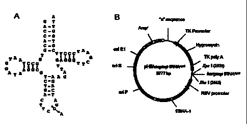
20 Figures 2A and 2B are diagrams depicting Suppressor tRNA and expression
vector constructs. Figure 2(A) Human Arginine tRNA: The sequence (noncoding
strand) and the clover leaf structure of the human arginine tRNA is shown. A
single
base change (shown with *) in the anticodon is required to convert the human
arginine
tRNA into an opal suppressor tRNA. Figure 2(B) HSV amplicon vector: Amp',
25 ampicillin resistant; "a", HSV-packaging signal; HSV-tk promoter, HSV-1
thymidine
kinase promoter; EBNA-1 modified EBV nuclear antigen gene; on P, EBV unique
latent replication origin; on S, HSV-1 replication origin.
Figures 3A -3D are a figures depicting the restoration of GFP 30 fluorescence
using hargsup tRNAOPaI. (A) GFP fluorescence detected in XPI2ROSV cells
30 cotransfected with the mhRGFP expression construct and the
6
CA 02318374 2000-07-13
WO 99/36519 PCT/US99/00717
pHEhargsup tRNAOPaI plasmid. Note bright green fluorescence in multiple
cells observed by fluorescence microscopy using a FITC filter. (B) Phase
contrast of the same field of transfected XP12ROSV cells as in A. (C)
XP12ROSV cells transfected with the mhRGFP vector alone. No significant
fluorescence is observed when the nonsense codon is not suppressed. (D)
Phase contrast of the same field as in (C).
Figures 4A and 4B are figures depicting Northern and Western analysis
of hRGFP expression. (A) Northern analysis. Total RNA (10 g/lane) from
XP12ROSV cells (Lane 1) or XP cells transfected with hRGFP gene (lane 2) or
1o with mhRGFP gene containing an opal nonsense mutation (CGC to TGA) at
amino acid 73 (lane 3) and probed for hRGFP cDNA (Top panel). GFP
transcripts of similar size were obtained. Positions of 18S rRNA is shown.
The same blot was then reprobed with Glyceraldehyde-Phosphate
Dehydrogenase (GAPDH) housekeeping gene (bottom panel). After
normalization of different RNA samples with the GAPDH gene, we observed
one third less abundance of the mhRGFP transcript compared to the hRGFP
transcript. (B) Detection of GFP protein by Western analysis. Cell lysates
were electrophoresed on a 10-20% SDS-PAGE gradient gel and probed with
the anti-GFP antibody. XP12ROSV cells expressing the hRGFP gene lone
(Lane 2) or coexpressing mhRGFP and hargsup tRNAOPaI (Lane 4) showed a
full-length 27 kDa GFP protein. Reduced levels of the full-length GFP protein
was observed in cells transfected with the mhRGFP and hargsup tRNAopaI
plasmids. No full-length GFP protein was detectable in cells expressing the
mhRGFP gene in the absence of suppressor tRNA (Lane 3). Nontransfected
XP12ROSV cells showed no non-specific staining (Lane 1).
Figure 5 is a graph depicting the partial correction of the DNA repair
deficient phenotype by suppressor tRNA. VA13, XP12ROSV and XP12ROSV
cells expressing the hargsup tRNAopaI were seeded at varying cell densities
from 2.5 x 10² to 1 x 10⁶, depending upon the cell line and UVC irradiation
dose used. The following day, cells were rinsed with PBS and irradiated with
UVC (254 nm) light at 0 to 6 J/m2. Approximately 10 to 12 days after
7
CA 02318374 2000-07-13
WO 99/36519 PCT/US99/00717
irradiation the cells were fixed and stained with crystal violet. The percent
colony-forming ability was determined by comparing the colony counts of the
irradiated plates with those of the unirradiated plates and plotted against
UVC dose used. A 4 to 35 fold increase in the colony forming ability at higher
UV doses was observed in XP12ROSV cells expressing the hargsup tRNAopal.
Figure 6 is a graph which depicts the correction of defective repair of
pSVCAT plasmid. XP12ROSV cells were transiently cotransfected with the
UVC irradiated pSVCAT and pHEhargsup tRNAopal plasmids. As controls
VA13 cells (positive control) and XP12ROSV cells (negative control) were
1o cotransfected with UVC irradiated pSVCAT plasmid and pHE vector alone.
After 72 hours, cell lysates were obtained and analyzed for CAT activity using
the liquid scintillation counting (LSC) method. CAT activity in cell extracts
was expressed as percent of the activity in cells transfected with
unirradiated
pSVCAT and plotted against UVC irradiation dose used to inactivate the
pSVCAT plasmid. XP12ROSV cells expressing the hargsup tRNAopal showed a
3 to 17 fold increased ability to reactivate UV irradiated pSVCAT plasmid
compared to XP12ROSV cells transfected the irradiated pSVCAT plasmid
alone.
Figures 7A and 7B are Northern and Western analysis of XPAC gene
expression. (A) Expression of XPAC transcript. Northern analysis of total
RNA (10 gg/lane) from normal control VA13 cells (Lane 1) and XP12ROSV
cells (Lane 2) probed with a truncated XPAC cDNA is shown in the
autoradiogram (top panel). The two bands in the VA13 cells (normal controls)
are due to alternative polyadenylation (Tanaka, K. et al., "Analysis of a
human
DNA excision repair gene involved in group A xeroderma pigmentosum and
containing a zinc-finger domain", Nature 348 73-78 (1990). The XPAC
transcript level was considerably reduced in the XP12ROSV cells. The same
blot was reproved for the GAPDH gene, to permit relative normalization of the
different RNA samples and shown in the bottom panel. (B) Western analysis
to detect XPAC proteins. Western analysis of DNA repair proficient VA13
cells (Lane 1), XP12ROSV cells (Lane 2) and XP12ROSV cells expressing the
8
CA 02318374 2000-07-13
WO 99/36519 PCT/US99/00717
suppressor tRNA (Lane 3) probed with anti-XPAC antibody. The XPAC
protein is easily observed in VA13 cells, but is not detected in either
XP12ROSV cells alone or XP12ROSV cells transfected with hargsup tRNAOPaI.
Figures 8A and 8B depict the transduction of XP12ROSV cells with
pHEhargsup tRNAoPaI amplicon vector packaged into HSV-1 virions. (A) GFP
fluorescent cells were observed when XP12ROSV cells expressing the mhRGFP
gene were transduced with the pHEhargsup tRNAOPaI amplicon vector,
suggesting a successful in vitro delivery of the suppressor tRNA by the
herpesvirus amplicon system and suppression of the nonsense mutation in the
to mhRGFP gene. (B). Phase contrast of the same field as in (A).
Figure 9 is a figure depicting human opal, amber and ochre suppressor
serine tRNAs designed according to the invention. (SEQ ID NOS:1-6) As is
illustrated, the suppressor tRNAs may be used in tandem using the restriction
splice sites indicated.
Figure 10 is a depiction of human opal suppressor serine tRNA and
human amber suppressor serine tRNAs designed according to the present
invention and a graphic illustration of the two suppressor tRNAs in tandem
using the splice sites indicated. (SEQ ID NOS:5-8)
Figure 11 is a diagram depicting the cloverleaf structure formed by the
novel human ochre suppressor serine tRNA of the invention. (SEQ ID NO:9)
Figure 12 is a diagram depicting the cloverleaf formation in the
anticodon region of yet another synthetic amber suppressor serine tRNA
formed in accordance with the present invention. (SEQ ID NO:10)
Figure 13 is a drawing depicting the cloverleaf formation of yet another
human serine opal suppressor tRNA illustrating the anticodon region in
accordance with the present invention. (SEQ ID NO:11)
Figure 14 is a drawing depicting the cloverleaf and anticodon regions
formed by yet another human opal suppressor tRNA by the present invention.
(SEQ ID NO:12)
9
CA 02318374 2000-07-13
WO 99/36519 PCT/US99/00717
DETAILED DESCRIPTION OF THE INVENTION
Atkinson and Martin in 1994 identified close to 180 unique point
mutations to nonsense codons identified in human genes from a search of
literature reports. These types of mutations result in muscular dystrophy,
Xeroderma pigmentosum, cystic fibrosis, hemophilia, anemia, hypothyroidism,
p53 squamal cell carcinoma, p53 hepatocellular carcinoma, p53 ovarian
carcinoma, esophageal carcinoma, osteocarcinoma, ovarian carcinoma,
esophageal carcinoma, hepatocellular carcinoma, breast cancer, hepatocellular
carcinoma, fibrous histiocytoma, ovarian carcinoma, SRY sex reversal,
1o triosephosphate isomerase-anemia, diabetes and rickets. The BRACA-1 and
BRACA-2 genes associated with breat cancer also have similar mutations
which may be treated accoding to the teachings herein. The present invention
in one embodiment includes methods for treating these diseases by reversing
the effects of mutations present that are associated with nonsense mutations
through introduction of the synthetic oligonucleotide suppressor tRNAs of the
invention.
The nucleotide sequences encoding several human tRNAs are known
and generally available to those of skill in the art through sources such as
Genbank. See also Sprinzl, Mathias et. Al., Nucleic Acids Research, volume
12, Supplement "compilation of tRNA Sequences" pgs, rl-r57 (1984);
Schimmel, P.R., et. Al. Editors, "Transfer-RNA: Structure, Properties, and
Recognition, Cold Spring Harbor Labs New York 1979.; Agris, P. F., (1983)
"The Modified Nucleosides of Transfer RNA, II, Alan R. Liss Inc., New York
(Buckland RA et al., "A cluster of tRNA genes into [DRNI, TRR3, DDRAN] on
the short arm of human chromosome 6", Genomics, 35 164-171 (1996)). tRNA's
have been shown to be highly conserved and are often functional across species
thus bacterial or other eucaryotic tRNA sequences are also potential sources
for the oligonucleotides of the invention. The determination of whether a
particular tRNA sequence will be functional in a desired mammalian cell can
be easily ascertained through routing experimentation and the assays and
methods discussed or incorporated herein. Further additional potential tRNA
CA 02318374 2010-04-28
WO 99/36519 PCTIUS99/00717
sequences as of yet unknown can be ascertained through the assays and
protocols discussed,
tRNA genes have strong promoters which are active in all cell types.
The promoters fb-r eukaryotic tRNA genes lie within the structural sequences
encoding the tRNA molecule itself. Although there are elements which
regulate transcriptional activity within the 5' upstream region, the length of
an active transcriptional unit may be considerably less than 500 base pairs
and thus accommodation within a delivery vector presents no problem. Once
io they have been transcribed and processed, tRNAs have low rates of
degradation. Finally gene therapy with a nonsense suppressor maintains the
endogenous physiological controls over the target gene which contains the
nonsense codon. One of skill in the art will appreciate that according to the
teachings herein, the oligonucleotides of the invention can be used for not
just
1s human genetic diseases caused by nonsense mutations but for gene therapy by
nonsense suppression to be applicable to mutations in a wide range of genes
for site-specific substitution of protein products.
Briefly an oligonucleotide is synthesized which comprises the structural
component of a tRNA gene functional in human cells. Thr sequence of this
20 oligonucleotide is designed based upon the known sequence with
substitutions
made in the anticodon region of the tRNA causing the specific tRNA to
recognize a nonsense or other specific mutation. For example as shown in
Figure 2 and according to the invention, the sequence of human serine tRNA
having an anticodon of TCG was modified to include a substitution of TCA the
25 complement of the opal mutation to cause the tRNA to recognize the opal
stop
codon rather than the traditional serine codon.
Importantly the sequences for the oligonucleotides of the invention
contain only the structural sequence encoding the tRNA molecule as well as a
small portion (around 20 nt)of the 3' flanking region. The 5' region is
omitted
30 to result in an oligonucleotide that is small and easier to handle (i.e.,
around
100 nucleotides long). The oligonucleotide sequence comprises the structural
II
CA 02318374 2000-07-13
WO 99/36519 PCT/US99/00717
component of the gene and includes around 15 bases from the 3' flanking
region and none of the 5' noncoding region. Traditional methods using
suppressor tRNAs to date have used entire suppressor tRNA molecules which
are isolated, cloned and then site-mutated to create the suppressor tRNA. The
present invention provides a much simpler method of designing suppressor
tRNAs, namely designing a oligonucleotide sequence comprising the structural
sequences encoding the tRNA and a portion of the 3' flanking region only. This
small oligonucleotide may then be synthetically synthesized, rather than
isolated, using standard genetic engineering techniques and these synthetic
i o suppressor tRNAs can be used according to the methods of the invention.
Once designed and shown to be functional according to the assays herein the
synthetic suppressor tRNA's can be used in a number of different techniques,
the most promising of which is gene therapy.
Suppressor tRNAs are presently being engineered as tools to address
basic biological questions. They are being used to site-specifically
incorporate
unnatural amino acids into proteins in vivo Noren C.J., Anthony-Cahill S.J.,
Griffith M.C. and Schultz P.G., "A general method for site-specific
incorporation of unnatural amino acids into proteins", Science 244:182-188
(1989)) and in combination with electrophysiological techniques, they have
provided a general method for structure-function studies of receptors,
channels
and transporters Nowak M.W., Kearney P.C., Sampson, J.R., Saks, M.E.,
Labarca G.C. et al., "Nicotinic receptor binding site probed with unnatural
amino acid incorporation in intact cells", Science 268:439-442 (1995)).
However, suppressor tRNAs are just now being investigated for the treatment
of human diseases. Nonsense mutations that occur in key regulatory genes
are often deleterious to the cell and responsible for several types of genetic
diseases. Atkinson and Martin (Atkinson J. and Martin R., "Mutations to
nonsense codons in human genetic disease: implications for gene therapy by
nonsense suppressor tRNAs", Nucleic Acids Res. 22:1327-1334 (1994))
surveyed and reported a list of 179 unique point mutations that resulted in
nonsense codons that were identified in human genes and caused human
12
CA 02318374 2000-07-13
WO 99/36519 PCT/US99/00717
genetic diseases. These are all potential targets for gene therapy (in vivo or
ex
vivo) for treatment of disease with suppressor tRNA technology. The potential
application of suppressor tRNA to gene therapy was first demonstrated for f-
thalassemia (Temple G.F., Dozy A.M., Roy K.L. and Kan Y.W., "Construction
of a functional human suppressor tRNA gene: an approach to gene therapy for
0-thalassemia", Nature 296:537-540 (1982)). A human amber suppressor
Lysine tRNA was able to suppress the nonsense mutation in mutated R-globin
gene, when both were simultaneously expressed in vivo in xenopus oocyte
system. The in vivo application of suppressor tRNAs for treatment of genetic
diseases caused by nonsense mutation was recently demonstrated in an mdx
mouse, which is an animal model for human Duchenne muscular dystrophy
with an ochre mutation in dystrophin gene (Li K, Zhang J., Buvoli M., Yan
X.D., Leinwand L. and He H., "Ochre suppressor transfer RNA restored
dystrophin expression in mdx mice", Life Sciences 61:205-209 (1997)). Direct
injection of plasmid DNA encoding the ochre suppressor tRNA into mdx mice
produced dystrophin positive fibers. The practical application of suppressor
tRNA, as therapeutic agents for gene therapy would largely depend on the
development of efficient vectors that can sustain gene expression.
Recently they have served as tools to address basic biological questions
such as study of cell-cell interactions during development (Kunes, S. &
Steller,
H., "Ablation of drosophila photoreceptor cells by conditional expression of a
toxin gene", Genes Develop 5, 970-983 (1991)), or to site-specifically
incorporate
unnatural amino acids into proteins in vivo (Noren, C.J., Anthony-Cahil, S.J.,
Griffith, M.C. & Schultz, P.G., "A general method for site-specific
incorporation
of unnatural amino acids into proteins", Science 244, 182-188 (1989)). In
combination with electrophysiological techniques, they have provided a
general method for structure-function studies of receptors, channels and
transporters (Nowak, M.W. et al., "Nicotinic receptor binding site probed with
unnatural amino acid incorporation in intact cells", Science 268, 439-442
(1995)).
13
CA 02318374 2000-07-13
WO 99/36519 PCT/US99/00717
Additional uses of the invention can include use of the suppressor
oligonucleotide tRNAs as triggering molecules to convert inactive toxin
molecule into an active toxin molecule. See for example Robinson et al.,
"Suppression of single and double nonsense mutations induced into the
diphtheria toxin A-Chain Gene: a potential binary system for toxin gene
therapy", Human Gene Therapy 6:137-143 (Feb. 1995).
Further the oligonucleotides of the invention can be used to institute
site-directed mutagenesis of protein products in vitro by introducing missense
mutations in gene products to identify structure and function relationships.
According to the invention, human opal, amber, and ochre suppressor
serine and arginine tRNAs have been designed which are approximately 100
nucleotides in length and can be introduced to cells to suppress mutations
resulting in nonsense codons where a serine or arginine should be present.
The oligonucleotides can be introduced directly to recipient cells or can be
ligated in tandem to increase efficacy of the oligonucleotide. In yet another
embodiment the suppressor tRNA of the invention may be introduced to the
cells using standard conventional genetic engineering techniques through use
of vectors. Because of the internal promoter sequences of tRNA encoding
sequences the tRNA sequence need not be included in a separate transcription
unit, although one may be provided.
In a preferred embodiment the nucleotide expression system of the
invention is included within an appropriate gene transfer vehicle which is
then
used to transduce cells to express the suppressor tRNA. The gene delivery
vehicle can be any delivery vehicle known in the art and can include simply
naked DNA which is facilitated by a receptor mediated transfection as well as
any of a number of vectors. Such vectors include but are not limited to
eukaryotic vectors, prokaryotic vectors (such as for example bacterial
vectors)
and viral vectors including but not limited to retroviral vectors, adenoviral
vectors, adeno-associated viral vectors, lentivirus vectors (human and other
including porcine), Herpes virus vectors, Epstein-Barr virus vectors, SV40
virus vectors, pox virus vectors, pseudotype virus vectors.
14
CA 02318374 2000-07-13
WO 99/36519 PCTNS99/00717
In a preferred embodiment, a packaging cell line is transduced with the
viral vector containing the nucleic acid sequence to be expressed to form a
producer cell line including the viral vector. The producer cells may then be
directly administered, whereby the producer cells generate viral particles
capable of transducing the recipient cells.
In a preferred embodiment, the viral vector is a retroviral or adenoviral
vector. Examples of retroviral vectors which may be employed include, but are
not limited to, Moloney Murine Leukemia Virus, spleen necrosis virus, and
vectors derived from retroviruses such as Rous Sarcoma Virus, Harvey
1o Sarcoma Virus, avian leukosis virus, human immunodeficiency virus,
myeloproliferative sarcoma virus, and mammary tumor virus.
Retroviral vectors are useful as agents to mediate retroviral-mediated
gene transfer into eukaryotic cells. Retroviral vectors are generally
constructed such that the majority of sequences coding for the structural
genes
of the virus are deleted and replaced by the gene(s) of interest. Most often,
the
structural genes (i.e., gag, pol, and env), are removed from the retroviral
backbone using genetic engineering techniques known in the art. This may
include digestion with the appropriate restriction endonuclease or, in some
instances, with Bal 31 exonuclease to generate fragments containing
appropriate portions of the packaging signal.
These new genes have been incorporated into the proviral backbone in
several general ways. The most straightforward constructions are ones in
which the structural genes of the retrovirus are replaced by a single gene
which then is transcribed under the control of the viral regulatory sequences
within the long terminal repeat (LTR). Retroviral vectors have also been
constructed which can introduce more than one gene into target cells. Usually,
in such vectors one gene is under the regulatory control of the viral LTR,
while
the second gene is expressed either off a spliced message or is under the
regulation of its own, internal promoter.
Efforts have been directed at minimizing the viral component of the
viral backbone, largely in an effort to reduce the chance for recombination
CA 02318374 2000-07-13
WO 99/36519 PCTNS99/00717
between the vector and the packaging-defective helper virus within packaging
cells. A packaging-defective helper virus is necessary to provide the
structural
genes of a retrovirus, which have been deleted from the vector itself.
In one embodiment, the retroviral vector may be one of a series of
vectors described in Bender, et al., J. Virol. 61:1639-1649 (1987), based on
the
N2 vector (Armentano, et al., J. Virol., 61:1647-1650) containing a series of
deletions and substitutions to reduce to an absolute minimum the homology
between the vector and packaging systems. These changes have also reduced
the likelihood that viral proteins would be expressed. In the first of these
vectors, LNL-XHC, there was altered, by site-directed mutagenesis, the
natural ATG start codon of gag to TAG, thereby eliminating unintended
protein synthesis from that point.
In Moloney murine leukemia virus (MoMuLV), 5' to the authentic gag
start, an open reading frame exists which permits expression of another
glycosylated protein (pPr80gag). Moloney murine sarcoma virus (MoMuSV)
has alterations in this 5' region, including a frameshift and loss of
glycosylation sites, which obviate potential expression of the amino terminus
of pPr80gag. Therefore, the vector LNL6 was made, which incorporated both
the altered ATG of LNL-XHC and the 5' portion of MoMuSV. The 5' structure
of the LN vector series thus eliminates the possibility of expression of
retroviral reading frames, with the subsequent production of viral antigens in
genetically transduced target cells. In a final alteration to reduce overlap
with
packaging-defective helper virus, Miller has eliminated extra env sequences
immediately preceding the 3' LTR in the LN vector (Miller, et al.,
Biotechniques, 7:980-990, 1989).
The paramount need that must be satisfied by any gene transfer system
for its application to gene therapy is safety. Safety is derived from the
combination of vector genome structure together with the packaging system
that is utilized for production of the infectious vector. Miller, et al. have
developed the combination of the pPAM3 plasmid (the packaging-defective
helper genome) for expression of retroviral structural proteins together with
16
CA 02318374 2000-07-13
WO 99/36519 PCT/US99/00717
the LN vector series to make a vector packaging system where the generation
of recombinant wild-type retrovirus is reduced to a minimum through the
elimination of nearly all sites of recombination between the vector genome and
the packaging-defective helper genome (i.e. LN with pPAM3).
In one embodiment, the retroviral vector may be a Moloney Murine
Leukemia Virus of the LN series of vectors, such as those hereinabove
mentioned, and described further in Bender, et al. (1987) and Miller, et al.
(1989). Such vectors have a portion of the packaging signal derived from a
mouse sarcoma virus, and a mutated gag initiation codon. The term "mutated"
io as used herein means that the gag initiation codon has been deleted or
altered
such that the gag protein or fragment or truncations thereof, are not
expressed.
In another embodiment, the retroviral vector may include at least four
cloning, or restriction enzyme recognition sites, wherein at least two of the
.
sites have an average frequency of appearance in eukaryotic genes of less than
once in 10,000 base pairs; i.e., the restriction product has an average DNA
size
of at least 10,000 base pairs. Preferred cloning sites are selected from the
group consisting of NotI, SnaBI, Sall, and XhoI. In a preferred embodiment,
the retroviral vector includes each of these cloning sites.
When a retroviral vector including such cloning sites is employed, there
may also be provided a shuttle cloning vector which includes at least two
cloning sites which are compatible with at least two cloning sites selected
from
the group consisting of NotI, SnaBI, Sall, and XhoI located on the retroviral
vector. The shuttle cloning vector also includes at least one desired gene
which is capable of being transferred from the shuttle cloning vector to the
retroviral vector.
The shuttle cloning vector may be constructed from a basic "backbone"
vector or fragment to which are ligated one or more linkers which include
cloning or restriction enzyme recognition sites. Included in the cloning sites
3o are the compatible, or complementary cloning sites hereinabove described.
Genes and/or promoters having ends corresponding to the restriction sites of
17
CA 02318374 2000-07-13
WO 99/36519 PCT/US99/00717
the shuttle vector may be ligated into the shuttle vector through techniques
known in the art.
The shuttle cloning vector can be employed to amplify DNA sequences
in prokaryotic systems. The shuttle cloning vector may be prepared from
plasmids generally used in prokaryotic systems and in particular in bacteria.
Thus, for example, the shuttle cloning vector may be derived from plasmids
such as pBR322; pUC 18; etc.
The vector includes one or more promoters. Suitable promoters which
may be employed include, but are not limited to, the retroviral LTR; the SV40
1o promoter; and the human cytomegalovirus (CMV) promoter described in
Miller, et al., Biotechniques, 7:(9):980-990 (1989), or any other promoter
(e.g.,
cellular promoters such as eukaryotic cellular promoters including, but not
limited to, the histone, pol III, and R-actin promoters). Other viral
promoters
which may be employed include, but are not limited to, adenovirus promoters,
TK promoters, and B19 parvovirus promoters. The selection of a suitable
promoter will be apparent to those skilled in the art from the teachings
contained herein.
The vector then is employed to transduce a packaging cell line to form a
producer cell line. Examples of packaging cells which may be transfected
include, but are not limited to the PE501, PA317, `Y2, 'P-AM, PA12, T19-14X,
VT-19-17-H2, TCRE, `YCRIP, GP+E-86, GP+envAM12, and DAN cell lines.
The vector containing the nucleic acid sequence encoding the agent which is
capable of providing for the inhibition, prevention, or destruction of the
growth
of the tumor cells upon expression of the nucleic acid sequence encoding the
agent may transduce the packaging cells through any means known in the art.
Such means include, but are not limited to, electroporation, the use of
liposomes, and CaPO4 precipitation.
The producer cells then are administered directly to or adjacent to
desired recipient cells.
In a preferred embodiment the invention comprises a viral vector which
commonly infects humans and packaging cell line which is human based. For
18
CA 02318374 2000-07-13
WO 99/36519 PCTIUS99/00717
example vectors derived from viruses which commonly infect humans such as
Herpes Virus, Epstein Barr Virus, may be used which do not express an active
a-galactosyl envelope.
In a most preferred embodiment the vector comprises a Herpes Simplex
Virus plasmid vector. Herpes simplex virus type-1 (HSV-1) has been
demonstrated as a potential useful gene delivery vector system for gene
therapy, Glorioso, J.C., "Development of Herpes Simplex Virus Vectors for
Gene Transfer to the Central Nervous System. Gene Therapeutics: Methods
and Applications of Direct Gene Transfer", Jon A. Wolff, Editor, 1994
1o Birkhauser Boston, 281-302; Kennedy, P.G., "The Use of Herpes Simplex Virus
Vectors for Gene Therapy in Neurological Diseases", Q J Med, Nov. 1993,
86(11):697-702; Latchman, D.S., "Herpes Simplex Virus Vectors for Gene
Therapy", Mol Biotechnol, Oct. 1994, 2(2):179-95.
HSV-1 vectors have been used for transfer of genes to muscle. Huard, J.,
"Herpes Simplex Virus Type 1 Vector Mediated Gene Transfer to Muscle", Gene
Therapy, 1995, 2, 385-392; and brain, Kaplitt, M.G., "Preproenkephalin
Promoter Yields Region-Specific and Long-Term Expression in Adult Brain
After Direct In Vivo Gene Transfer Via a Defective Herpes Simplex Viral
Vector", Proc Natl Acad Sci USA, Sep 13, 1994, 91(19):8979-83, and have been
used for murine brain tumor treatment, Boviatsis, E.J., "Long-Term Survival of
Rats Harboring Brain Neoplasms Treated With Ganciclovir and a Herpes
Simplex Virus Vector That Retains an Intact Thymidine Kinase Gene", Cancer
Res, Nov 15, 1994, 54(22):5745-51; Mineta, T., "Treatment of Malignant
Gliomas Using Ganciclovir-Hypersensitive, Ribonucleotide Reductase-Deficient
Herpes Simplex Viral Mutant", Cancer Res, Aug 1, 1994, 54(15):3963-6.
Helper virus dependent mini-viral vectors have been developed for
easier operation and their capacity for larger insertion (up to 140 kb),
Geller,
Al, "An Efficient Deletion Mutant Packaging System for Defective Herpes
Simplex Virus Vectors: Potential Applications to Human Gene Therapy and
Neuronal Physiology", Proc Natl Acad Sci USA, Nov 1990, 87(22):8950-4;
19
CA 02318374 2010-04-28
WO 99/36519 PCT/US99/00717
Frenkel, N., "The Herpes Simplex Virus Amplicon: A Versatile Defective Virus
Vector", Gene Therapy. 1. Supplement 1, 1994. Replication incompetent HSV
amplicons have been constructed in the art, one example is the pHSVlac vector
by Geller et al, Science, 241, Sept. 1988. .
These HSV amplicons contain large deletions of the HSV genome to provide
space for insertion of exogenous DNA. Typically they comprise the HSV-1
packaging site, the HSV-1 "ori S" replication site and the IE 4/5 promoter
sequence. These virions are dependent on a helper virus for propagation.
Primarily two types of mutant helper viruses have been developed to
minimize recombination. Other complementary HSV helper virus systems are
contemplated herein and are within the scope of those of skill in the art. One
such system which has been developed is a temperature-sensitive mutant. An
HSV temperature-sensitive (TS) mutant has been developed with a TS
mutation in the IE3 gene. Davison et al, 1984, J. Gen. Viral., 65:859-863.
Consequently this virus has an IE phenotype, does not replicate DNA, does not
significantly alter cellular physiology, and does not produce progeny virus at
37 C. Virus is grown at the permissive temperature of 37 C. TS mutants
however have had a tendency to revert to wild type.
In contrast a second helper virus system is a deletion mutant with the
majority of the IE3 gene simply deleted. These do not revert to wild type.
Therefore HSV-1 vectors packaged using a deletion mutant as helper virus is
the most preferred helper virus of the invention. See for example Patterson et
al., 1990, J. Gen. Virol., 71:1775-1783. Other replication incompetent helper
viruses can be used and one of skill in the art will appreciate that other
mutations in the IE genes or other genes which result in a replication
incompetent helper virus which will provide the appropriate replication and
expression functions and which are coordinated with the helper cell line and
vector are contemplated within this invention. Any cell line can be used for
this step so long as it is capable of expressing the IE3 or replication
dependent
gene, or obtaining a helper cell line which has already been transformed and
is
commercially available. Any cell line can be used by introducing pHE and the
CA 02318374 2000-07-13
WO 99/36519 PCT/US99/00717
plasmid containing the IE3 gene simultaneously. Next, the vector is delivered
to the helper cell line by electroporation, calcium phosphate DNA transfection
or any other suitable method. Any cell line can be used by introducing pHE
and the plasmid containing the IE3 gene simultaneously. The cells are next
infected with a helper virus IE3 deletion mutant or other corresponding
deletion mutant which is replication incompetent. The IE3 gene or other such
gene in the helper cell line complements the helper virus resulting in a
productive HSV-1 infection and the resulting virus stock consists of HSV-1
particles containing either vector DNA or helper virus DNA, all of which are
1o replication incompetent. Further information about helper cell lines and
the
methodology is disclosed in Geller et al., PNAS, 87:8950-8954, November 1990,
"An Efficient Deletion Mutant Packaging System for Defective Herpes Simplex
Virus Vectors: Potential Applications to Human Gene Therapy and Neuronal
Physiology". The invention comprises a HSV mini vector which combines a
replication incompetent HSV amplicon with other viral sequences such as
those from Epstein-Barr virus, human papillomavirus, or bovine
papillomavirus type 1 which allow the vector to be maintained in the cell in
episomal form achieving a 10 times greater titer, and a very large DNA insert
capacity.
One embodiment of the present invention involves a helper virus-
dependent mini-viral vector comprising: (a) the HSV-1 "a" sequence for the
package/cleavage signal and an "ori S" replication origin for the replication
packaging of the plasmid (in response to signals to replicate and package from
the helper virus); (b) an Epstein-Barr virus (EBV) nuclear antigen (EBNA-1)
gene and an EBV latent origin of replication (ori P) which allow the vector to
be maintained in episomal form within the nucleus for replication without
integration to the host genome and for even replication into each of two
dividing cells; preferably (c) genes from prokaryotic cells for propagation of
the
vector in E. coli (a selectable marker gene such as the ampicillin resistance
or
tetracycline resistance gene and the col. El ori) and (d) a sequence encoding
a
nonsense suppressor tRNA. Optionally the vector may also comprise
21
CA 02318374 2010-04-28
WO 99/36519 PCTIUS99/00717
prokaryotic genes that provide for a second selectable marker such as the
genes for positive Hygromycin selection. As described in United States Patent
Number 5,830,727.:
In this particular embodiment the packaging function of mini-vector
DNA into Herpes simplex viral capsids is provided by a helper virus and a
helper cell line.
In yet another embodiment the HSV vector can be engineered to
produce a helper free viral vector as in Mann et al,, "Construction of a Retro-
Virus Packaging Mutant and its Use to Produce Helper-Free Defective
1o Retrovirus", 33 Sal., p. 153-159, May 1983, Journal of Virology, September
1989, pp. 3822-3829, September 1989; Samuiski "Helper Free Stocks of
Recombinant Adeno-Associated Viruses: Normal Integration Does Not Require
Viral Gene Expression"; and Kohn et al., "High Efficiency Gene Transfer Into
Mammalian Cells: Generation of Helper-Free Recombinant Retrovirus With
Broad Mammalian Host Range", PNAS, 81:6349-6353, October 1984. See also
Okasinki, U.S. Patent No. 4,970,155 "HSV HELPER VIRUS INDEPENDENT
VECTOR".
According to the invention several tRNA synthetic suppressors have
been synthesized including a Human ochre suppressor serine tRNA (SEQ ID
NO: 1), a human amber suppressor serine tRNA (SEQ ID NO:2), a human
serine opal suppressor tRNA (SEQ ID NO:3), and a human opal suppressor
arginine tRNA (SEQ ID NO:4). These oligonucleotides have been shown to
function as active suppressor tRNAs in human cells, providing for read
through of nonsense mutations. The invention thus includes these
oligonucleotides as well as functional equivalents thereof. Vectors have also
been designed which incorporate the oligonucleotides and successfully deliver
these to recipient cells according to the invention including
pHEhargsuptRNAoPa1.
The design of additional oligonucleotides based upon the teachings
herein as well as nucleotide delivery vehicles incorporating the same are
22
CA 02318374 2010-04-28
WO 99/36519 PCTIUS99/00717
simply optimization of routine experimental procedures and are intended to be
within the scope of this invention.
The following examples serve to illustrates the
teachings herein and are not intended to limit the invention in any way.
EXAMPLES
Xeroderma pigmentosum (XP), is an autosomal recessive disease with a
marked predisposition to sunlight-induced skin cancer and other neurological
1o abnormalities, (Cleaver, J.E., "Defective repair replication of DNA in
xeroderma pigmentosum", Nature 218, 652-656 (1968)). The disease is
characterized by extreme sensitivity to ultraviolet radiation and defective
DNA nucleotide excision repair (NER), (Hanawalt, P.C., "DNA repair comes of
age", Mutation Res 336, 101-113 (1995); Copeland, N.E., Hanke, C.W. &
Michalak, J.A., "The molecular basis of xeroderma pigmentosum", Dermatol
Surg 23, 447-455 (1997)). Cell fusion studies to complement DNA repair
defects has led to the identification of seven genetic complementation groups
of
XP, designated XPA - XPG (Cleaver, J.E. & Kraemer, A.L., "Xeroderma
Pigmentosum", The Metabolic Basis of Inherited Disease Vol. IT' (eds Scriver,
C.R., L., B.A., Sly, W.S. & Valle, D.) 2949-2971 (McGraw-Hill, New York,
1989)), suggesting that at least seven different gene products are involved in
the NER pathway. All of the relevant human genes (XPA, XPB, XPC, XPD,
XPE, XPF and XPG) have been identified and mapped to specific chromosomal
locations, (Cleaver, J.E. & Kraemer, A.L., "Xeroderma Pigmentosum", The
Metabolic Basis of Inherited Disease Vol. IT' (eds Scriver, C.R., L., B.A.,
Sly,
W.S. & Valle, D.) 2949-2971 (McGraw-Hill, New York, 1989); Tanaka, K. et al.,
"Analysis of a human DNA excision repair gene involved in group A xeroderma
pigmentosum and containing a zinc-finger domain", Nature 348 73-78 (1990);
Satokata, I., Iwai, K,, Matsuda, T., Okada, Y. & Tanaka, K., "Genomic
3o characterization of the human DNA excision repair-controlling gene XPAC",
23
CA 02318374 2000-07-13
WO 99/36519 PCTIUS99/00717
Gene 136, 345-348 (1993); Boulikas, T., "Xeroderma pigmentosum and
molecular cloning of DNA repair genes", Anticancer Res 16, 693-708 (1996)).
The cloning and characterization of these DNA repair genes has greatly
increased the understanding of NER mechanisms and the relationship
between NER molecular defect and the clinical manifestations of XP disease,
(Nishigori, C., Moriwaki, S.-i., Takebe, H., Tanaka, T. & Imamura, S., "Gene
alterations and clinical characteristics of xeroderma pigmentosum group A
patients in Japan", Arch Dermatol 130, 191-197 (1994)). This disease was
chosen to demonstrate the novel genetic approach to correct the diseased
phenotype.
A common approach to correct the XP phenotype defect would be to
introduce relevant DNA repair genes into cells derived from XP patients.
Several in vitro studies have demonstrated restoration of normal DNA repair
phenotype and improved survival after UV irradiation of XP cells following
expression of the normal DNA repair gene (Mezzina, M. et al., "Correction by
the ERCC2 gene of UV sensitivity and repair deficiency phenotype in a subset
of trichothiodystrophy cells", Carcinogenesis 15, 1493-1498 (1994); Gozukara,
E.M. et al., "The human DNA repair gene, ERCC2 (XPD), corrects ultraviolet
hypersensitivity and ultraviolet hypermutability of a shuttle vector
replicated
in Xeroderma pigmentosum", Cancer Res 54, 3837-3844 (1994); Levy, D.D.,
Saijo, M., Tanaka, K., Kraemer, K.H., "expression of a transfected DNA repair
gene (XPA) in xeroderma pigmentosum group A cells restores normal DNA
repair and mutagenesis of UV-treated plasmids", Carcinogenesis 16, 1557-1563
(1995); Yagi, T. et al., "Complete restoration of normal DNA repair
characteristics in group F xeroderma pigmentosum cells by over-expression of
transfected XPF cDNA", Carcionogenesis 19, 55-60 (1998)). Other
investigators have complemented the defect by introducing DNA repair genes
from other organisms such as mouse, yeast, and Drosophila, (Tanaka, K.,
Satokata, I., Ogita, Z., Uchida, T. & Okada, Y., "Molecular cloning of a mouse
3o DNA repair gene that complements the defect of group-A xeroderma
24
CA 02318374 2000-07-13
WO 99/36519 PCT/US99/00717
pigmentosum", Proc Natl Acad Sci, USA, 86, 5512-5516 (1989); Lambert, C., et
al., "A yeast DNA repair gene partially complements defective excision repair
in mammalian cells", EMBO J 7, 3245-3253 (1988); Shimamoto, T., et al.,
"Expression and functional analyses of the Dxpa gene the drosophila homolog
of the human excision repair gene XPA", JBiol Chem 270, 22452-22459
(1995)). Another useful approach would be to correct the genetic defect in the
DNA repair gene or its transcript and thereby restore a normal phenotype.
Molecular genetic studies have defined novel deletions, splicing, point
mutations and nonsense mutations in the XP group A complementing (XPAC)
gene, (Satokata, I. et al., "Characterization of a splicing mutation in group
A
xeroderma pigmentosum", Proc Natl Acad Sci, USA 87, 9908-9912 (1990);
Satokata, I., et al., "Three nonsense mutations responsible for group A
xeroderma pigmentosum", Mutation Res 273, 193-202 (1992); Satokata, I.,
Tanaka, K. & Okada, Y., "Molecular basis of group A xeroderma pigmentosum:
a missense mutation and two deletions located in a zinc finger consensus
sequence of the XPAC gene", Hum Genet 88, 603-607 (1992); Satokata, I.,
Tanaka, K., Yuba, S. & Okada, Y., "Identification of splicing mutations of the
last nucleotides of exons a nonsense mutation and a missense mutation of the
XPAC gene as causes of group A xeroderma pigmentosum", Mutation Res 273,
203-212 (1992)). The genetic defect caused by nonsense mutations could be
repaired by expressing suppressor tRNAs that recognize the premature
termination signal, thereby resulting in the insertion of its corresponding
amino acid and restoration of normal phenotype in accordance with the
teachings of the present invention.
Materials and Methods
Cells lines. DNA repair proficient human fibroblast cell line (VA13, an SV40
transformed human W138 fibroblast cell line, ATCC, Gaithersburg, MD) and
an SV40 transformed cell line established from a XP patient (XP12ROSV; a
kind gift from Dr. Tanaka, Japan), were used for study. The cells were grown
CA 02318374 2010-04-28
WO 99/36519 PCTIUS99/00717
in Dulbecco's modified Eagle's medium (DMEM; Gibco BRL, Gaithersburg,
MD) supplemented with 1% glutamine and 10% fetal bovine serum (DIV
medium). Cells were grown at 37 C in a humidified C02 incubator. E5 helper
cells are green monkey kidney cells that express the IE3 gene and allow the
replication of the IE3-deleted helper virus (kindly provided by P. Johnson,
San
Diego).
Transfection. Using FuGENE 6 reagent (Boehringer Manheim, Germany)
transfection was carried out as per manufacturer instructions with minor
modifications. Cells (-1.5-2 x 10⁵) were seeded in 60 mm tissue culture plates
to and allowed to attach for 16-20 h. FuGENE 6 transfection reagent (12 l) was
incubated for 5 minutes at room temperature (RT) in 88 l opti-MEM medium
(Gibco BRL.), then added to the plasmid DNA (4 g) and further incubated for
minutes at RT. This transfection mixture was added to the cells grown in
complete DMEM medium (2 ml). After 14-16 hours at 37 C, the cells were
15 washed once with Hanks balanced salt solution (HBSS) and then fed with
complete medium and further incubated at 37 C for a total of 72 hours after
transfection. Transfected cells were selected in the presence of hygromycin
(100-150 g/ml).
Construction of human arginine opal suppressor tRNA (hargsup
tRNAopa1) and subcloning into a herpes simplex virus (HSV amplicon
vector. Based on the reported nucleotide sequence of human arginine tRNA
gene (Buckland RA et al., "A cluster of tRNA genes into [DRNI, TRR3,
DDRAN] on the short arm of human chromosome 6", Genomics, 35 164-171
(1996)), the arginine opal suppressor tRNA was constructed that contained the
structural sequences encoding the tRNA molecule and 15 bases from the 3'
flanking region. The 3' flanking region contains clusters of T residues in the
noncoding strand that act as the signal to terminate transcription. The
sequence (noncoding strand) and the clover leaf structure of the human
arginine tRNA is as shown in Fig. 2A. A single base change (shown with * in
Fig. 2A) in the anticodon was introduced to convert the human arginine tRNA
26
CA 02318374 2000-07-13
WO 99/36519 PCT/US99/00717
into an opal suppressor tRNA. A pair of overlapping oligonucleotides: 5'-
GCGCTCGAGAAAACGAAC
CCCACTTAACCACGAAGGGATTCGAACCCTCAATCTTCTGATC-3' and 5'-
GCGGGT
ACCGACCACGTGGCCTAATGGATAAGGCGTCTGACTTCAGATCAGAAGAT
TGAGGG-3' (synthesized by Integrated DNA Technologies, Inc., Coralville, IA)
were annealed and filled in using T7 DNA polymerase (Sequenase Version 2,
United States Biologicals) enzyme and a reaction mixture containing all four
deoxynucleotides. The double stranded oligonucleotide was digested with Kpn
1o I/Xho I and ligated at the same restriction sites into an HSV amplicon
(Wang,
S., Young, W.-B., Jacobson, C. & Link, C.J., "A novel Herpesvirus amplicon
system for in vivo gene therapy", Gene Ther 4, 1132-1141 (1997)) vector that
was modified to remove the CMV promoter and the poly A sequences. The
resulting expression vector pHEhargsup tRNAopa1 is as shown in Fig. 2B. The
expression vector contains an EBV on P and EBNA sequence to maintain the
plasmid episomally and a hygromycin resistance gene to permit selection of
transfected mammalian cells. The plasmid also contains the HSV- 1 lytic
replication origin (ori S) and a HSV-1 terminal packaging signal "A" sequences
for replication and packaging into HSV-virions in the presence of transacting
helper virus (Wang, S., Young, W.-B., Jacobson, C. & Link, C.J., "A novel
Herpesvirus amplicon system for in vivo gene therapy", Gene Ther 4, 1132-
1141 (1997); Spaete, R.R. & Frenkel, H., "The herpes simplex virus amplicon:
A new eucaryotic defective-virus cloning-amplifying vector", Cell 30, 295-304
(1982); Wang, S. & Vos, J., "A hybrid infectious vector based on Epstein-Barr
virus and Herpes simplex virus type I for gene transfer into Human Gene", J
Virol 70, 8422-8430 (1996)).
Introducing an opal (TGA) mutation at a defined position within a
humanized red-shifted green fluorescent protein (hRGFP) gene. An
opal mutation was introduced at position Arg(CGC)730pa1(TGA) in the
hRGFP gene (Levy, J.P., Muldoon, R.R., Zolotukin, S. & Link, C.J., "Retroviral
27
CA 02318374 2010-04-28
WO 99/36519 PCT/US99100717
transfer and expression of humanized, red shifted green fluorescent protein
into human tumor cells", Nature Biotechnol 14, 610-614 (1996)). The mutation
was introduced by PCR and sequenced to confirm the mutation. The opal-
mutated hRGFP was subcloned into the pHE700 HSV amplicon vector (Wang,
S., Young, W.-B., Jacobson, C. & Link, C.J., "A novel Herpesvirus amplicon
system for in vivo gene therapy", Gene Ther 4, 1132-1141 (1997)), under the
control of the CMV promoter (mnRGFP).
Northern Analysis. Total RNA was isolated from cultured cells using the
to RNeasy Total RNA kit (Qiagen, Santa Clarita, CA). RNA samples (10 g) were
separated by electrophoresis through 1% formaldehyde agarose gels and
transferred onto nytran membrane filters (Schleicher & Schuell, Keene, NH).
The filters were pre-hybridized for 2 hours at 42 C in hybrisol I solution
(Oncor, Gaithersburg, MD) and then hybridized overnight at 42 C with probes
labeled with a (Atkinson, J. & Martin, R,, "Mutations to nonsense codons in
human genetic disease: implications for gene therapy by nonsense suppressor
tRNAs", Nucleic Acids Res 22, 1327-1334 (1994)) PdCTP using the Random
Primed DNA labeling kit (Boehringer Mannheim). The filters were washed
twice with 2 x SSC, 0.1% SDS at RT, then 0.1 x SSC and 0.1% SDS at 55 C for
30 minutes and autoradiographed.
Western Analysis. Cultured cells were washed twice with phosphate-
buffered saline (PBS) and then lysed in 50 mM Tris-HCI, pH 7.5, 0.15 M NaCl,
1% Nonidet P-40, 0.1% SDS and containing protease inhibitors
phenylmethylsulfonylfluoride (100 g/ml), aprotinin (1 g/ml) and leupeptin (1
g/ml). The cell lysate was incubated on ice for 20 minutes, centrifuged at
10,000 x g for 10 minutes at 4 C and the supernatant collected. Samples (30
g) were mixed with 6 l or 5 x sample buffer (Laemmli, 1970), boiled for 3
minutes and separated by SDS-PAGE. Protein samples were electroblotted
onto nitrocellulose membrane and probed with specific antibodies. The GFP
28
CA 02318374 2010-04-28
WO 99/36519 PCTIUS99/00717
and XPAC proteins were detected by Enhanced Chemiluminescence
(Amersham, England), using anti-GFP (Clontech, Palo Alto, CA) and anti
XPAC (Santa Cruz Biotechnology, Inc., Santa Cruz, CA) polyclonal antibodies
respectively and horseradish peroxidase (HRP)-conjugated anti-rabbit
immunoglobulin as second antibody.
Clonogenic assay. For UV survival experiments, three cell lines- VA13,
XP12ROSV and XP12ROSV expressing the hargsup tRNA0Pal were plated in
triplicates in 60 mm plates at different cell densities ranging from 2.5 x 10²
to
1 x 10⁶ in 60 mm petri plates, depending on the cell line and UVC dose used.
The next day the cells were rinsed with PBS and exposed to UVC dose (254
nm, Spectroline, model XX-15G, Spectronics Corp, NY) ranging from 0-6j/M2.
The PBS buffer was removed and replaced with complete DMEM medium
containing vitamins and nonessential amino acids. Approximately 10-12 days
1s after irradiation the colonies were fixed with formaldehyde and stained
with
crystal violet (0.05% in water). The percent colony-forming ability was
determined by comparing the colony counts of the irradiated plates with those
of the unirradiated plates. Separate experiments were independently repeated
two to three times.
Reactivation assay using plasmid carrying chloramphenicol acetyl
transferase (CAT) reporter gene. Plasmid pSVCAT (control vector;
Promega, Madison, WI) expressing the CAT gene under the control of SV
promoter was used for plasmid reactivation studies. CAT assays were
performed using the manufacturer's protocol with minor modifications.
Plasmid DNA (20 g/ml) in water was treated with 254 nm UVC radiation at
dose ranging from 0-600 J/m2, ethanol precipitated and resuspended in
suitable volume for transfection. XP12ROSV cells (2x10⁵) were seeded in 60
mm plate and on the next day were cotransfected with pHEhargsup tRNAoPa1
and UVC irradiated pSVCAT plasmid DNA in a 10:1 ratio (4 g of total DNA).
As control for CAT plasmid reactivation, DNA repair proficient VA13 cells
29
CA 02318374 2010-04-28
WO 99/36519 PCTIUS99/00717
(positive control) and XP12ROSV cells (negative control) seeded in 60 mm
plate were cotransfected with the modified pHE vector and the UVC irradiated
pSVCAT plasmid in the ratio of 10:1. After 72 hours, the cells were harvested
and lysed by three freeze-thaw cycles. Lysates were analyzed for CAT activity
using a liquid scintillation counting (LSC) method. Briefly, reaction mixture
containing protein lysates, [15C] chloramphenicol (Du Pont New England
Nuclear, Wilmington, DE; NEC 408A, 0.15 ,tCi) and n-butyryl CoA (25 g as
substrate (final reaction volume of 125 l) was incubated at 37 C for 3 hours.
The reaction products were extracted with 300 l of mixed xylene. The upper
io xylene phase was collected and extracted twice more by adding 100 l of
0.25
M Tris-HCI, pH 8Ø A fixed amount of the. upper xylene phase that represents
the butyrylated chloramphenicol product was added to the scintillation fluid
(5
TM .
ml; OptiScint "HiSafe"l LKB, England) and counted by liquid scintillation
spectrometry. Individual transfections were carried out in duplicate and
separate experiments were independently repeated two to three times. The
ability to reactivate the UVC irradiated plasmid was defined as the ratio of
the
CAT activity in extracts from cells transfected with the UV irradiated plasmid
to that in cells transfected with the non-irradiated pSVCAT plasmid.
Packaging of pHEhargsup tRNAopal amplicon vector. E5 cells (2 x 10⁶
cells/10 cm dish) were transfected with the pHEhargsup tRNAOPaI vector. Two
days following transfection the cells were placed under selection with
hygromycin B (150 g/ml). The hygromycin-resistant E5 cells were super-
infected with 1 MOI of Cgalk3 helper virus (kindly provided by P. Johnson,
San Diego) in 1 ml of Opti-MEM medium. The viruses were allowed to adsorb
on the cells for 2 hours at 37 C in a humidified, 5% C02 incubator and then 9
ml of DEM medium with 10% FBS was added and the cells further incubated
for 50-60 hours. The viral supernatant containing cell debris was collected.
The cells were lysed by freeze-thaw method (repeated three times), then
centrifuged at 2400 x g to pellet the cell debris. The titer of Cga1A3 helper
CA 02318374 2000-07-13
WO 99/36519 PCT/US99/00717
virus was determined by 5-bromo-4-chloro-3-indolyl-f -D-galactopyranoside (X-
gal) staining, as described previously (Wang, S., Young, W.-B., Jacobson, C. &
Link, C.J., "A novel Herpesvirus amplicon system for in vivo gene therapy",
Gene Ther 4, 1132-1141 (1997)). To demonstrate in vitro transfer of the
pHEhargsup tRNA0Pa1 amplicon vector, XP12ROSV cells expressing the
mhRGFP gene were transduced with different amounts of the viral
supernatant diluted in opti-MEM and incubated at 37 C in a humidified 5%
CO2 incubator. After 24 hours the cells were visualized for restoration of GFP
fluorescence.
Results
Restoration of GFP fluorescence by suppressor tRNA. To test the
functional activity of the hargsup tRNA0Pai, mhRGFP was used as a reporter
gene. An opal mutation introduced at position Arg73 Opal in hRGFP gene
completely prevents detectable GFP fluorescence. However, cotransfection of
the plasmid containing the hargsup tRNAOPal with the plasmid containing the
mhRGFP gene in XP12ROSV cells, was able to suppress the opal mutation in
mhRGFP and restore GFP fluorescence (Fig. 3A and 3B). Control
transfections with mhRGFP plasmid alone did not exhibit any GFP
fluorescence (Fig. 3C and 3D).
Expression of GFP transcripts and protein. To determine the expression
of the hRGFP and mhRGFP transcripts in transfected XP12ROSV cells,
northern blots were probed with the hRGFP cDNA. Transcripts of similar size
were observed in both the transfected cell lines (Fig. 4A, top panel). After
normalization with the GAPDH housekeeping gene (Fig. 4A, bottom panel),
mhRGFP transcripts were approximately one third less abundant compared to
the hRGFP transcripts. Western analysis using anti-GFP antibody showed
that XP12ROSV cells cotransfected with mhRGFP and hargsup tRNAopaI
3o expressed a full-length GFP protein similar in size to the hRGFP protein
(Fig.
31
CA 02318374 2000-07-13
WO 99/36519 PCT/US99/00717
4B). Thus providing direct evidence that hargsup tRNAopa1 can suppress the
nonsense mutation in the mhRGFP gene and produce a full-length functional
protein. Although equal amounts of the protein were loaded (data not shown),
an approximate 10-fold reduction in signal intensity was observed in the
XP12ROSV cells transfected with both the mhRGFP and hargsup tRNAOpa1
compared to hRGFP alone. Two observations account for the much lower
detected hRGFP protein found in the cells co-transfected with both the
mhRGFP and hargsup tRNAOPaI. The level of mhRGFP transcript targets for
the suppressor tRNA were about one third less in abundance relative to
1o expression of a housekeeping gene (Fig. 4A). However, the reduction in
protein level was significantly greater than the decrease in RNA transcripts
indicating an additional effect from relatively low suppression efficiency.
XP12ROSV cells transfected with the mhRGFP gene alone should produce a
truncated (-8 kDa) GFP protein but this was not detected because of the gel
system used.
Correction of XPA NER deficiency phenotype in XP cell lines by
nonsense suppressor tRNA as measured by post UV irradiation cell
survival. To demonstrate in vitro that suppressor tRNAs can repair genetic
defects caused by nonsense mutations, a DNA repair-deficient XP fibroblast
cell line -XP12ROSV was selected. A C to T transition at nucleotide 619 in
exon 5 of the XPAC gene altered Arg-207 codon (CGA) to a nonsense codon
(TGA). Thus resulting in the truncated XPAC protein and disruption of the
functional activity of the XPAC gene, (Satokata, I., et al., "Three nonsense
mutations responsible for group A xeroderma pigmentosum", Mutation Res
273, 193-202 (1992)). This cell line has a low colony forming ability after UV
irradiation. These cells were transfected with the pHEhargsup tRNAopa1
plasmid and selected with hygromycin. The selected cells were irradiated with
UVC (254 nm) at doses ranging from 0-6j/M2. Clonogenic assay was
performed and a 4 to 35 fold increase in the colony-forming ability at higher
32
CA 02318374 2000-07-13
WO 99/36519 PCT/US99/00717
UV doses was observed in XP12ROSV cells expressing the hargsup tRNAopal
(Fig. 5). The results demonstrated an improved UV survival of the XP12ROSV
cells due to partial correction in the DNA repair deficiency phenotype by the
introduction of the suppressor tRNA.
UV irradiated CAT plasmid reactivation studies. An alternative in vitro
test to determine if suppressor tRNAs can suppress the XPAC gene nonsense
mutation in XP12ROSV cells and restore DNA repair activity is the use of
expression vectors. Plasmid vectors expressing the bacterial CAT gene has
1o been widely used to measure DNA repair and mutagenesis in human cells
(Kraemer, K.H., Protic-Sabljic, M., Bredberg, A. & Seidman, M.M., "Plasmid
vectors for study of DNA repair and mutagenesis", Curr Probl Derm 17, 166-
181 (1987)). The XP12ROSV cells transiently transfected with the UV
irradiated pSVCAT plasmid were much more sensitive to inhibition of CAT
expression compared to the normal VA13 cells (Fig. 6). However, transient
expression of suppressor tRNA in XP12ROSV cells results in an increased
ability to reactivate UV irradiated pSVCAT plasmid by efficiently suppressing
the nonsense mutation in XPAC gene and restoring the DNA repair activity.
A 3 to 17 fold increase in the CAT activity at higher UV doses was observed in
XP cells transiently transfected with both the pHEhargsup tRNAopal plasmid
and UV damaged pCAT plasmid (Fig. 6).
Levels of XPAC transcript and XPAC protein in XP cells. Northern
analysis of XP12ROSV cells showed a very low abundance of the XPAC
transcript compared to the normal DNA repair proficient VA13 cells (Fig. 7A,
top panel). The results obtained were similar to those reported earlier
(Satokata, I., et al., "Three nonsense mutations responsible for group A
xeroderma pigmentosum", Mutation Res 273, 193-202 (1992)). To correlate the
partial phenotypic correction in XP12ROSV cells by suppressor tRNA with the
presence of full-length protein, Western analysis was performed. Fig. 7B
shows that XPAC protein while detectable in the normal VA13 cells, was not
33
CA 02318374 2000-07-13
WO 99/36519 PCT/US99/00717
detectable in XP12ROSV cells transfected with the hargsup tRNAopal.
Furthermore, no truncated XPAC polypeptide was observed in the XP12ROSV
cells.
Transduction of XP12ROSV cells expressing mhRGFP with
pHEhargsup tRNAopa1 amplicon vector packaged into HSV-1 virions.
The pHEhargsup tRNAOPaI amplicon vector packaged into HSV-1 virions could
successfully transduce and suppress nonsense mutation in the mhRGFP gene
expressed in XP12ROSV cells, as visualized by restoration of GFP fluorescence
(Fig. 8A and 8B). At 40 MOI of helper virus, restoration of GFP fluorescence
was observed in approximately 5% of cells. However substantial cytotoxicity
was evident by 24 to 48 hours after infection. At lower MOI of infection no
GFP positive cells were observed. GFP fluorescence was not detected with the
above cell line was transduced with the pHE700TK vector packaged into HSV-
1 virions, used as negative control or when XP12ROSV cells were transduced
with the pHEhargsup tRNAOPaI amplicon vector alone (data not shown).
Discussion.
Using Xeroderma Pigmentosum group A cells as a disease model, we
have been able to demonstrate in vitro partial restoration of the DNA repair
activity of the nonsense mutated XPAC gene. The XP12ROSV cell line used
for study contains a homozygous nonsense mutation in the XPAC gene
(Satokata, I., et al., "Three nonsense mutations responsible for group A
xeroderma pigmentosum", Mutation Res 273, 193-202 (1992)). An arginine
(CGA) codon had been mutated to an opal (TGA) codon in the XPAC gene,
hence for our study we chose to construct a human arginine opal suppressor
tRNA. To date, no tRNA suppressors had previously been derived from the
human arginine tRNA. The designed suppressor tRNA is small in size as the
5' flanking sequences have been deleted while 15 bases from 3' flanking
sequences were retained to signal transcription termination. The construction
of small-sized tRNAs by using a pair of oligonucleotides has an advantage over
34
CA 02318374 2000-07-13
WO 99/36519 PCT/US99/00717
the more tedious and time consuming method of site-directed mutagenesis to
convert tRNA into a suppressor tRNA. Using our method we have constructed
several different functional suppressor tRNAs such as human serine amber
suppressor and tyrosine ochre suppressor tRNA. A high readthrough
efficiency using amber or ochre mutated hRGFP as a reporter gene was also
observed by FACS analysis of GFP fluorescence (data not shown).
The functional activity of the hargsup tRNAOPaI was first established
using an opal mutated humanized red-shifted green fluorescent protein as a
reporter gene. A high efficiency of nonsense suppression (>80%) was
1o demonstrated by the restoration of hRGFP fluorescence and by FACS analysis
for GFP positive cells (data not shown). Expression of the hargsup tRNAOPa1 in
XP cell lines produced a 4 to 35 fold increase in cell survival after UV
irradiation and increased ability of XP cells to reactivate UV-irradiated
pSVCAT plasmid.
Suppressor tRNAs may cause readthrough of the natural termination
codons. The C terminal extended proteins may have codominant negative
properties, or they may have severely limited activity or they may be
subjected
to premature degradation. All of which may be deleterious to the cell.
However, the cell may be able to safeguard itself from such harmful effects
because of multiple translational stop codons at the end of the gene and also
due to the inefficient suppression by the different suppressor tRNAs.
Preliminary studies from our toxicity assay revealed that stable expression of
suppressor tRNA in XP cells did not alter the cycle as determined by FACS
analysis (data not shown) or cause a direct cytotoxic affect that could be
detected in clonogenic assays.
The practical application of suppressor tRNA, as therapeutic agents for
gene therapy will be highly dependent on the development of efficient vectors
that can sustain long term gene expression in the appropriate target tissues
or
cells. Several types of vectors are under development for this purpose. Use of
recombinant retroviruses to deliver DNA repair genes such as XPD (Carreau,
M., et al., "Functional retroviral vector for gene therapy of xeroderma
CA 02318374 2000-07-13
WO 99/36519 PCT/US99/00717
pigmentosum group D patients", Hum Gene Ther 6, 1307-1315 (1995); Quilliet,
X., et al., "Long-term complementation of DNA repair deficient human
primary fibroblasts by retroviral transduction of the XPD gene", Mutation Res
364, 161-169 (1996)), XPA, XPB and XPC genes (Zeng, L. et al., "Retrovirus-
mediated gene transfer corrects DNA repair defect of xeroderma pigmentosum
cells of complementation groups A, B and C", Gene Ther 4, 1077-1084 (1997) in
the XP cells with the corresponding defective DNA repair gene has been
reported. Functional expression of the transgene and correction of DNA repair
activity was observed in the transduced cells. To obtain efficient delivery of
1o the suppressor tRNA we chose to use a novel HSV amplicon vector, pHE,
(Wang, S., Young, W.-B., Jacobson, C. & Link, C.J., "A novel Herpesvirus
amplicon system for in vivo gene therapy", Gene Ther 4, 1132-1141 (1997) so
that the plasmid containing the suppressor tRNA could be amplified and
packaged into infectious HSV-1 virions in the presence of transacting helper
virus. The infectious pHE vector has efficient transgene expression in a
variety of human cell lines. The designed suppressor tRNA is only about 0.1
kb in size. Thus approximately 15 copies of the tRNA might be packaged into
a single 152 kb viral genome. Although we were able to package the
suppressor tRNA into the herpes genome, less than 5% of the transduced
XP12ROSV cells expressing the mhRGFP demonstrated green fluorescence
(Fig. 7). The cellular toxicity and low suppression efficiency is likely
secondary
to helper virus proteins that shutoff host protein synthesis. Previous studies
have demonstrated that the HSV host shutoff genes such as vhs (UL41) and
ICP47 (UL54) can inhibit host cell protein synthesis (Kwong, A.D., Kruper,
J.A. & Frenkel, N., "Herpes simplex virus host shutoff function", J Virol 62,
912-921 (1988); Hardwicke, M.A. & Sandri-Goldin, R.M., "The Herpes simplex
virus regulatory protein ICP27 contributes to the decrease in cellular mRNA
levels during infection", J Virol 68, 4797-4810 (1994)). For example,
expression of the vhs gene facilitates the degradation of host mRNA (Kwong,
3o A.D., Kruper, J.A. & Frenkel, N., "Herpes simplex virus host shutoff
function",
36
CA 02318374 2000-07-13
WO 99/36519 PCT/US99/00717
J Virol 62, 912-921 (1988). It is possible that these or other helper virus
proteins reduced the mRNA stability or expression of the mhRGFP gene and
resulted in fewer targets for the suppressor tRNA. Studies are in progress to
use the recently developed helper-free packaging system to eliminate the
presence of viral host shutoff genes and reduce or eliminate virus
cytotoxicity
(Fraefel, C. et al., "Helper virus-free transfer of herpes simplex virus type
1
plasmid vectors into neural cells", J Virol 70, 7190-7 (1996).
According to the invention, several other human suppressor serine
tRNAs have been synthesized and shown to be functional in accordance with
io the teachings herein. These are disclosed at Figures 8-13.
37
CA 02318374 2000-07-13
WO 99/36519 PCTIUS99/00717
SEQUENCE LISTING ^
<110> Panchal, Rekha G.
Link, Charles J. G
<120> Suppressor tRNA Oligonucletides and Methods of Use for
Same
<130> suppressor tRNAs
0
<140>
<141>
<150> 60/071,416
<151> 1998-01-14
<160> 14
<170> Patentln Ver. 2.0
<210> 1
<211> 118 _
<212> DNA C
<213> Artificial Sequence
<220> ^
<223> Description of Artificial Sequence: synthetic
0
<400> 1 ^
gcgcggtacc agtaaaaaaa gcacgccgta gtcggcagga ttcgaacctg cgcggggaga 60
ccccaatgga tttgaagtcc atcgccttaa ccactcggcc acgactacca gctgcgcg 118
u
<210> 2
<211> 119
<212> DNA
<213> Artificial Sequence
G
<220> ^
<223> Description of Artificial Sequence: synthetic G
0
<400> 2 0
1
CA 02318374 2000-07-13
WO 99/36519 PCTIUS99/00717
cgcgccatgg tcattttttt cgtgcggcat cagccgtcct aagcttggac gcgcccctct 60
ggggttacct aaacttcagg tagccggaat tggtgagccg gtgctgatgg tcgaccgcg 119
G
<210> 3 C
<211> 118 ^
<212> DNA C
<213> Artificial Sequence =
<220>
<223> Description of Artificial Sequence: synthetic _
<400> 3
gcgcctcgag agtaaaaaaa gcacgccgta gtcggcagga ttcgaacctg cgcggggaga 60
ccccaatgga tttagagtcc atcgccttaa ccactcggcc acgactacgg taccgcgc 118
<210> 4
<211> 118 ^
<212> DNA ^
<213> Artificial Sequence
-
<220>
<223> Description of Artificial Sequence: synthetic
01
<400> 4
cgcggagctc tcattttttt cgtgcggcat cagccgtcct aagcttggac gcgcccctct 60
ggggttacct aaatctcagg tagcggaatt ggtgagccgg tgctgatgcc atggcgcg 118
<210> 5 ^
<211> 118 ^
<212> DNA ^
<213> Artificial Sequence ^
<220> ^
<223> Description of Artificial Sequence: synthetic
`
<400> 5
gcgcgctagc agtaaaaaaa gcacgccgta gtcggcagga ttcgaacctg cgcggggaga 60
ccccaatgga tttaaagtcc atcgccttaa ccactcggcc acgactacct cgaggcgc 118
<210> 6 ^
<211> 118 ^
2
CA 02318374 2000-07-13
WO 99/36519 PCT/US99/00717
<212> DNA
<213> Artificial Sequence ^
C
<220> ^
<223> Description of Artificial Sequence: synthetic r
<400> 6
cgcgcgatcg tcattttttt cgtgcggcat cagccgtcct aagcttggac gcgcccctct 60
ggggttacct aaatttcagg tagcggaatt ggtgagccgg tgctgatgga gctccgcg 118
<210> 7 _
<211> 118 ^
<212> DNA ^
<213> Artificial Sequence
<220>
<223> Description of Artificial Sequence: synthetic
^
<400> 7 ^
gcgcggtacc agtaaaaaaa gcacgccgta gtcggcagga ttcgaacctg cgcggggaga 60
ccccaatgga tttgaagtcc atcgccttaa ccactcggcc acgactacca gctggcgc 118
<210> 8
<211> 119
<212> DNA ^
<213> Artificial Sequence ^
<220> -
<223> Description of Artificial Sequence: synthetic
<400> 8 ^
cgcgccatgg tcattttttt cgtgcggcat cagccgtcct aagcttggac gcgcccctct 60
ggggttacct aaacttcagg tagccggaat tggtgagccg gtgctgatgg tcgaccgcg 119
<210> 9 71
<211> 118
<212> DNA
<213> Artificial Sequence
<220> ^
<223> Description of Artificial Sequence: synthetic
3
CA 02318374 2000-07-13
WO 99/36519 PCT/US99/00717
<400> 9
gcgcctcgag agtaaaaaaa gcacgccgta gtcggcagga ttcgaacctg cgcggggaga 60 ^
ccccaatgga tttagagtcc atcgccttaa ccactcggcc acgactacgg taccgcgc 118
-
<210> 10
<211> 118
<212> DNA
<213> Artificial Sequence
<220>
<223> Description of Artificial Sequence: synthetic
<400> 10
cgcggagctc tcattttttt cgtgcggcat cagccgtcct aagcttggac gcgcccctct 60
ggggttacct aaatctcagg tagcggaatt ggtgagccgg tgctgatgcc atggcgcg 118
<210> 11
<211> 82
<212> DNA ^
<213> Artificial Sequence
<220>
<223> Description of Artificial Sequence: synthetic
<400> 11
gtagtcgtgg ccgagtggtt aaggcgatgg actttaaatc cattggggtc tccccgcgca 60
ggttcgaatc ctgccgacta cg 82
<210> 12 ^
<211> 82 ^
<212> DNA ^
<213> Artificial Sequence
<220>
<223> Description of Artificial Sequence: synthetic
<400> 12
gtagtcgtgg ccgagtggtt aaggcgatgg actctaaatc cattggggtc tccccgcgca 60
ggttcgaatc ctgccgacta cg 82
4
CA 02318374 2000-07-13
WO 99/36519 PCT/US99/00717
<210> 13 ^
<211> 82
<212> DNA
<213> Artificial Sequence ^
^
<220> ^
<223> Description of Artificial Sequence: synthetic ^
<400> 13
gtagtcgtgg ccgagtggtt aaggcgatgg acttcaaatc cattggggtc tccccgcgca 60
ggttcgaatc ctgccgacta cg 82 G
L
<210> 14
<211> 73
<212> DNA
<213> Artificial Sequence
^
<220> C
<223> Description of Artificial Sequence: synthetic
^
<400> 14 ^
gaccacgtgg cctaatggat aaggcgtctg acttcagatc agaagattga gggttcgaat 60
cccttcgtgg tta 73 0
C
5