Note: Descriptions are shown in the official language in which they were submitted.
CA 02319749 2000-07-31
WO 99/38440 PCTNS99/01838
1
METHODS AND APPARATUS FOR RETRACTING TISSUE
FIELD OF THE INVENTION
The present invention is directed to surgical apparatus and associated
methods for retracting tissue during surgical procedures. More particularly,
the
present invention is directed to simplified, in situ surgical retracting
apparatus
and methods for their use within resMcted surgical fields or in connection
with
delicate animal tissue where traditional retracting devices are unwieldy or
unsuitable.
BACKGROUND OF THE INVENTION
Surgical apparatus for retracting animal tissue during invasive surgical
procedures are well known in the art. In their most simple form, surgical
retractors can comprise a hand-held device, typically made of surgical-grade
stainless steel or other rigid material, having tissue-engaging projections
which
the surgeon or surgical assistant utilizes to engage and displace tissue
manually
1 S firm the surgical field of operation. More complex retracting devices
include
parallel tissue engaging appendages projecting finm scissor-like or screw jack
structures which can be adjusted by the surgeon to separate the tissue-
engaging
paddles from one another after they have been inserted into a surgical
incision.
As those skilled in the art appreciate, the projecting scissor- or screw-
adjusting
structures must be sufficiently large to allow the tissue-contacting proj
ections or
jaws to open sufficiently in order to facilitate surgical access to the field
of
operation. Whether manipulated by hand or adjusted in place, these prior art
surgical retracting apparatus all include bulky structures projecting out from
the
surgical field within the patient's body. One of the most dramatic examples of
such a medical device is the surgical retractor used for "cracking the chest"
during open heart surgery where the patient's rib cage is separated at the
CA 02319749 2000-07-31
WO 99/38440 PCTNS99/01838
2
sternum and held open by the retractor while the surgeon or surgical team
accesses the patient's heart.
Recently, advancing trends in medical practice have lead to the
development of "minimally invasive" surgical procedures designed to reduce or
substantially eliminate the majority of physical trauma experience by the
patient. An exemplary minimally invasive procedure is the limited or
"minithoracotomy" wherein one or more small incisions are made in the rib
cage rather than splitting the patient's chest open. In such a minimally
invasive
approach, smaller external retractors are positioned within the incisions and
mechanically opened to separate the chest wall tissue and provide access to
the
interior chest cavity and the heart. The surgeon is then able to make
incisions in
the heart tissue in order to provide access to internal heart structures.
Surgery upon the heart itself is illustrative of the drawbacks associated
with conventional, prior art surgical retractors. Because of their relatively
bulky
proj ecting operating structures, conventional retractors are not well suited
for
use within the crowded confines of small, minimally invasive surgical
incisions.
Even hand-held retractors require the projecting manipulating handles to
extend
through the incision which further reduces the space available for surgical
operation while obstructing visual access to the surgical field. As a result,
conventional approaches to retraction of heart tissue itself typically involve
temporarily sewing the heart tissue encroaching on the incision to adjacent
tissue within the chest cavity and drawing the sutures tight to hold the
encroaching tissue out of the way. Upon completion of the surgical procedure,
' the securing sutures are removed to allow the tissue to return to its normal
position where the incision can be closed permanently. Compounding matters,
surgical procedures executed deep within the heart, such as a mitral valve
replacement, are difficult to visualize due to inadequate lighting. Unlike
conventional open heart surgery where the heart is exposed to ambient lighting
~~.,~,.~.-~ N~~~~,.~rtttv U1 :10- 3- 0 : CA 02319749 2000-07-31
_ _. RICSON-. +49 89 23994d~~ : ~
10-03-2000 99906688
_ 3
and external spot sources of light, minimally invasive procedures provide
arcrss to the interior of the heart through relatively narrow channels
s which are difficult to illuminate, particularly with conventional retractors
in place.
U.S. Patent No. 3,807,393 to McDonald discloses an early surgical
retractor having a pair of stet Like sides engaged by cooperating ratchet
and pawl arrangements. An illuminator may be mounted on the retractor.
o GB Patent No. 1,151,993 to Harrower discloses a wound edge
protxctor having a pair of thin, flexible rings connected by a din sheet of
flexible material. Tize rings can be flexed for insertion in a wound
UpLtllIlg with the flexible material protecting the wound edges.
Accordingly, is view of the foregoing, it is an object of the present
i~ ItiW tlltiUll to provide surgical apparatus Fwd associated methods of use
for
retracting tissue which eliminate many of the drawbacks associated with
prier ;art surgical retracrnrs.
it is an additional object of the present invention to provide such
Burg ical apparatus and methods far retracting tissue which atraumatically
2ti pntvide the surgeon with unobstructed access and enhanced viewing of the
~ur~;ical field.
It is yet another object of the present invention to provide surgical
apparatus for retracting tissue which do not require bulky external handles
car projecting operating mechanisms.
2s SUMMARY' OF THE INVENTION
These and other objects arc achieved by the surgical apparatus and
asscx;iatExl methods of the pre~nt invention which retract animal tissue
{human, mammalian or otherwise) in an atraumatic, non-obstructive
~nauner. In a broad aspect, the surgical apparatus for retracting animal
icy tis5uc: includes a member having a loop shape which may be either open
AMENDED SHEET
. . .... ~, ~..va:.nv.nav v t : 1 a - .i- U : CA 0 2 319 7 4 9 2 0 0 0 - 0 7 -
31 RKSUN-r +49 89 23994~.~.: # R
10-03-2000 99906688 I
3a
car closed. The loop-shaped member has an inner periphery defuzing an
interior surt'ace and an outer periphery defining an exterior surface. The
s ir~i~rr peripheral surface defines an aperture through which surgical access
and mewing are achieved. The member itself is adjustable between a
compressed condition in which the aperture is substantially closed and an
expaaded condition in which the aperEwre is open and the surrounding
a issue is retracted or displaced out of the way. The member may be
~o resiliently deformable and biased to the open, expanded condition or it
may be provided with a mechanical adjuster. In the resiliently deformable
AMENDED SHEET
CA 02319749 2000-07-31
_ WO 99!38440 PCTNS99/01838
4
embodiment of the present invention, the member may be constructed to be
sufficiently flexible to enable opposing portions of the interior surface to
be
brought into contact with each other when the member is fully compressed, for
example, by the surg~n's hand. To assist in this functionality or to focus the
stresses within the compressed member, portions of the member may be
provided with one or more flex portions having less resiliency than the
remaining portions of the member. Exemplary flex portions include notches,
slots, creases and equivalent permanent deformations in the member.
Alternatively, materials having relatively enhanced flexibility may be
incorporated directly into the member to form the flex portions. Those skilled
in the art will appreciate that flex portions may be provided in both the open-
and closed-loop embodiments of the present invention.
Alternatively, where it is desired to provide the member with a
mechanical adjuster it is preferred that the member be of the open-loop
configuration. This configuration will result in the member having opposing
ends. In the exemplary embodiment of the present invention illustrated herein,
the first end of the member is slidably received in a slot provided on the
second
end thereof. Further, the second end is provided with mechanical adjuster
which allows the surgeon to precisely control the degree of compression or
expansion provided by the surgical apparatus. This can be accomplished by
utilizing a rotatable toothed mechanical adjuster which engages corresponding
notches or teeth formed along a first length of member adjacent to the first
end.
These teeth can be formed on the inner peripheral surface or along edge
surfaces
of the member.
As an additional feature of the present invention, the loop-shaped
member may be provided with a variety of tissue-engaging structures.
Preferably, these are formed or disposed upon the outer peripheral surface of
the
member. Exemplary tissue-engaging structures include projecting annular
CA 02319749 2000-07-31
WO 99/38440 PCTNS99/01838
ridges or depressed grooves formed in the outer periphery of the member.
These structures aid in retaining the position of the surgical apparatus
within an
incision by more positively engaging the tissue surfaces defined by the
incision
as the radially outwardly directed force of the expanded member is directed
into
5 the displaced animal tissue.
To utilize the surgical apparatus of the present invention as a tissue
retractor in accordance with the teachings thereof, the surgeon or operator
simply forms an incision in the patient's tissue and the positions the
surgical
apparatus in its compressed, insertion position within the incision. The
apparatus is then expanded to the tissue-supporting condition either through
its
own, intrinsic resilient biasing or through precise mechanical adjustment.
Once
expanded, the interior peripheral surface of the member defines an open,
access
aperture through which additional surgical instruments or devices may be
inserted while the surgical field is open to view. Those skilled in the art
will
appreciate that the unique configuration of the surgical apparatus of the
present
invention provides maximal visualization and access to the surgical field
through the complete elimination of projecting operating structures such as
handles, transverse scissors and screw jacks. Removal of the apparatus is
equally simple involving the manual or mechanical compression of the member
to its collapsed or closed position which disengages the outer peripheral
surface
or tissue-engaging structure from the surrounding tissue of the incision so
that
the apparatus can be withdrawn.
It will also be appreciated by those skilled in the art that the closed-loop
' embodiment of the present invention can be configured in various intended
expanded diameters as appropriate for the intended surgical procedures. For
example, mitral valve surgery may involve an expanded diameter on the order
of two or three centimeters. Alternatively, abdominal surgery may require a
significantly larger expanded diameter. Similarly, the resiliency of the
CA 02319749 2004-03-02
6
deformable, closed-loop embodiment can be modified for compatibility with the
intended animal tissue. For example, retracting muscle tissue may require a
significantly more resilient apparatus than would be desirable for retracting
brain
tissue. Similar sizing and resiliency concerns are appropriate for the open-
s loop member configuration of the present invention as well. However, where
the open loop embodiment is provided with a mechanical adjuster, the ability
to
precisely control the expanded diameter may address this need.
Additionally, surgical apparatus may be provided with additional
features including self contained or directed light sources or surgical
instrument
retainers. For example, light-emitting elements coupled to an external power
source may be incorporated into the member. Alternatively, chemiluminescent
light sources may be incorporated. For example, the ember may be formed with
first and second chamber divided by a rupturable structure. At least one of
the
chamber would include a transluscent wall defining at least a portion of the
inner
periphery of the member. Individual components of a chemiluminescent system as
known in the art may be incorporated in each of the first and second chambers.
As
a result, when the surgical apparatus is compressed, the rupturable structure
is
broken, allowing the chemiluminescent chemical components to mix and generate
light.
To assist the surgeon utilizing the surgical apparatus of the present
invention by retaining additional surgical apparatus, instruments, sutures, or
the
like, the member may be provided with one or more retaining structures. These
can include resiliently deformable slots, grooves or projections into which
various
surgical devices can be temporarily fixed. In this manner, the operating
surgeon can utilize the apparatus of the present invention to maintain control
of
multiple surgical instruments while providing clear access to the surgical
field.
CA 02319749 2004-03-02
6a
In accordance with an aspect of the present invention, there is provided a
surgical apparatus for retracting animal tissue, said apparatus comprising:
a resilient, deformable member having a loop shape defining an
interior surface and an exterior surface, and being deformable about a region
of
said member having less resiliency than other regions of said member between a
compressed insertion position wherein opposing regions of said interior
surface
contact each other when said member is fully compressed and an expanded tissue-
supporting position.
In accordance with another aspect of the present invention, there is
provided a surgical apparatus for retracting animal tissue, said apparatus
comprising:
a resilient, deformable member having a loon shape defining an
interior surface and an exterior surface, said member being provided with at
least
one flex portion having less resiliency than remaining portions of said member
and
being deformable about said at least one flex portion between a compressed
insertion position wherein opposing portions of said interior surface contact
each
other when said member is fully compressed and an expanded tissue-supporting
position.
In accordance with a further aspect of the present invention, there is
provided a surgical apparatus for retracting heart tissue, said apparatus
comprising:
a member having an inner periphery and an outer periphery, and
an aperture definable within said inner periphery;
said member being adjustable between a compressed condition in
which said aperture is substantially closed and an expanded condition in which
said aperture is substantially open and has sufficient strength to keen said
heart
tissue in a substantially atraumatic retracted position.
In accordance with another aspect of the present invention, there is
provided a device for retracting heart tissue comprising:
a body member having a closed-loop shape;
said body member being deformable between a compressed condition and
an expanded condition;
CA 02319749 2004-03-02
6b
said body member having sufficient resilience to substantially
atraumatically retract heart tissue when said body member is positioned within
a
surgical field;
said body member having sufficient flexibility to enable opposite inside
interior surfaces of said body member to come into contact with each other
when
said body member is fully compressed by a human hand.
In accordance with a further aspect of the present invention, there is
provided a surgical apparatus for retracting tissue, said apparatus
comprising:
a member having an inner periphery and an outer periphery, said member
being resiliently biased to an expanded condition and provided with at least
one
flex portion having less resiliency than remaining portions of said member;
an aperture definable within said inner periphery; and
said member being adjustable about said at least one flex portion between a
compressed condition in which said aperture is substantially closed and said
1 S expanded condition in which said aperture is substantially open.
In accordance with another aspect of the present invention, there is
provided a surgical apparatus for retracing tissue, said apparatus comprising:
a member having an inner periphery and an outer periphery;
an aperture definable within said inner periphery;
a light source disposed on said member, said light source being
chemiluminescent; and
said member being adjustable between a compressed condition in which
said aperture is substantially closed and an expanded condition in which said
aperture is substantially open.
In accordance with a further aspect of the present invention, there is
provided a surgical apparatus for retracting tissue, said apparatus
comprising:
a member having an inner periphery and an outer periphery;
an aperture definable within said inner periphery;
a mechanical adjuster for moving said member between said compressed
condition and said expanded condition, said member being an open loop having a
CA 02319749 2004-03-02
6c
first and second opposing ends, and a plurality of notches formed along a
first
length of said member adjacent to said first end; and
said mechanical adjuster being disposed on said second end of said member
and including:
a slot for slidably receiving said first end and said first length of said
member thereby defining said aperture;
a toothed drive for engaging said plurality of notches; and
said member being adjustable between a compressed condition in which
said aperture is substantially closed and an expanded condition in which said
aperture is substantially open and applies an outward radial force to said
tissue.
Other aspect, features, and advantages of the present invention will
20
30
become apparent to those persons having ordinary skill in the art to which the
CA 02319749 2000-07-31
- WO 99/38440 PG"T/US99/01838
7
present invention pertains from the ~ following description taken in
conjunction
with the accompanying drawings.
BRIEF DESCRIPTION OF THE DRAWINGS
FIG. 1 is a perspective view of an exemplary tissue surgical apparatus
illustrating the principles of the present invention;
FIG. 2 is a view of the exemplary tissue apparatus of Fig. 1 in a
compressed condition;
FIGS. 3A, 3B, and 3C are sequential schematic views illustrating a
surgical procedure for retracting tissue in accordance with the present
invention;
FIG. 4 is a cross-sectional view of the exemplary apparatus of FIG. l,
taken along line 4-4, illustrating particular features of the apparatus,
including
exemplary tissue-engaging structures and flex portions;
FIG. S is an enlarged fragmentary cross-sectional view of the exemplary
apparatus taken along line 5-5 of FIG. 3C, particularly illustrating the
exemplary tissue-engaging structure;
FIG. 6 is an enlarged fragmentary cross-sectional view similar to that of
FIG. 5, showing,the exemplary tissue-engaging structure of the apparatus in an
alternative surgical procedure;
FIG. 7 is a perspective view an alternative exemplary embodiment of the
apparatus of the present invention illustrating an exemplary light source;
FIG. 8 is a side view of the apparatus of FIG. 7;
FIG. 9 is a perspective view of an alternative apparatus of the present
_ invention, particularly illustrating an additional exemplary light source;
FIG. 10 is a cross-sectional view of the apparatus of FIG. 9 taken along
line 9-9;
FIGS. 11 A and 11 B are enlarged fragmentary cross-sectional views of
the apparatus of FIG. 9, respectively illustrating the apparatus in an
expanded
CA 02319749 2000-07-31
WU 99!38440 PCTNS99/01838
8
condition and in a compressed condition and the associated action of an
exemplary chemiluminscent light source;
FIG. 12 is a perspective view of an additional exemplary embodiment of
the apparatus of the present invention, particularly illustrating a sensing
apparatus;
FIG. 13 is a perspective view of an additional exemplary apparatus of
the present invention;
FIG. 14 is a cross-sectional view of the apparatus of FIG. 13 taken along
line 14-14;
FIG. 15 is a perspective view of another exemplary alternative
embodiment of the apparatus of the present invention;
FIG. 16 is a schematic view of an open loop apparatus additionally
provided with a mechanical adjuster;
FIG. 17 is a perspective view of the apparatus of FIG. 16;
FIG. 18 is a cross-sectional view of the apparatus of FIG. 17, taken
along line 18-18 and particularly illustrating an exemplary mechanical
adjuster;
FIGS. 19A and 19B are schematic views illustrating steps of an
exemplary method of retracting tissue in a surgical procedure utilizing the
apparatus of FIG. 16;
FIG. 20 is a partial cross-sectional view of a heart from an anterior
perspective;
FIG. 21 is a schematic cross-sectional view of a heart, illustrating
- apparatus of the present invention in a cardiac surgical procedure; and
FIG. 22 is a perspective view of an apparatus in accordance with yet
another embodiment of the present invention, with a portion of the surgical
apparatus shown in cross section.
CA 02319749 2000-07-31
WO 99138440 PCTNS99/01838
9
DETAILED DESCRIPTION OF EXEMPLARY EMBODIMENTS
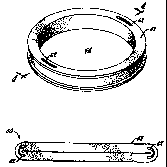
Referring to FIG. 1 of the drawings, an exemplary embodiment of the
surgical apparatus 50 of the present invention is illustrated. Apparatus 50
includes a member 52 with an aperture 54 defined therein. Apparatus 50 is
expandable and is shown in an expanded condition in FIG. 1. Alternatively,
apparatus 50 may be thought of as being compressible, with the apparatus
shown in a compressed condition in FIG. 2. Expandable or compressible, in
either case the deformable or shape-transformable apparatus 50 is adjustable
or
positionable in a state or condition in a range defined by the expanded
condition
of FIG. 1 and the compressed condition of FIG. 2. As shown, aperture 54 is
substantially open when apparatus 50 is in an expanded condition and
substantially closed when apparatus 50 is in a compressed condition. According
to an exemplary embodiment of apparatus 50, member 52 is made from resilient
bio-compatible material such that apparatus 50 is biased to return to the
expanded condition from a compressed condition. That is, when compressed,
member 52 may apply force radially in an outward direction. An exemplary
configuration of member 52 is a closed-loop shape through an open-loop
configuration is within the scope of the present invention.
FIGS. 3A-3C illustrate an exemplary method of utilizing apparatus 50
to retract tissue in a surgical procedure. After an incision 56 is made in
tissue
58 as shown in FIG. 3A, apparatus 50 may be inserted into and/or positioned
within incision 56 while in a compressed condition, as shown in FIG. 3B. Once
in position, apparatus 50 may then be expanded to an expanded condition within
' incision 56, thereby urging tissue 58 apart and opening incision 56. This
expansion of apparatus 50 may be accomplished by releasing apparatus 50 with,
for example, forceps. By such an expansion, an opening 60 is defined by the
opened incision 56 within the now retracted tissue 58 and by aperture 54 of
apparatus 50. Accordingly, a surgeon has unobstructed vision and access to the
CA 02319749 2000-07-31
WO 99/38440 PCT/US99/01838
surgical field and may perform a surgical procedure through opening 60. As
shown in FIG. 3C, during use, apparatus 50 need not take a substantially non-
compressed shape (as shown in FIG. 1 ) but need only take a shape in which
aperture 54 is not entirely closed so that opening 60 is definable and
available
5 for further surgical steps.
With fiuther reference to FIGS. 1 and 2, according to an exemplary
embodiment of apparatus 50, member 52 may have flex portions 62 formed
therein to facilitate the compression of apparatus 50. Flex portions 62 are
preferably configured as portions of member 52 having less resiliency or
10 elasticity than other portions of member 52. For example, as particularly
shown
in FIG. 4, flex portions 62 may be configured as slots within member 52,
specifically axial slots. Accordingly, when apparatus 50 is compressed,
member 52 folds outwardly at flex portions 62 rather than at some other
location along member 52. As illustrated, it is preferable to form a pair of
diametrically opposed flex portions 62 in member 52 so that apparatus 50 is
compressible to the compressed condition shown in FIG. 2 in which apparatus
50 is substantially flattened, oblong, or elliptical in shape.
With continued reference to FIGS. 1 and 4 and additional reference to
FIG. 5, apparatus 50 may be configured with tissue-engaging structure 64 for
engaging the tissue 58 exposed by and surrounding an incision to aid in
retaining apparatus 50 in position within the incision. In accordance with an
exemplary embodiment of the invention, tissue-engaging structure 64 includes
ridges 66 formed on an outer surface of member 52. A channel 68 is defined
' between ridges 66. With the provision of tissue-engaging structure 64,
apparatus 50 may be positioned within an incision so that tissue 58 is
substantially engaged by tissue-engaging structure 64, with the tissue 58
received within channel 68 and retained by ridges 66. Accordingly, tissue-
CA 02319749 2000-07-31
WO 99/38440 PCT/US99/01838
11
engaging structure 64 augments the outward radial resiliency of member 52 in
retaining apparatus 50 within an incision.
Alternatively, referencing FIG. 6, if apparatus 50 is positioned within an
incision made in relatively thick tissue (that is, in tissue having a
thickness
greater than a thickness of channel 68), tissue-engaging structure 64 may
still
aid in retaining apparatus 50 within the incision by engaging with the surface
of
the tissue 58. Because of the expandable characteristics of apparatus 50,
ridges
66 are urged against the tissue 58, thereby retarding or preventing movement
of
apparatus 50 in the axial direction shown by arrow A. To reduce or eliminate
the risk of injuring the tissue 58, tissue-engaging structure 64 is preferably
configured as atraumatic as possible while still retaining tissue-engaging
characteristics.
Apparatus 50 may be configured in any desired shape. In the exemplary
embodiment shown in FIGS. 1--6, apparatus 50 is substantially cylindrical when
in the expanded condition. However, apparatus 50 may be configured so as to
be elliptical or ovoid when in the expanded condition. Alternatively,
apparatus
50 may be configured to assume an irregular configuration specifically
designed
for a particular type of incision or surgical application. In the cylindrical
embodiment of apparatus 50, ridges 66 of tissue-engaging structure 64 are in
the
form of annular extensions of end surfaces of member 52.
Referencing FIGS. 7 and 8, apparatus 50 may include a light source for
providing light 70 to a surgical field. Many surgical procedures in which
apparatus 50 may be applied are in remote or awkward locations in the body.
' Accordingly, light sources external to the body in the surgical theater may
not
illuminate the entire surgical field or may cast distracting shadows. The
provision of the light source on apparatus 50 itself, which is essentially
adjacent
to or directly above the surgical field; substantially eliminates any shadows
or
non-illuminated areas within the surgical field.
CA 02319749 2000-07-31
WO 99/38440 PCT/US99/Q1838
12
The exemplary light source illustrated in FIGS. 7 and 8 includes a light-
emitting element 72 coupled to a lead 74. In accordance with an exemplary
embodiment, a plurality of light-emitting elements 72 may be spaced around an
inner periphery 76 of member 52. Providing a plurality of spaced light-
emitting
elements 72 eliminates any shadows which may be created by surgical
instruments or other apparatus positioned within the surgical field through
aperture 54 of apparatus 50. Leads 74 may be secured to member 52 through,
for example, ridges 66, with leads 74 being disposed in channel 68 on an outer
periphery 78 of member 52 as shown in FIG. 8. Leads 74 may then be attached
to electronics within the surgical theater to provide power to light-emitting
elements 72. Leads 74, as well as light-emitting elements 72, are configured
on
member 52 so as to minimize the effect of compressing apparatus 50; for
example, leads 74 may be attached to member 52 near ends of flex portion 62 as
shown in FIG. 7. Leads 74 may be electrical conductors, fiber optics, or other
suitable media. If leads 74 are configured as fiber optics, light-emitting
elements 72 are defined as the end of the fiber optics from which light is
emitted.
An alternative embodiment of the light source is exemplified in FIG. 9,
in which a light-emitting element is configured as a luminescent structure 80
disposed within an inner periphery of member 52. Luminescent structure 80
preferably includes a twin-chambered housing 82 having first and second
chambers 84a, 84b divided by a breakable, rupturable, or piercable divider 86.
Luminescent structure 80 has a translucent inner peripheral wall 88. First and
- second chambers 84a, 84b respectively contain first and second chemical
solutions 90a, 90b which, when mixed together, generate light through
chemiluminescence.
FIG. 11A shows an enlarged sectional view of apparatus 50 in an
expanded condition with divider 86 in tact. To activate chemiluminescence,
~-~-~ ~~~~~om.rttv V1 :1U- 3- 0 : CA 02319749 2_00__0_=07=_3.1_~~~, ~,~ ~9
~3g~"Q~." a
10-03-2000 99906688 ~
I3
divider 86 i~ punctured, preferably when apparatus 50 is con ipresscd as
sh«wn in FIG. IIB {also see FIG. 2 for reference of a compressed
~cmditiun), allowing first and second chemical solutions 94a, 90b to mix.
'rhr. mixing of chemical solutions 90a, 90b may be facilitated by agitating
apparatus 50. Apparatus 50 may then be positioned within an incision as
described above and shown in FIGS. 3B and 3C. When expanded, light
generated by the ehemilumineseence illuminates the surgical field through
o aperture 54. The puncturing or breaking of divider 8G may be
~c:cumplished by a physical structure disposed within housing 82 or by the
incrt;ased pressure of chemical solutions 90a, 90b against divider 8G
caused by the compression of apparatus 50. As compared to the use of
the previously described light-emitting apparatus, luminesrxnce structure
is ~iU iv self-contained and does not require external manipulation or power
murces for tight generation once apparatus 50 is positioned within an
inc; i~u~n.
In accord nee with an exemplary embodiment of Luminescent
5irueture 80, divider 8b may have a break point or portion 92 near flex
~n p~irtic~ns G2 to facilitate the piercing or breaking of divider 86 when
apparatus 5!) is compressed as shown in FIGS. lIA and IIB. Break
portion 92 rnay be defined by a thinned portion of divider 86. In addition,
is nlxy be preferable to extend translucent inner peripht'ral wall 88 over a
bottom end portion of housing 82 as shown so that light is directed to the
2s surgical field through said end portinn_ In some applications, it may be
~!'C~E:f3~?~e tU have a high opacity of top end portion 96 of housing 82 so
~Jia~ light is not directed coward dle surgeon. Further, inner peripheral
wall 88 may be configured so that light is directed only substantially
downward toward the surgical field by, for example, an internal louver
3a ,~rrangenlent. A commercial supplier of chemiluminescent structures is
AMENDED SHEET
.. ._."....,~~ "~ .«- ,s- a : CA 02319749_2000=07-31~~NY ~g 89 1:389~-~~-~~~-
10-03-2000 _ ___ _ _ _ . w. ..L 99906688
- 13a
Umnigluw Corporation of Novato, California, which produces non-toxic
light structures undrr the name of CYALUME°' which radiate light in
s more than one v:olor for durations on the order of eight hours.
AMENDED SHEET
CA 02319749 2000-07-31
WO 99/38440 PCTNS99/01838
14
Yet another exemplary embodiment of apparatus 50 is illustrated in FIG.
12 in which monitoring or sensing apparatus 98 coupled to leads 100 is
disposed on member 52. Leads 100 may be connected with electrical
equipment in the surgical theater for carrying electrical signals between
sensing
apparatus 98 and the external equipment. Sensing apparatus 98 may carry out
diagnostic functions and may include, for example, heat sensors for monitoring
a temperature in the surgical field. Sensing apparatus 98 may also include
video-monitoring equipment to provide a video image of the surgical field.
Sensing apparatus 98 may be configured within member 52 such that individual
elements of the sensing apparatus are disposed on either the inner or the
outer
peripheral surface 76 or 78 of member 52 as shown. An alternative to physical
heads 100 may be a wireless communication configuration in which sensing
apparatus 98 includes a transmitter and/or a receiver for communicating with
electrical equipment by means of electromagnetic waves. Such a wireless
embodiment eliminates any unnecessary and possibly obstructive electrical
leads in the surgical field. .
With reference to FIGS. 13 and 14, apparatus 102 of the present
invention is exemplified in an alternative embodiment. Apparatus 102, like
apparatus 50 illustrated above, includes a member 104. Flex portions 106 are
formed in member 104 to facilitate the compression of apparatus 102.
However, rather than forming axial slots within the member of the surgical
apparatus as described above, flex portions 106 in accordance with this
embodiment are formed as radial slots in member 104. Alternatively, notches
or cavities may be formed in place of slots. Apparatus 102 may also include
tissue-engaging stricture 108 comprised of an annular ridge 110 and axial
projections 112. Annular ridge 110 functions analogously to ridges 66
described above. Axial projections 112 provide an increased outer annular
surface area for engaging tissue of an incision, and facilitate the removal of
-~~- ~~~~ ~wu.r~r.!v u1 :1U- 3- o ; CA 02319749 2000-07-31 , ,_
..... ... . ", , _~ ,..,.,ARKSOh-. +4~ oy '-'~99' ' 99906688
10-03-2000
apparatus 102 from an incision by providing an elemettt onto _ which
(iarceps ur hemostats may grasp. Alternatively, apparatus 102 may
~ncludc: a wb 114 to facilitate the removal of apparatus 102 from an
incision with forceps or hemostats. Tab 1I4, which is preferably flexible,
s ma~~ bc~ attached to apparatus 102 through a slot of flex portion 106.
Yet another embodiment of the tissue surgical apparatus of the
present invention is shown in FIG. 15. Like apparatus 50 described
abewe. surgical apparatus 1I6 is compressible and, accordingly,
expandable. Apparatus 116 includes a member 118 which is configured
to ca have a concave outer annular periphery 120 which functions as tissue-
c:ngagmg means as described above. Such a concave co~guration is
substantially atrautnatic and does not necessarily have flex portions to
fecititate the compressing of apparatus 116. An inner periphery 122 of
member 118 may be substantially cylindrical or, as shown, convex.
is With reference to FIGS. 16, 17, and 18, a further exemplary
embodiment of a tissue surgical apparatus 124 according to the priaciples
o!' the present invention is shown. Apparatus 124 includes a member 126
arid an adjusting portion 128 attached to an end of member 126 for
expanding and for compressing apparatus 124. A plurality of slots or
~o notches !30 are formed in an inner periphery I32 along a first length h of
member 126, and an elongate projection 134 is formed oa inner periphery
132 along a second length 1z of member I26 {which will be discussed
hel«w). Adjusting portion 128 includes a slot 136 for slidably receiving
the first length h of member I26, and a rotatable drive 138 for driving the
first length h of member 126 through slot 136. Drive I38 has teeth 140
tier engaging car meshing with notc:h~ 130 of member 126, and a head 142
1>r allowing a physician to actuate drive i38. An aperture 144 is defined
within member 126 when the ~lrst length h of member 126 is received in
~lca 136.
AMENDED SHEE I
CA 02319749 2000-07-31
WO 99/38440 PCTNS99/01838
16
FIGS. 19A and 19B illustrate apparatus 124 utilized in a surgical
procedure. As shown in FIG. 19A, apparatus 124 is positioned within an
incision 56 made in tissue 58 while in a compressed condition. Apparatus 124
is then expanded to an expanded condition, as shown in FIG. 19B, thereby
retracting tissue 58 and defining an opening 146 within member 126 through
which the surgical field may be viewed. The expansion of apparatus 124 may
be accomplished by the surgeon engaging head 142 with a complementary
surgical implement 148 and rotating drive 138. Teeth 140 mesh with notches
130 as drive 138 is rotated, as shown in FIG. 18. After the surgical procedure
is
completed, apparatus 124 may then be compressed for removal from incision
56. An example of one of the alternatives to the arrangement of teeth 140 and
notches 130 is configuring drive 138 with a worm gear.
With fiu~ther reference to FIGS. 17 and 18, apparatus 124 may include
tissue-engaging structure 106 formed on an outer periphery of member 126. As
described above in relation to the exemplary embodiment illustrated in FIGS. 5
and 6, tissue-engaging structure 150 engages with tissue 58 which defines
incision 56, thereby securing or aiding to secure surgical apparatus 124
within
incision 56. Tissue-engaging structure 150 may include annular ridges 152 and
a channel 154 defined between ridges 152. When the first length II of member
126 is received within slot 136 of adjusting portion 138, channel 154 slidably
receives projection 134 therein. The engagement of channel 154 with projection
134 provides guidance as the first length h slides through slot 136 and
provides
added torsional stability and rigidity to apparatus 124. Apparatus 124 may
also
' include a locking mechanism for locking or temporarily retaining the first
length
1, of member 126 at a desired positioned within slot 136 so that apparatus 124
maintains a desired size during a surgical procedure.
The exemplary surgical apparatus of the present invention may be
utilized in any number of surgical procedures in which the retraction of
tissue,
CA 02319749 2000-07-31
WO 99/38440 PCT/US99/01838
17
that is, the formation of an opening, would be advantageous. For example, one
of the exemplary applications of the surgical apparatus is in heart valve
replacement surgery. As surgical procedures become beneficially more
minimally invasive (that is, less traumatic to the patient), medical products
used
in such procedures need to provide surgeons with tools enabling them to
perform surgical procedures less invasively. This is particularly true in
heart
valve replacement surgery. An exemplary application of the surgical apparatus
of the present invention used in heart valve replacement surgery will be
discussed below.
Referencing FIG. 20, a heart 156, which is shown in an anterior partial
section, has four chambers, including a right atrium 158, a left atrium 160, a
right ventricle 162, and a left ventricle 164. Tissue of the heart 156
includes
myocardium 166, which is essentially the muscle of the heart, and a septum 168
which separates the right chambers of the heart from the left chambers.
(Specifically, the muscular ventricular septum separates the ventricles, and
the
membranous septum generally separates the atria.) Valves define openings to
the chambers and include a mitral valve 170 positioned between the left atrium
160 and the left ventricle 164 and an aortic valve 172 positioned between the
left ventricle 174 and the aorta.
One of the most common valve replacement surgeries is the replacement
of the mitral valve 170. Although different procedures exists for replacing
the
mitral valve 170, one procedure involves accessing the mitral valve 170
through
the right and left atria 158, 160, respectively. This is due to the
physiological
' position of the heart 156 within the chest cavity of a patient, with the
right
chambers of the heart being anteriorly positioned and the left chambers being
posteriorly positioned. With reference to FIG. 21, to perform this mitral
valve
replacement surgery, after accessing the heart 156, a surgeon makes an
incision
through the myocardium 166 to provide access to the right atrium 158. A
CA 02319749 2000-07-31
WO 99!38440 PCT/US99/01838
18
surgical apparatus in accordance with the present invention, for example,
apparatus 50', may then be positioned within incision and expanded as
described above (see FIGS. 3B and 3C) to define an opening 60'. An incision
may then be made in the septum 168 between the right atrium 158 and the left
atrium 160, with another apparatus 50" positioned within this incision,
thereby
defining opening 60" between the atria. Accordingly, the surgeon may now
access the mitral valve 170 through openings 60', 60" and perform procedures
required to replace the valve.
In addition to the surgical procedure described above, the utilization of
surgical apparatus of the present invention is widely applicable to many other
procedures in which any type of tissue may be retracted. Further, the surgical
apparatus need not necessarily be positioned within an incision but may be
placed adjacent, for example, an organ so that the outward radial force
applied
by the surgical apparatus may urge or retract the organ from obstructing a
surgical field. In addition, rather than working through the opening defined
in
the aperture when in an expanded condition, the surgical apparatus may be
positioned in or near an incision so that a surgeon is able to work adjacent
to the
outer periphery of the members, that is, in a opening defined by the outer
periphery of the member and the surrounding tissue. In a commercial
embodiment, the surgical apparatus may be included in kits which include other
related items for use in a particular procedure. The member of the surgical
apparatus may include structure such as sewing holes to aid the surgeon in
retaining tissue. The surgical apparatus may be configured so as to be
"specialized" for a particular type of incision or tissue, particularly the
tissue-
engaging structures. The surgical apparatus may be of varying size (including
. thickness, depth, width, height, diameter, etc.). Further, the tissue
surgical
apparatus may include features designed to aid the surgeon during the
procedure.
CA 02319749 2000-07-31
WO 99/38440 PCT/US99/01838
19
In this regard, reference is made to FIG. 22 which illustrates an
exemplary embodiment of a surgical apparatus 174 including a member 176. A
retaining structure 178 or, more preferably, a plurality of retaining
structures
178 are disposed around an inner periphery 180 of member 176. Retaining
structures 178 may be configured as clips including a central channel 182 for
receiving a surgical implement 184 or leads coupled to external surgical
apparatus which a may be used during an operation. Retaining structures 178
are preferably made from a resilient material so as to temporarily retain the
surgical implements 184, and may be integral with or attached to member 176.
Body member 176 may have an inner channel 186 formed therein. Inner
channel 186 facilitates the compression of member 176 and rnay be used to
house leads attached to a light source (see FIGS. 7 and 8). In addition, inner
channel 186 may house a spring apparatus which biases member 176 to assume
an expanded position. With further reference to FIGS. 9 and 10, inner channel
186 may house luminescent structure 80, with inner periphery 180 being
translucent.
As mentioned, the surgical apparatus of the present invention may be
made from a resilient bio-compatible material. Examples of such material
which may be used in the manufacture of the surgical apparatus include
polyacetal, polycarbonate, polyester, polyethylene, polyphenylsulfone,
polypropylene, polysulfone, polyurethane, polyvinyl chloride, and silicone. In
addition, apparatus 124 illustrated in FIGS. 17 and 18 may be made from
materials including stainless steel and titanium. Rather than making, for
' example, apparatus 50 from a solid resilient material, apparatus 50 may
include
an inner or imbedded spring apparatus (as mentioned above) which is biased to
an expanded condition. Such a spring apparatus may be made from stainless
steel or titanium.
CA 02319749 2000-07-31
wo 99r~s~o pcrius~roisas
Those skilled in the art will understand that the embodiments of the
present invention described above exemplify the principles of the invention
and
do not limit the scope of the invention to those embodiments of the surgical
apparatus specifically illustrated in the drawings and described above. The
5 exemplary embodiments provide a foundation from which numerous alternatives
and modifications may be made, which alternatives and modifications are also
within the scope of the present invention as defined in the appended claims.