Note: Descriptions are shown in the official language in which they were submitted.
CA 02321070 2000-08-15
WO 99/42813 PCT/US99/03807
-1-
METHOD AND APPARATUS FOR SYNTHESIS OF ARRAYS OF
DNA PROBES
FIELD OF THE INVENTION
This invention pertains generally to the field of biology and particularly
to techniques and apparatus for the analysis and sequencing of DNA and related
polymers.
BACKGROUND OF THE INVENTION
The sequencing of deoxyribonucleic acid (DNA) is a fundamental tool of
modern biology and is conventionally carried out in various ways, commonly by
processes which separate DNA segments by electrophoresis. See, e.g., Current
Protocols In Molecular Biology, Vol. 1, Chapter 7, "DNA Sequencing," 1995. The
sequencing of several important genomes has already been completed (e.g.,
yeast, E.
coli), and work is proceeding on the sequencing of other genomes of medical
and
agricultural importance (e.g., human, C. elegans, Arabidopsis). In the medical
context, it will be necessary to "re-sequence" the genome of large numbers of
human
individuals to determine which genotypes are associated with which diseases.
Such
sequencing techniques can be used to determine which genes are active and
which
inactive either in specific tissues, such as cancers, or more generally in
individuals
exhibiting genetically influenced diseases. The results of such investigations
can allow
identification of the proteins that are good targets for new drugs or
identification of
appropriate genetic alterations that may be effective in genetic therapy.
Other
applications lie in fields such as soil ecology or pathology where it would be
desirable
to be able to isolate DNA from any soil or tissue sample and use probes from
CA 02321070 2000-08-15
WO 99/42813 PCTIUS99/03807
-2-
ribosomal DNA sequences from all known microbes to identify the microbes
present in
the sample.
The conventional sequencing of DNA using electrophoresis is typically
laborious and time consuming. Various alternatives to conventional DNA
sequencing
have been proposed. One such alternative approach, utilizing an array of
oligonucleotide probes synthesized by photolithographic techniques is
described in
Pease, et al., "Light-Generated Oligonucleotide Arrays for Rapid DNA Sequence
Analysis," Proc. Natl. Acad. Sci. USA, Vol. 91, pp. 5022-5026, May 1994. In
this
approach, the surface of a solid support modified with photolabile protecting
groups is
illuminated through a photolithographic mask, yielding reactive hydroxyl
groups in the
illuminated regions. A 3' activated deoxynucleoside, protected at the 5'
hydroxyl with
a photolabile group, is then provided to the surface such that coupling occurs
at sites
that had been exposed to light. Following capping, and oxidation, the
substrate is
rinsed and the surface is illuminated through a second mask to expose
additional
hydroxyl groups for coupling. A second 5' protected activated deoxynucleoside
base is
presented to the surface. The selective photodeprotection and coupling cycles
are
repeated to build up levels of bases until the desired set of probes is
obtained. It may
be possible to generate high density miniaturized arrays of oligonucleotide
probes using
such photolithographic techniques wherein the sequence of the oligonucleotide
probe at
each site in the array is known. These probes can then be used to search for
complementary sequences on a target strand of DNA, with detection of the
target that
has hybridized to particular probes accomplished by the use of fluorescent
markers
coupled to the targets and inspection by an appropriate fluorescence scanning
microscope. A variation of this process using polymeric semiconductor
photoresists,
which are selectively patterned by photolithographic techniques, rather than
using
photolabile 5' protecting groups, is described in McGall, et al., "Light-
Directed
Synthesis of High-Density Oligonucleotide Arrays Using Semiconductor
Photoresists,"
Proc. Natl. Acad. Sci. USA, Vol. 93, pp. 13555-13560, November 1996, and G.H.
McGall, et al., "The Efficiency of Light-Directed Synthesis of DNA Arrays on
Glass
Substrates," Journal of the American Chemical Society 119, No. 22, 1997, pp.
5081-
5090...
CA 02321070 2000-08-15
WO 99/42813 PCTIUS99/03807
-3-
A disadvantage of both of these approaches is that four different
lithographic masks are needed for each monomeric base, and the total number of
different masks required are thus four times the length of the DNA probe
sequences to
be synthesized. The high cost of producing the many precision
photolithographic
masks that are required, and the multiple processing steps required for
repositioning of
the masks for every exposure, contribute to relatively high costs and lengthy
processing
times.
SUMMARY OF THE INVENTION
In accordance with the present invention, the synthesis of arrays of DNA
probe sequences, polypeptides, and the like is carried out rapidly and
efficiently using
patterning processes. The process may be automated and computer controlled to
allow
the fabrication of a one or two-dimensional array of probes containing probe
sequences
customized to a particular investigation. No lithographic masks are required,
thus
eliminating the significant costs and time delays associated with the
production of
lithographic masks and avoiding time-consuming manipulation and alignment of
multiple masks during the fabrication process of the probe arrays.
In the present invention, a substrate with an active surface to which
DNA synthesis linkers have been applied is used to support the probes that are
to be
fabricated. To activate the active surface of the substrate to provide the
first level of
bases, a high precision two-dimensional light image is projected onto the
substrate,
illuminating those pixels in the array on the substrate active surface which
are to be
activated to bind a first base. The light incident on the pixels in the array
to which
light is applied deprotects OH groups and makes them available for binding to
bases.
After this development step, a fluid containing the appropriate base is
provided to the
active surface of the substrate and the selected base binds to the exposed
sites. The
process is then repeated to bind another base to a different set of pixel
locations, until
all of the elements of the two-dimensional array on the substrate surface have
an
appropriate base bound thereto. The bases bound on the substrate are
protected, either
with a chemical capable of binding to the bases or with a layer(s) of
photoresist
covering all of the bound bases, and a new array pattern is then projected and
imaged
onto the substrate to activate the protecting material in those pixels to
which the first
CA 02321070 2000-08-15
WO 99/42813 PCTIUS99/03807
-4-
new base is to be added. These pixels are then exposed and a solution
containing the
selected base is applied to the array so that the base binds at the exposed
pixel
locations. This process is then repeated for all of the other pixel locations
in the
second level of bases. The process as described may then be repeated for each
desired
level of bases until the entire selected two-dimensional array of probe
sequences has
been completed.
The image is projected onto the substrate utilizing an image former
having an appropriate light source that provides light to a micromirror device
comprising a two-dimensional array of electronically addressable micromirrors,
each of
which can be selectively tilted between one of at least two separate
positions. In one of
the positions of each micromirror, the light from the source incident on the
micromirror is deflected off an optical axis and away from the substrate, and
in a
second of the at least two positions of each micromirror, the light is
reflected along the
optical axis and toward the substrate. Projection optics receive the light
reflected from
the micromirrors and precisely image the micromirrors onto the active surface
of the
substrate. Collimating optics may be used to collimate the light from the
source into a
beam provided directly to the micromirror array or to a beam splitter, wherein
the
beam splitter reflects a portion of the beam to the micromirror array and
transmits
reflected light from the micromirror array through the beam splitter. The
light directly
reflected from the micromirrors or transmitted through the beam splitter is
directed to
projection optics lenses which image the micromirror array onto the active
surface of
the substrate. Because the selectively addressable micromirrors in the
micromirror
array may either fully reflect or fully deflect the light provided to them,
the image of
the micromirror array exhibits a very high contrast between the "on" and "off"
pixels.
The micromirrors may also be capable of being indexed to more than two
positions, in
which case additional optics may be provided to allow exposure of more than
one
substrate using a single micromirror array device. In addition, the
micromirrors are
capable of reflecting light at any wavelength without damage to them, allowing
short
wavelength light, including light in the range of ultraviolet to near
ultraviolet light, to
be utilized from the light source.
The micromirror array is operated under control of a computer which
provides appropriate pixel address signals to the micromirror array to cause
the
CA 02321070 2000-08-15
WO 99/42813 PCT/US99/03807
-5-
appropriate micromirrors to be in their "reflect" or "deflect" positions. The
appropriate micromirror array pattern for each activation step in each level
of bases to
be added to the probes is progranuned into the computer controller. The
computer
controller thus controls the sequencing of the images presented by the
micromirror
array in coordination with the reagents provided to the substrate.
In one embodiment, the substrate may be transparent, allowing the
image of the micromirror array to be projected through the surface of the
substrate that
is opposite to the active surface. The substrate may be mounted within a flow
cell,
with an enclosure sealing off the active surface of the array, allowing the
appropriate
reagents to be flowed through the flow cell and over the active surface of the
array in
the appropriate sequence to build up the probes in the array.
Further objects, features and advantages of the invention will be
apparent from the following detailed description when taken in conjunction
with the
accompanying drawings.
BRIEF DESCRIPTION OF THE DRAWINGS
In the drawings:
Fig. 1 is a schematic view of an array synthesizer apparatus in
accordance with the present invention.
Fig. 2 is a schematic view of another array synthesizer apparatus in
accordance with the present invention.
Fig. 3 is a more detailed schematic view of a general telecentric array
synthesizer apparatus in accordance with the invention.
Fig. 4 is an illustrative ray diagram for the refractive optics of the
apparatus of Fig. 3.
Fig. 5 is a schematic view of a further embodiment of an array
synthesizer apparatus in accordance with the invention in which telecentric
reflective
optics are utilized.
Fig. 6 is an illustrative ray diagram for the reflective optics of the
apparatus of Fig. 5.
Fig. 7 is a top plan view of a reaction chamber flow cell which may be
utilized in the array synthesizer apparatus of the invention.
CA 02321070 2000-08-15
WO 99/42813 PCT/US99/03807
-6-
Fig. 8 is a cross-sectional view through the reaction chamber flow cell
of Fig. 7 taken generally along the lines 8-8 of Fig. 7.
Fig. 9 is an illustrative view showing the coating of a substrate with a
photolabile linker molecule.
Fig. 10 is an illustrative view showing the photo-deprotection of the
linker molecule and the production of free OH groups.
Fig. 11 is an illustrative view showing the coupling of markers to free
OH groups produced by the photo-deprotection of the linker molecules.
Fig. 12 is an illustrative view showing the coupling of DMT-nucleotide
to free OH groups produced from photo-deprotection of the linker molecules.
Fig. 13 is an illustrative view showing acid deprotection of DMT-
nucleotides.
Fig. 14 is an illustrative view showing the hybridization of poly-A probe
labeled with fluorescein with poly-T oligonucleotide synthesized from DMT-
nucleotide-CEPs.
DETAILED DESCRIPTION OF THE INVENTION
With reference to the drawings, an exemplary apparatus that may be
used for DNA probe array synthesis, polypeptide synthesis, and the like is
shown
generally at 10 in Fig. 1 and includes a two-dimensional array image former 11
and a
substrate 12 onto which the array image is projected by the image former 11.
For the
configuration shown in Fig. 1, the substrate has an exposed entrance surface
14 and an
opposite active surface 15 on which a two-dimensional array of nucleotide
sequence
probes 16 are to be fabricated. For purposes of illustration, the substrate 12
is shown
in the figure with a flow cell enclosure 18 mounted to the substrate 12
enclosing a
volume 19 into which reagents can be provided through an input port 20 and an
output
port 21. However, the substrate 12 may be utilized in the present system with
the
active surface 15 of the substrate facing the image former 11 and enclosed
within a
reaction chamber flow cell with a transparent window to allow light to be
projected
onto the active surface. The invention may also use an opaque or porous
substrate.
The reagents may be provided to the ports 20 and 21 from a conventional base
synthesizer (not shown in Fig. 1).
CA 02321070 2000-08-15
WO 99/42813 PCT/US99/03807
-7-
The image former 11 includes a light source 25 (e.g., an ultraviolet or
near ultraviolet source such as a mercury arc lamp), an optional filter 26 to
receive the
output beam 27 from the source 25 and selectively pass only the desired
wavelengths
(e.g., the 365 nm Hg line), and a condenser lens 28 for forming a collimated
beam 30.
Other devices for filtering or monochromating the source light, e.g.,
diffraction
gratings, dichroic miurors, and prisms, may also be used rather than a
transmission
filter, and are generically referred to as "filters" herein. The beam 30 is
projected
onto a beam splitter 32 which reflects a portion of the beam 30 into a beam 33
which is
projected onto a two-dimensional micromirror array device 35. The micromirror
array
device 35 has a two-dimensional array of individual micromirrors 36 which are
each
responsive to control signals supplied to the array device 35 to tilt in one
of at least two
directions. Control signals are provided from a computer controller 38 on
control lines
39 to the micromirror array device 35. The micromirrors 36 are constructed so
that in
a first position of the mirrors the portion of the incoming beam of light 33
that strikes
an individual micromirror 36 is deflected in a direction oblique to the
incoming beam
33, as indicated by the arrows 40. In a second position of the mirrors 36, the
light
from the beam 33 striking such mirrors in such second position is reflected
back
parallel to the beam 33, as indicated by the arrows 41. The light reflected
from each of
the mirrors 36 constitutes an individual beam 41. The multiple beams 41 are
incident
upon the beam splitter 32 and pass through the beam splitter with reduced
intensity and
are then incident upon projection optics 44 comprised of, e.g., lenses 45 and
46 and an
adjustable iris 47. The projection optics 44 serve to form an image of the
pattern of
the micromirror array 35, as represented by the individual beams 41 (and the
dark
areas between these beams), on the active surface 15 of the substrate 12. The
outgoing
beams 41 are directed along a main optical axis of the image former 11 that
extends
between the micromirror device and the substrate. The substrate 12 in the
configuration shown in Fig. 1 is transparent, e.g., formed of fused silica or
soda lime
glass or quartz, so that the light projected thereon, illustratively
represented by the
lines labeled 49, passes through the substrate 12 without substantial
attenuation or
diffusion.
A preferred micromirror array 35 is the Digital Micromirror Device
(DMD) available connnercially from Texas Instruments, Inc. These devices have
CA 02321070 2000-08-15
WO 99/42813 PCT/US99/03807
-8-
arrays of micromirrors (each of which is substantially a square with edges of
10 to 20
m in length) which are capable of forming patterned beams of light by
electronically
addressing the micromirrors in the arrays. Such DMD devices are typically used
for
video projection and are available in various array sizes, e.g., 640 x 800
micromirror
elements (512,000 pixels), 640 x 480 (VGA; 307,200 pixels), 800 x 600 (SVGA;
480,000 pixels); and 1024 x 768 (786,432 pixels). Such arrays are discussed in
the
following article and patents: Larry J. Hornbeck, "Digital Light Processing
and
MEMs: Reflecting the Digital Display Needs of the Networked Society," SPIE/EOS
European Symposium on Lasers, Optics, and Vision for Productivity and
Manufacturing I, Besancon, France, June 10-14, 1996; and U.S. Patents
5,096,279,
5,535,047, 5,583,688 and 5,600,383. The micromirrors 36 of such devices are
capable of reflecting the light of normal usable wavelengths, including
ultraviolet and
near ultraviolet light, in an efficient manner without damage to the mirrors
themselves.
The window of the enclosure for the micromirror array preferably has anti-
reflective
coatings thereon optinzized for the wavelengths of light being used. Utilizing
commercially available 600 x 800 arrays of micromirrors, encoding 480,000
pixels,
with typical micromirror device dimensions of 16 microns per mirror side and a
pitch
in the array of 17 microns, provides total micromirror array dimensions of
13,600
microns by 10,200 microns. By using a reduction factor of 5 through the optics
system
44, a typical and readily achievable value for a lithographic lens, the
dimensions of the
image projected onto the substrate 12 are thus about 2,220 microns by 2040
microns,
with a resolution of about 2 microns. Larger images can be exposed on the
substrate
12 by utilizing multiple side-by-side exposures (by either stepping the flow
cell 18 or
the image projector 11), or by using a larger micromirror array. It is also
possible to
do one-to-one imaging without reduction as well as enlargement of the image on
the
substrate, if desired.
The projection optics 44 may be of standard design, since the images to
be formed are relatively large and well away from the diffraction liunit. The
lenses 45
and 46 focus the light in the beam 41 passed through the adjustable iris 47
onto the
active surface of the substrate. The projection optics 44 and the beam
splitter 32 are
arranged so that the light deflected by the micromirror array away from the
main
optical axis (the central axis of the projection optics 44 to which the beams
41 are
CA 02321070 2000-08-15
WO 99/42813 PCT/US99/03807
-9-
parallel), illustrated by the beams labeled 40 (e.g., 100 off axis) fall
outside the
entrance pupil of the projection optics 44 (typically 0.515 = 0.1; 10
corresponds to an
aperture of 0.17, substantially greater than 0.1). The iris 47 is used to
control the
effective numerical aperture and to ensure that unwanted light (particularly
the off-axis
beams 40) are not transmitted to the substrate. Resolution of dimensions as
small as
0.5 microns are obtainable with such optics systems. For manufacturing
applications,
it is preferred that the micromirror array 35 be located at the object focal
plane of a
lithographic I-line lens optimized for 365 nm. Such lenses typically operate
with a
numerical aperture (NA) of 0.4 to 0.5, and have a large field capability
The micromirror array device 35 may be formed with a single line of
micromirrors (e.g., with 2,000 mirror elements in one line) which is stepped
in a
scanning system. In this manner the height of the image is fixed by the length
of the
line of the micromirror array but the width of the image that may be projected
onto the
substrate 12 is essentially unlimited. By moving the stage 18 which carries
the
substrate 12, the mirrors can be cycled at each indexed position of the
substrate to
define the image pattern at each new line that is imaged onto the substrate
active
surface.
Various approaches may be utilized in the fabrication of the DNA
probes 16 on the substrate 12, and are adaptations of microlithographic
techniques. In
, a "direct photofabrication approach," the glass substrate 12 is coated with
a layer of a
chemical capable of binding the nucleotide bases. Light is applied by the
projection
system 11, deprotecting the OH groups on the substrate and making them
available for
binding to the bases. After development, the appropriate nucleotide base is
flowed
onto the active surface of the substrate and binds to the selected sites using
normal
phosphoramidite DNA synthesis chemistry. The process is then repeated, binding
another base to a different set of locations. The process is simple, and if a
combinatorial approach is used the number of permutations increases
exponentially.
The resolution limit is presented by the linear response of the deprotection
mechanism.
Because of the limitations in resolution achievable with this method, methods
based on
photoresist technology may be used instead, as described, e.g., in McGall, et
al.,
supra. In the indirect photofabrication approach, compatible chemistries exist
with a
two-layer resist system, where a first layer of, e.g., polyimide acts as a
protection for
CA 02321070 2000-08-15
WO 99/42813 PCT/US99/03807
-10-
the underlying chemistry, while the top imaging resist is an epoxy-based
system. The
imaging step is common to both processes, with the main requirement being that
the
wavelength of light used in the imaging process be long enough not to excite
transitions
(chemical changes) in the nucleotide bases (which are particularly sensitive
at 280 nm).
Hence, wavelengths longer than 300 nm should be used. 365 nm is the I-line of
mercury, which is the one used most conunonly in wafer lithography.
Another form of the array synthesizer apparatus 10 is shown in a
simplified schematic view in Fig. 2. In this arrangement, the beamsplitter 32
is not
used, and the light source 25, optional filter 26, and condenser lens 28 are
mounted at
an angle to the main optical axis (e.g., at 20 to the axis) to project the
beam of light
30 onto the array of micromirrors 36 at an angle. The micromirrors 36 are
oriented to
reflect the light 30 into off axis beams 40 in a first position of the mirrors
and into
beams 41 along the main axis in a second position of each mirror. In other
respects,
the array synthesizer of Fig. 2 is the same as that of Fig. 1.
A more detailed view of a preferred array synthesizer apparatus which
uses the off-axis projection arrangement of Fig. 2 is shown in Fig. 3. In the
apparatus
of Fig. 3, the source 25 (e.g., 1,000 W Hg arc lamp, Orie16287, 66021),
provided
with power from a power supply 50 (e.g., Orie168820), is used as the light
source
which contains the desired ultraviolet wavelengths. The filter system 26 is
composed,
for example, of a dichroic mirror (e.g., Orie166226) that is used to absorb
infrared
light and to selectively reflect light of wavelengths ranging from 280 to 400
nm. A
water-cooled liquid filter (e.g., Orie16127) filled with deionized water is
used to
absorb any remaining infrared. A colored glass filter (Oriel 59810) or an
interference
filter (Oriel 56531) is used to select the 365 nm line of the Hg lamp 25 with
a 50%
bandwidth of either 50 nm or 10 nm, respectively. An F/I two element fused
silica
condenser (Oriel 66024) is used as the condenser 28, and with two plano-convex
lenses
52 (Melles Griot O1LQP033 and Melles Griot O1LQP023), forms a Kohler
illumination
system. This illumination system produces a roughly collimated uniform beam 30
of
365 nm light with a diameter just large enough to encompass the 16 mm x 12 mtn
active area of the micromirror array device 35. This beam 30 is incident onto
the
device 35 at an angle of 20 measured from the normal to the face of the
device. The
micromirror array device 35 is located approximately 700 mm away from the last
CA 02321070 2000-08-15
WO 99/42813 PCT/US99/03807
-11-
filter. When the micromirrors are in a first position, the light in the beam
30 is
deflected downwardly and out of the system. For example, in this micromirror
device
the mirrors in their first position may be at an angle of -10 with respect to
the normal
to the plane of the micromirrors to reflect the light well away from the
optical axis.
When a micromirror is controlled to be deflected in a second position, e.g.,
at an angle
of + 10 with respect to the normal to the plane of the micromirrors, the
light reflected
from such micromirrors in the second position emerges perpendicularly to the
plane of
the micromirror array in the beam 41. The pattern formed by the light
reflected from
the micromirrors in their second position is then imaged onto the active
surface 15 of a
glass substrate 12 enclosed in a flow cell 18 using a telecentric imaging
system
composed of two doublet lenses 45 and 46 and an adjustable aperture 47. Each
of the
doublet lenses 45 and 46 is composed of a pair of plano-convex lenses (e.g.,
Melles
Griot O1LQP033 and 01LQP037) put together with the curved surfaces nearly
touching. The first doublet lens is oriented so that the shorter focal length
(O1LQP033)
side is towards the micromirror array device 35, and the second doublet is
oriented so
that its longer focal length (O1LQP037) side is toward the micromirror array
device 35.
Doublets composed of identical lenses may be used, in which case either side
may face
the micromirror array device. The adjustable aperture 47, also called a
telecentric
aperture, is located at the back focal plane of the first doublet. It is used
to vary the
angular acceptance of the optical system. Smaller aperture diameters
correspond to.
improve contrast and resolution but with correspondingly decreased intensity
in the
image. As illustrated in Fig. 3, a standard DNA synthesizer 55 supplied with
the
requisite chemicals can be connected by the tubes 20 and 21 to the flow cell
18 to
provide the desired sequence of chemicals, either under independent control or
under
control of the computer 38. A typical diameter for the aperture 47 is about 30
nm. An
illustrative ray diagram showing the paths of light through the lenses 45 and
46 is
shown in Fig. 4 for this type of refractive optical system. Fans of rays
originating at
the center of the object (the micromirror device face), at the edge, and at an
intermediate location are shown. The optical system forms an inverted image of
the
face of the micromirror array device.
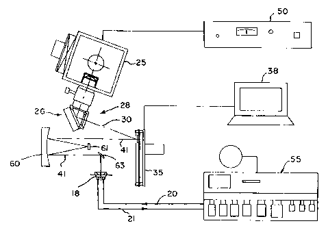
Another embodiment of the array synthesizer apparatus using reflective
optics is shown in Fig. 5. An exemplary system utilizes a 1,000 W Hg arc lamp
25 as
CA 02321070 2000-08-15
WO 99/42813 PCT/US99/03807
-12-
a light source (e.g., Orie16287, 66021), with a filter system formed of a
dichroic
mirror (e.g., Oriel 66228) that absorbs infrared light and selectively
reflects light of
wavelengths ranging from 350 to 450 nm. An F/1 two element fused silica
condenser
lens (Oriel 66024) is used to produce a roughly collimated beam of light 30
containing
the 365 nm line but excluding undesirable wavelengths around and below 300 nm.
A
Kohler illumination system may optionally also be used in the apparatus of
Fig. 5 to
increase uniformity and intensity. The beam 30 is incident onto the
micromirror array
device 35 which has an active area of micromirrors of about 16 mm x 12 mm and
which is located about 210 nm from the snout of the UV source 25, with the
beam 30
striking the planar face of the micromirror device 35 at an angle of 20 with
respect to
a normal to the plane of the array. The light reflected from the micromirrors
in a first
position of the micromirrors, e.g., -10 with respect to the plane of the
array, is
directed out of the system, whereas light from micromirrors that are in a
second
position, e. g. ,+ 10 with respect to the plane of the array, is directed in
the beam 41
toward a reflective telecentric imaging system composed of a concave mirror 60
and a
convex mirror 61. Both mirrors are preferably spherical and have enhanced UV
coating for high reflectivity. After executing reflections from the mirrors 60
and 61,
the beam 41 may be incident upon another planar mirror 63 which deflects the
beam
toward the flow cell 18. The light reflected from the micromirrors is imaged
onto the
active surface of a glass substrate enclosed in the flow cell 18. A
telecentric aperture
(not shown in Fig. 5) may be placed in front of the convex mirror. The beam 41
first
strikes the concave mirror, then the convex mirror, and then the concave
mirror again,
with the planar nrirror 63 optionally being used to deflect the light 90 to
direct it to
the flow cell 18. For the system shown, the concave mirror 60 may have a
diameter of
152.4 mm, and a spherical nlirror surface radius of 304.8 mm (ES F43561), and
the
convex mirror may have a diameter of 25 nun, and a spherical mirror surface
radius of
152.94 mm (ES F45625). Ideally, the radius of curvature of the concave mirror
is
twice that of the convex mirror. Such reflective optical systems are well
known and
conventionally used in optical lithography in "MicroAlign" type systems. See,
e.g.,
A. Offner, "New Concepts in Projection Mask Aligners," Optical Engineering,
Vol.
14, pp. 130-132 (1975), and R.T. Kerth, et al., "Excimer Laser Projection
CA 02321070 2000-08-15
WO 99/42813 PCT/US99/03807
-13-
Lithography on a Full-Field Scanning Projection System," IEEE Electron Device
Letters, Vol. EDL-7(5), pp. 299-301 (1986).
Fig. 6 illustrates image formation for the optical system of Fig. 5. Fans
of rays originating in the center of the object (the micromirror array
device), at the
edge, and at an intermediate position are shown in Fig. 6. The rays reflect
first from
the concave mirror 60, then from the convex mirror 61, then from the concave
mirror
60 again, to form an inverted image of the face of the micromirror array
device. The
planar mirror 63 is not included in the diagram of Fig. 6. A telecentric
aperture (not
shown) may be placed in front of the convex mirror.
The refractive or reflective optical systems are both preferably
"symmetric" to minimize aberrations such as coma and spherical aberration via
cancellation. The foregoing reflective system has a higher numerical aperture
which
yields higher intensity. Both of the telecentric optical systems of Figs. 3
and 5 are 1:1
imaging systems. A reflective system has the potential advantages of
eliminating
chromatic aberration and providing higher resolution, as well as being compact
and
less expensive. A preferred system for doing 1:1 imaging would be a Wynne-
Dyson
type system which combines concave mirror with lenses and prisms. It has a
very high
numerical aperture which enhances intensity. See, e.g., F.N. Goodall, et al.,
"Excimer Laser Photolithography with 1:1 Wynne-Dyson Optics," SPIE Vol. 922,
Optical/Laser Microlithography, 1988; and B. Ruff, et al., "Broadband Deep-UV
High
NA Photolithography System," SPIE Vol. 1088, Optical/Laser Microlithography II
(1989).
More detailed views of a reaction chamber flow cell 18 that may be
utilized with the apparatus of the invention is shown in Figs. 7 and 8. The
exemplary
flow cell 18 in these figures includes an aluminum housing 70, held together
by bolts
71, having an inlet 73 connected to an input port line 20 and an outlet 75
converted to
an output port line 21. As illustrated in the cross-sectional view of Fig. 8,
the housing
70 includes a lower base 78 and an upper cover section 79 which are secured
together
over the substrate with the bolts 71. The substrate 12, e.g., a transparent
glass slide, is
held between the upper plate 79 and a cylindrical gasket 81 (e.g., formed of
Kal
RezTM), which in turn is supported on a nonreactive base block 82 (e.g.,
TeflonTM),
with an inlet channel 85 extending from the inlet 73 to a sealed reaction
chamber 88
CA 02321070 2000-08-15
WO 99/42813 PCT/US99/03807
-14-
formed between the substrate 12 and the base block 82 that is sealed by the
gasket, and
with an outlet channe189 extending from the reaction chamber 88 to the outlet
75. The
bolts 71 can be screwed and unscrewed to detachably secure the substrate 12
between
the cover section and the base to allow the substrate to be replaced with
minimal
displacement of the base of the flow cell. Preferably, as shown in Fig. 8, a
rubber
gasket 90 is mounted at the bottom of the plate 79 to engage against the
substrate at a
peripheral region to apply pressure to the substrate against the gasket 81. If
desired,
the flow cell may also be used as a hybridization chamber during readout.
An exemplary process for forming DNA probes is illustrated with
respect to the schematic diagrams of Figs. 9-14. Fig. 9 illustrates the
coating of the
substrate 12, having a silane layer 95 forming the active surface 15 thereof,
with the
photolabile linker molecule MENPOC-HEG coated on the silane layer using
standard
phosphoramidite chemistry. MENPOC-HEG-CEP=18-O-[(R,S)-(1-(3,4-
(Methylenedioxy)-6-nitrophenyl)ethoxy)carbonyl]-3,6,9,12,15,18-hexaoxaoctadec-
1-yl
O'-2-cyanoethyl-N,N-Diisopropylphosphoramidite. The silane layer was made from
N-
(3-(triethoxysilyl)-propyl)-4-hydroxybutyramide. At the step shown in Fig. 9,
the
substrate can be exposed to light and active free OH groups will be exposed in
areas
that have been exposed to light.
Fig. 10 illustrates the photo-deprotection of the MENPOC-HEG linker
and the production of free OH groups in the area 100 that is exposed to light.
Fig. 11
illustrates the coupling of FluorePrimeT"" fluorescein amidite to free OH
groups
produced from photo-deprotection of MENPOC-HEG. Fig. 12 illustrates the
coupling
of DMT-nucleotide to free OH groups produced from photo-deprotection of
MENPOC-HEG linker. Fig. 13 illustrates the step of acid deprotection of DMT-
nucleotides in the area 100 exposed to light. Fig. 14 illustrates the
hybridization of
poly-A probe labeled with fluorescein with poly-T oligonucleotides synthesized
from
DMT-nucleotide-CEPs.
It is understood that the invention is not confmed to the particular
embodiments set forth herein as illustrative, but embraces all such modified
forms
thereof as come within the scope of the following claims.