Note: Descriptions are shown in the official language in which they were submitted.
CA 02323922 2000-09-14
WO 99/47082 PCT/US99/05757
PERCUTANEOUS PROSTHETIC SPINAL DISC NUCLEUS
AND METHOD OF MANUFACTURE
Cross-Reference to Co-Pending_Ap lication
This is a continuation-in-part of co-pending application Serial No.
08/870,866 filed on June 6, 1997.
1 o BACKGROUND OF THE INVENTION
The present invention relates to a prosthetic spinal disc nucleus. More
particularly, it relates to a percutaneously implantable, capsule-shaped
intradiscal
prosthesis and a method of manufacture therefor.
The vertebral spine is the axis of the skeleton upon which all of the body
parts "hang". In humans, the normal spine has seven cervical, twelve thoracic
and
five lumbar segments. The lumbar segments sit upon the sacrum, which then
attaches to the pelvis, in tum supported by the hip and leg bones. The bony
vertebral bodies of the spine are separated by intervertebral discs, which act
as
joints, but allow known degrees of flexion, extension, lateral bending and
axial
2 0 rotation.
The typical vertebra has a thick interior bone mass called the vertebral body,
with a neural (vertebral) arch that arises from a posterior surface of the
vertebral
body. Each narrow arch combines with the posterior surface of the vertebral
body
and encloses a vertebral foramen. The vertebral foramina of adjacent vertebrae
are
2 5 aligned to form a vertebral canal, through which the spinal sac, cord and
nerve
rootlets pass. The portion of the neural arch that extends posteriorly and
acts to
protect a posterior side of the spinal cord is known as the lamina. Projecting
from
the posterior region of the neural arch is a spinous process. The centra of
adjacent
vertebrae are supported by the intervertebral disc.
3 0 The intervertebral disc primarily serves as a mechanical cushion between
the vertebral bones, permitting controlled motions within vertebral segments
of the
axial skeleton. The normal disc is a unique, mixed structure, comprised of
three
component tissues: The nucleus pulposus ("nucleus"), the anulus fibrosus
("anulus"), and two opposing vertebral end plates. The two vertebral end
plates are
CA 02323922 2000-09-14
WO 99147082 PCT/US99105757
each composed of thin cartilage overlying a thin layer of hard, cortical bone
which
attaches to the spongy, richly vascular, cancellous bone of the vertebral
body. The
end plates thus serve to attach adjacent vertebrae to the disc. In other
words, a
transitional zone is created by the end plates between the malleable disc and
the
bony vertebrae.
The anulus of the disc is a tough, outer fibrous ring that binds together
adjacent vertebrae. This fibrous portion, which is much like a laminated
automobile
tire, is generally about 10 to 15 millimeters in height and about 1 S to 20
millimeters
in thickness. The fibers of the anulus consist of 15 to 20 overlapping
multiple plies,
and are inserted into the superior and inferior vertebral bodies at roughly a
30 angle
in both directions. This configuration particularly resists torsion, as about
half of
the angulated fibers will tighten when the vertebrae rotate in either
direction,
relative to each other. The laminated plies are less firmly attached to each
other.
Immersed within the anulus, positioned much like the liquid core of a golf
ball, is the nucleus. The healthy nucleus is largely a gel-like substance
having a
high water content, and similar to air in a tire, serves to keep the anulus
tight yet
flexible. The nucleus-gel moves slightly within the anulus when force is
exerted on
the adjacent vertebrae with bending, lifting, etc.
The nucleus and the inner portion of the anulus have no direct blood supply.
2 0 In fact, the principal nutritional source for the central disc arises from
circulation
within the vertebral body. Microscopic, villous-like fingerlings of the
nuclear and
anular tissue penetrate the vertebral end plates and allow fluids to pass from
the
blood across the cell membrane of the fingerlings and then inward to the
nuclear
tissue. These fluids are primarily body water and the smallest molecular
weight
2 5 nutrients and electrolytes.
The natural physiology of the nucleus promotes these fluids being brought
into and released from the nucleus by cyclic loading. When fluid is forced out
of
the nucleus, it passes again through the end plates and then back into the
richly
vascular vertebral bodies. The cyclic loading amounts to daily variations in
applied
3 0 pressure on the vertebral column (e.g., body weight and muscle pull)
causing the
nucleus to expel fluids, followed by periods of relaxation and rest, resulting
in fluid
absorption or swelling by the nucleus. Thus, the nucleus changes volume under
2
CA 02323922 2000-09-14
WO 99/47082 PCT/US99/05757
loaded and non-loaded conditions. Further, the resulting tightening and
loosening
effect on the anulus stimulates normal anulus collagen fibers to remain
healthy or
to regenerate when torn, a process found in all normal ligaments related to
body
joints. Notably, the ability of the nucleus to release and imbibe fluids
allows the
spine to alter its height and flexibility through periods of loading or
relaxation.
Normal loading cycling is thus an effective nucleus and inner anulus tissue
fluid
pump, not only bringing in fresh nutrients, but perhaps more importantly,
removing
the accumulated, potentially autotoxic by-products of metabolism.
The spinal disc may be displaced or damaged due to trauma or a disease
process. A disc herniation occurs when the anulus fibers are weakened or torn
and
the inner tissue of the nucleus becomes permanently bulged, distended, or
extruded
out of its normal, internal anular confines. The mass of a herniated or
"slipped"
nucleus can compress a spinal nerve, resulting in leg pain, loss of muscle
control,
or even paralysis. Alternatively, with discal degeneration, the nucleus loses
its
water binding ability and deflates, as though the air had been let out of a
tire.
Subsequently, the height of the nucleus decreases, causing the anulus to
buckle in
areas where the laminated plies are loosely bonded. As these overlapping
laminated
plies of the anulus begin to buckle and separate, either circumferential or
radial
anular tears may occur, which may contribute to persistent and disabling back
pain.
2 o Adjacent, ancillary spinal facet joints will also be forced into an
overriding
position, which may create additional back pain.
Whenever the nucleus tissue is herniated or removed by surgery, the disc
space will narrow and may lose much of its normal stability. In many cases, to
alleviate pain from degenerated or herniated discs, the nucleus is removed and
the
two adjacent vertebrae surgically fused together. While this treatment
alleviates the
pain, all discal motion is lost in the fused segment. Ultimately, this
procedure
places greater stresses on the discs adjacent to the fused segment as they
compensate for the lack of motion, perhaps leading to premature degeneration
of
those adjacent discs. A more desirable solution entails replacing in part or
as a
3 0 whole the damaged nucleus with a suitable prosthesis having the ability to
complement the normal height and motion of the disc while stimulating the
natural
disc physiology.
3
CA 02323922 2000-09-14
WO 99/47082 PCT/US99/05757
Restoring the nutrition-flushing cycle of a natural disc is important for a
prosthetic spinal disc nucleus to be successful. Vascular circulation and
nerve
supply to the disc is limited to the outer layers of the anulus, never
penetrating more
than a few millimeters, or about five of the anular plies. Most of the
nutrition for
the inner anulus and nucleus is provided by diffusion through the end plates
of the
vertebral bodies and by the important pumping action between the partially
loaded
and fully loaded conditions of the disc. If the nutritional cycle is impeded,
a variety
of degenerative changes may occur. Nutrition to the inner disc slowly ceases,
resulting in intradiscal build-up of acids and autotoxins, and other changes.
This
is followed by nuclear and anular fiber degeneration, shrinkage of the
nucleus,
segmental laxity, spur formation, disc space collapse and perhaps spontaneous
fusion. Additionally, significantly disabling back pain may develop.
As an alternative to vertebral fusion, various prosthetic discs have been
developed. The first prostheses embodied a wide variety of ideas, such as ball
bearings, springs, metal spikes and other perceived aids. These prosthetic
discs
were designed to replace the entire intervertebral disc space and were large
and
rigid. Beyond the questionable applicability of these devices is the inherent
difficulties encountered during implantation. Due to their size and
inflexibility,
these devices required an anterior implantation approach as the barn'ers
presented
2 0 by the lamina and, more importantly, the spinal cord and nerve rootlets
during
posterior implantation could not be avoided. Recently, smaller and more
flexible
prosthetic nucleus devices have been developed. With the reduction in
prosthesis
size, the ability to work around the spinal cord and nerve rootlets during
posterior
implantation has become possible.
2 5 One such application utilizes a hydrogel-based material as a replacement
for
the natural nucleus. For example, Bao et al., U.S. Patent No. 5,047,055,
discloses
a prosthetic nucleus far a vertebral disc made of a hydrogel material. Prior
to
implant, the hydrogel material is implanted into the intradiscal space in a
dehydrated state. The hydrogel material then hydrates to a shape conforming to
the
3 0 natural nucleus. Similarly, Bao et al., U.S. Patent No. 5,192,326,
describes a
prosthetic nucleus comprised of a solid hydrogel core or a multiplicity of
hydrogel
beads surrounded by a membrane. Once again, this prosthesis is implanted into
the
4
CA 02323922 2000-09-14
WO 99/47082 PCT/US99/05757
disc space in a dehydrated state, subsequently hydrating to a shape conforming
to
the natural nucleus.
While posterior implantation is available with the devices described in the
two Bao patents, several drawbacks exist. For example, because the prosthesis
is
purposefully designed to match the shape of the nucleus cavity, accurate
orientation
of the prosthetic disc within the nucleus cavity prior to hydration is
difficult to
ascertain. Additionally, the Bao devices rely solely upon the natural anulus
to
constrain expansion of the hydrogel core. Obviously, with most applications,
the
anulus is already damaged, and any additional forces placed upon the anulus by
the
1 o prosthesis may impede healing and even cause further deterioration.
Similarly,
implantation of the Bao devices inherently requires imparting an opening
through
the anulus. Because the Bao devices rely exclusively on the anulus for
expansion
constraint, there is a distinct possibility that the prosthesis may migrate
out from the
nucleus cavity through the hole in the anulus. Further, the hydrogel bead-
based
prosthesis requires molding hydrogel beads to a size of 40-120 m. Beyond the
costs
associated with creating an appropriately sized mold, the spherical-shaped
beads
inherently result in undesirable spacing between individual beads. In other
words,
upon hydration, the hydrogel beads are not compactly stacked, resulting in a
prosthesis that rnay not provide necessary intradiscal support.
2 0 Degenerated, painfully disabling interspinal discs are a major economic
and
social problem for patients, their families, employers and the public at
large. Any
significant means to correct these conditions without further destruction or
fusion
of the disc may therefore serve an important role. Other means to replace the
function of a degenerated disc have major problems such as complex surgical
2 5 procedures, unproven efficacy, place unnecessary and possibly destructive
forces
on an already damaged anulus, etc. Therefore, a substantial need exists for an
easily-implantable prosthetic spinal disc nucleus that restores the size, load-
bearing
ability and pumping action of a normal disc while minimizing any additional
trauma
to the disc space.
5
CA 02323922 2003-10-28
77596-6
SUMMARY OF THE INVENTION
The present invention provides an elongated
prosthetic spinal disc nucleus for implantation deep inside
a nucleus cavity of a human disc space and a method of
manufacturing such a prosthesis. The nucleus cavity is
defined by an opposing pair of vertebral bodies, forming
opposing endplates, and an annulus. The prosthesis is
comprised of a substantially inelastic constraining jacket
maintaining an amorphous polymer core.
The constraining jacket is preferably flexible but
inelastic, having a generally fixed maximum volume that is
less than a volume of the nucleus cavity. The maximum
volume of the constraining jacket is determined by a
generally fixed circumference and length. Further, the
constraining jacket defines a height corresponding to a
plane substantially perpendicular to the opposing endplates.
The amorphous polymer core is flowable in at least
a first state. The amorphous polymer core is disposed
within the constraining jacket and is configured such that
upon insertion, the amorphous polymer core fills an initial
volume of the constraining jacket and creates an internal
pressure within the constraining jacket. The constraining
jacket, in turn, is configured to transition from the
initial volume toward the maximum volume, increasing
substantially in height in response to the internal
pressure.
More particularly, the present invention provides
a prosthetic spinal disc nucleus for implantation into a
nucleus cavity of a spinal disc, the nucleus cavity defined
by an opposing pair of vertebral bodies, forming opposing
end plates, and an annulus, the prosthetic spinal disc
nucleus comprising:
6
CA 02323922 2003-10-28
77596-6
a substantially inelastic constraining jacket
having a generally fixed maximum volume determined by a
generally fixed circumference and length, the maximum volume
being less than a volume of the nucleus cavity, wherein the
constraining jacket defines a height corresponding to a
plane substantially perpendicular to the opposing end
plates, and further wherein, upon implantation, the
constraining jacket is configured such that the opposing end
plates force the constraining jacket to an initial implant
volume in which the constraining jacket is oval-like cross-
section; and
an amorphous polymer core inserted into the
constraining jacket, the polymer core configured to be
flowable in at least a first state such that upon insertion,
the amorphous polymer core fills the initial implant volume
of the constraining jacket and creates an internal pressure
within the constraining jacket, the constraining jacket
being configured to transition from the initial implant
volume toward the maximum volume, increasing substantially
in height in response to the internal pressure.
In one preferred embodiment, the amorphous polymer
core is a hydrogel configured to expand from an unhydrated
state to a hydrated state. With this embodiment, the
maximum volume of the constraining jacket is greater than a
volume of the hydrogel in the unhydrated state, but less
than a theoretical, unconstrained volume of the hydrogel in
the hydrated state. The internal pressure within the
constraining jacket is a swelling pressure of the hydrogel
transitioning from the unhydrated state to the hydrated
state.
The preferred method of manufacturing a prosthetic
spinal disc nucleus in accordance with the present invention
includes providing a substantially inelastic constraining
7
CA 02323922 2003-10-28
77596-6
jacket and an amorphous polymer core that is flowable in at
least a first state. The constraining jacket has a
generally fixed maximum volume determined by a fixed
circumference and length and defines a height corresponding
to a transverse plane of the nucleus cavity. The maximum
volume of the constraining jacket is less than a volume of
the nucleus cavity.
The amorphous polymer core, in a flowable state,
is inserted into the constraining jacket and fills an
initial volume of the constraining jacket. An internal
pressure is generated within the constraining jacket. The
constraining jacket transitions from the initial volume
toward the maximum volume and increases substantially in
height in response to the internal pressure.
One preferred application includes implanting a
properly sized constraining jacket into a nucleus cavity of
a damaged disc space. The amorphous polymer core, in a
flowable state, is then inserted into the constraining
jacket, via a syringe or small diameter catheter. This
insertion preferably occurs percutaneously. In an
alternative embodiment, the amorphous polymer core is placed
within the constraining jacket prior to implant.
More particularly, according to the present
invention there is provided a method of manufacturing a
prosthetic spinal disc nucleus implanted into a nucleus
cavity of a spinal disc, the method comprising: providing a
substantially inelastic constraining jacket having a
generally fixed maximum volume determined by a generally
fixed circumference and length, the maximum volume being
less than a volume of the nucleus cavity, and wherein the
constraining jacket defines a height corresponding to a
transverse plane of the nucleus cavity, and further wherein
the constraining jacket is configured such that upon
7a
CA 02323922 2003-10-28
77596-6
implantation, the constraining jacket is forceable to an
initial implant volume in which the constraining jacket is
oval-like in cross-section; providing an amorphous polymer
core that is flowable in at least a first state; inserting
the amorphous polymer core in the first state into the
constraining jacket such that the amorphous polymer core
fills the initial implant volume of the constraining jacket;
and generating an internal pressure within the constraining
jacket, wherein the constraining jacket transitions from the
initial implant volume toward the maximum volume, increasing
substantially in height in response to the internal
pressure.
Following implant, the prosthetic spinal disc
nucleus of the present invention re-establishes near-normal
disc height and near-normal annulus position and function.
Additionally, by utilizing an amorphous polymer core, the
prosthetic spinal disc nucleus is compliant such that the
prosthesis will conform to the available internal shape of
the nucleus cavity, although it does not encompass the
entire cavity. Finally, the constraining jacket serves to
direct and constrain the amorphous polymer core, minimizing
transverse forces on an interior of the anulus.
BRIEF DESCRIPTION OF THE DRAWINGS
FIG. 1 is a perspective view of a prosthetic
spinal disc nucleus, including a cut-away view showing a
portion of a core, in accordance with the present invention;
FIG. 2 is a front sectional view of the prosthetic
spinal disc nucleus along the line 2-2 of FIG. 1;
FIG. 3 is a posterior view of a spinal segment
including a degenerated discal area;
7b
CA 02323922 2003-10-28
77596-6
FIG. 4 is a posterior view of the spinal segment
of FIG. 3 showing a flap that has been cut through an
annulus;
7c
CA 02323922 2000-09-14
WO 99147082 PCT/US99105757
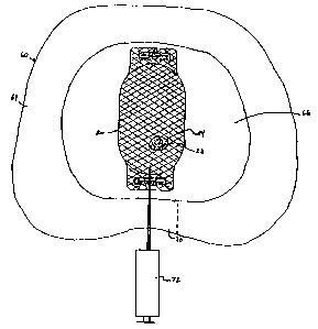
FIG. 5 is a top, sectional view of a human disc space having a prosthetic
spinal disc nucleus in accordance with the present invention implanted;
FIG. 6 is a posterior view of a spinal segment including a degenerated discal
area;
FIG. 7 is a posterior view of the spinal segment of FIG. 6 showing two flaps
that have been cut through an anulus;
FIG. 8 is a top, sectional view of a human disc space having two prosthetic
spinal disc nuclei implanted by an alternative method in accordance with the
present
invention;
1 o FIG. 9 is a perspective view of an alternative embodiment of a prosthetic
spinal disc nucleus, including a cut-away view showing a portion of a core, in
accordance with the present invention;
FIG. 10 is a front sectional view of the prosthetic spinal disc nucleus of
FIG.
9 along the line 10-10; and
~ 5 FIGS. 11-15 illustrate steps of fabricating the alternative prosthetic
spinal
disc nucleus of FIG. 9 in accordance with the present invention.
DETAILED DESCRIPTION OF THE INVENTION
A preferred embodiment of a prosthetic spinal disc nucleus 20 is shown in
2 o FIG. 1. The prosthetic spinal disc nucleus 20 is a capsule-shaped body
comprised
of an amorphous polymer core 22 and a constraining jacket 24. The constraining
jacket 24 is defined by an anterior end 26 and a posterior end 28, and is
secured
around the amorphous polymer core 22 by an anterior closure 30 located at the
anterior end 26 and a posterior closure 32 located at the posterior end 28.
2 5 Various components of the prosthetic spinal disc nucleus 20 are described
in greater detail below. Generally speaking, however, the amorphous polymer
core
22 is preferably configured to be flowable in at least a first state. The
amorphous
polymer core 22 is inserted into the constraining jacket 24, generating an
internal
pressure. The constraining jacket 24 is configured to be flexible, but
substantially
3 0 inelastic such that the prosthetic spinal disc nucleus 20 increases in a
desired
direction in response to the internal pressure.
8
CA 02323922 2000-09-14
WO 99/47082 PCT/US99/05757
A. Amorphous Polymer Core 22 As A Hydrogel
In a preferred embodiment, the amorphous polymer core 22 is a hydrogel
configured to imbibe fluids, expanding from an unhydrated state to a hydrated
state.
In this regard, the hydrogel material is preferably formulated as a mixture of
hydrogel polyacrylonitrile. In particular, acrylamide and acrylonitrile (block
co-
polymer) are used. Alternatively, the hydrogel material used for the amorphous
polymer core 22 can be any hydrophilic acrylate derivative with a unique
multiblock copolymer structure or any other hydrogel material having the
ability to
deform and reform in a desired fashion in response to placement and removal of
loads. Even further, a biologically-safe polymer or elastomer that can imbibe
fluids
while maintaining its structure under various stresses is acceptable. For
example,
the amorphous polymer core 22 can be formulated as a mixture of polyvinyl
alcohol
and water. In one preferred embodiment, the hydrogel material used for the
amorphous polymer core 22 is manufactured under the trade name HYPAN~ by
Hymedix International, Inc. of Dayton, NJ.
in one preferred embodiment, the hydrogel material of the amorphous
polymer core 22 is in a powder form. In other words, the amorphous polymer
core
22 preferably consists of a plurality of fine, irregularly shaped grains of
hydrogel
material. The grains are non-spherical. With this configuration, each of the
grains
2 0 of hydrogel material has a width on the order of 8x IO-' inches.
Acceptable powder
hydrogel material is available, for example, under the tradename HYPAN~ from
Hymedix International, Inc. of Dayton, NJ. The hydrogel powder may be used as
supplied by the manufacturer, or may be processed to generally orientate the
shape
of the individual grains. In a preferred embodiment, the individual grains
have a
2 S flat side, and are defined by a height less than a length and a width. For
example,
each of the flattened hydrogel powder grains will preferably have a height,
length
and width aspect ratio of approximately 1:5:5. With this configuration, the
flattened
hydrogel powder grains will lie against one another when compacted, and have a
tendency to slide. The shape of individual grains of the amorphous polymer
core
3 0 22 may be fixrther controlled, as described in greater detail below.
While each grain of hydrogel material of the amorphous polymer core 22
does have a discernable shape, the overall amorphous polymer core 22 does not.
9
CA 02323922 2000-09-14
WO 99!47082 PCT/US99105757
Therefore, the amorphous polymer core 22 has a fluid-like attribute such that
in at
least one state the amorphous polymer core 22 will flow. For example, in the
preferred embodiment wherein the amorphous polymer core 22 is a powdered
hydrogel, the individual grains are relatively small such that the powder as a
whole
"flows". This flowable attribute can be enhanced by coating the individual
grains
with a low friction material, such as polyvinyl alcohol or polyacrylonitrite.
While the amorphous polymer core 22 has been preferably described as
consisting of a dry, hydrogel powder, other forms are acceptable. For example,
the
amorphous polymer core 22 may consist of a hydrogel powder, as described
above,
suspended in a viscous liquid. In one preferred embodiment, the viscous liquid
is
glycerine, although other similar fluid carriers able to suspend hydrogel
powder can
be used. Even further, the amorphous polymer core 22 may be a fluid hydrogel,
consisting of dry hydrogel powder, as described above, dissolved in a solvent,
such
as Dimethyl Sulfoxide (DMSO). Other solvents able to keep the hydrogel polymer
chains mobile are also available. The resulting fluid hydrogel is non-
thixotropic.
Prior to exposure to water (such as in a disc space), the fluid hydrogel
flows.
However, upon contact with water, the solvent is replaced by water, causing
the
fluid hydrogel to permanently congeal or solidify. Thus, upon hydration, the
fluid
hydrogel will fuse into solid form. It should be understood that the solid
form of
2 0 the fluid hydrogel will still have a conformability characteristic, such
that the
amorphous polymer core 22 will deform slightly in response to various loads.
Regardless of exact form, where a hydrogel material is used, the amorphous
polymer core 22 expands from a dehydrated state (prior to implant) to a
hydrated
state (following implant). In the dehydrated state, the amorphous polymer core
22
flows, such that it can be poured or injected into the constraining jacket 24,
as
described below.
B. Constraining Jacket 24
Completely surrounding the amorphous polymer core 22 is the constraining
jacket 24. The constraining jacket 24 is preferably a capsule-shaped tube made
of
3 0 a tightly-woven, high molecular weight, high tenacity polymeric fabric. In
a
preferred embodiment, high molecular weight polyethylene is used as the weave
material for the constraining jacket 24. However, polyester or any other high
CA 02323922 2000-09-14
WO 99147082 PCTIUS99/05757
molecular weight, high tenacity polymeric material can be employed, and carbon
fiber yarns, ceramic fibers, metallic fibers, etc., are also acceptable. While
the
constraining jacket 24 is itself flexible, the material comprising the
constraining
jacket 24 is not. In other words, the material making up the constraining
jacket 24
has virtually no stretch.
The constraining jacket 24 is preferably made of fibers that have been
highly orientated along their length. As a result, the constraining jacket 24
material,
while flexible, has little elasticity or stretch and a generally fixed maximum
volume.
The maximum volume of the constraining jacket 24 is defined by a generally
fixed
length and circumference. Additionally, with reference to FIG. 2, the
constraining
jacket 24 defines a height and a width. The height of the constraining jacket
24
corresponds to a transverse plane of a nucleus cavity (not shown) and is
represented
by the "x" plane in FIG. 2. Conversely, the width of the constraining jacket
24
corresponds to the sagittal plane of the nucleus cavity and is represented by
the "y"
plane in FIG. 2.
The preferred woven construction of the constraining jacket 24 creates a
plurality of small openings 34, as shown in FIG. 2. The plurality of small
openings
34 are Large enough to allow bodily fluids to interact with the amorphous
polymer
core 22 otherwise maintained within the constraining jacket 24. However, the
2 0 plurality of small openings 34 are small enough to prevent the individual
particles
of the amorphous polymer core 22 from escaping. Preferably, the plurality of
small
openings 34 have an average diameter smaller than the particle size of the
individual
grains of the amorphous polymer core 22, or about 8x10⁻³ inches, although
other
dimensions are acceptable. While the constraining jacket 24 is described as
having
2 5 a weave configuration, any other configuration having a semi-permeable or
porous
attribute can be used, such as a self sealing membrane.
The preferred woven construction of the constraining j acket 24 also
provides a textured outer surface for purchase within the disc space, as
described in
greater detail below. Thus, the constraining jacket 24 prevents the prosthetic
spinal
3 0 disc nucleus 20 from spontaneously dislodging from the disc space.
Additionally,
the constraining jacket 24 material preferably allows for tissue ingrowth.
11
CA 02323922 2000-09-14
WO 99/47082 PCT/US99/05757
C. Construction of Prosthetic Spinal Disc Nucleus 20 With Hydrogel
Material
In one embodiment, the prosthetic spinal disc nucleus 20 of the present
invention is constructed by selecting the constraining jacket 24 sized to fit
within
a disc space (described below). The posterior end 28 of the constraining
jacket 24
is sewn closed by the posterior closure 32, which is a stitching comprised of
the
same high-tenacity polymeric material, such as high molecular weight
polyethylene,
as is used for the constraining jacket 24. The amorphous polymer core 22 (in
an
unhydrated state) is poured into the constraining jacket 24 at the open,
anterior end
26. The anterior end 26 is then closed by the anterior closure 30. Following
closure
of the anterior end 26 of the constraining jacket 24, the prosthetic spinal
disc
nucleus 20 is massaged to horizontally orientate the amorphous polymer core
22,
partially flattening and narrowing the prosthetic spinal disc nucleus 20 in
preparation for implantation.
As an alternative to pouring the amorphous polymer core 22 (in an
unhydrated state) into the constraining jacket 24, the amorphous polymer core
22,
due to a flowable attribute in at least a first state, may instead be injected
within the
constraining jacket 24 by a syringe or small diameter catheter. This approach
is
described in more detail below. Generally speaking, however, the constraining
2 0 jacket 24 is sealed at both the anterior end 26 and posterior end 28. A
syringe or
small diameter catheter is passed through an outer wall of the constraining
jacket
24 and an appropriate volume of the amorphous polymer core 22 is injected. To
facilitate injection, the constraining jacket 24 may include a self sealing
mechanism.
The self sealing mechanism may assume a variety of forms, including a normally
closed tube extending from the constraining jacket 24 that expands or opens
with
applied pressure (such as when the amorphous polymer core 22 is forced
therethrough). Alternatively, the self sealing mechanism may be a spiral tube
that
is normally closed until pressure is applied.
Regardless of whether the amorphous polymer core 22 is placed into the
3 0 constraining jacket 24 before or after implant, an important concern is
the actual
amount or total volume of the amorphous polymer core 22 relative to the volume
of the constraining jacket 24. The constraining jacket 24 has a generally
fixed
12
CA 02323922 2000-09-14
WO 99/47082 PCT/US99/05757
maximum volume. In a preferred embodiment, the volume of the amorphous
polymer core 22 in an unhydrated state fills approximately 60% - 80% of the
available internal volume of the constraining jacket 24. Alternatively, the
percent
volumetric filling can be altered, either slightly higher or lower. As
described in
greater detail below, the volume of the amorphous polymer core 22, where a
hydrogel material is used, will expand greatly upon hydration. Thus, while the
volume of amorphous polymer core 22 in the dehydrated state is less than the
internal volume of the constraining jacket 24, the theoretical volume of the
amorphous polymer core 22 in an unconstrained, hydrated state is greater than
the
internal volume of the constraining jacket 24.
In addition to varying the volume of the amorphous polymer core 22 placed
within the constraining jacket 24, other adjustments can be made to better
meet the
needs of a particular disc space. For example, the hydrogel material used for
the
amorphous polymer core 22 can be selected to have a higher or lower swelling
behavior. Alternatively, the grains comprising the amorphous polymer core 22
can
be coated with a hygroscopic film to increase overall flow by lowering the
coefficient of friction between individual grains.
As described above, the generally fixed maximum volume of the
constraining jacket 24 is greater than a volume of the hydrogel material used
for the
2 0 amorphous polymer core 22 in an unhydrated state. Conversely, the
generally fixed
maximum volume of the constraining jacket 24 is less than the volume of the
amorphous polymer core 22 if allowed to hydrate fully without constraint.
Thus,
because the amorphous polymer core 22 has a natural hydrated volume greater
than
that of the constraining jacket 24, the constraining jacket 24 will be tight
about the
2 5 amorphous polymer core 22 when hydrated, as described in greater detail
below.
In this manner, the volume differential between the constraining jacket 24 and
the
amorphous polymer core 22 in a hydrated state serves to extend the useful life
of the
prosthetic spinal disc nucleus 20. In particular, the constraining jacket 24
effectively prevents the amorphous polymer core 22 from reaching a natural
3 0 hydration level. Consequently, the amorphous polymer core 22 will have a
constant
affinity for imbibing additional fluid.
13
CA 02323922 2000-09-14
WO 99147082 PCTIUS99105757
In final form, the prosthetic spinal disc nucleus 20 is preferably sized to
conform to the approximate length of a sagittal diameter and an approximate
height
of an adult human disc nucleus cavity. For example, in one preferred
embodiment,
the prosthetic spinal disc nucleus 20 will have, in final form, a length in
the range
of approximately 10 to 35 millimeters and an outer diameter in the range of
approximately 3 to 15 millimeters. The preferred prosthetic spinal disc
nucleus 20
is 25 millimeters in length and IO millimeters in outer diameter. It is
realized that
not all human disc nucleus cavities are of the same size. Therefore, the
prosthetic
spinal disc nucleus 20 can be constructed to assume a wide variety of
dimensions.
The appropriate size of the prosthetic spinal disc nucleus 20 for a particular
patient
is determined by various diagnostic procedures prior to and during surgery.
Basically, the properly dimensioned prosthesis is a function of the patient's
size and
spinal level. By providing a different prosthetic spinal disc nucleus 20 with
varying
dimensions, the space requirements reflected by any spinal segment, human or
animal, are satisfied.
D. Implantation and Function of The Prosthetic Spinal Disc Nucleus 20
With Hydrogel Material
In one preferred embodiment, the prosthetic spinal disc nucleus 20 is
preferably percutaneously implanted into a damaged disc space 60, shown in
FIGS.
2 0 3-5. The disc space 60 separates two adjacent vertebrae 62, defining
opposing
endplates (not shown), and includes an anulus 64 and a nucleus cavity 66 (FIG.
5).
Implantation is preferably performed via a posterior approach, although it
should
be understood that an anterior or oblique technique may also be employed. With
the posterior method, a unilateral laminotomy in a targeted lamina area 68 may
be
2 5 required. As shown in FIG. 4, a flap 70 is created in the anulus 64, and,
if
necessary, excess material is removed from the nucleus cavity 66 (FIG. S) to
create
room for the prosthetic spinal disc nucleus 20. The appropriate volume of the
nucleus cavity 66 is estimated and the prosthetic spinal disc nucleus 20 is
selected.
More particularly, the surgeon evaluates the disc space 60 in terms of
3 0 pressure, volume, degree of disc distention or other visual clues. With
this
information in mind, an appropriately sized constraining jacket 24 (FIG. 1) is
selected and placed through the flap 70. Notably, the opening provided by the
flap
14
CA 02323922 2000-09-14
WO 99/47082 PCT/US99105757
70 can be very small because the constraining jacket 24 is "empty" (i.e., does
not
initially contain the amorphous polymer core 22) and can therefore be compact
for
insertion through the opening provided by the flap 70. As shown in FIG. 5, the
constraining jacket 24 is orientated essentially transverse across the disc
space 60.
With the constraining jacket 24 properly oriented, the amorphous polymer core
22
is injected into the constraining jacket 24.
Percutaneous injection of the amorphous polymer core 22 is achieved
through use of a syringe or catheter 72 which is directed to pass through the
constraining jacket 24. The preferred hydrogel material of the amorphous
polymer
1 o core 22, in an unhydrated state, is injected into the constraining jacket
24. A variety
of methods are available for forcing the amorphous polymer core 22 into the
constraining jacket 24. For example, where the amorphous polymer core 22 is
comprised of a powder hydrogel material, pressurized carbon dioxide can be
used
to force the powder hydrogel into the constraining jacket 24. Alternatively,
with
hydrogel powder suspended in a liquid, or a fluid hydrogel, the amorphous
polymer
core 22 can be forced through the syringe 72 with manually applied pressure.
Once the amorphous polymer core 22 has been deposited, the syringe or
catheter 72 is removed. In this regard, the constraining jacket 24 is
preferably
configured to essentially be self sealing such that insertion and removal of
the
2 0 syringe or catheter 72 does not damage or otherwise impart a hole into the
constraining jacket 24 large enough to allow particles of the amorphous
polymer
core 22 to escape. Even further, the constraining jacket 24 may be provided
with
a self sealing mechanism (described above) to allow efficient introduction and
removal of the syringe or catheter 72.
2 5 While the preferred method has described implantation of a single spinal
prosthetic disc nucleus 20 via.injection of the amorphous polymer core 22,
other
approaches are equally acceptable. For example, the amorphous polymer cure 22
and the constraining jacket 24 may be implanted as a single device. In other
words,
the prosthetic spinal disc nucleus 20 may be constructed {i.e., the amorphous
3 0 polymer core 22 placed into the constraining jacket 24) prior to implant
into the disc
space 60. Even further, the prosthetic spinal disc nucleus 20 may be implanted
in
pairs into the damaged disc space 60 as shown in FIGS. 6-8. With this
approach,
CA 02323922 2000-09-14
WO 99/47082 PCTIUS99/05757
a pair of flaps 70a and 70b (FIG. 7) are created in the anulus 64 to provide
for
passage for two of the prosthetic spinal disc nuclei 20.
The flaps 70a and 70b have a height less than a minor axis dimension of the
prosthetic spinal disc nucleus 20. In a preferred embodiment, the flaps 70a
and 70b
have a length of about 12 millimeters and a height of about 6 millimeters for
use
with a prosthetic body 20 having a minor axis diameter of 7 millimeters.
Importantly, because the prosthetic spinal disc nucleus 20 can be massaged to
a
flattened shape, the flaps 70a and 70b need not encompass the entire height of
the
anulus 64. Although in this example, a pair of flaps 70a and 70b are
illustrated and
z 0 discussed, a single flap may alternatively be used.
The vertebrae 62 adjacent the damaged disc space 60 are then slightly
separated. This slight separation can be achieved by inserting an inflatable
jack (not
shown) through one of the flaps 70a or 70b and jacking apart the adjacent
vertebrae
62. Once separation sufficient to insert a prosthetic spinal disc nuclei 20 is
achieved, the flap 70a or 70b not occupied by the jack has one of the
prosthetic
spinal disc nucleus 20 inserted via a tapered holding tube. The jack is then
deflated
and removed, and a second prosthetic spinal disc nucleus 20 is placed through
the
remaining flap 70a or 70b.
With the alternative implantation approach, each one of the prosthetic spinal
2 0 disc nuclei 20 is orientated essentially transverse across the disc space
60 as shown
in FIG. 8. Once implanted, the amorphous polymer core 22 (FIG. 1) of the
prosthetic spinal disc nuclei 20 begins to hydrate, imbibing surrounding
fluids. To
promote an increase in the rate of hydration, saline or similar fluid is
injected or
flushed into the nucleus cavity 66. Finally, the flaps 70a and 70b are sewn
into their
2 5 original position.
Regardless of the number of prosthetic spinal disc nuclei 20 implanted or
whether the amorphous polymer core 22 is placed within the constraining jacket
24
before or after the constraining jacket 24 is positioned within the disc space
60,
upon insertion the amorphous polymer core 22 will flow to approximately fill
the
3 0 constraining jacket 24 (FIGS. 5 and 8). As the hydrogel hydrates, or
transitions
from the unhydrated state to the hydrated state, an internal pressure is
created within
the constraining jacket 24. More particularly, the hydrogel-based amorphous
16
CA 02323922 2000-09-14
WO 99/47082 PCT/US99/05757
polymer core 22 generates a swelling pressure as it expands within the
constraining
j acket 24. Because the constraining j acket is located between adj acent
vertebrae 62,
the resulting cross-sectional shape of the constraining jacket 24 is a
flattened oval.
With reference to FIG. 2, then, the amorphous polymer core 22 swells to fill
this
shape, or initial volume, of the constraining jacket 24. Notably, this initial
volume
is less than the generally fixed maximum volume of the constraining jacket 24
because the constraining jacket 24 is not circular in cross-section, but
instead is
elliptical. From this point, as the amorphous polymer core 22 continues to
swell
(and generate the internal pressure), the constraining jacket 24 transitions
from the
initial volume toward the maximum volume, increasing substantially in height
("x"
in FIG. 2). The increase in height of the prosthetic spinal disc nucleus 20,
in turn,
forces the adjacent vertebrae 62 to lift apart and separate to a natural
level.
The particulate, high surface to volume nature of the amorphous polymer
core 22 allows for a faster hydration of the prosthetic spinal disc nucleus 20
than if
a single, integral core body were provided, since water and body fluids will
be
quickly distributed throughout the amorphous polymer core 22. This rapid
hydration promotes a quick expansion of the disc space 60, a rapid rise in
disc
height with a tightening of the circumferential, ligamentous anulus 64 and an
early
establishment of a barrier to dislodgment of the prosthetic spinal disc
nucleus 20.
2 0 Following hydration, the preferred powdered hydrogel material of the
amorphous polymer core 22 permits a small amount of slippage between
individual
grains and therefore a limited flow of the total core within the constraining
jacket
24 as the disc space 60 is wedged during bending motions. Due to the unique
design
of the amorphous polymer core 22, the prosthetic spinal disc nucleus 20 is
2 5 compliant, able to conform to the available internal shape of the nucleus
cavity 66
defined by opposing end plates (not shown). Thus, the amorphous polymer core
22
allows for natural movements between adjacent vertebrae 62 as the viscosity of
the
amorphous polymer core 22 will not change as a function of shear. Even after
swelling, the amorphous polymer core 22 maintains a degree of deformability,
so
3 0 that the prosthetic spinal disc nucleus 20 will slightly change its shape
in response
to physiological loads and conditions.
17
CA 02323922 2000-09-14
WO 99/47082 PCTIUS99/05757
Following implantation, the prosthetic spinal disc nucleus 20 functions as
an intervertebral spacer and a cushion, and restores the normal fluid pumping
action
of the disc space 60. By employing a flexible woven material for the
constraining
jacket 24, the amorphous polymer core 22 is allowed to deform and reform in a
controlled fashion in response to physiological loads. As the amorphous
polymer
core 22 imbibes fluid, the constraining jacket 24 has sufficient flexibility
to allow
the amorphous polymer core 22 to expand. However, the strength and flexibility
characteristics of the material used for the constraining jacket 24 are such
that the
general capsule shape of the prosthetic spinal disc nucleus 20 will always be
maintained. Further, the constraining jacket 24 prevents undesirable creep of
the
amorphous polymer core 22 due to the substantially inelastic construction.
The prosthetic spinal disc nucleus 20 will deform and reform in response to
the placement and removal of loads on the disc space 60. The prosthetic spinal
disc
nucleus 20 flattens in response to placement of physiologic loads on the
spine, thus
assuming a more flattened shape, and acts as a cushion against various loads
placed
upon it. As these loads are decreased (e.g., when the patient reclines), the
amorphous polymer core 22 reforms, as a whole, back to a more circular cross-
sectional shape. Effectively then, the constraining jacket 24 directs the
amorphous
polymer core 22 to reform, as a whole, vertically within the nucleus cavity
66. This
2 o controlled reformation pushes apart or further separates the adjacent
vertebrae 62
(FIGS. 5 and 8), as would a normal nucleus.
The prosthetic spinal disc nucleus 20 also restores the natural fluid pumping
action of the disc space 60. The hydrated prosthetic spinal disc nucleus 20
occupies
a certain percentage, but not all of, the nucleus cavity 66. As loads on the
disc
2 5 space 60 increase, the prosthetic spinal disc nucleus 20 cushions the
vertebral end
plates (not shown) and slowly deforms. As a result, the volume within the
nucleus
cavity 60 decreases. Notably, because the prosthetic spinal disc nucleus 20
does not
occupy the entire nucleus cavity 66, there is room for the prosthetic spinal
disc
nucleus 20 to deform, and the reduction in volume of the nucleus cavity 66 is
3 0 allowed to take place as would otherwise occur with a normal nucleus. In
this
regard, the amorphous polymer core 22 will flatten or deform as a whole, but
not
decrease in volume in response to the load so that the prosthetic spinal disc
nucleus
18
CA 02323922 2000-09-14
WO 99147082 PCT/US99/05757
20 now occupies a larger percentage of the nucleus cavity 66. As a result of
the
reduction in space, fluids otherwise found in the nucleus cavity 66 are forced
out of
the disc space 60, thus flushing out the accumulated acids or autotoxins
contained
therein. Due to the preferred granule nature of the amorphous polymer core 22,
more unbound or loosely bound water will flow into and out of the amorphous
polymer core 22 then if a singular block material were used.
Conversely, when the load is removed or decreased, the prosthetic spinal
disc nucleus 20 reforms to a more circular cross-sectional shape. This entails
an
increase in the vertical direction (relative to the spine in an upright
position),
1 o causing the vertebral end plates (not shown) to separate, creating an
increased
volume in the nucleus cavity 66. It will be remembered that the amorphous
polymer core 22 does not increase in volume, but simply reforms. As a result,
bodily fluid, containing beneficial nutrients, fills the now-increased volume
of the
nucleus cavity 66, revitalizing the overall disc space 60. The prosthetic
spinal disc
nucleus 20 acts in concert with the natural disc space 60 to restore the
natural
pumping action of the disc space 60.
Notably, the prosthetic spinal disc nucleus 20 of the present invention
independently absorbs the force/pressure placed upon the disc space 60. Thus,
the
anulus 64 is not required to support the force/pressure generated by swelling
of the
2 o amorphous polymer core 22 during hydration. The anulus 64 does not provide
any
circumferential support to the prosthetic spinal disc nucleus 20.
E. Alternative Prosthetic Spinal Disc Nucleus Utilizing Hydrogel
Material
An alternative embodiment of a prosthetic spinal disc nucleus 120 is shown
2 5 in FIGS. 9 and 10. The prosthetic spinal disc nucleus 120 is highly
similar to that
previously described, in that it is comprised of an amorphous polymer core 122
and
a constraining jacket 124. The constraining jacket I24 is identical to the
constraining jacket 24 (FIG. 1) previously described, and includes an anterior
end
126, a posterior end 128, an anterior closure 130 and a posterior closure 132.
The
3 0 amorphous polymer core I22, however, is defined by a plurality of hydrogel
microchips. The plurality of hydrogel nucrochips 122 are preferably made from
the
same hydrogel material set forth above. Unlike the previously described
amorphous
19
CA 02323922 2000-09-14
WO 99/47082 PCTNS99/05757
polymer core 22 (FIG. 1}, however, the plurality of hydrogel microchips 122
are
manufactured to have a certain shape.
FIGS. 11-15 illustrate the manufacturing of the prosthetic spinal disc
nucleus 120. First, a block 140 of hydrogel material is provided. The material
making up the block 140 of hydrogel is preferably polyacrylonitrile, although
other
materials may also be useful. The block 140 of hydrogel material can be cast
in any
shape. In a preferred embodiment, the block 140 of hydrogel material is a cast
or
extruded rod of polymer approximately one millimeter in diameter.
Alternatively,
other dimensions may also be useful.
1 o The block 140 of hydrogel material is fed into a holding channel (not
shown) associated with a milling machine 142, as shown in FIG. 11. In a
preferred
embodiment, the milling machine 142 is a rotating hobbing mill 142 having a
number of cutting edges 144. As the block 140 of hydrogel material is fed
toward
the milling machine 142, the cutting edges 144 cut the block 140 of hydrogel
material, creating the plurality of hydrogel microchips 122. Because the block
140
of hydrogel is preferably amorphous and semi-rigid, the cutting edges 144 are
able
to easily cut the hydrogel material, resulting in a relatively uniform shape.
In a preferred embodiment, each of the plurality of hydrogel microchips 122
is approximately wedge-shaped. For example, as shown in FIG. 12A, each of the
2 o plurality of hydrogel microchips 122 is a crescent-shaped wedge, defined
by a
convex surface 146 and a concave surface 148. Alternatively, as shown in FIG.
12B, each of a plurality of hydrogel microchips 122 may have a more oval
contour,
including a slight concavity on one surface 150. Even further, as shown in
FIG.
12C, each of a plurality of hydrogel microchips 122 can alternatively be an
2 5 elongated body, having opposing relatively flat surfaces.
As shown by the above-described figures, the plurality of hydrogel
microchips 122 can assume any of a number of wedge-shaped configurations.
Preferably, however, the particular shape generated facilitates tight stacking
between each of the plurality of hydrogel microchips 122. In this regard, the
final
3 o shape of each of the plurality of hydrogel microchips I22 is not spherical
so that at
least a portion of the outer surface is not convex. With this design, the
plurality of
CA 02323922 2000-09-14
WO 99/47082 PCT/US99/05757
hydrogel microchips 122 can be closely compacted within the constraining j
acket
124 (FIG. 11), as described in greater detail below.
Following the cutting process, the plurality of hydrogel microchips 122 are
placed into a tumbler apparatus 152, as shown in FIG. I3. In a preferred
embodiment, the tumbler apparatus 152 includes a drum 154 driven by an
obliquely-mounted motor shaft 156. Alternatively, other similar devices may
also
be used.
The plurality of hydrogel microchips 122 are first dry tumbled in the
tumbler apparatus 152 so as to slightly dull their outer surface. Thus, the
tumbling
process abrades and polishes each of the plurality of hydrogel microchips 122,
smoothing any sharp points or edges.
Any excess material removed during the dry tumbling process is separated
from the drum 154, such as by a simple blowing process. Alternatively, a
microfilter can be provided to filter the fine particulates from the plurality
of
hydrogel microchips 122 otherwise maintained in the drum 154. Following the
dry
tumbling process, the plurality of hydrogel microchips 122 may be slightly
flattened
between rotating rollers (not shown) to increase a packing density of the
plurality
of hydrogel microchips 122.
In the final stages of tumbling, the plurality of hydrogel microchips 122 are
2 o tumble-coated with another, softer, low friction formulation of hydrogel.
The
hydrogel coating may be any suitable, stable, appropriately hygroscopic
material.
For example, the coating may be a separate polymer having characteristics
different
from the material of the plurality of hydrogel microchips 122, such as a
different
shear behavior. Regardless of exact form, the polymer coating facilitates
2 5 deformation or sliding between individual particles of the plurality of
hydrogel
microchips 122. As a result, a total mass formed by the plurality of hydrogel
microchips 122 exhibits a deformable attribute, and is able to conform to
minor
variations within a nucleus cavity. In a preferred embodiment, a lower
friction
polyvinyl alcohol or polyacrylonitrile is used as the coating, although other
similar
3 0 materials may also be useful. The coating is formed as a fine, aquatic
slurry that is
slowly added to the drum 154 while continuously tumbling the plurality of
hydrogel
microchips 122. The coating material naturally adheres to the plurality of
hydrogel
21
CA 02323922 2000-09-14
WO 99/47082 PCT/US99/05757
microchips 122, forming a thin film. Following an appropriate dwell period,
each
of the plurality of hydrogel microchips 122 individually become thinly coated
with
the coating material, creating a bonded, smooth surface.
Once properly coated, the plurality of hydrogel microchips 122 are
subjected to warm, filtered air and slowly dehydrated. In a preferred
embodiment,
forced air at a temperature of less than 100C is blown on the plurality of
hydrogel
microchips 122 while the drum 154 continues to rotate. The polishing, tumble
coating and dehydration process results in coarse, free-flowing microchips,
each
having an approximately wedge shape.
It should be recognized that adjustments can be made in several parameters
in order to achieve the desired static and dynamic behavior of the plurality
of
hydrogel microchips 122. For example, the viscosity and swelling behavior of
the
initial block 140 (FIG. 13) of hydrogel; the size and shape of each of the
plurality
of hydrogel microchips 122; the coefficient of friction and swelling behavior
of the
coating gel; and the thickness of the coating layer may be altered to achieve
desired
performance characteristics.
Following the tumbling process, the plurality of hydrogel microchips 122
are placed within the constraining jacket 124, as shown in FIG. 16. As
previously
described, the constraining jacket 124 is preferably a high molecular weight,
polyethylene-woven jacket. Prior to placement of the plurality of hydrogel
microchips 122, the constraining jacket 124 is closed at the posterior end 128
by the
posterior closure I32. Any excess material at the posterior end 128 is removed
by
a thermal cut, fusing posterior closure 132.
The plurality of hydrogel microchips 122 (FIG. 13) are poured into the
2 5 constraining jacket 124 at the open, anterior end 126. The anterior end
126 is then
closed and any excess material is removed from the anterior end 126 by a
thermal
cut, fusing the anterior closure 130.
F. Alternative Prosthetic Spinal Disc Nucleus Utilizing Non-
Hydrophilic Polymer
3 0 As described above, the preferred prosthetic spinal disc nucleus 20 (FIG.
1 )
employs a hydrogel material for the amorphous polymer core 22 (FIG. 1). It
should
be recognized, however, that non-hydrophilic, biocompatible polymers may also
be
22
CA 02323922 2000-09-14
WO 99/47082 PCT/US99/05757
useful. In particular, a non-hydrophilic polymer that is flowable (or can be
maintained flowable) in a first state and cured or non-flowable in a second
state can
be used. It should be understood that the term "non-hydrophilic" as used in
this
specification, encompasses not only hydrophilic materials, but also materials
with
a slight affinity to water. Thus, any material that cannot imbibe and maintain
a
significant amount of water relative to an overall volume of the material is
considered "non-hydrophilic". The "flowable" first state can be achieved in a
number of different manners, such as by retaining the polymer in a solvent
that later
is released, use of a catalyst, heating the polymer to a molten state, etc.
For
l0 example, silicone rubber (RTV) with acetic acid is flowable; once exposed,
however, the acid is released and the silicone rubber cures.
While the non-hydrophilic polymer does not imbibe a significant amount of
fluid, the resulting prosthetic spinal disc nucleus is basically identical to
the
preferred prosthetic spinal disc nucleus 20 shown in FIGS. 1 and 2. In other
words,
the substantially inelastic constraining jacket 24 is implanted into the disc
space,
and the amorphous polymer core 22 is percutaneously inserted into the
constraining
jacket 24, such as by a syringe. With the alternative embodiment, the non-
hydrophilic polymer used for the amorphous polymer core 22 is inserted into
the
constraining jacket 24 in the first, flowable state, filling an initial volume
of the
2 0 . constraining jacket 24 (which is less than the generally fixed maximum
volume).
As additional material is forced into the constraining jacket 24, a filling
pressure
is developed, causing the constraining jacket 24 to transition from the
initial volume
to the generally fixed maximum volume, increasing substantially in height ("x"
in
FIG. 2). In other words, the constraining jacket transitions from a flattened,
oval
2 5 shape to a more circular cross-section. This structural characteristic of
the
constraining jacket 24 is identical to the previous embodiments and results in
necessary spacing between adjacent vertebrae. Once filling of the constraining
jacket 24 is complete, the amorphous polymer core 22 cures, preferably
remaining
somewhat compliant. In the cured state, the prosthetic spinal disc nucleus 20
3 o functions identically to the previous embodiments, acting in concert with
the disc
space to pump fluids into and out of the nucleus cavity.
23
CA 02323922 2000-09-14
WO 99/47082 PCT/US99/05757
The prosthetic spinal disc nucleus of the present invention: a) restores the
height of the damaged disc space; b) restores and tightens the natural anulus
to stop
further degeneration and permit its healing; c) restores the normal load-
unload
cycling and thus flushes out toxic by-products, bringing in fresh nutrients to
the disc
space; d) allows a near-normal range of motion; e) relieves the movement-
induced
discogenic pain of the vertebral segment; and f) allows the use of a minimal,
posterior surgical procedure that provides both cost and medical benefits. In
short,
the prosthetic spinal disc nucleus of the present invention has the ability to
elevate
the disc space from the inside, as does the normal, highly hygroscopic
nucleus. It
1 o will tighten the ligamentous anulus and therefore promote the health and
repairability of anular fibers. Beyond these functions, the prosthetic spinal
disc
nucleus of the present invention has the unique ability to conform to contours
of the
available internal nucleus cavity. Further, the prosthetic spinal disc nucleus
will
exhibit shear behavior under load, imitating the normal, constrained rheology
of the
natural disc nucleus. Finally, hospital inventory costs are greatly reduced in
that the
final size of the prosthetic spinal disc nucleus need not be determined until
actual
surgery. The surgeon then simply chooses an appropriately sized constraining
jacket and subsequently inserts a sufficient amount of the amorphous polymer
core.
Although the present invention has been described with reference to
2 0 preferred embodiments, workers skilled in the art will recognize that
changes may
be made in form and detail without departing from the spirit and scope of the
invention. For example, other methods of sealing the ends of the constraining
jacket exist, such as heat, ultrasound, crimp ring seals or spin entanglement.
Additionally, more than a single layer of material may be used to maintain the
2 5 integrity of the amorphous polymer core. In other words, a plurality of
jackets can surround the amorphous polymer core with one layer providing
efficient
filtering of the amorphous polymer core and assure full containment, and a
second
layer providing strength.
24